Chapter 20 + 21
Study these flashcardsFree · spaced repetition · iPhone, Android, Mac & web
All cards
1
The oral cavity is lined by a for protection.
The oral cavity is lined by a stratified squamous epithelium for protection.
2
The most common cancer of the oral cavity is
The most common cancer of the oral cavity is squamous cell carcinoma.
3
ORAL MUCOSA = and the
ORAL MUCOSA = stratified squamous epithelium and the underlying CT (lamina propria)
4
The 3 parts of the oral mucosa are
1
2.
3.
1
2.
3.
The 3 parts of the oral mucosa are
1. Epithelial lining
2. Lamina propria
3. Muscularis mucosae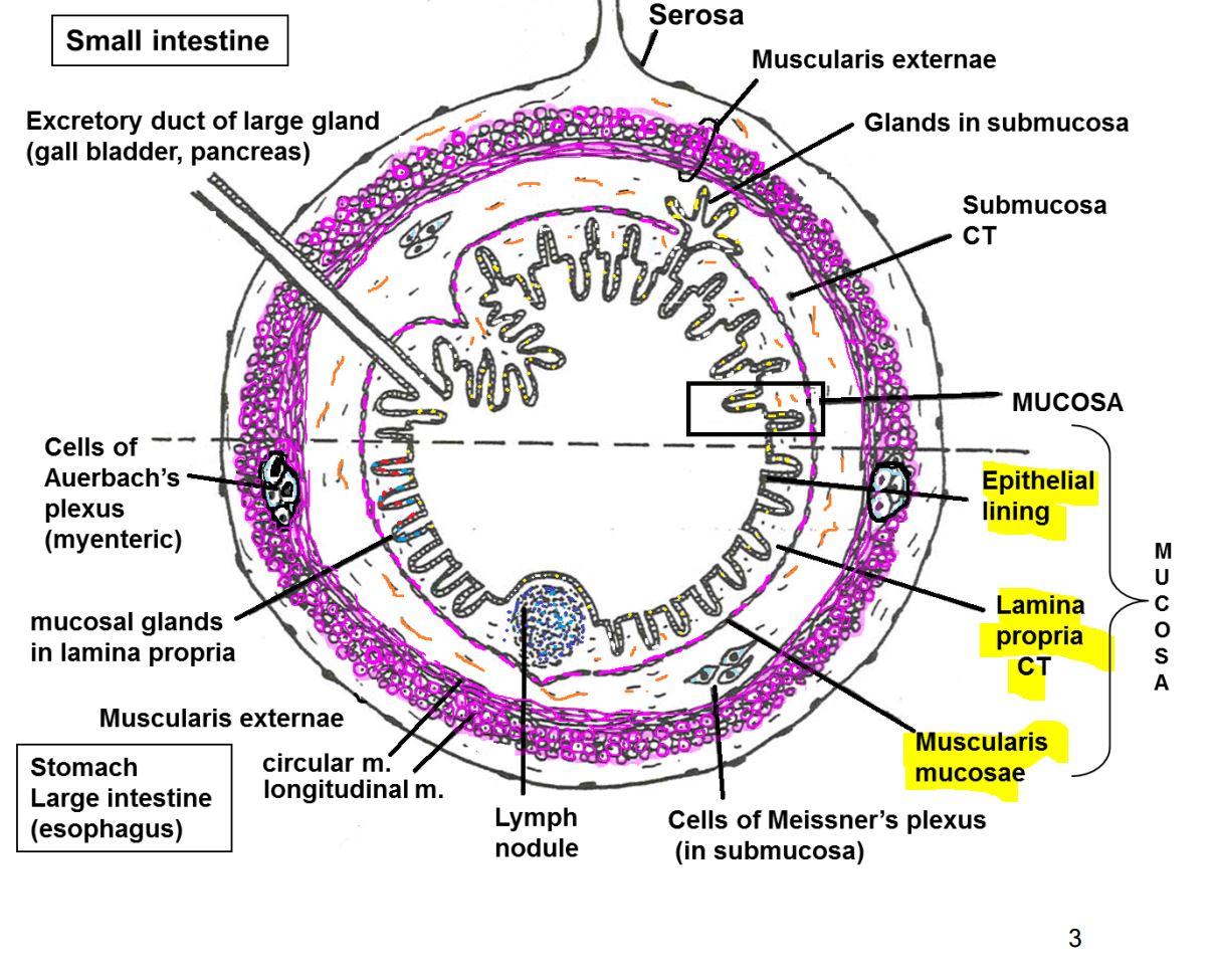
1. Epithelial lining
2. Lamina propria
3. Muscularis mucosae
5
Masicatory = over
Masicatory = over bone
6
Lining mucosa = over like inside of and
Lining mucosa = over soft structures like inside of cheek and lip
7
Specialized mucosa = over
Specialized mucosa = over anterior 2/3 of the tongue
8
Masticatory mucosa -- lines the and
Masticatory mucosa -- lines the gingiva (gums) and hard palate. 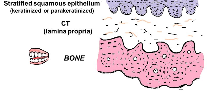
9
In the surface cells produce keratin but the cells retain
shrunken nuclei.
shrunken nuclei.
In parakeratinized epithelium the surface cells produce keratin but the cells retain
shrunken nuclei.
shrunken nuclei.
10
Lining mucosa -- lines the oral side of the , , , , and
Lining mucosa -- lines the oral side of the lip, cheeks, floor of the mouth, inferior
surfaces of the tongue, and soft palate.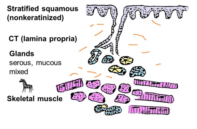
surfaces of the tongue, and soft palate.
11
LIP -- junction between the and
- basically a bag of containing a core of
- basically a bag of containing a core of
LIP -- junction between the skin and oral mucosa
- basically a bag of epithelium containing a core of skeletal muscle.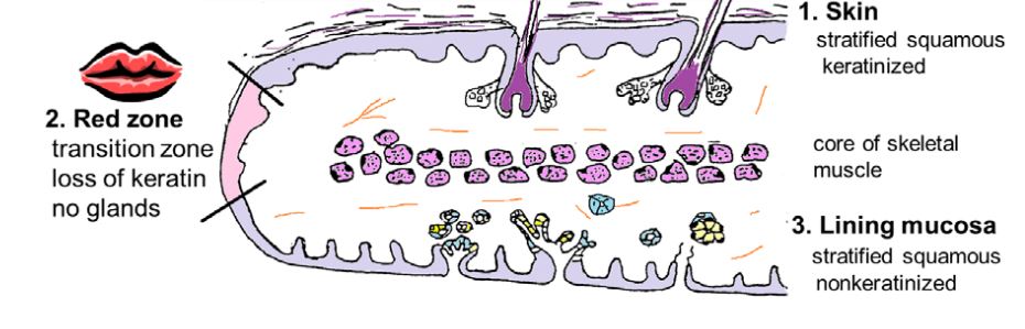
- basically a bag of epithelium containing a core of skeletal muscle.
12
Specialized mucosa - lines the of the . It contains all the basic
layers of the lining mucosa with the addition of and .
layers of the lining mucosa with the addition of and .
Specialized mucosa - lines the dorsal 2/3 of the TONGUE. It contains all the basic
layers of the lining mucosa with the addition of lingual papilla and taste buds.
layers of the lining mucosa with the addition of lingual papilla and taste buds.
13
Taste buds are a chemoreceptor with a
Taste buds are a chemoreceptor with a 10 day lifespan
14
What are the 4 types of papillae
1. Fungiform papillae
2. Filliform papillae
3. Circumvallate papillae
4. Foliate papillae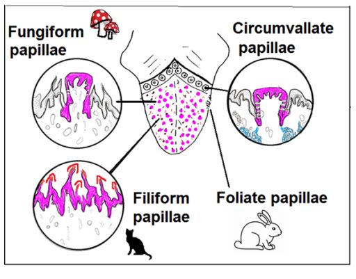
2. Filliform papillae
3. Circumvallate papillae
4. Foliate papillae
15
The tongue is unique in that it has interlacing bundles of muscle in
planes of orientation.
planes of orientation.
The tongue is unique in that it has interlacing bundles of skeletal muscle in 3
planes of orientation.
planes of orientation.
16
The serous glands of the mucosa are referred to as
Von Ebner glands
17
3 layers of mucosa are
1. Epithelium
2. Lamina propria
3. Muscularis mucosae
2. Lamina propria
3. Muscularis mucosae
18
Epithelium - stratified squamous ( ) or simple columnar ( )
Epithelium - stratified squamous (protection) or simple columnar (secretory or absorptive)
19
Lamina propria - highly , may contain
Lamina propria - highly cellular connective tissue (usually loose), may contain glands
20
Muscularis mucosae - thin layer of , present from to .
Muscularis mucosae - thin layer of smooth muscle, present from esophagus to anal canal.
21
SUBMUCOSA - containing and , may be present
SUBMUCOSA - loose CT containing large blood vessels and nerves, glands may be present
22
MUSCULARIS EXTERNAE (Muscularis Propria) - usually ( in part
of the esophagus), commonly arranged in an and an .
of the esophagus), commonly arranged in an and an .
MUSCULARIS EXTERNAE (Muscularis Propria) - usually smooth muscle layers (skeletal in part
of the esophagus), commonly arranged in an inner circular and an outer longitudinal layer.
of the esophagus), commonly arranged in an inner circular and an outer longitudinal layer.
23
ADVENTITIA ( , ) or SEROSA ( plus , )
ADVENTITIA (CT, fibrosa) or SEROSA (CT plus simple squamous epithelium, mesothelium)
24
Exocrine glands – includes glands that extend down into the
(present in ), and glands that extend further down into the (present
only in the and )
(present in ), and glands that extend further down into the (present
only in the and )
Exocrine glands – includes glands that extend down into the lamina propria of the mucosa
(present in all regions), and glands that extend further down into the submucosa (present
only in the esophagus and duodenum)
(present in all regions), and glands that extend further down into the submucosa (present
only in the esophagus and duodenum)
25
Exocrine glands mostly secrete (except
glands that secrete and )
glands that secrete and )
Exocrine glands mostly secrete mucus (except fundic
glands that secrete acid and digestive enzymes)
glands that secrete acid and digestive enzymes)
26
Lymphoid tissue (nodules and infiltrate) can appear transiently anywhere, but always
present in the and .
present in the and .
Lymphoid tissue (nodules and infiltrate) can appear transiently anywhere, but always
present in the ileum and appendix (MALT, GALT).
present in the ileum and appendix (MALT, GALT).
27
Meissner’s nerve plexus in and Auerbach’s nerve plexus (myenteric) in the
.
.
Meissner’s nerve plexus in submucosa and Auerbach’s nerve plexus (myenteric) in the
muscularis externa.
muscularis externa.
28
The enteric nervous system is made of the
The enteric nervous system is made of the Meissner's and Auerbach's nerve plexuses
29
Auerbach’s plexus mostly regulates
, while Meissner’s plexus is more involved in . Ganglion cells are derived from the
, while Meissner’s plexus is more involved in . Ganglion cells are derived from the
Auerbach’s plexus mostly regulates
smooth muscle contraction (motility), while Meissner’s plexus is more involved in gland
secretion. Ganglion cells are derived from the neural crest.
smooth muscle contraction (motility), while Meissner’s plexus is more involved in gland
secretion. Ganglion cells are derived from the neural crest.
30
Enteroendocrine cells ( ) - scattered or in the
or throughout the tract, derived from the
or throughout the tract, derived from the
Enteroendocrine cells (APUD, argentaffin) - scattered endocrine or paracrine cells in the
surface epithelium or glands throughout the tract, derived from the neural crest
surface epithelium or glands throughout the tract, derived from the neural crest
31
The esophagus can be identified by the combination of a with a .
The esophagus can be identified by the combination of a stratified
squamous epithelium (nonkeratinized) with a muscularis mucosa.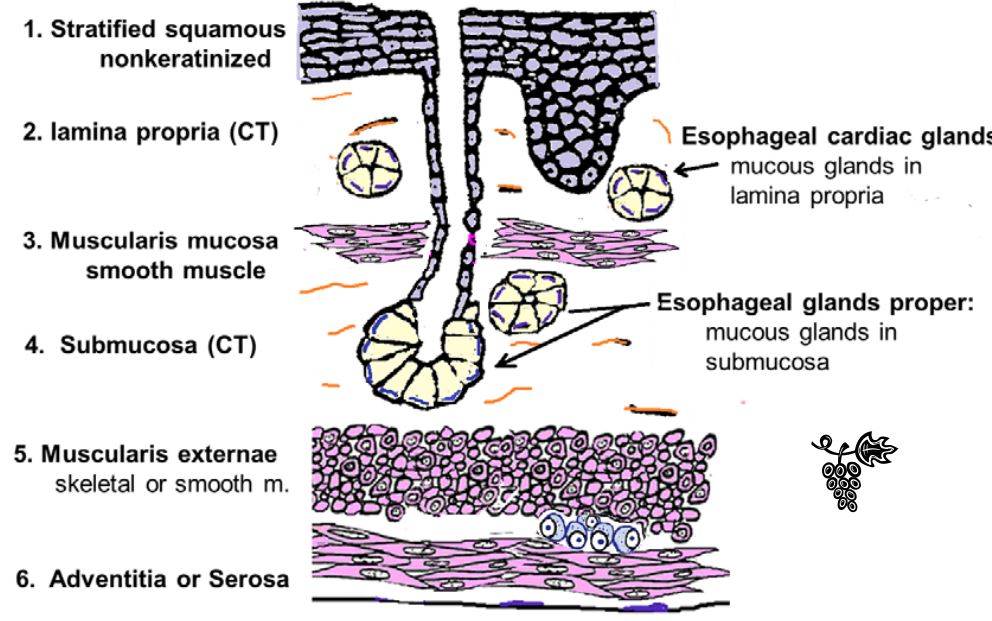
squamous epithelium (nonkeratinized) with a muscularis mucosa.
32
The muscularis externae differs from that of the rest of the digestive tract in that the upper
third contains , the middle third contains both , and
the lower third contains only . Muscle is arranged into and
layers.
third contains , the middle third contains both , and
the lower third contains only . Muscle is arranged into and
layers.
The muscularis externae differs from that of the rest of the digestive tract in that the upper
third contains skeletal muscle, the middle third contains both smooth and skeletal, and
the lower third contains only smooth muscle . Muscle is arranged into inner circular and
outer longitudinal layers.
third contains skeletal muscle, the middle third contains both smooth and skeletal, and
the lower third contains only smooth muscle . Muscle is arranged into inner circular and
outer longitudinal layers.
33
– dilated tortuous veins develop in the submucosa and mucosa of
the esophagus in patients with long standing . Rupture of these
vessels is a common cause of death in .
the esophagus in patients with long standing . Rupture of these
vessels is a common cause of death in .
Esophageal varices – dilated tortuous veins develop in the submucosa and mucosa of
the esophagus in patients with long standing portal hypertension. Rupture of these
vessels is a common cause of death in alcoholic cirrhosis.
the esophagus in patients with long standing portal hypertension. Rupture of these
vessels is a common cause of death in alcoholic cirrhosis.
34
:
This is a complication of long-standing from the stomach. The of the distal portion of the esophagus
undergoes to a epithelium like that of the intestines (absorptive
simple columnar with goblet cells). This epithelium can become dysplastic and develop
into an .
This is a complication of long-standing from the stomach. The of the distal portion of the esophagus
undergoes to a epithelium like that of the intestines (absorptive
simple columnar with goblet cells). This epithelium can become dysplastic and develop
into an .
Barrett’s esophagus:
This is a complication of long-standing reflux of acid from the stomach. The stratified squamous epithelium of the distal portion of the esophagus
undergoes metaplasia to a glandular epithelium like that of the intestines (absorptive
simple columnar with goblet cells). This epithelium can become dysplastic and develop
into an adenocarcinoma.
This is a complication of long-standing reflux of acid from the stomach. The stratified squamous epithelium of the distal portion of the esophagus
undergoes metaplasia to a glandular epithelium like that of the intestines (absorptive
simple columnar with goblet cells). This epithelium can become dysplastic and develop
into an adenocarcinoma.
35
Cancer of the esophagus – malignant, prognosis
Cancer of the esophagus – highly malignant, poor prognosis
36
. Squamous cell carcinoma
- develops from , most
- develops from , most
. Squamous cell carcinoma
- develops from surface epithelium, most common
- develops from surface epithelium, most common
37
Adenocarcinoma
- develops from the glands in the of the
esophagus, associated with
- develops from the glands in the of the
esophagus, associated with
Adenocarcinoma
- develops from the glands in the lower portion of the
esophagus, associated with Barrett’s esophagus
- develops from the glands in the lower portion of the
esophagus, associated with Barrett’s esophagus
38
Generally the the cancer cells migrate in the organ, the the prognosis
Generally the deeper the cancer cells migrate in the organ, the worse the prognosis
39
Criteria for GI carcinomas (4)
(1) Fidelity to parent cell
(2) Location and number of mitotic figures
(3) Metastasis
(4) Location of cancer cells
(2) Location and number of mitotic figures
(3) Metastasis
(4) Location of cancer cells
40
The stomach coverts digested foot into
The stomach coverts digested foot into chyme
41
The stomach has ,
longitudinal folds of the mucosa and submucosa, which usually disappear when
longitudinal folds of the mucosa and submucosa, which usually disappear when
The stomach has rugae,
longitudinal folds of the mucosa and submucosa, which usually disappear when the
stomach is full.
longitudinal folds of the mucosa and submucosa, which usually disappear when the
stomach is full.
42
Although the surface epithelial cells and many glandular cells of the
stomach secrete mucus they are classified as goblet cells (different shape).
stomach secrete mucus they are classified as goblet cells (different shape).
Although the surface epithelial cells and many glandular cells of the
stomach secrete mucus they are NOT classified as goblet cells (different shape).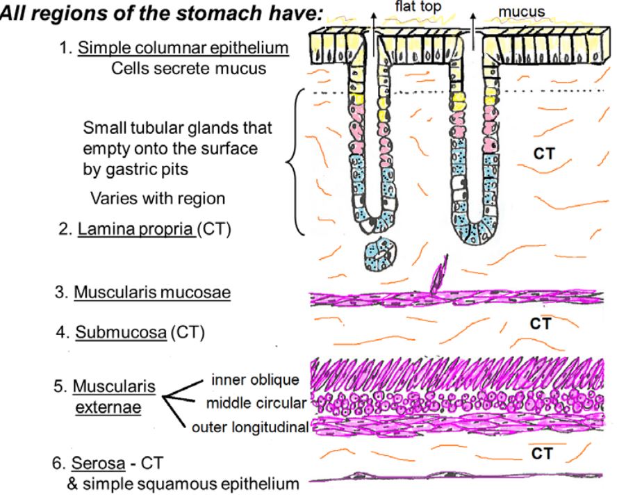
stomach secrete mucus they are NOT classified as goblet cells (different shape).
43
Cardiac glands = secrete
Cardiac glands = secrete mucus 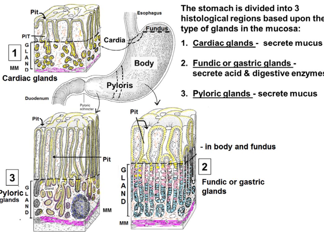
44
Fundic or gastric glands and
Fundic or gastric glands secrete acid and digestive enzymes 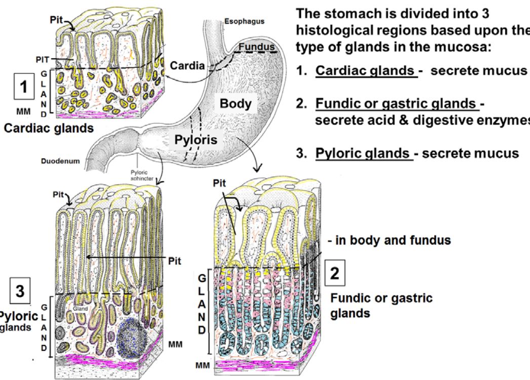
45
Pyloric glands
Pyloric glands secrete mucus 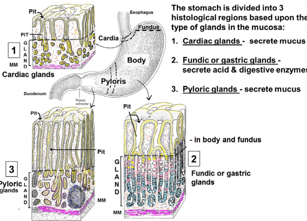
46
Pariteal cells are and are filled with
Pariteal cells are acidophilic and are filled with mitochondria
47
Parietal cells have deep folds of cell membrane that form
Parietal cells have deep folds of cell membrane that form intracellular canalicui
48
Parietal cells secrete and
Parietal cells secrete HCL and gastric intrinstic factor
49
Chief cells are . They are filled with and . Main function is to secrete .
Chief cells are basophilic. They are filled with RER and secretory vessicles. Main function is to secrete pepsinogen.
50
The enteroendocrine cells are between cells and derived. They are or secreting cells.
The enteroendocrine cells are hidden between cells and neural crest derived. They are endocrine or paracrine secreting cells.
51
replace surface and gland cells
Stem cells replace surface and gland cells
52
There are subtle differences in appearance (shading) between the and the . Evident at junction.
There are subtle differences in appearance (shading) between the surface mucous cells and the mucous cells in the gland. Evident at pit/ gland junction. 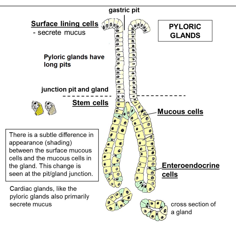
53
To identify a stomach cell, look for
(1) , (surface description)
(2) (cell type) with (not goblet)
(3) Many small mucosal glands into lamina propria
(1) , (surface description)
(2) (cell type) with (not goblet)
(3) Many small mucosal glands into lamina propria
To identify a stomach cell, look for
(1) Flat top, no villi (surface description)
(2) Simple columnar (cell type) with mucous cells (not goblet)
(3) Many small mucosal glands extending down into lamina propria
(1) Flat top, no villi (surface description)
(2) Simple columnar (cell type) with mucous cells (not goblet)
(3) Many small mucosal glands extending down into lamina propria
54
Cardiac stomach is next to (of ) (cell type)
Cardiac stomach is simple columnar next to stratified squamous (of esophagus) (cell type)
55
Fundic stomach is with glands with and cells filling mucosa
Fundic stomach is simple columnar with colorful glands with Parietal and Chief cells filling mucosa
56
Pyloric stomach is with . Pits are long and there is a it appearance from top of gland to pit.
Pyloric stomach is simple columnar with no goblets. Pits are long and there is a difference it appearance from top of gland to pit.
57
Gastritis – an inflammation of the
. In chronic
gastritis and increase in number. The
mucosa can atrophy and undergo
to an intestinal
epithelium. Further dysplasia can
lead to .
. In chronic
gastritis and increase in number. The
mucosa can atrophy and undergo
to an intestinal
epithelium. Further dysplasia can
lead to .
Gastritis – an inflammation of the
stomach mucosa. In chronic
gastritis lymphocytes and plasma
cells increase in number. The
mucosa can atrophy and undergo
metaplasia to an intestinal
epithelium. Further dysplasia can
lead to carcinoma.
stomach mucosa. In chronic
gastritis lymphocytes and plasma
cells increase in number. The
mucosa can atrophy and undergo
metaplasia to an intestinal
epithelium. Further dysplasia can
lead to carcinoma.
58
Loss of parietal cells can cause
and decreased
acid of the
and decreased
acid of the
Loss of parietal cells can cause
pernicious anemia and decreased
acid of the stomach (achlorhydria).
pernicious anemia and decreased
acid of the stomach (achlorhydria).
59
Ulcer – defined as a breach in the
mucosa extending through the
various . Ulcers
can occur throughout the GI tract,
but are most common in the
and where
large amounts of acid are present.
mucosa extending through the
various . Ulcers
can occur throughout the GI tract,
but are most common in the
and where
large amounts of acid are present.
Ulcer – defined as a breach in the
mucosa extending through the
various stomach layers. Ulcers
can occur throughout the GI tract,
but are most common in the
stomach and duodenum where
large amounts of acid are present.
mucosa extending through the
various stomach layers. Ulcers
can occur throughout the GI tract,
but are most common in the
stomach and duodenum where
large amounts of acid are present.
60
Gastric Carcinoma (adenocarcinoma) - highly carcinomas. A confined to the mucosa is better than one that extends down into the layer.
Gastric Carcinoma (adenocarcinoma) - highly malignant carcinomas. A neoplasm confined to the mucosa is better than one that extends down into the muscularis externa layer.
61
The small intestine, the portion of the gut, digests food and absorbs
the end-products of digestion.
the end-products of digestion.
The small intestine, the longest portion of the gut, digests food and absorbs
the end-products of digestion.
the end-products of digestion.
62
The has Brunner’s glands in the
submucosa to help
submucosa to help
The duodenum has Brunner’s glands in the
submucosa to help neutralize the acidic chyme.
submucosa to help neutralize the acidic chyme.
63
The has Peyer’s patches,
a permanent
a permanent
The ileum has Peyer’s patches,
a permanent accumulation of lymphatic nodules.
a permanent accumulation of lymphatic nodules.
64
4 modifications of the small intestine that increase absoprition?
1. Plicase circulares
2. Villi
3. Crypts of Lieberkuhn
4. Microvill
2. Villi
3. Crypts of Lieberkuhn
4. Microvill
65
– permanent folds of the mucosa and submucosa
Plicae circulares – permanent folds of the mucosa and submucosa
66
– finger-like projections of the mucosa
Villi – finger-like projections of the mucosa
67
–downward invaginations
continuous with the villi, in the mucosa
continuous with the villi, in the mucosa
Crypts of Lieberkühn (intestinal glands) –downward invaginations
continuous with the villi, in the mucosa
continuous with the villi, in the mucosa
68
– on the surface simple columnar epithelium
Microvilli (striated border) – on the surface simple columnar epithelium
69
Brunner's glands look like , Peyer's patches look like
Brunner's glands look like grapes, Peyer's patches look like pumpkins
70
The 3 complents of the mucosa of the small intestine are
1. Villi
2. Surface epithelium
3. Crypts of Lieberkuhn
2. Surface epithelium
3. Crypts of Lieberkuhn
71
- projections of the epithelium and lamina propria
Villi - projections of the epithelium and lamina propria
72
Surface epithelium of Stomach
- with absorptive and
- Cells are replaced
- with absorptive and
- Cells are replaced
Surface epithelium of Stomach
- Simple columnar with absorptive and goblet cells.
- Cells are replaced every few days.
- Simple columnar with absorptive and goblet cells.
- Cells are replaced every few days.
73
Crypts of Lieberkuhn (intestinal glands)
– of surface epithelium into lamina propria layer of mucosa
- contains , , , , and .
– of surface epithelium into lamina propria layer of mucosa
- contains , , , , and .
Crypts of Lieberkuhn (intestinal glands)
– Invaginations of surface epithelium into lamina propria layer of mucosa
- contains absorptive, goblet, Paneth, enteroendocrine, and stem cells.
– Invaginations of surface epithelium into lamina propria layer of mucosa
- contains absorptive, goblet, Paneth, enteroendocrine, and stem cells.
74
Submucosa – only the has glands in the submucosa ( glands)
Submucosa – only the duodenum has glands in the submucosa (Brunner’s glands)
75
Goblet cells increase in number from the to the
Goblet cells increase in number from the duodenum to the ileum
76
Central lacteals are in middle of
Central lacteals are lympathic vessels in middle of villus
77
Paneth cells are granules that
Paneth cells are acidophilic granules that secrete lysozyme
78
The lamina propria is a loose CT
The lamina propria is a highly cellular loose CT
79
DUODENUM - ID
Small intestine with
gland in
submucosa
Small intestine with
gland in
submucosa
DUODENUM - ID
Small intestine with
Brunner’s gland in
submucosa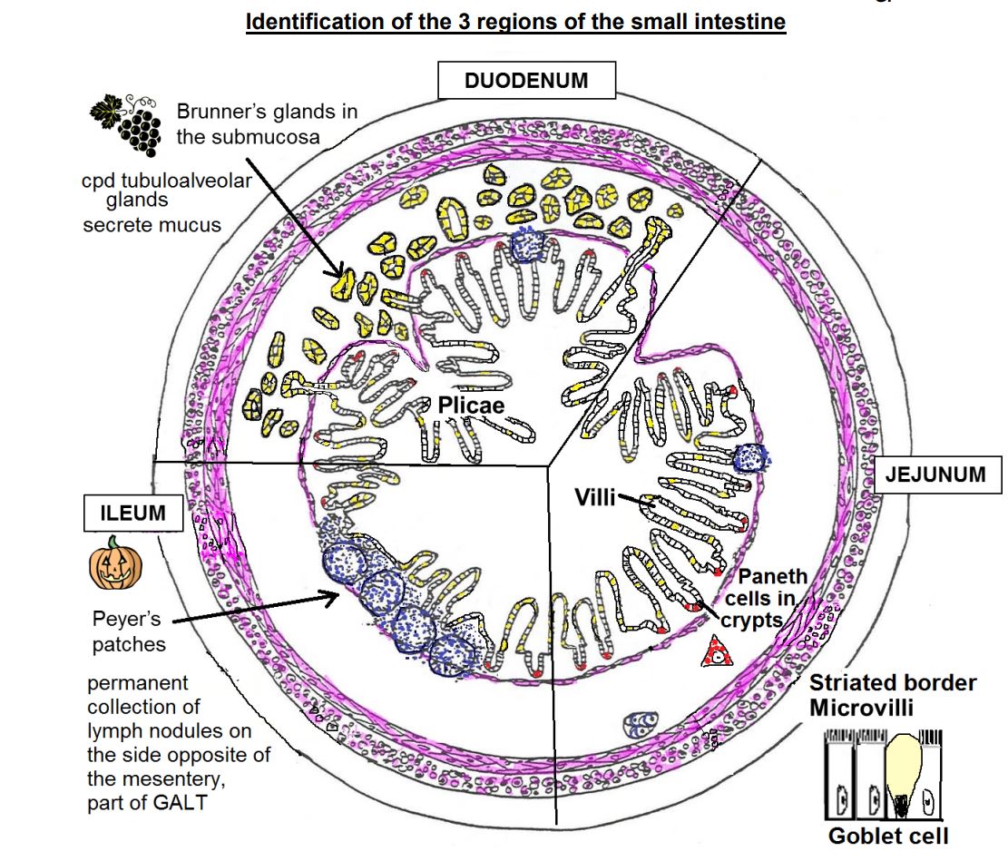
Small intestine with
Brunner’s gland in
submucosa
80
JEJUNUM -ID
Small intestines
No
No
- Boring
Small intestines
No
No
- Boring
JEJUNUM -ID
Small intestines
No Brunner’s glands
No Peyer’s patches
- Boring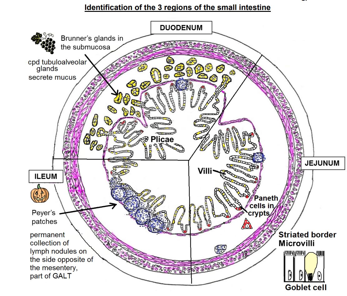
Small intestines
No Brunner’s glands
No Peyer’s patches
- Boring
81
ILEUM
Small intestine with
patches
Small intestine with
patches
ILEUM
Small intestine with
Peyer’s patches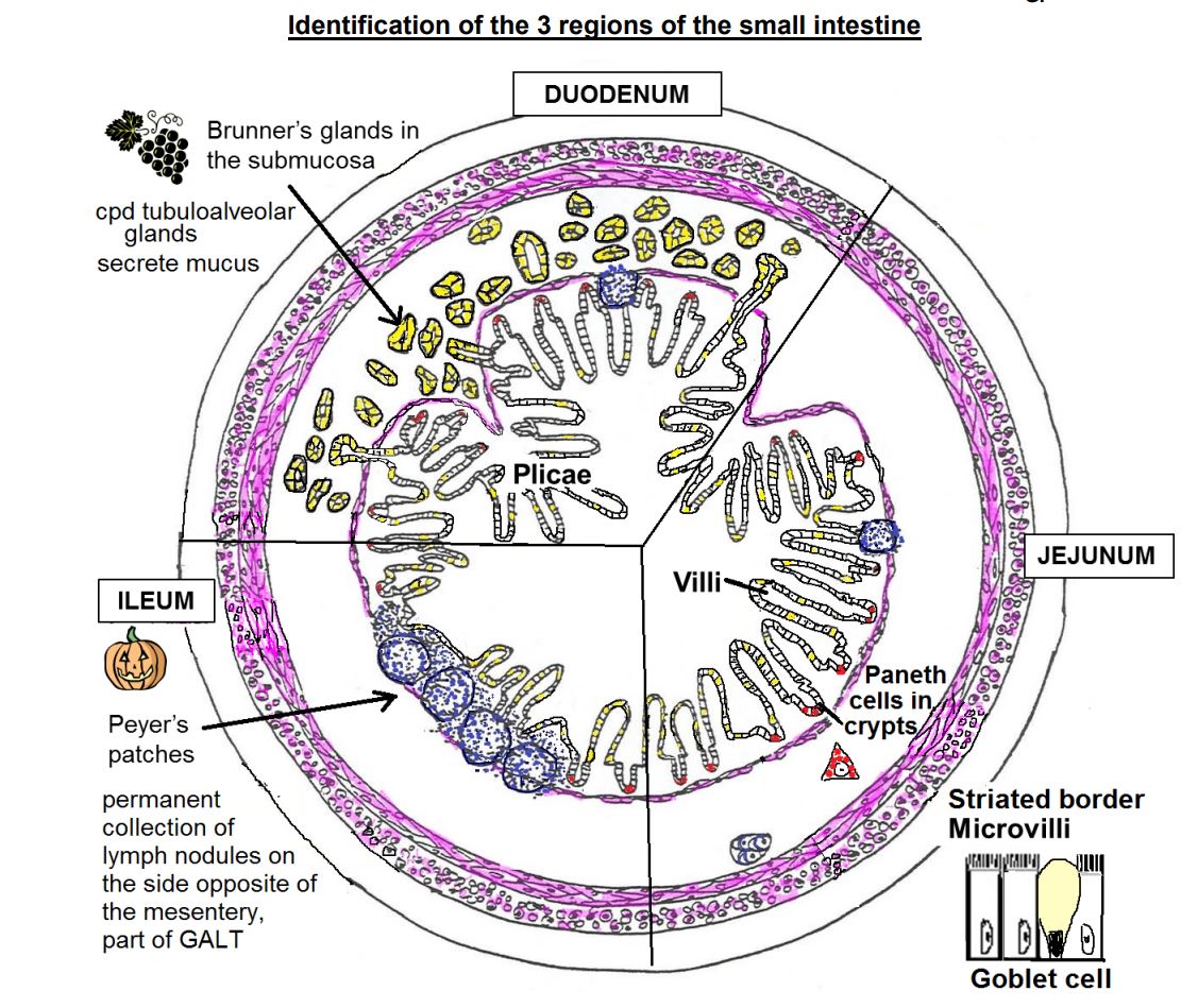
Small intestine with
Peyer’s patches
82
Malabsorption is a general term used to describe a number of clinical conditions in
which nutrients, vitamins, and minerals are inadequately absorbed by the GI tract. This
is present in diseases in which there is an atrophy and reduction in the number of in the small intestines. Sensitivity to is one of the many causes of
malabsorption
which nutrients, vitamins, and minerals are inadequately absorbed by the GI tract. This
is present in diseases in which there is an atrophy and reduction in the number of in the small intestines. Sensitivity to is one of the many causes of
malabsorption
Malabsorption is a general term used to describe a number of clinical conditions in
which nutrients, vitamins, and minerals are inadequately absorbed by the GI tract. This
is present in diseases in which there is an atrophy and reduction in the number of villi
(and microvilli) in the small intestines. Sensitivity to gluten is one of the many causes of
malabsorption
which nutrients, vitamins, and minerals are inadequately absorbed by the GI tract. This
is present in diseases in which there is an atrophy and reduction in the number of villi
(and microvilli) in the small intestines. Sensitivity to gluten is one of the many causes of
malabsorption
83
Crohn’s disease – is a chronic idiopathic inflammatory bowel disease that typically
affects the and/or . It is characterized by involvement of discontinuous
segments of the intestine ( lesions). In the diseased sections, inflammation
penetrates all the layers of intestine (transmural), causing a thickened, rigid wall and
narrowed lumen. The intestine develops ulcers that appear as narrow fissures that
penetrate deeply into the gut wall
affects the and/or . It is characterized by involvement of discontinuous
segments of the intestine ( lesions). In the diseased sections, inflammation
penetrates all the layers of intestine (transmural), causing a thickened, rigid wall and
narrowed lumen. The intestine develops ulcers that appear as narrow fissures that
penetrate deeply into the gut wall
Crohn’s disease – is a chronic idiopathic inflammatory bowel disease that typically
affects the ileum and/or colon. It is characterized by involvement of discontinuous
segments of the intestine (skip lesions). In the diseased sections, inflammation
penetrates all the layers of intestine (transmural), causing a thickened, rigid wall and
narrowed lumen. The intestine develops ulcers that appear as narrow fissures that
penetrate deeply into the gut wall
affects the ileum and/or colon. It is characterized by involvement of discontinuous
segments of the intestine (skip lesions). In the diseased sections, inflammation
penetrates all the layers of intestine (transmural), causing a thickened, rigid wall and
narrowed lumen. The intestine develops ulcers that appear as narrow fissures that
penetrate deeply into the gut wall
84
Meckel’s diverticulum – remnant of , connection of to . This common malformation of small intestine can lead to bleeding,
obstruction and perforation.
obstruction and perforation.
Meckel’s diverticulum – remnant of vitelline duct, connection of yolk sac to midgut
(future ileum). This common malformation of small intestine can lead to bleeding,
obstruction and perforation.
(future ileum). This common malformation of small intestine can lead to bleeding,
obstruction and perforation.
85
The large intestine is composed of the , , , and
The large intestine is composed of the cecum, colon, rectum, and anal canal.
86
Most of the large intestine has , bands of muscle derived from
the outer longitudinal layer of the .
the outer longitudinal layer of the .
Most of the large intestine has taenia coli, three bands of muscle derived from
the outer longitudinal layer of the muscularis externae.
the outer longitudinal layer of the muscularis externae.
87
In the simple columnar epithelium of the large intestine, there are absorptive cells called . There ends up being a decreasing amount of and an increasing about of
In the simple columnar epithelium of the large intestine, there are absorptive cells called enterocytes . There ends up being a decreasing amount of enterocytes and an increasing about of goblet cells.
88
The crypts of lieberkuhn are simple tubular organs with (1) (2) (3)
The crypts of lieberkuhn are simple tubular organs with (1) Goblet cells (2) Enteroendocrine (3) Stem cells
89
The muscularis externae layer is the layer with an layer in bands
The muscularis externae layer is the inner circular m. layer with an outer muscle layer in 3 bands (taenia coli)
90
The large intestine can be identified because it has a with . It is simple columnar with . There is also taenia coli present but not in every area of large intestine
The large intestine can be identified because it has a flat top with glands. It is simple columnar with multiple goblet cells. There is also taenia coli present but not in every area of large intestine
91
Where are the 3 places that the taenia coli are not found?
1. Rectum
2. Anal canal
3. Appendix
2. Anal canal
3. Appendix
92
The appendix has a similar strcuture to the colon EXCEPT 1. 2. 3.
The appendix has a similar strcuture to the colon EXCEPT 1. Small size 2. Continuous ring of lymph nodules (GALT) 3. No taenia coli
93
Embryological transition from to to ( line)
with new blood supply and innervation. The also marks the location of the
original at the end of the hindgut.
with new blood supply and innervation. The also marks the location of the
original at the end of the hindgut.
Embryological transition from hindgut to endoderm to ectoderm (pectinate or dentate line)
with new blood supply and innervation. The pectinate line also marks the location of the
original anal membrane at the end of the hindgut.
with new blood supply and innervation. The pectinate line also marks the location of the
original anal membrane at the end of the hindgut.
94
of rectum to of anal canal and then to (white line of ) continuous with skin. (cell types)
Simple columnar of rectum to nonkeratinized stratified
squamous epithelium of anal canal and then to keratinized stratified squamous
epithelium (white line of Hilton) continuous with skin. (cell types)
squamous epithelium of anal canal and then to keratinized stratified squamous
epithelium (white line of Hilton) continuous with skin. (cell types)
95
. Appendicitis – usually an of the
. Appendicitis – usually an acute inflammation of the appendix
96
Carcinoid Tumors ( cell tumors) – uncommon tumor from neural crest
derived enteroendocrine cells in the . They can secrete a variety of substances,
including , , , and
derived enteroendocrine cells in the . They can secrete a variety of substances,
including , , , and
Carcinoid Tumors (Neuroendocrine cell tumors) – uncommon tumor from neural crest
derived enteroendocrine cells in the appendix. They can secrete a variety of substances,
including serotonin, gastrin, somatostatin, and histamine.
derived enteroendocrine cells in the appendix. They can secrete a variety of substances,
including serotonin, gastrin, somatostatin, and histamine.
97
Ulcerative colitis – is a chronic idiopathic inflammatory bowel disease typically limited
to the and . It commonly extends proximally in a continuous fashion from
the rectum. Inflammation, usually limited to the & layers, results in
many small ulcers. cells, which normally are not present, appear in the
(Paneth cell ). Long-standing ulcerative colitis may lead to cancer.
to the and . It commonly extends proximally in a continuous fashion from
the rectum. Inflammation, usually limited to the & layers, results in
many small ulcers. cells, which normally are not present, appear in the
(Paneth cell ). Long-standing ulcerative colitis may lead to cancer.
Ulcerative colitis – is a chronic idiopathic inflammatory bowel disease typically limited
to the colon and rectum. It commonly extends proximally in a continuous fashion from
the rectum. Inflammation, usually limited to the mucosa & submucosa layers, results in
many small ulcers. Paneth cells, which normally are not present, appear in the crypts
(Paneth cell metaplasia). Long-standing ulcerative colitis may lead to colon cancer.
to the colon and rectum. It commonly extends proximally in a continuous fashion from
the rectum. Inflammation, usually limited to the mucosa & submucosa layers, results in
many small ulcers. Paneth cells, which normally are not present, appear in the crypts
(Paneth cell metaplasia). Long-standing ulcerative colitis may lead to colon cancer.
98
Malignant neoplasm in the large intestine thought to develop
from that have undergone
malignant change
from that have undergone
malignant change
Malignant neoplasm in the large intestine thought to develop
from polyps that have undergone
malignant change
from polyps that have undergone
malignant change
99
Ischemic bowel disease – a
of blood flow to the
large and/or small intestines can result in
death. Within 1-4 days, growth of
intestinal bacteria can produce
and perforation of the bowel.
of blood flow to the
large and/or small intestines can result in
death. Within 1-4 days, growth of
intestinal bacteria can produce
and perforation of the bowel.
Ischemic bowel disease – a
significant obstruction of blood flow to the
large and/or small intestines can result in
death. Within 1-4 days, growth of
intestinal bacteria can produce gangrene
and perforation of the bowel.
significant obstruction of blood flow to the
large and/or small intestines can result in
death. Within 1-4 days, growth of
intestinal bacteria can produce gangrene
and perforation of the bowel.
100
, is caused
by a loss of enteric neurons in a section of colon. This can lead to obstruction and
by a loss of enteric neurons in a section of colon. This can lead to obstruction and
Toxic megacolon (Hirschsprung’s disease), is caused
by a loss of enteric neurons in a section of colon. This can lead to obstruction and gangrene.
by a loss of enteric neurons in a section of colon. This can lead to obstruction and gangrene.