LIVER AND BILIARY PASSAGES
Study these flashcardsFree · spaced repetition · iPhone, Android, Mac & web
All cards
1
The liver has both and functions
The liver has both exocrine and endocrine (i.e. clotting factors) functions
2
The liver occupies the , and (regions)
The liver occupies the right hypochondrium, epigastrium and left
hypochondrium (regions)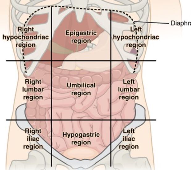
hypochondrium (regions)
3
Diaphragmatic surface – ,
Diaphragmatic surface – convex, smooth
4
Vicersal surface- related to the , , ,
Vicersal surface- related to the right colic flexure, right kidney, stomach, duodenum
5
Two lobes of the liver
near the costal margin
posterior, near the diaphragm and closer to the
hepatic vessels
near the costal margin
posterior, near the diaphragm and closer to the
hepatic vessels
Two lobes of the liver
Quadrate: near the costal margin
Caudate: posterior, near the diaphragm and closer to the
hepatic vessels
Quadrate: near the costal margin
Caudate: posterior, near the diaphragm and closer to the
hepatic vessels
6
The quadrate and caudate lobes are part of the
The quadrate and caudate lobes are part of the right lobe
7
The gallbladder is near the , near the
The gallbladder is near the quadrate lobe, near the
costal margin
costal margin
8
The gallbladder is between the and the
at the
at the
The gallbladder is between the right
costal margin and the
right linea semilunaris
at the transpyloric
plane (L1).
costal margin and the
right linea semilunaris
at the transpyloric
plane (L1).
9
The liver separates the liver into two lobes and is one of the peritoneal folds
The falciform liagment liver separates the liver into two lobes and is one of the peritoneal folds
10
TEST QUESTION The quaradrate and caudate lobes are part of the
TEST QUESTION The quaradrate and caudate lobes are part of the right lobe 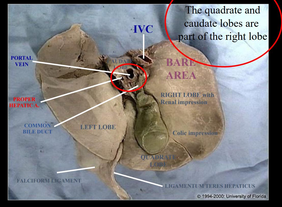
11
The liver is covered by the peritoneum EXCEPT at the
The liver is covered by the peritoneum EXCEPT at the
“bare”
area.
“bare”
area.
12
The bare space is associated with the
The bare space is associated with the IVC
13
When the falciform ligament (Obliterated )
gets to the visceral surface of the liver it changes names:
•Coronary ligaments:
• Triangular ligaments:
gets to the visceral surface of the liver it changes names:
•Coronary ligaments:
• Triangular ligaments:
When the falciform ligament (Obliterated umbilical vein)
gets to the visceral surface of the liver it changes names:
•Coronary ligaments: anterior/posterior
• Triangular ligaments: right/left
gets to the visceral surface of the liver it changes names:
•Coronary ligaments: anterior/posterior
• Triangular ligaments: right/left
14
Liagamentum teres forms the
Liagamentum teres forms the obliterated umbilical vein
15
In the fetus the sends blood from the umbilical vein
directly to the inferior vena cava.
directly to the inferior vena cava.
In the fetus the ductus venosus sends blood from the umbilical vein
directly to the inferior vena cava.
directly to the inferior vena cava.
16
• BLOOD SUPPLY Liver
• BLOOD SUPPLY Liver
• Portal vein
• Hepatic arteries (celiac trunk)
• Hepatic veins (into the IVC)
• Portal vein
• Hepatic arteries (celiac trunk)
• Hepatic veins (into the IVC)
17
NERVE SUPPLY of liver
• from the
• from the
NERVE SUPPLY of liver
• Hepatic plexus from the celiac plexus
• Hepatic plexus from the celiac plexus
18
Anterior = . Posterier =
Anterior = quadrant vein. Posterier = caudate.
19
Relationship of Gallbladder
Inferior:
Anterior =
Posterior =
Inferior:
Anterior =
Posterior =
Relationship of Gallbladder
Inferior: duodenum and billary tree
Anterior = inferior border of liver and anterior abdominal wall
Posterior = transverse colon and proximal dudodenum
Inferior: duodenum and billary tree
Anterior = inferior border of liver and anterior abdominal wall
Posterior = transverse colon and proximal dudodenum
20
Right hepatic duct + left hepatic duct =
Right hepatic duct + left hepatic duct = common hepatic duct
21
• Common hepatic duct + cystic duct =
• Common hepatic duct + cystic duct = common bile duct.
22
• Common bile duct + main pancreatic duct = to to
• Common bile duct + main pancreatic duct = hepatopancreatic
ampulla to greater duodenal papilla to 2nd part of
duodenum.
ampulla to greater duodenal papilla to 2nd part of
duodenum.
23
The ampullary portion of the bile duct is guarded by the
The ampullary portion of the bile duct is guarded by the
Sphincter of Oddi
Sphincter of Oddi
24
The bare area is on the surface of the right lobe with the
The bare area is on the viseral surface of the right lobe with the IVC
25
What is the blood supply of the gallbladder and bile duct?
Cystic artery (right hepatic artery [common hepatic artery] {celiac trunk})
26
What is the nerve supply of the bile duct?
Vagus (parasympathetic)
Greater splanchnic nerve (sympathetic)
Greater splanchnic nerve (sympathetic)
27
What are the 3 borders of the Hepatocystic Triangle of Calot?
1. Inferior border of liver (superiorly)
2. Common hepatic (medial)
3. Cystic duct (laterally)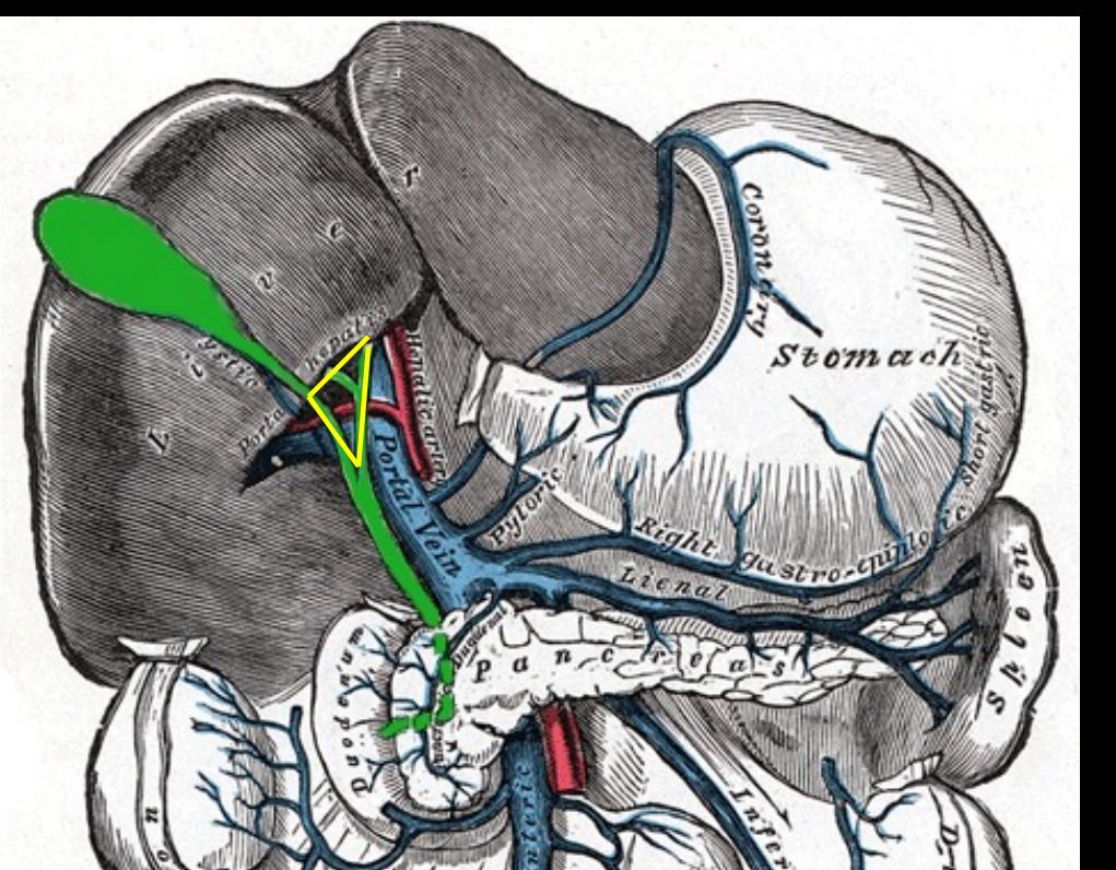
2. Common hepatic (medial)
3. Cystic duct (laterally)
28
What are the contents of the Hepatocystic Triangle of Calot?
1, Lymph node of calot
2, Cystic artery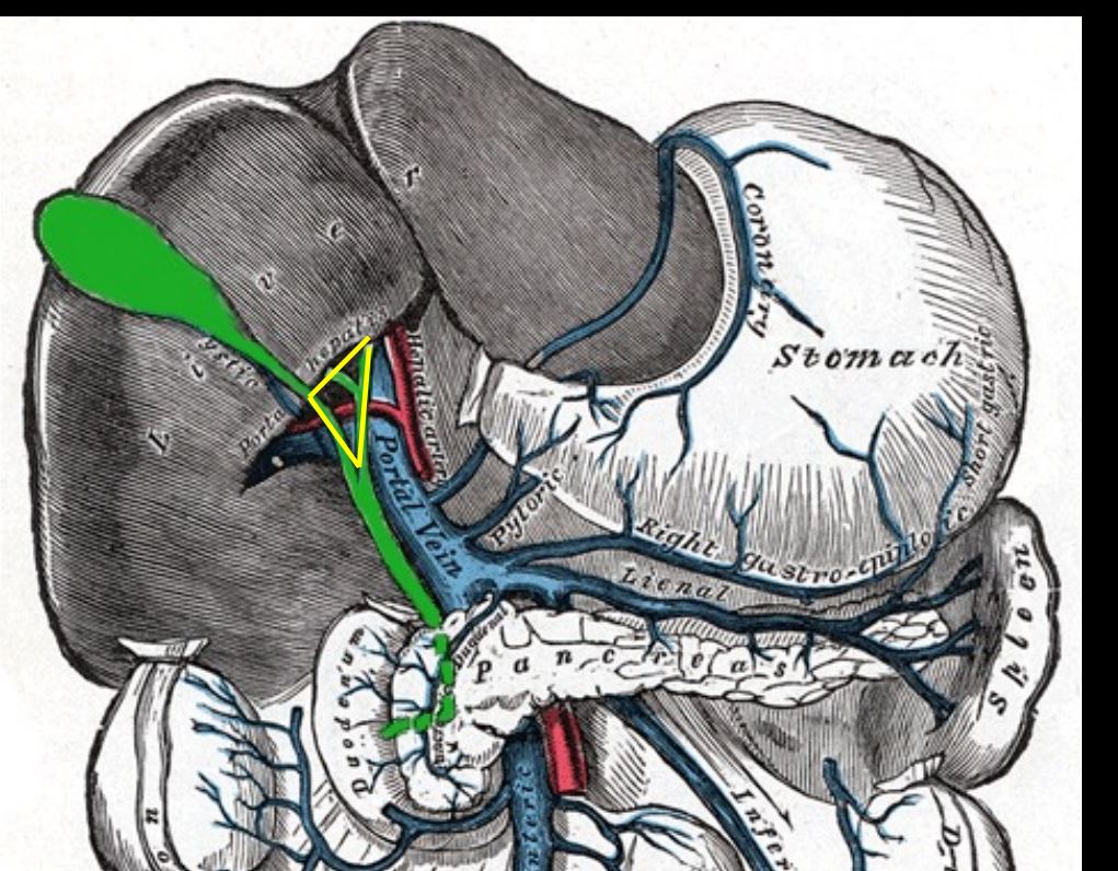
2, Cystic artery
29
Trainagle of calot is important for
Trainagle of calot is important for laproscopic surgery