Thorax Study Guide
Study these flashcardsFree · spaced repetition · iPhone, Android, Mac & web
All cards
1
Aortic valve Auscultation: over the
Aortic valve Auscultation: over the right ICS2
2
Pulmonary valve Auscultation: over the
Pulmonary valve Auscultation: over the left ICS2
3
Bicusid valve: : 6-7 cm from the median plane generally in
Bicusid valve: apex of the heart: 6-7 cm from the median plane generally in ICS 5
4
Tricuspid valve Auscultation: of the lower end of the body of the at
Tricuspid valve Auscultation: left ½ of the lower end of the body of the sternum at ICS4
5
Secondary pulmonic:
Secondary pulmonic: Left ICS3
6
Tricuspid valve
On the between the and
Behind the opposite
On the between the and
Behind the opposite
Tricuspid valve
On the right between the RA and RV
Behind the right ½ of the sternum opposite ICS4
On the right between the RA and RV
Behind the right ½ of the sternum opposite ICS4
7
Bicuspid/mitral valve
On the left between the and
Behind the opposite
On the left between the and
Behind the opposite
Bicuspid/mitral valve
On the left between the LA and LV
Behind the left ½ of sternum opposite costal cartilage 4
On the left between the LA and LV
Behind the left ½ of sternum opposite costal cartilage 4
8
Aortic valve: between and
opposite
opposite
Aortic valve: between LV and aorta
Left ½ of sternum opposite ICS3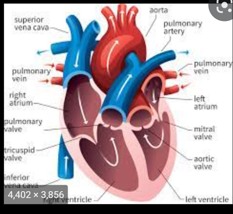
Left ½ of sternum opposite ICS3
9
Pulmonary valve: between and
Behind the medial end of the
Behind the medial end of the
Pulmonary valve: between RV and pulmonary artery
Behind the medial end of the 3rd left costal cartilage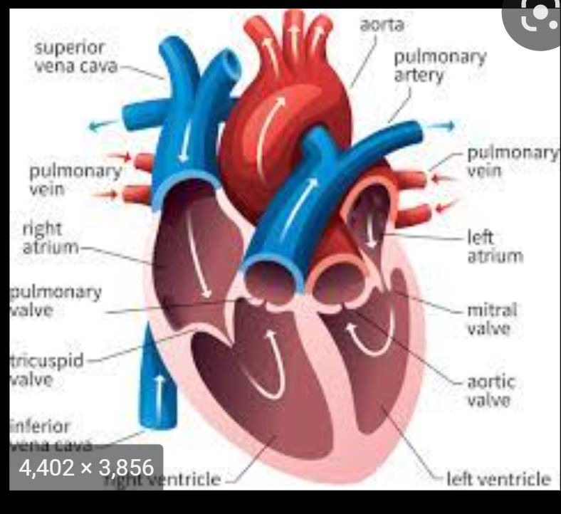
Behind the medial end of the 3rd left costal cartilage
10
The left lung has ( ) and ( )
The left lung has one fissue (oblique) and two lobes (superior and inferior)
11
What are the three special features of the left lung?
1. Cardiac notch at costal cartilage 4,5,6
2. Lingula
3. Groove for aortic arch and descending aorta
2. Lingula
3. Groove for aortic arch and descending aorta
12
Where are the cardiac notch and the lingula?
Superior lobe
13
The right lung has fissures ( and ) and lobes ( )
The right lung has 2 fissures (oblique and horzontal) and 3 lobes (superior, middle, and inferior)
14
What are the 3 special features of the right lung?
1. Tracheal and esophageal area
2. Groove for SVC and IVC
3. Groove for azygous vein
2. Groove for SVC and IVC
3. Groove for azygous vein
15
The pulmonary artery is to the bronchi in the left lung
The pulmonary artery is superior to the bronchi in the left lung 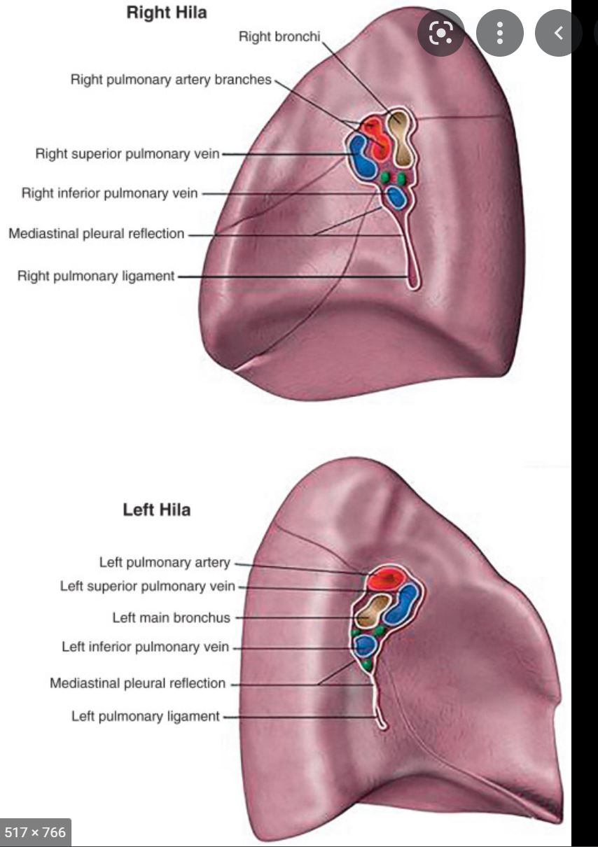
16
The pulmonary artery is to the bronchi in the right lung
The pulmonary artery is anterior to the bronchi in the right lung 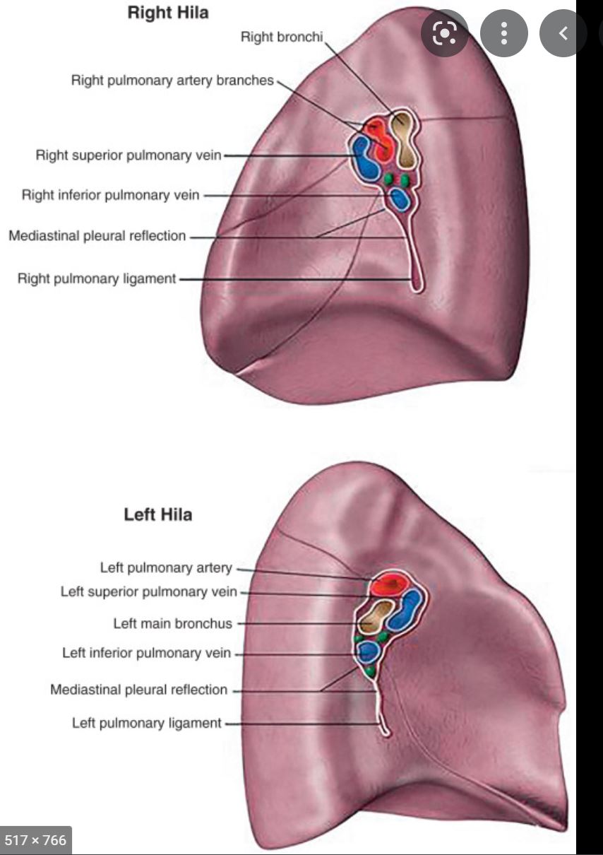
17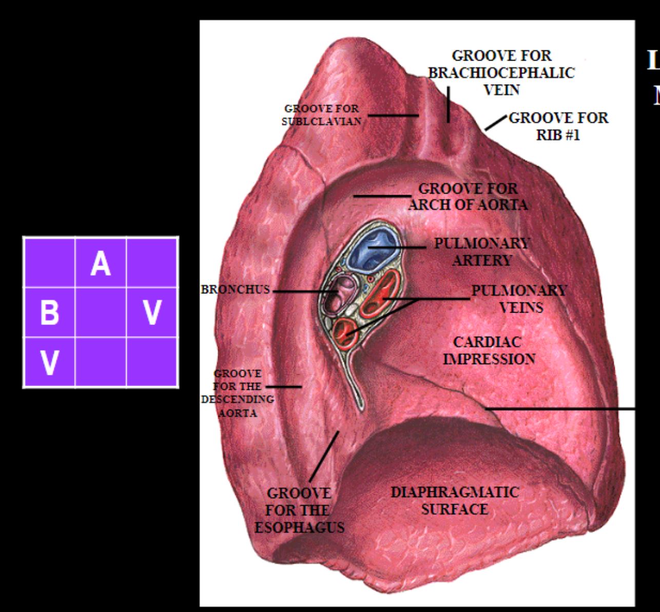
Relationship between hilum vessels left lung
18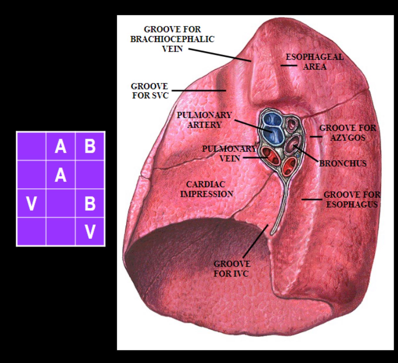
Relationship between hilum vessels right lung
19
Oblique fissure begins 2.5 cm (1 inch) from apex of lungs and ends at
Oblique fissure begins 2.5 cm (1 inch) from apex of lungs and ends at costal cartilage 6
20
Horizontal fissure is located at
Horizontal fissure is located at costal cartilage 4
21
Aortic valve Auscultation: over the
Aortic valve Auscultation: over the right ICS2
22
Thoracic duct begins at as the
Thoracic duct begins at L2 as the cisterna chyli
23
Entire left side of the body and right abdomen and leg→ → , the thoracic duct crosses over to the —> enters the and behind the subclavian artery —>
Entire left side of the body and right abdomen and leg→ cisterna chyli→ thoracic duct
at T5/6, the thoracic duct crosses over to the left behind the esophagus—> enters the left internal jugular vein and subclavian vein behind the subclavian artery —> SVC
at T5/6, the thoracic duct crosses over to the left behind the esophagus—> enters the left internal jugular vein and subclavian vein behind the subclavian artery —> SVC
24
Only the right head, neck, thorax, and arm drains into the —> —>
Only the right head, neck, thorax, and arm drains into the right lymphatic duct —> R internal jugular vein —> SVC
25
Blood supply to heart is by branches of right and left coronary arteries from the
Blood supply to heart is by branches of right and left coronary arteries from the ascending aorta 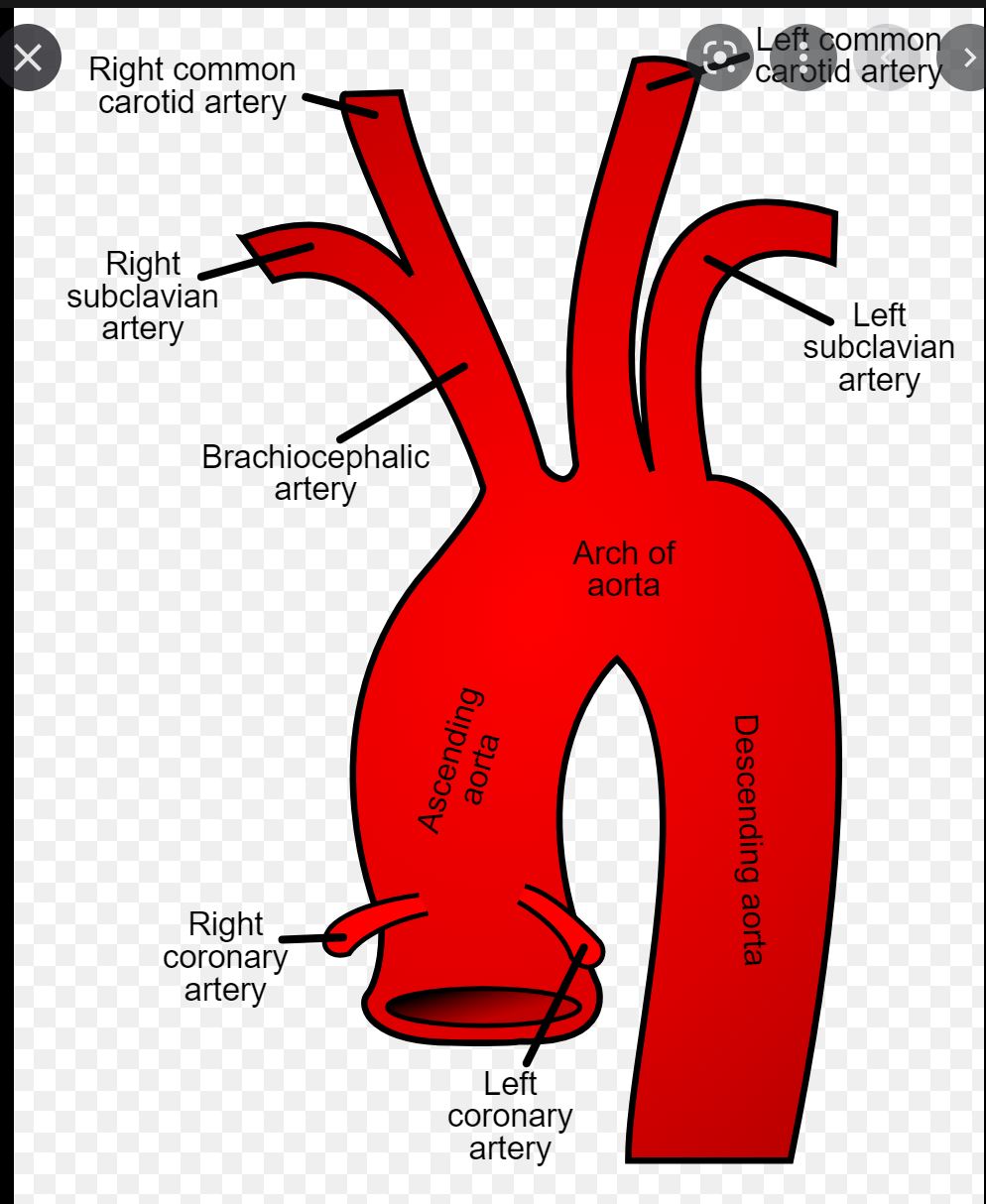
26
What are the 3 things in the right coronary artery?
1. SA nodal artery (supplies SA node)
2. AV nodal artery (supplies AV node)
3. Posterior interventricular (posterior desending a. supplies right + left ventricle +posterior 1/3 of septum)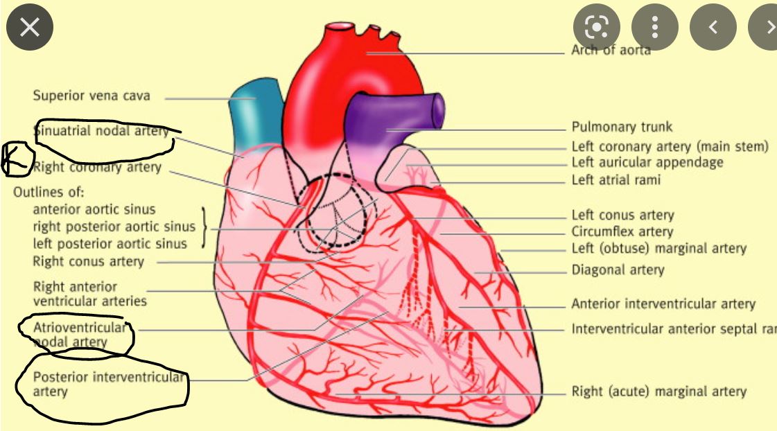
2. AV nodal artery (supplies AV node)
3. Posterior interventricular (posterior desending a. supplies right + left ventricle +posterior 1/3 of septum)
27
SA nodal a. - supplies the
SA nodal a. - supplies the SA node
28
AV nodal a. - supply the
AV nodal a. - supply the AV node
29
Posterior interventricular —> posterior descending a. supplies part of and and
Posterior interventricular —> posterior descending a. supplies part of right and left ventricle and posterior 1/3 of interventricular septum
30
What are the two branches of the left coronary artery?
1. Left anterior desending LAD
2. Circumflex artery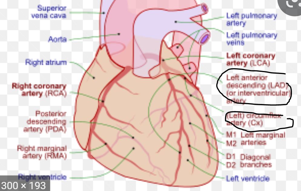
2. Circumflex artery
31
What are the 4 things supplied by the left anterior desecending?
1. Anterior left ventricular wall
2. Anterior 2/3 of interventricular septum
3. Bundle of His
4. Apex
2. Anterior 2/3 of interventricular septum
3. Bundle of His
4. Apex
32
What are the 2 things supplied by the circumflex artery?
1. Left border of heart via marginal artery
2. Posterior-inferior left ventricular wall
2. Posterior-inferior left ventricular wall
33
Where is the SA node located?
wall of the RA, superior to the crista terminalae at the junction of the anteromedial aspect of the SVC and right auricle || notes:Mnemonic: The SA node is always right. It thinks that it is superior to Cristae. It puts itself before and center to superior people and right people.
34
What is the blood supply of the SA node?
Branch of the right coronary artery
35
Where is the AV node located?
interatrial septum on the ventricular side of the orifice of the coronary sinus || notes:Mnmenonic: AV node has an infection interatrial septum on the ventricular side of its sinus
36
What is the blood supply of the AV node?
branch of the right coronary artery
37
Bronchial arteries supply the
Bronchial arteries supply the conductive airway (non respiratory)
38
What is the blood supply of the right bronchi?
One on the right from the third posterior intercostal artery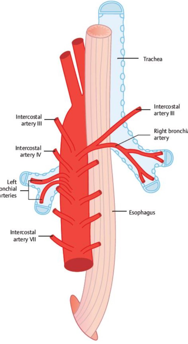
39
What is the blood supply of the left bronchi?
2 on the left come from the thoracic aorta
40
Pulmonary arteries to the terminal portion of the
Pulmonary arteries to the terminal portion of the bronchioles
41
Where does the right bronchial vein drain?
Azygus v.
42
Where does the left bronchial vein drain?
Hemi-azygus v.
43
Aspirated foreign bodies will enter the (because it is shorter, wider, and more vertical) -> will go to lower base of
Aspirated foreign bodies will enter the right main bronchus (because it is shorter, wider, and more vertical) -> will go to lower base of right inferior lobe
44
What is the costal margin?
Costal cartilage 7-10
45
Where is the angle of Luis?
T4
46
Where is the xiphoid process?
T10/11
47
Boundaries of the Superior Thoracic Aperature
Posteriorly – T1 vertebral body
Anteriorly – superior border of manubrium
Laterally – first rib and costal cartilages
Anteriorly – superior border of manubrium
Laterally – first rib and costal cartilages
48
Contents of the superior thoracic aperature?
• Trachea
• Esophagus
• Vagus (CN X) and Phrenic (C3-C5) nerves
• Apex of Lungs (cupula)
• Esophagus
• Vagus (CN X) and Phrenic (C3-C5) nerves
• Apex of Lungs (cupula)
49
The superior thoracic aperature (inlet) is also called the?
Clinical outlet
50
Thoracic outlet syndrome: caused by pancoast tumor, extra cervical rib, or compression of —> compression of
Thoracic outlet syndrome: caused by pancoast tumor, extra cervical rib, or compression of scalene muscles—> compression of brachial plexus
51
Boundaires of the inferior thoracic aperature (outlet)
-Posteriorly – T12 vertebral body
-Posterolaterally – 12th ribs
-Anterolaterally – costal margin (costal cartilages 7-10)
-Anteriorly – Xiphoid Process
-Posterolaterally – 12th ribs
-Anterolaterally – costal margin (costal cartilages 7-10)
-Anteriorly – Xiphoid Process
52
Contents of inferior throacic aperature (outlet)?
1. Caval opening (T8)
2. Esophogeal opening (T10)
3. Aortic opening (T12)
2. Esophogeal opening (T10)
3. Aortic opening (T12)
53
- only “true hole” because its going through the
Caval Opening (T8)- only “true hole” because its going through the central tendon.
54
Contents of Caval Opening
IVC
Phrenic nerves (C3-5)
Lymphatic vessels
Phrenic nerves (C3-5)
Lymphatic vessels
55
Contents of esophogeal opening
esophagus
Right and left vagus nerves (CN 10)
Esophageal branches of the left gastric artery and vein
Right and left vagus nerves (CN 10)
Esophageal branches of the left gastric artery and vein
56
Contents of aortic opening?
Thoracic aorta
Thoracic duct
Great splanchnic nerves (T5-9)
Azygous vein
Thoracic duct
Great splanchnic nerves (T5-9)
Azygous vein
57
External Intercostal muscles- ( and )
for
for
External Intercostal muscles- (down and forward)
for INSPIRATION
for INSPIRATION
58
Internal Intercostal Muscles- fibers running and
For
For
Internal Intercostal Muscles- fibers running down and backwards
For EXPIRATION
For EXPIRATION
59
Innermost intercostal muscles- fibers run and -
for
for
Innermost intercostal muscles- fibers run down and backwards-
for EXPIRATION
for EXPIRATION
60
What are the 3 branches of the innermost muscles?
1. Innermost proper- between ribs
2. Subcostalis- angle
3. Sternocostalis transversus thoracis- near the sternum
2. Subcostalis- angle
3. Sternocostalis transversus thoracis- near the sternum
61
Inferior to every rib, there’s a costal groove that has the neurovascular nerve bundles (VAN) [ → ]
Inferior to every rib, there’s a costal groove that has the neurovascular nerve bundles (VAN) [superior→ inferior]
62
Rib 1 doesn’t have a but has groove for
Rib 1 doesn’t have a costal groove but has groove for subclavian v and a
63
VAN travels between the and muscles
VAN travels between the internal and innermost intercostal muscles
64
Thorocentesis- make sure to go at most , or )When adminsitering nerve block, you to hit the nerve.
Thorocentesis- make sure to go at most inferior part of intercostal space, or superior to the inferior rib )When adminsitering nerve block, you WANT to hit the nerve.
65
Layers of Penetration for Mid-Axillary
Skin
Superficial fascia
Deep fascia
Serratus anterior muscle
External intercostal m.
Internal intercostal m
Innermost intercostal m
Innermost
Subcostalis
Sternocostalis transversus thoracic
Endothoracic fascia *
Parietal pleura
Pleural cavity
Visceral pleura
Lungs
Superficial fascia
Deep fascia
Serratus anterior muscle
External intercostal m.
Internal intercostal m
Innermost intercostal m
Innermost
Subcostalis
Sternocostalis transversus thoracic
Endothoracic fascia *
Parietal pleura
Pleural cavity
Visceral pleura
Lungs
66
In a thoracentensis, you would want to enter mid-axillary (up to )
In a thoracentensis, you would want to enter mid-axillary (up to ICS 9)
67
- CT layer that prevents the parietal pleura from rubbing against the innermost intercostal m. It is thickened over the apex of the lung at the cupula =
Endothoracic fascia- CT layer that prevents the parietal pleura from rubbing against the innermost intercostal m. It is thickened over the apex of the lung at the cupula = Sibsons fascia.
68
Trachea deviates to (when there is empty space):
Atelectasis
Pneumonectomy - removing a lung
Large pleural fibrosis
Pulmonary agenesis -absense of lung
Atelectasis
Pneumonectomy - removing a lung
Large pleural fibrosis
Pulmonary agenesis -absense of lung
Trachea deviates to affected side (when there is empty space):
Atelectasis
Pneumonectomy - removing a lung
Large pleural fibrosis
Pulmonary agenesis -absense of lung
Atelectasis
Pneumonectomy - removing a lung
Large pleural fibrosis
Pulmonary agenesis -absense of lung
69
Trachea deviates to the (when space is being filled and pressure increases):
Tension pneumothorax
Pleural effusions- water buildup around lung
Hemothorax
Large mass
Tension pneumothorax
Pleural effusions- water buildup around lung
Hemothorax
Large mass
Trachea deviates to the opposite side (when space is being filled and pressure increases):
Tension pneumothorax
Pleural effusions- water buildup around lung
Hemothorax
Large mass
Tension pneumothorax
Pleural effusions- water buildup around lung
Hemothorax
Large mass
70
Pneumothorax- air in the pleural space causing the lung to collapse around the hilus
- may push towards the
(ex: can result after stab wound)- air escapes when breathing [loss of negative pressure] and can cause lung collapse
- may push towards the
(ex: can result after stab wound)- air escapes when breathing [loss of negative pressure] and can cause lung collapse
Pneumothorax- air in the pleural space causing the lung to collapse around the hilus
- may push mediastinal contents (ex: heart and great vessels) towards the other lung
(ex: can result after stab wound)- air escapes when breathing [loss of negative pressure] and can cause lung collapse
- may push mediastinal contents (ex: heart and great vessels) towards the other lung
(ex: can result after stab wound)- air escapes when breathing [loss of negative pressure] and can cause lung collapse
71
Phrenic- and to hilum
Phrenic- lateral and anterior to hilum 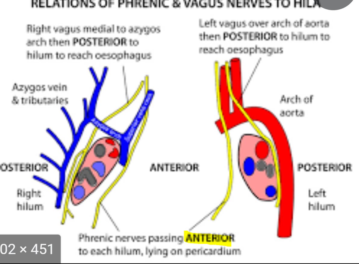
72
Vagus- to hilum
Vagus- posterior to hilum 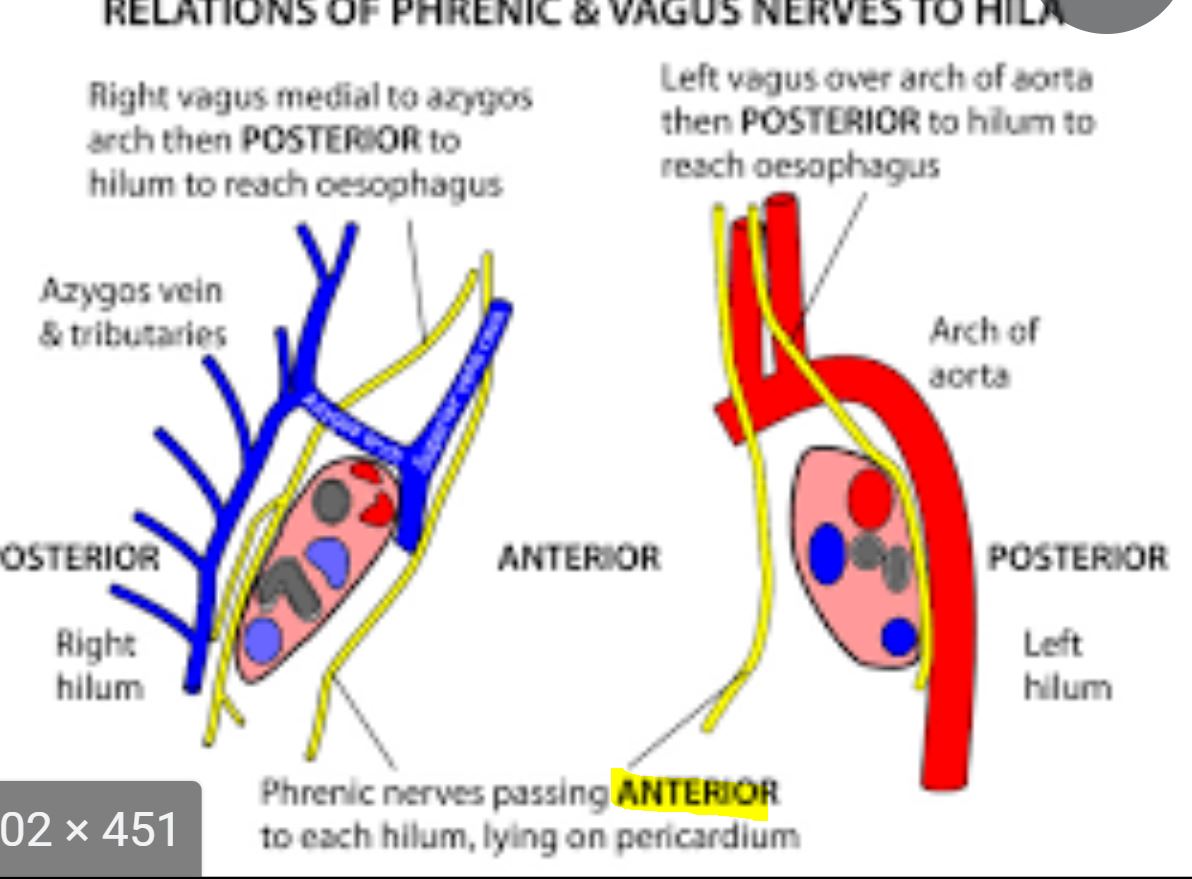
73
Phrenic: runs to root/ hilum of the lungs. Located between and
Phrenic: runs anterior to root/ hilum of the lungs. Located between parietal pleura and pericardium
74
Where is the right phrenic nerve?
Right (lateral) of SVC and RA
75
Where is the left phrenic nerve?
Travels over the arch of the aorta then POSTERIOR to the hilum to reach the esophagus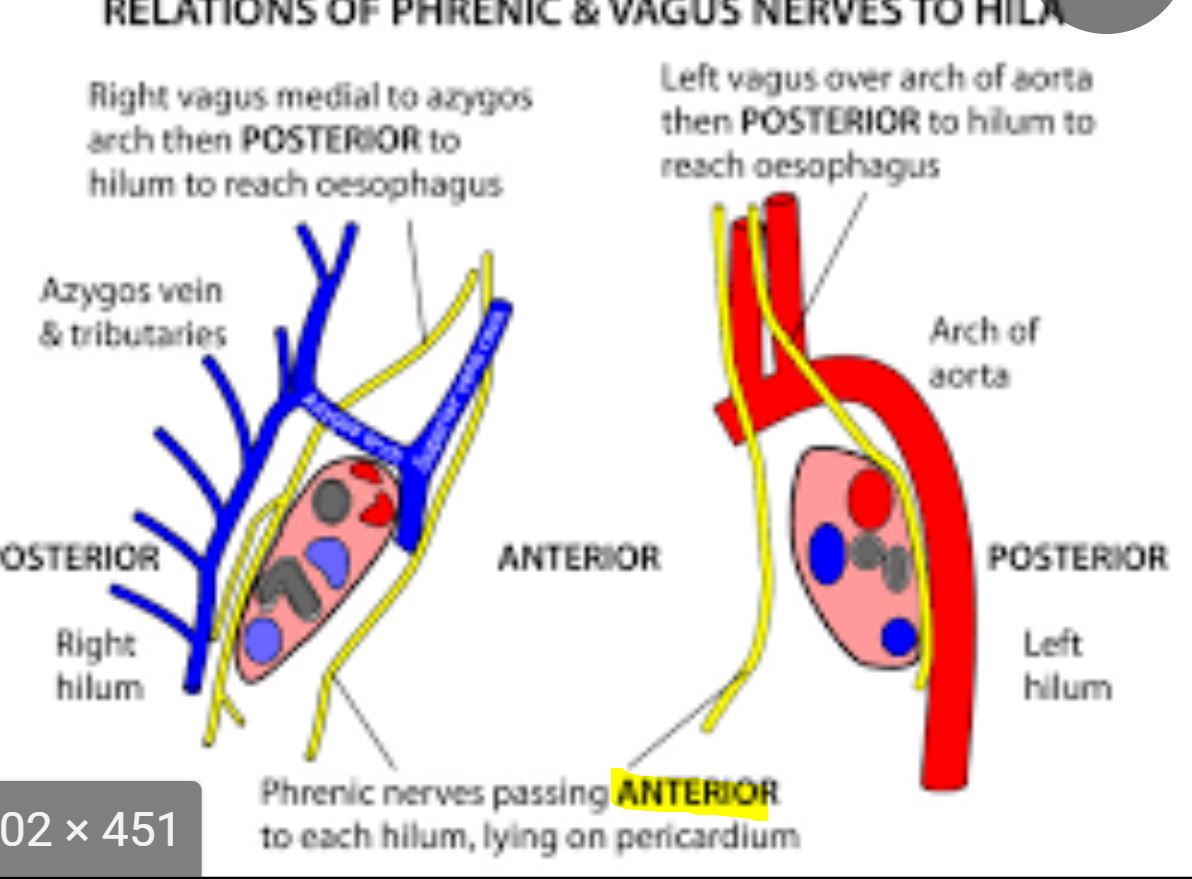
76
Right Vagus Relationships
Posterior to root of right lung
Posterolateral to brachiocephalic trunk
Medial to right phrenic
Lateral to trachea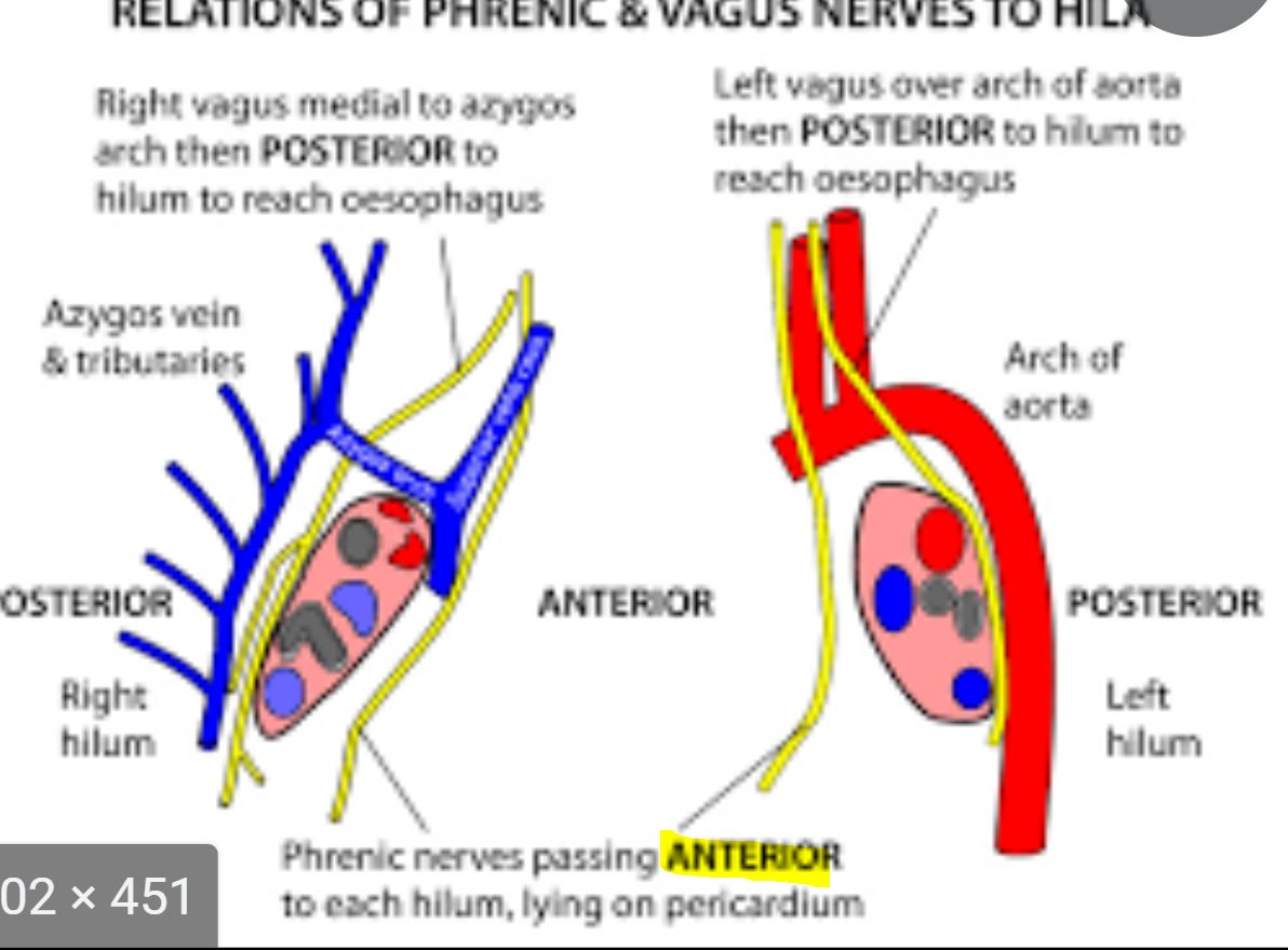
Posterolateral to brachiocephalic trunk
Medial to right phrenic
Lateral to trachea
77
Left Vagus relationships
Between the LCC and left subclavian
Crosses the left side of the aortic arch
Posterior to the root of the lung
Crossed by the left phrenic below the aortic arch || notes:-Stuck in the middle (LCC and left subclav)
-Cross the arch (aortic arch)
-Gets behind the root (of the left lung)
-Crossed by phrenic below (aorta)
Crosses the left side of the aortic arch
Posterior to the root of the lung
Crossed by the left phrenic below the aortic arch || notes:-Stuck in the middle (LCC and left subclav)
-Cross the arch (aortic arch)
-Gets behind the root (of the left lung)
-Crossed by phrenic below (aorta)
78
Mass extending anteriorly will occlude the , mass extending posteriorly will occlude the
Mass extending anteriorly will occlude the phrenic nerve, mass extending posteriorly will occlude the vagus nerve
79
What are the two approaches to pericardiocentesis?
1. Entering below the xyphoid process
2. Entering in the ICS at the left infrasternal angle
2. Entering in the ICS at the left infrasternal angle
80
If you enter below the xyphoid process for a pericardiocentesis, which layers do you enter?
Skin -> subcutaneous tissue- > rectus abdominus m. -> rectus sheet -> rectus muscle (now in pericardium-see pericarial layers)
81
If entering in the ICS- at the left infrasternal angle through the cardiac notch of the left lung at sternal margin 4,5,6, what vessel would you be concerned about injuring? What are the layers to penetrate?
-Internal thoracic vessels
-Skin -> subcutaneous tissue -> external intercostal membrane -> costochonral joint -> intercostal muscles (now in pericardium - see pericardial layers)
-Skin -> subcutaneous tissue -> external intercostal membrane -> costochonral joint -> intercostal muscles (now in pericardium - see pericardial layers)
82
Layers of penetration for pericardium?
Fibrous pericardium -> SEROUS- parietal layer -> pericardial cavity -> visceral layer (AKA epicardium) - - -> HEART (epicardium -> myocardium -> endocardium -> chambers of heart)
83
Left recurrent laryngeal nerve , distal/lateral to the (at the angle of Louis where it may be damaged by pathology (aneurism of aorta, malignancy…) resulting in
Left recurrent laryngeal nerve hooks under the arch of the aorta, distal/lateral to the ligamentum arteriosum (at the angle of Louis where it may be damaged by pathology (aneurism of aorta, malignancy…) resulting in paralysis of the left larynx and vocal cord
84
The right recurrent laryngeal nerve be affected because it arises from the in the root of the neck and it hooks under the
The right recurrent laryngeal nerve will not be affected because it arises from the right vagus nerve in the root of the neck and it hooks under the right subclavian artery.
85
Either the right or left recurrent laryngeal nerves may be damaged during
Either the right or left recurrent laryngeal nerves may be damaged during thyroid gland surgery
86
The mediastinum contains all of the structures of the thorax EXCEPT for the , , and
The mediastinum contains all of the structures of the thorax EXCEPT for the lungs, pleural membrane, and pleural cavity
87
Structures of Mediastinum
Sternum→ Ascending aorta→ SVC→ arch of aorta→ pulmonary artery and bronchi (underneath the arch of the aorta , between ascending and descending aorta) → descending aorta→ trachea → esophagus → vertebrae
88
Anterior Arteries of Thoracic Wall
ICS 1-6 – anterior intercostal arteries from internal
thoracic artery
ICS 7-9 – anterior intercostal arteries from
musculophrenic artery (branch of internal thoracic)
ICS 10-11 – no anterior intercostal arteries
thoracic artery
ICS 7-9 – anterior intercostal arteries from
musculophrenic artery (branch of internal thoracic)
ICS 10-11 – no anterior intercostal arteries
89
ant thor wall =
ant thor wall = internal thoracic a.
90
Posterior Arteries of Thoracic Wall
ICS 1 – highest intercostal artery (AKA 1st posterior intercostal artery)
from costocervical trunk
ICS 2-11 – posterior intercostal arteries from thoracic aorta
ICS 12 – subcostal artery from thoracic aorta
from costocervical trunk
ICS 2-11 – posterior intercostal arteries from thoracic aorta
ICS 12 – subcostal artery from thoracic aorta
91
post thor wall = EXCEPT
post thor wall = thoracic aorta EXCEPT ICS 1
-> costocervical trunk
-> costocervical trunk
92
Lungs Arteries: ,
Lungs Arteries: bronchial arteries (two on left, one on right), pulmonary arteries
93
Trachea: , , ,
Trachea: superior thyroid a, inferior thyroid a, bronchial a, internal thoracic a
94
Bronchi: ,
Bronchi: bronchial arteries, pulmonary arteries
95
Esophogous: , , , ,
Esophogous: inferior thyroid a, bronchial a, esophogeal a, phrenic a, left gastric-> esophageal
96
Pericardium: ,
Pericardium: pericardiophrenic, musculophrenic
97
Heart: and
Heart: right coronary and left coronary a
98
The anterior supply of thorax is drained the by the (into the )
The anterior supply of thorax is drained the by the Internal Thoracic Vein (into the Brachiocephalic vein)
99
The posterior supply of the thorax is drained by the
The posterior supply of the thorax is drained by the azygos system
100
Azygos veins drain deoxygenated blood from posterior walls of the thorax and abdomen into the
Azygos veins drain deoxygenated blood from posterior walls of the thorax and abdomen into the SVC
101
ICS 1 - highest intercostal vein → brachiocephalic vein
ICS 2-3 - highER intercostal vein → azygos
ICS 4-11 - azygos
ICS 12 - (subcostal vein) → azygos
Posterior right side Azygous
ICS 1 - highest intercostal vein → brachiocephalic vein
ICS 2-3 - highER intercostal vein → azygos
ICS 4-11 - azygos
ICS 12 - (subcostal vein) → azygos
102
Posterior left side azygous
ICS 1 - highest → brachiocephalic vein
ICS 2-4 - accessory hemiazygos → azygos vein
ICS 5-12 - hemiazygos vein → azygos vein
ICS 2-4 - accessory hemiazygos → azygos vein
ICS 5-12 - hemiazygos vein → azygos vein
103
What are the 6 structures of the right atrium?
Fossa ovalis
Pectinate muscle (and smooth edge- crista terminalis)
Opening of tricuspid valve
Opening of coronary sinus- Thebesian valve
Opening of SVC/IVC
Opening of anterior cardiac veins || notes:For Pecs, try corn serve veins
Pectinate muscle (and smooth edge- crista terminalis)
Opening of tricuspid valve
Opening of coronary sinus- Thebesian valve
Opening of SVC/IVC
Opening of anterior cardiac veins || notes:For Pecs, try corn serve veins
104
What are the 5 structures of the right ventricle?
Tricuspid valve
Pulmonary trunk
Papillary muscle
Moderator band/septomarginal trabecula from the anterior papillary muscle
Chordae tendinae || notes:Try Pulling Paps Moderate Cancer
Pulmonary trunk
Papillary muscle
Moderator band/septomarginal trabecula from the anterior papillary muscle
Chordae tendinae || notes:Try Pulling Paps Moderate Cancer
105
What are the 3 structures of the left atrium?
Opening of pulmonary veins
Opening of bicuspid valve
Auricles (Right and left and Have pectinate muscles)
Opening of bicuspid valve
Auricles (Right and left and Have pectinate muscles)
106
Bicuspid valve
Opening of ascending aorta
Chordae tendinae
Papillary muscles || notes:Buy AA cancer Paps
What are the four structures of the left ventricle?
Bicuspid valve
Opening of ascending aorta
Chordae tendinae
Papillary muscles || notes:Buy AA cancer Paps
107
In the ventricles the trabeculae carnae is all the structures except the , , and
In the ventricles the trabeculae carnae is all the structures except the cusps, chordae tendinae, and septomarginal trabecula
108
The pectinate muscle in found in the , and and only
The pectinate muscle in found in the RA, and right and left auricle only
109
What are the 3 functions of the chordae tendinae?
1. Prevent back flow of blood into atrium
2. Prevent eversion of valves during ventricular contraction
3. Attach papillary muscles to cusps of AV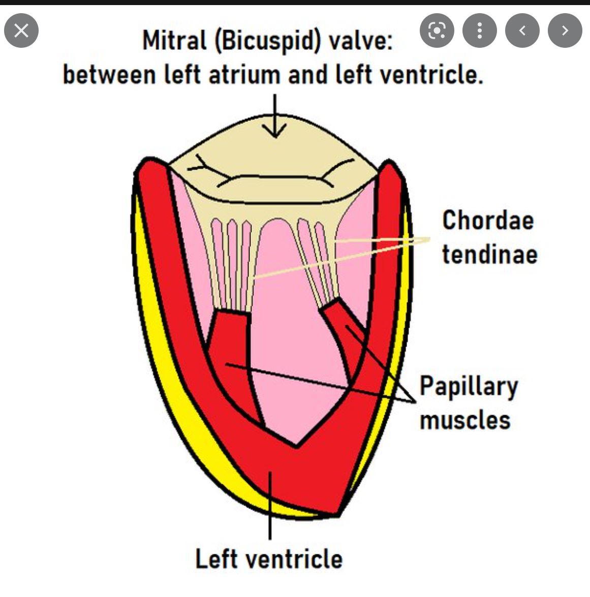
2. Prevent eversion of valves during ventricular contraction
3. Attach papillary muscles to cusps of AV
110
What are the boundaries of the mediastinum?
Superior: thoracic inlet and root of the neck
Inferior: diaphragm
Anterior: sternum
Posterior: vertebral column
Inferior: diaphragm
Anterior: sternum
Posterior: vertebral column
111
Entire mediastinum is (superior , inferior )
Entire mediastinum is T1-T12 (superior T1-4, inferior T5-12)
112
The superior mediastinum is located posterior to the and anterior to the first
The superior mediastinum is located posterior to the manubrium and anterior to the first 4 thoracic vertebrae
113
Contents of the Superior Mediastinum
Phrenic Nerves
Vagus Nerves
Thoracic Duct
Left Recurrent Laryngeal (not right)
Brachiocephalic Vein (via SVC)
Aortic Arch (and branches)
Thymus
Trachea
Lymph Node
Esophagus
Vagus Nerves
Thoracic Duct
Left Recurrent Laryngeal (not right)
Brachiocephalic Vein (via SVC)
Aortic Arch (and branches)
Thymus
Trachea
Lymph Node
Esophagus
114
Brachiocephalic veins= +
Brachiocephalic veins= subclavian+ internal jugular
115
SVC=
SVC= R+L brachiocephalic veins
116
Where is the inferior mediastenum located?
Below T4
117
What are the 3 components of the Inferior Mediastenum?
1. Anterior (posterior sternum to ant percardial sac)
2. Middle
3. Posterior (posterior pericardial sac and diaphram + T12)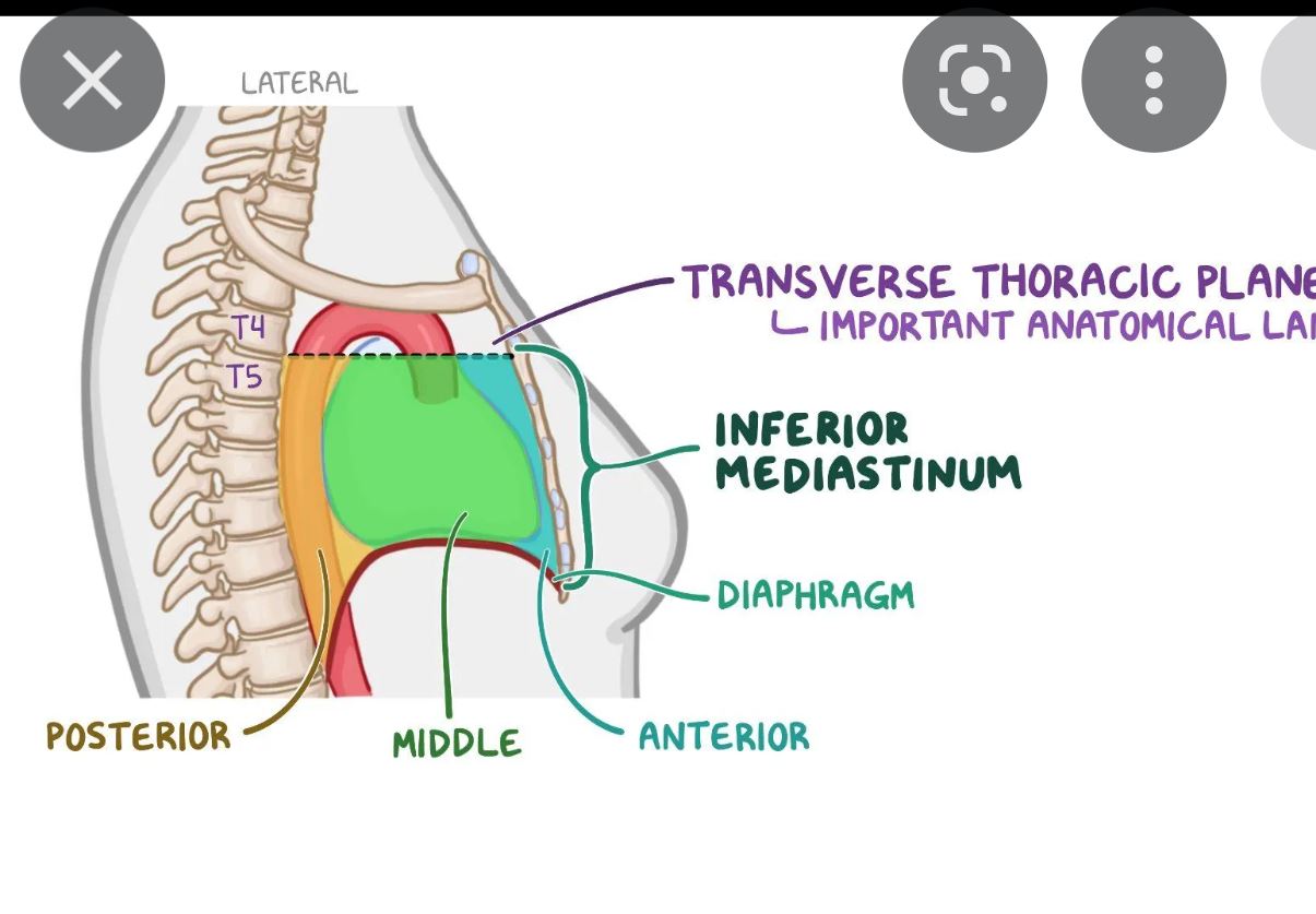
2. Middle
3. Posterior (posterior pericardial sac and diaphram + T12)
118
What are the 3 contents of the anterior inferior mediastenum?
1. Fat
2. Thymus
3. Lymph nodes
2. Thymus
3. Lymph nodes
119
What are the 5 contents of the middle inferior mediastenum?
1. Heart
2. Pericardiym
3. Main bronchi
4. Great vessels
5. Phrenic nerve
2. Pericardiym
3. Main bronchi
4. Great vessels
5. Phrenic nerve
120
What are the 6 contents of the posterior mediastenum?
1. Esophogous
2. Thoracic aorta
3. Thoracic ducts
4. Azygous vein
5. Vagus nerve
6. Splanchnic
2. Thoracic aorta
3. Thoracic ducts
4. Azygous vein
5. Vagus nerve
6. Splanchnic
121
What 6 structures enter the heart?
1. Pulmonary veins from the lungs to the LA
2. Usually an upper and lower vein
3. SVC enters the RA
4. IVC enters the RA
5. Anterior cardiac veins enter the RA
6. Coronary sinus enters the RA
2. Usually an upper and lower vein
3. SVC enters the RA
4. IVC enters the RA
5. Anterior cardiac veins enter the RA
6. Coronary sinus enters the RA
122
What two structures leave the heart?
1. Ascending aorta leaves LV
2. Pulmonary trunk and pulmonary arteries leave RV (Crosses arota and divides into right and left pulm arteries at angle of louis. Left travels over left bronchus. Right is posterior to asending aorta and SVC. Pulm trunk is connected by ligamentum arteriousis.)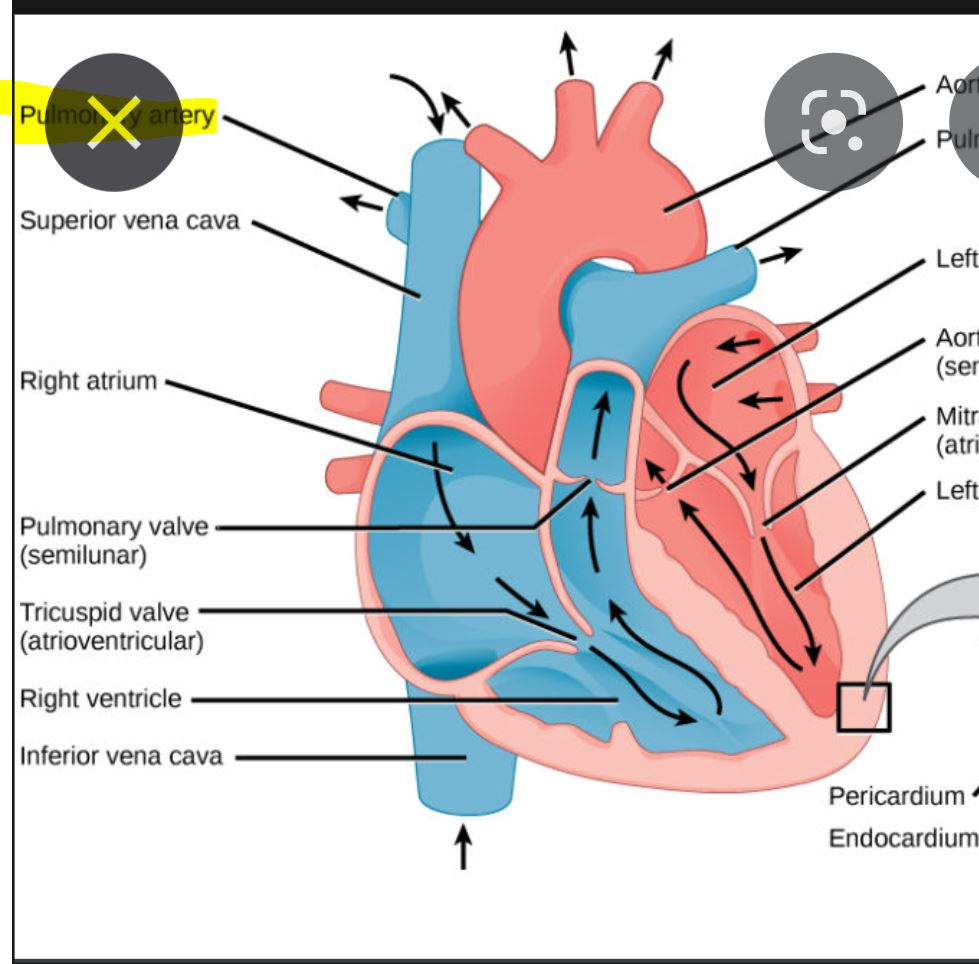
2. Pulmonary trunk and pulmonary arteries leave RV (Crosses arota and divides into right and left pulm arteries at angle of louis. Left travels over left bronchus. Right is posterior to asending aorta and SVC. Pulm trunk is connected by ligamentum arteriousis.)
123
All of the veins of the heart drain into the which drain into the ***Two exceptions
All of the veins of the heart drain into the coronary sinus which drain into the right atrium ***Two exceptions
124
What are the two venous drainage of the heart exceptions?
Anterior cardiac veins- drains directly into RA
Venae chordae minimae- mostly drains directly into RA
Venae chordae minimae- mostly drains directly into RA
125
Right lung- pulmonary artery is to primary bronchus
Right lung- pulmonary artery is anterior to primary bronchus 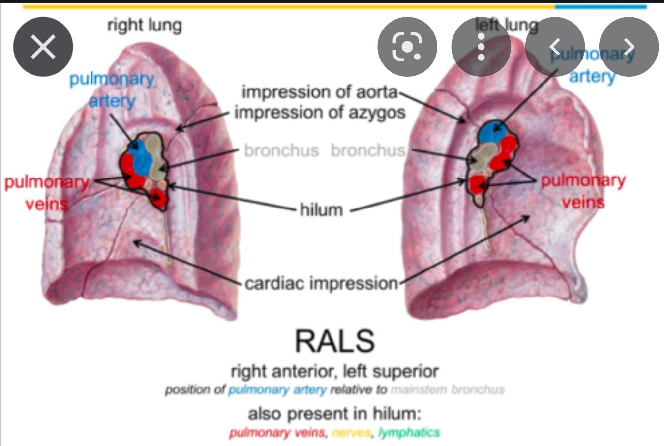
126
Left lung- pulmonary artery is to primary bronchus
Left lung- pulmonary artery is superior to primary bronchus
127
Bronchus
Wider, shorter, and more vertical
Before entering hilum of lung it gives off the
On entering the hilum, it divides into a and lobar bronchus
Wider, shorter, and more vertical
Before entering hilum of lung it gives off the
On entering the hilum, it divides into a and lobar bronchus
Right Bronchus
Wider, shorter, and more vertical
Before entering hilum of lung it gives off the superior lobar bronchus
On entering the hilum, it divides into a middle and inferior lobar bronchus
Wider, shorter, and more vertical
Before entering hilum of lung it gives off the superior lobar bronchus
On entering the hilum, it divides into a middle and inferior lobar bronchus
128
Bronchus
Narrower, longer, and more horizontal
It passes to the left and in (see pic below)
On entering the hilum of the left lung, it divides into a and bronchus
Narrower, longer, and more horizontal
It passes to the left and in (see pic below)
On entering the hilum of the left lung, it divides into a and bronchus
Left Bronchus
Narrower, longer, and more horizontal
It passes to the left below the arch of the aorta and in front of the esophogous (see pic below)
On entering the hilum of the left lung, it divides into a superior and inferior lobar bronchus
Narrower, longer, and more horizontal
It passes to the left below the arch of the aorta and in front of the esophogous (see pic below)
On entering the hilum of the left lung, it divides into a superior and inferior lobar bronchus
129
Primary bronchus-
Secondary bronchi-
Tertiary bronchi-
Secondary bronchi-
Tertiary bronchi-
Primary bronchus- right and left
Secondary bronchi- lobar
Tertiary bronchi- segmental
Secondary bronchi- lobar
Tertiary bronchi- segmental
130
What is the nerve supply to the diaphram?
Phrenic nerve (C3,4,5)
131
A is a pleural cavities that separate the visceral and parietal pleura.
A pleural recess is a pleural cavities that separate the visceral and parietal pleura.
132
Pleural recesses allow for the expansion of lung during
Pleural recesses allow for the expansion of lung during inspiration
133
Parietal pleura:
- and
arteries
- TO PAIN
- and
arteries
- TO PAIN
Parietal pleura:
- Internal thoracic artery and
intercostal arteries
- VERY SENSITIVE TO PAIN
- Internal thoracic artery and
intercostal arteries
- VERY SENSITIVE TO PAIN
134
Visceral pleura:
- arteries
- TO PAIN
- arteries
- TO PAIN
Visceral pleura:
- Bronchial arteries
- INSENSITIVE TO PAIN
- Bronchial arteries
- INSENSITIVE TO PAIN
135
The parietal pleura becomes continuous w/ visceral pleura at the of the lungs and forms a “cuff” that surrounds the structures entering/ leaving the lung at the root
The parietal pleura becomes continuous w/ visceral pleura at the HILUM of the lungs and forms a “cuff” that surrounds the structures entering/ leaving the lung at the root
136
at the junction of the costal and diagphragmatic parietal pleura, at the mid axillary line.
Costodiaphragmatic recess: at the junction of the costal and diagphragmatic parietal pleura, at the mid axillary line.
137
The is lowest area of the pleural cavity into which the lungs expand during inspiration
The costodiaphragmatic recess is lowest area of the pleural cavity into which the lungs expand during inspiration
138
Where does the costodiaphragmatic recess extend?
Anteriorly ICS 8, midaxillar ICS 9, and posteriorly ICS 10
139
What does blundting of the costophrenic angle in the costodiaphragmatic recesss indicate?
Fluid in lungs
140
: lie along the anterior margin of the pleura
Costomediastinal recess:: lie along the anterior margin of the pleura
141
What are the branches of the aortic arch?
1. Brachiocephalic trunk (branches into right subcalvian and common carotid)
2. Left subclavian artery
3. Left common carotid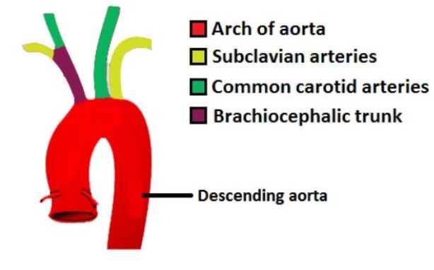
2. Left subclavian artery
3. Left common carotid
142
Atypical ribs-
Atypical ribs- 1,2,10, 11,12
143
Rib 7 is , rib 12 is
Rib 7 is longest, rib 12 is shortest
144
Rib dislocation- displacement of costal cartilage from sternum at (contact sports)
Rib dislocation- displacement of costal cartilage from sternum at ribs 2-7 (contact sports)
145
Rib separation- dislocation of the between the rib and its costal cartilage
Rib separation- dislocation of the Costochondral joint between the rib and its costal cartilage
146
The trachea Begins as the contiinuation of the larynx at the lower border of the
The trachea Begins as the contiinuation of the larynx at the lower border of the cricoid cartilage (C6)
147
The trachea descends into the midline of the neck anterior to the esophogous entering the superior mediastinum and deviating at the angle of Louis before it divides
The trachea descends into the midline of the neck anterior to the esophogous entering the superior mediastinum and deviating slightly to the right of the median plane at the angle of Louis before it divides
148
The trachea ends in the thorax by dividing into the right and left primary bronchi at the at the
The trachea ends in the thorax by dividing into the right and left primary bronchi at the sternal angle (T4-5) at the Carina
149
Angle of the carina is about degrees. If its greater than that -> may indicate the presence of a
Angle of the carina is about 90 degrees. If its greater than that -> may indicate the presence of a mediastinal mass
150
Posterior surface of trachea is flat and is covered by
Posterior surface of trachea is flat and is covered by trachialis muscle
151
What 5 structures are anterior to the trachea?
1. Sternum
2. Thymus
3. Arch of aorta
4. Brachiochephalic trunk origin
5. Left common corotid
2. Thymus
3. Arch of aorta
4. Brachiochephalic trunk origin
5. Left common corotid
152
What structure is posterior to the trachea?
Esophogous
153
What are the 4 blood supplies to the trachea?
1. Superior thyroid artery
2. Inferior thyroid artery
3. Bronchial artery
4. Internal thoracic artery
2. Inferior thyroid artery
3. Bronchial artery
4. Internal thoracic artery
154
What is the nerve supply of the trachea?
Vagus and sympathetic chain
155
Esophogous: it is a median structure (median plane) that lies posterior to and
Esophogous: it is a median structure (median plane) that lies posterior to trachea and left atrium
156
The is the the main posterior relationship of the base of the heart
The esophogus is the the main posterior relationship of the base of the heart
157
The esophogus deviates to the to the main bronchus and it enters the diaphragm at anterior to the aorta with the vagus and the esophageal branch of the left gastric artery and vein
The esophogus deviates to the left, posterior to the left main bronchus and it enters the diaphragm at T10 anterior to the aorta with the vagus and the esophageal branch of the left gastric artery and vein
158
What is the nerve supply of the trachea?
1. Vagus nerve
2. Sympathetic fibers
3. Pain fibers
2. Sympathetic fibers
3. Pain fibers
159
Pericardium- fibrous sac that encloses heart and vessels
Located in , posterior to the and , and anterior to
Located in , posterior to the and , and anterior to
Pericardium- fibrous sac that encloses heart and vessels
Located in middle mediastinum, posterior to the body of the sternum and 2-6 costal cartilages, and anterior to T5-8 vertebrae
Located in middle mediastinum, posterior to the body of the sternum and 2-6 costal cartilages, and anterior to T5-8 vertebrae
160
Transverse Sinus: located behind the and , and in front of the and
Important during bypass- you can stick your finger between great arteries and pulmonary trunk
Important during bypass- you can stick your finger between great arteries and pulmonary trunk
Transverse Sinus: located behind the aorta and pulmonary trunk, and in front of the left atrium and SVC
Important during bypass- you can stick your finger between great arteries and pulmonary trunk
Important during bypass- you can stick your finger between great arteries and pulmonary trunk
161
Oblique Sinus: between the and
Oblique Sinus: between the pulmonary veins and IVC
162
Nerve supply to Pericardium:
Fibrous pericardium is supplied by
- - located between the parietal pleura and fibrous pericardium)
-
- The visceral layer is supplied by the
Fibrous pericardium is supplied by
- - located between the parietal pleura and fibrous pericardium)
-
- The visceral layer is supplied by the
Nerve supply to Pericardium:
Fibrous pericardium is supplied by
-Phrenic n (C3-5)- located between the parietal pleura and fibrous pericardium)
-Vagus n
- The visceral layer is supplied by the coronary plexus
Fibrous pericardium is supplied by
-Phrenic n (C3-5)- located between the parietal pleura and fibrous pericardium)
-Vagus n
- The visceral layer is supplied by the coronary plexus
163
To decompress a cardiac temponade, do a pericariocentesis- place the needle at the through the of the L lung
To decompress a cardiac temponade, do a pericariocentesis- place the needle at the left infrasternal angle through the cardiac notch of the L lung
164
Heart:
-Base is and apex is
-It is related to sternum, the costal cartilages and the medial ends of the ribs on the left
-Posteriorly, it extends from
-Base is and apex is
-It is related to sternum, the costal cartilages and the medial ends of the ribs on the left
-Posteriorly, it extends from
Heart:
-Base is posterior and apex is inferolateral
-It is related to sternum, the costal cartilages and the medial ends of the ribs 3-5 on the left
-Posteriorly, it extends from T6-9
-Base is posterior and apex is inferolateral
-It is related to sternum, the costal cartilages and the medial ends of the ribs 3-5 on the left
-Posteriorly, it extends from T6-9
165
What are the borders of the heart?
Right border: RA
Left border: LV
Apex: tip of left ventricle, found in ICS5 about midclavicular line
Superior border: right and left auricles and the conus arteriosus (area from which pulmonary trunk arises) of the RV
Inferior border: diaphragm by the RV
Left border: LV
Apex: tip of left ventricle, found in ICS5 about midclavicular line
Superior border: right and left auricles and the conus arteriosus (area from which pulmonary trunk arises) of the RV
Inferior border: diaphragm by the RV
166
Blood flow through the heart
IVC/SVC -> RA -> tricuspid valve -> RV -> pulmonary valve ->pulmonary artery -> lungs -> pulmonary vein -> LA -> mitral valve -> left ventricle -> aortic valve -> aorta -> body
167
Innervation of the heart: cardiac plexus formed by branches of and
Innervation of the heart: cardiac plexus formed by branches of vagus and sympathetic trunks