nervous system
Study these flashcardsFree · spaced repetition · iPhone, Android, Mac & web
All cards
1
The Nervous system contains two classes of cells: and
The Nervous system contains two classes of cells: NEURONS and GLIA
2
Neurons are excitable cells that recieve, integrate stimuli, and conduct electrical impulses along processes, passing information through
Neurons are excitable cells that recieve, integrate stimuli, and conduct electrical impulses along processes, passing information through synapses
3
Neurons are differeniated
Neurons are terminally differeniated
4
support the neuron
Glial cells support the neuron
5
Glial cells , , and
Glial cells protect, myelinate, and modulate the environment
6
What are the 4 glia of the CNS
1. Oligodendrocytes
2. Astrocytes
3. Ependymal cells
4. Microglia
2. Astrocytes
3. Ependymal cells
4. Microglia
7
What are the two glia of the PNS?
Schwann and satellite cells
8
A collection of cell bodies plus axons is called what in the CNS? PNS?
In the CNS = Gray matter
In the PNS = Sensory, Spinal, Dorsal root Ganglia, Autonomic, Sympathetic, and Parasympathetic
In the PNS = Sensory, Spinal, Dorsal root Ganglia, Autonomic, Sympathetic, and Parasympathetic
9
The collection of axons with NO cell bodies is called what in the CNS? PNS?
In CNS = White Matter
In PNS = Spinal nerves and roots, peripheral nerves, most cranial nerves
In PNS = Spinal nerves and roots, peripheral nerves, most cranial nerves
10
Action potential travels from to
Action potential travels from dendrites to axon terminal
11
are short, tapering processes that may branch. They are an extension of the cell body.
Dendrites are short, tapering processes that may branch. They are an extension of the cell body.
12
is made up of the nucleus plus the cytoplasm
Cell body is made up of the nucleus plus the cytoplasm
13
What is found in the cytoplasm of the cell body?
-Protein-maker organelles Golgi apparatus, ribosomes RER (Nissl), and Mitochondria
14
What are the cytosketeal components of the cell body?
-Microtubules, microfilaments, neurofilaments, synpatic vessels, lysosomes
15
The nucleus of a neuron is a large nucleus with a
The nucleus of a neuron is a large euchromatic nucleus with a prominant nucleolus
16
is an elevated region of the cell body beginning at the axon.
Axon hillox is an elevated region of the cell body beginning at the axon.
17
The axon hillox is distinct because it is where the cell
The axon hillox is distinct because it is where the cell stop making proteins
18
The is a nerve fiber
The axon is a nerve fiber
19
The axon is myelinated by
The axon is myelinated by glia
20
What does the axon not contain?
Organelles to make new proteins (no Golgi, RER, ribosomes)
21
WHat does the axon contain?
Mitochondria, cytokeletal components, and synpatic vesicles
22
The Nervous system contains two classes of cells: and
The Nervous system contains two classes of cells: NEURONS and GLIA
23
When does exocytosis of the axon terminal occur?
On arrival of nerve impulse
24
What part of the neuron is considered the metabolic/tropic center?
The cell body
25
What is axoplasmic transport?
Bidirectional flow guided by microtubules
26
What is anterograde transport? Is it slow or fast?
-Cell body to terminal
-Brings newly synthesized materials down the axon
-Can be slow or fast
-Brings newly synthesized materials down the axon
-Can be slow or fast
27
What is retrograde transport? Is it slow or fast?
-From terminal to cell body
-Only slow
-Only slow
28
The Nervous system contains two classes of cells: and
The Nervous system contains two classes of cells: NEURONS and GLIA
29
Which part of the neuron is presonsible for recieving and integrating stimuli?
Cell body/Soma/Perikaryon
30
What does the cell body NOT contain?
Glycogen
31
Describe the nucelus of the cell body?
Large and lightly stained with distict nucleolus
32
What can be seen in the cytoplasm of the cell body histologically?
Lipfuschin pigment, basophillic clumps called Nissl
33
Cell bodies are present in the ______ of the PNS?
Ganglia
34
Cell bodies are present in the _______ of the CNS?
Gray matter
35
What part of the neuron is responsible for integrating stimuli and increasing surface area?
The dendrites
36
What do dendrites look like histologically?
Usually not visible, except close to cell body where they are near Nissl bodies
37
Axons in the PNS are surrounded or myelinated by ______?
Schwann cells
38
Axons in the CNS are myelinated by _________?
Oligodendrocytes
39
What histological feasture do axons lack?
Nissl
40
What do axons look like histiologically?
-Hard to identify but easier if sheath
41
What forms nerves in the PNS?
Bundles of axons with Schwann cells and connective tissue form nerves
42
– collection of Cell bodies (and attached axons)
GANGLIA – collection of Cell bodies (and attached axons)
43
What is a multipolar neruon?
Many dendrites and single axon coming from cell body 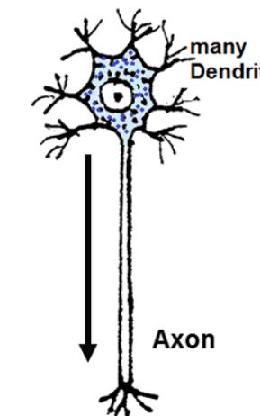
44
What is the function of a mulitpolar neruon?
-Most common
-Almost all CNS
-All motor neruons and most interneruons
-Almost all CNS
-All motor neruons and most interneruons
45
What are 2 examples of mulitpolar neurons?
-Lower motor neurons to skeletal muscle
-Autonomic nerurons: sympathetic and parasympathetic
-Autonomic nerurons: sympathetic and parasympathetic
46
What is a bipolar nueron?
A signle dendrite and single axon from opposite poles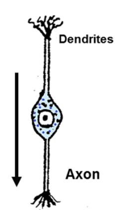
47
What is the function of bipolar neruons?
-Receptor neurons for special sensory only
48
What are 3 examples of functions of bipolar neuronss?
1. vision
2. hearing and balance
3. smell
2. hearing and balance
3. smell
49
What is a pseudounipolar neuron?
A single process from a cell body that divides a short distance between two axons. Only one top is near periphery 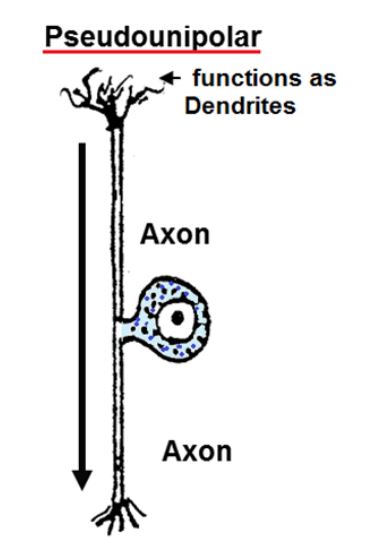
50
What is the function of the pseudounipolar neuron?
-General sensory (touch, pain, vibration)
51
What is an example of where pseudounipolar neruons are found?
Found in dorsal root ganglia and some cranial ganglia
52
Sensory neurons – carry impulses from receptors TO the
Sensory neurons – carry impulses from receptors TO the CNS
53
Motor neurons – carry impulses from CNS or autonomic ganglia to
Motor neurons – carry impulses from CNS or autonomic ganglia to their target or
effector cells
effector cells
54
Skeletal muscle, cardiac
muscle (Purkinje fibers also), smooth muscle and glands are all examples of
muscle (Purkinje fibers also), smooth muscle and glands are all examples of
Skeletal muscle, cardiac
muscle (Purkinje fibers also), smooth muscle and glands are all examples of effector cells
muscle (Purkinje fibers also), smooth muscle and glands are all examples of effector cells
55
Interneurons - interconnect other neurons, completely , 99.9% of all
neuron
neuron
Interneurons - interconnect other neurons, completely within CNS, 99.9% of all
neuron
neuron
56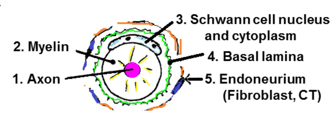
Image occlusion: name the hidden labels
57
- collection of axons (no cell bodies)
NERVES - collection of axons (no cell bodies)
58
Schwann cells surround what?
All axons of peripheral nerves
59
Satellite cells surround what?
All cell bodies in the PNS
60
Rember SAME DAVE
Senesory is afferent, motor is efferent
61
The autonomic nervous system is also known as the
Visceromotor system
62
The dorsal roots ONLY transmit incoming while
the ventral roots ONLY transmit outgoing .
the ventral roots ONLY transmit outgoing .
The dorsal roots ONLY transmit incoming sensory impulses while
the ventral roots ONLY transmit outgoing motor impulses.
the ventral roots ONLY transmit outgoing motor impulses.
63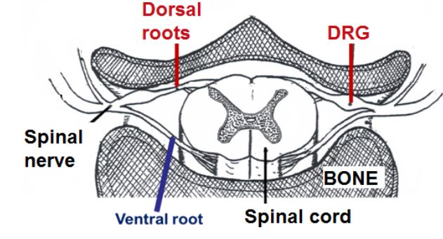
Image occlusion: name the hidden labels
64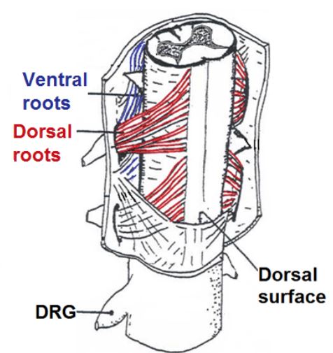
Image occlusion: name the hidden labels
65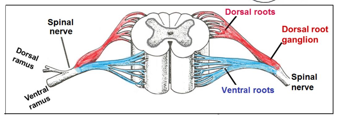
Image occlusion: name the hidden labels
66
The is a specialized synapse between a lower motor neuron (LMN) and a skeletal muscle fiber.
The motor-end plate is a specialized synapse between a lower motor neuron (LMN) and a skeletal muscle fiber.
67
What does the motor end plate look like histologically?
It is only visible with special stains
68
Stimuli, like touch, are converted into electrical impulses at the of sensory
neurons.
neurons.
Stimuli, like touch, are converted into electrical impulses at the very distal tips of sensory
neurons.
neurons.
69
, , and information
excites unspecialized “naked” nerve endings while more discriminative senses excite
nerve endings surrounded or encapsulated by fancy connective tissue structures.
excites unspecialized “naked” nerve endings while more discriminative senses excite
nerve endings surrounded or encapsulated by fancy connective tissue structures.
Touch, pain, and temperature information
excites unspecialized “naked” nerve endings while more discriminative senses excite
nerve endings surrounded or encapsulated by fancy connective tissue structures.
excites unspecialized “naked” nerve endings while more discriminative senses excite
nerve endings surrounded or encapsulated by fancy connective tissue structures.
70
Which encapsulated receptor is a strech receptor?
Muscle spindle
71
Which encapsulated receptor is a touch receptor?
Meissner's touch corpuscle
72
Which encapsulated receptor deals with vibration?
Pacinian corpuscle
73
Meissner's touch corpusle has what shape?
Peanut
74
Pacinian corpuscle has what shape?
Onion
75
Muscle spindles have what shape?
Spindle shapped
76
All ganglia contain what 2 things
1. cell bodies of neurons attached to axons
2. glial cells
2. glial cells
77
All axons (fibers) in the PNS are surrounded by which provide structural and
metabolic support.
metabolic support.
All axons (fibers) in the PNS are surrounded by Schwann cells which provide structural and
metabolic support.
metabolic support.
78
What are the two ways that a schwann cell surrounds an axon?
1. Schwann surrounds many axons within the clefts of its cytoplasm (non-myelinated)
2. Schwann cell wraps itself around an axon into a myelin sheath
2. Schwann cell wraps itself around an axon into a myelin sheath
79
At the junction between adjacent Schwann cells
is a myelin free region called the
is a myelin free region called the
At the junction between adjacent Schwann cells
is a myelin free region called the Node of Ranvier.
is a myelin free region called the Node of Ranvier.
80
The myelin sheath electrically insulates
the axon and the speed of nerve impulse conduction.
the axon and the speed of nerve impulse conduction.
The myelin sheath electrically insulates
the axon and increases the speed of nerve impulse conduction.
the axon and increases the speed of nerve impulse conduction.
81
In addition to producing
myelin, Schwann cells secrete a
myelin, Schwann cells secrete a
In addition to producing
myelin, Schwann cells secrete a basal lamina.
myelin, Schwann cells secrete a basal lamina.
82
What are the 3 things that around each axon in the PNS?
1. Schwann cells (+ or = meyelin)
2. Basal lamina
3. CT wrapping (endoneurium)
2. Basal lamina
3. CT wrapping (endoneurium)
83
In an H&E preparation, the myelin, composed mostly of lipid, looks like
In an H&E preparation, the myelin, composed mostly of lipid, looks like empty space
84
Image occlusion: name the hidden labels
85
Peripheral nerves contain a mixture of ....
1. Motor (efferent)
2. Sensory (afferent)
2. Sensory (afferent)
86
What are the three layers of connective tissue wrapping of a peripheral nerve?
1. Endoneurium - around one axon
2. Perinerium - around bundle of axons (most substantial)
3. Epineurium - around several bundles
2. Perinerium - around bundle of axons (most substantial)
3. Epineurium - around several bundles
87
Our slides for this lab do not contain
Our slides for this lab do not contain epinerium
88
The blood/nerve barrier is formed by tight junctions betwen specialized fibroblasts forming and tight junctions between
The blood/nerve barrier is formed by tight junctions betwen specialized fibroblasts forming perineurium and tight junctions between endothelial cells of capillaries
89
This regeneration or regrowth of the
severed axons is guided by the , their basal lamina, and the endoneurium.
severed axons is guided by the , their basal lamina, and the endoneurium.
This regeneration or regrowth of the
severed axons is guided by the Schwann cells, their basal lamina, and the endoneurium.
severed axons is guided by the Schwann cells, their basal lamina, and the endoneurium.
90
Since ALL peripheral nerves are MIXED, carrying motor
and sensory information then if a nerve is severed, but MAY return if the
axons successfully regrow and find their targets.
and sensory information then if a nerve is severed, but MAY return if the
axons successfully regrow and find their targets.
Since ALL peripheral nerves are MIXED, carrying motor
and sensory information then if a nerve is severed, ALL functions are lost but MAY return if the
axons successfully regrow and find their targets.
and sensory information then if a nerve is severed, ALL functions are lost but MAY return if the
axons successfully regrow and find their targets.
91
The ANS is a functional subdivision of the nervous system, including portions of the and
.
.
The ANS is a functional subdivision of the nervous system, including portions of the CNS and
PNS.
PNS.
92
How do dorsal root ganglia and preganglionic roots appear histologically?
Ganglia are big and will be the only thing on the slide. It is very packed with Different sized cells . 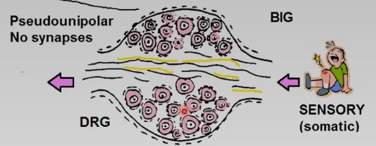
93
How do sympathetic ganglion appear histologically?
Take up slide and loosely packed and evenly dispersed with SAME sized cells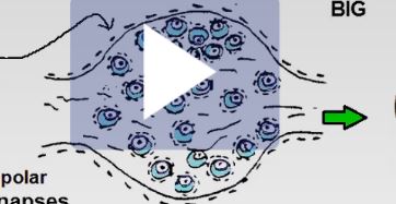
94
How do parasympathetic ganglion appear?
Tiny cells, seen inside of others = LITTLE TITTIES 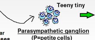
95
Autonomic neuron (sympathetic and parasympathetic) -> ->
Autonomic neuron (sympathetic and parasympathetic) -> autonomic ganglion -> Cardiac, glands, smooth muscle
96
Grey matter cell bodies
Grey matter CONTAINS cell bodies
97
White matter cell bodies
White matter DOESN'T CONTAIN cell bodies
98
On fresh dissection, how does white matter appear?
White due to large amount of myelinated lipid fibers
99
Sympathetic and parasympathetic do not SHARE
Sympathetic and parasympathetic do not SHARE CELL STRUCTURES
100
What is an onligodendrocyte?
-Myelinates in CNS
-Lipid from myelin is responsible for "white" matter
-Lipid from myelin is responsible for "white" matter
101
What is an astrocyte?
-Cell processes that are associates with nerouns and capillaries
-Part of blood brain barrier
-Modulate ionic environment of neurons
-Has intemediate filaments
-Part of blood brain barrier
-Modulate ionic environment of neurons
-Has intemediate filaments
102
What are ependymal cells?
-Cilated cells lining the ventricles of the brain and central canal of spine
-Modidied eoendymal cells secrete CSF into ventricles
-Modidied eoendymal cells secrete CSF into ventricles
103
What are microglia ?
-Derived from monocytes (not true glia)
-Active in phagocytosis, antigen sensing, and wound healing
-Active in phagocytosis, antigen sensing, and wound healing
104
What is mulitple sclerosis? What type of glial cell is impacted?
-Inflammation and demyelination of actions, Oligodendrocytes
105
Which of the glial cells of the CNS proliferates a glial scar in response to CNS damage that inhibits regeneration?
Astrocytes
106
Which of the glial cells is related to gliobastoma (a highly maligant neoplasm)?
Astrocytes
107
Grey matter appears as a of the spinal cord
Grey matter appears as a butterfly in the center of the spinal cord 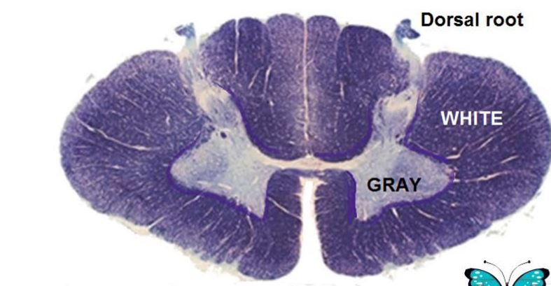
108
White matter the grey matter
White matter surrounds the grey matter
109
What do axons look like in cross section?
Dots surrounded by empty space 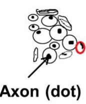
110
What does grey matter look like in cross section?
Cell bodies are stained and their is nissil substance (basophillic clumps). Glial cells surround the body. 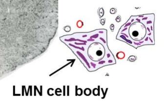
111
What is the cerebral cortext?
-Thin outer gray area, 3-6 layers
-The PYRAMIDAL cell is the LARGEST in the cerebral cortex
-The PYRAMIDAL cell is the LARGEST in the cerebral cortex
112
What is the cerebellar cortex?
-Thin outer gray area, 3 layers
-the PURKINJE cells is the largest in the cerebellar cortex
-the PURKINJE cells is the largest in the cerebellar cortex
113
When does the neural tube and neural crest form?
Week 3
114
What is the neural tube? What does it give rise to?
-Hollow tube, initially open at both ends
-Gives rise to the CNS
-Gives rise to the CNS
115
What is the neural crest?
-Collection of cells not incorporated into the neural tube that migrate away
-Gives rise to the PNS
-Gives rise to the PNS
116
What are the two neural tube defects?
Spina bifida and ancephaly
117
IF the CELL BODY is in the CNS then it is derived from the
IF the CELL BODY is in the CNS then it is derived from the neural TUBE
118
What is the only support cell of the CNS that is not in the neural tube?
The microglial cells BECAUSE they are derived from monocytes
119
2. IF the CELL BODY is in the PNS (ganglia) then it is derived from
2. IF the CELL BODY is in the PNS (ganglia) then it is derived from neural CREST
120
All neurons with cell bodies in the brian and spinal cord come from the
All neurons with cell bodies in the brian and spinal cord come from the neural tube