Chapter 18
Study these flashcardsFree · spaced repetition · iPhone, Android, Mac & web
All cards
1
What are the 5 types of biosignaling?
1. communication
2. Signaling via molecules
3. intercellular communication
4. vesicles
5. communication
1. communication
2. Signaling via molecules
3. intercellular communication
4. vesicles
5. communication
What are the 5 types of biosignaling?
1. Direct cell-to-cell communication
2. Signaling via plasma membrane bound molecules
3. Receptor mediated intercellular communication
4. Extracelluar vesicles
5. Interkingdom communication
1. Direct cell-to-cell communication
2. Signaling via plasma membrane bound molecules
3. Receptor mediated intercellular communication
4. Extracelluar vesicles
5. Interkingdom communication
2
What is an example of direct cell-to-cell communication?
Gap junction
3
Two types of receptor-mediated intercellular communication are receptors and receptors
Two types of receptor-mediated intercellular communication are surface receptors and cytoplasmic/nuclear receptors
4
Examples of extracellular vesicles include , , ,
vesicles.
vesicles.
Examples of extracellular vesicles include oncosomes, microvesicles, exosomes,
bacterial outer membrane vesicles.
bacterial outer membrane vesicles.
5
modes of action not well known at this
time, but soon will become important – especially in the fields
of chronic diseases, chronic infections, acute infections,
cardiovascular diseases and mental illnesses
time, but soon will become important – especially in the fields
of chronic diseases, chronic infections, acute infections,
cardiovascular diseases and mental illnesses
Inter-kingdom communication modes of action not well known at this
time, but soon will become important – especially in the fields
of chronic diseases, chronic infections, acute infections,
cardiovascular diseases and mental illnesses
time, but soon will become important – especially in the fields
of chronic diseases, chronic infections, acute infections,
cardiovascular diseases and mental illnesses
6
“A key is useless without a matching lock, and
an is useless
without a in its target cell.
an is useless
without a in its target cell.
“A key is useless without a matching lock, and
an extracellular signaling molecule is useless
without a matching receptor in its target cell.
an extracellular signaling molecule is useless
without a matching receptor in its target cell.
7
: a molecule on the cell surface, in the
cytoplasm or in the nucleus that interacts with
other molecules and produces a response.
cytoplasm or in the nucleus that interacts with
other molecules and produces a response.
Receptor: a molecule on the cell surface, in the
cytoplasm or in the nucleus that interacts with
other molecules and produces a response.
cytoplasm or in the nucleus that interacts with
other molecules and produces a response.
8
What is an example of a fast response speed for a ligand and receptor?
Changes in protein
function
function
9
What is an example of a slow response speed for a ligand and receptor?
Change in transcription
and protein synthesis
and protein synthesis
10
a molecule that binds to its
receptor
receptor
Ligand: a molecule that binds to its
receptor
receptor
11
a ligand that causes the receptor to activate a response in the cell.
Agonist: a ligand that causes the receptor to activate a response in the cell.
12
a ligand that causes thereceptor to become inactive.
Antagonist: a ligand that causes thereceptor to become inactive.
13
What are 8 examples of endogenous ligands?
(1) Cytokines
(2) Hormones
(3) Toxins
(4) Neurotransmitters
(5) Drugs
(6) Bacterial metabolties
(7) Odor molecules
(8) Light
(2) Hormones
(3) Toxins
(4) Neurotransmitters
(5) Drugs
(6) Bacterial metabolties
(7) Odor molecules
(8) Light
14
What are the three opiod agonists of µ-receptor, endogenous ligand β endorphin, endomorphine-1.2?
1. Heroin
2. Morphine
3. Fentanyl
2. Morphine
3. Fentanyl
15
Beta blockers function as .
Beta blockers function as antagonists.
16
What are two examples of inhibitors of Beta-adrenergic receptors?
1. Propranolol
2. Atenolol
2. Atenolol
17
What are the endogenous ligands for β endorphin, endomorphine-1.2?
adrenaline/epinephrine and noradrenaliine/norepinephrine
18
Where are membrane bound receptors located? How do they work?
-They are embedded in the membrane with ligand facing the
-Binding on the outside activates the receptor, sending a singal of the cell
-They are embedded in the membrane with ligand facing the
-Binding on the outside activates the receptor, sending a singal of the cell
Where are membrane bound receptors located? How do they work?
-They are embedded in the membrane with ligand facing the extracellular face
-Binding on the outside activates the receptor, sending a singal inside of the cell
-They are embedded in the membrane with ligand facing the extracellular face
-Binding on the outside activates the receptor, sending a singal inside of the cell
19
Intracellular receptors: ligands can transverse the plasma membrane and bind
receptors
receptors
Intracellular receptors: hydrophobic ligands can transverse the plasma membrane and bind
receptors inside the cell
receptors inside the cell
20
Cytoplasmic receptors - a type of receptor that bind ligands in cytoplasm, becomes activated, then move into the to
Cytoplasmic receptors - a type of intraceullar receptor that bind ligands in cytoplasm, becomes activated, then move into the nucleus to regulate gene transcription
21
Nuclear receptors - a type of receptor were the ligand enters the nucleus to bind its receptor, activates it, then bind & promote
Nuclear receptors - a type of intracellular receptor were the ligand enters the nucleus to bind its receptor, activates it, then bind DNA & promote transcription of target genes
22
receptors - Involved in rapid synaptic signaling between
electrically excitable cells.
electrically excitable cells.
Ligand-gated Ion channel receptors - Involved in rapid synaptic signaling between
electrically excitable cells.
electrically excitable cells.
23
G-protein coupled receptors (GPCR) - transmembrane protein, regulate the activity of a target protein through a ( )
G-protein coupled receptors (GPCR) - Seven-pass transmembrane protein, Indirectly regulate the activity of a target protein through a trimeric GTP-binding protein (G-protein)
24
Enzyme-linked receptors - transmembrane protein, Function as or directly associated with they activate
Enzyme-linked receptors - Single-pass transmembrane protein, Function directly as enzyme or directly associated with enzymes they activate
25
Ligand-gated ion channels are what type of receptor?
Ionotropic receptors
26
G-protein-coupled receptors are what type of receptor?
Metabotropic
27
What type of cells are ligand-gated channels most important for?
Electrically excitable cells (nerves and muscles)
28
How do ligand gated challenels transduce signals?
They transduce chemical signals into electrical signals
29
What type of response do ligand-gated channels allow for (slow or fast)?
Fast
30
What are the 5 steps for ligand channel functioning?
1. Before binding, it is in a conformation
2. Ligand binds to ligand site
3. Channel so that ions can pass and induce a response
4. Ligand
5. Protein conformation of channel
1. Before binding, it is in a conformation
2. Ligand binds to ligand site
3. Channel so that ions can pass and induce a response
4. Ligand
5. Protein conformation of channel
What are the 5 steps for ligand channel functioning?
1. Before binding, it is in a closed conformation
2. Ligand binds to ligand binding site
3. Channel conformation changes so that ions can pass and induce a response
4. Ligand dissociates
5. Protein conformation of channel closes again
1. Before binding, it is in a closed conformation
2. Ligand binds to ligand binding site
3. Channel conformation changes so that ions can pass and induce a response
4. Ligand dissociates
5. Protein conformation of channel closes again
31
What type of ion channels are used by neurotransmitters?
Ligand-gated ion channels
32
What are the four steps of neurotrasmitter signaling?
1. Action potential at axon
2. Vesicles loaded with neurotransmitters fuse with and release
3. Ligand binds to site at channel
4. Ions pass through and alter the , triggering voltage gated channels to respond
1. Action potential at axon
2. Vesicles loaded with neurotransmitters fuse with and release
3. Ligand binds to site at channel
4. Ions pass through and alter the , triggering voltage gated channels to respond
What are the four steps of neurotrasmitter signaling?
1. Action potential at presynaptic axon
2. Vesicles loaded with neurotransmitters fuse with presynaptic membrane and release ligand
3. Ligand binds to ligand binding site at post-synaptic membrane channel
4. Ions pass through and alter the voltage of membrane, triggering voltage gated channels to respond
1. Action potential at presynaptic axon
2. Vesicles loaded with neurotransmitters fuse with presynaptic membrane and release ligand
3. Ligand binds to ligand binding site at post-synaptic membrane channel
4. Ions pass through and alter the voltage of membrane, triggering voltage gated channels to respond
33
Na+ channel that allows positive ions to flow cell membrane
Na+ channel that allows positive ions to flow into cell DEPOLARIZING membrane
34
1. Cl- channels - allow negative ions to flow membrane
2. K+ channels - allow positive ions to flow membrane
2. K+ channels - allow positive ions to flow membrane
1. Cl- channels - allow negative ions to flow in HYPERPOLARIZING membrane
2. K+ channels - allow positive ions to flow out HYPERPOLARIZING membrane
2. K+ channels - allow positive ions to flow out HYPERPOLARIZING membrane
35
TEST QUESTION: What are cholinergic receptors? Where are they found?
Receptors activated by . They are found in the and
Receptors activated by . They are found in the and
TEST QUESTION: What are cholinergic receptors? Where are they found?
Receptors activated by acetylcholine (ACh). They are found in the central and peripheral NS.
Receptors activated by acetylcholine (ACh). They are found in the central and peripheral NS.
36
TEST QUESTION: What are the two subtypes of cholinergic receptors? What are they named after?
and . They are named after the drugs they with.
and . They are named after the drugs they with.
TEST QUESTION: What are the two subtypes of cholinergic receptors? What are they named after?
Nictinic and muscarinic. They are named after the drugs they interact with.
Nictinic and muscarinic. They are named after the drugs they interact with.
37
TEST QUESTION: Nicotinic receptors (nAChR) are
TEST QUESTION: Nicotinic receptors (nAChR) are ionotropic ligand-gated ion channels
38
TEST QUESTION: Nicotinic receptors (nAChR) are made of subunits, arranged around a
TEST QUESTION: Nicotinic receptors (nAChR) are made of 5 subunits, arranged symmetrically around a central pore
39
TEST QUESTION: What are the two types of nicotinic receptors (nAChRs)
1. type
2. type
1. type
2. type
TEST QUESTION: What are the two types of nicotinic receptors (nAChRs)
1. Neuronal type
2. Muscle type
1. Neuronal type
2. Muscle type
40
TEST QUESTION: Neuronal-type nicotinic receptors (Nn): found in both and : located from other types of receptors
TEST QUESTION: Neuronal-type nicotinic receptors (Nn): found in both CNS and PNS: located “up-stream” from other types of receptors
41
TEST QUESTION: Muscle-type nicotinic receptors (Nm): are found on neuromuscularjunctions: responsible for .
TEST QUESTION: Muscle-type nicotinic receptors (Nm): are found post-synaptically on neuromuscularjunctions: responsible for skeletal muscle contractions.
42
TEST QUESTION: Muscarinic receptors (mAChR) respond to both and and are metabotropic receptors, or , that use to
propagate the signal.
propagate the signal.
TEST QUESTION: Muscarinic receptors (mAChR) respond to both Ach and muscarine and are metabotropic receptors, or G-protein-coupled, that use 2nd messenger system to
propagate the signal.
propagate the signal.
43
TEST QUESTION: How many subtypes of muscarinic receptors are there
subtypes, (We need to know subtypes , , and for the exam)
subtypes, (We need to know subtypes , , and for the exam)
TEST QUESTION: How many subtypes of muscarinic receptors are there
5 subtypes, M1-M5 (We need to know subtypes 1, 2, and 3 for the exam)
5 subtypes, M1-M5 (We need to know subtypes 1, 2, and 3 for the exam)
44
TEST QUESTION: A M1 receptor is protein-coupled. It increases . It is found in the .
TEST QUESTION: A M1 receptor is Gq protein-coupled. It increases exocrine gland secretions. It is found in the CNS.
45
TEST QUESTION: A M2 receptor is protein-coupled. It increases
TEST QUESTION: A M2 receptor is Gi protein-coupled. It increases heart parasympathetic effects.
46
TEST QUESTION: A M3 receptor is protein coupled. It increases
TEST QUESTION: A M3 receptor is Gq protein coupled. It increases classic parasympathetic effects.
47
Briefly glance at chart in case she frames a question around sympathetic versus parasympathetic systems and one of the receptors she talks about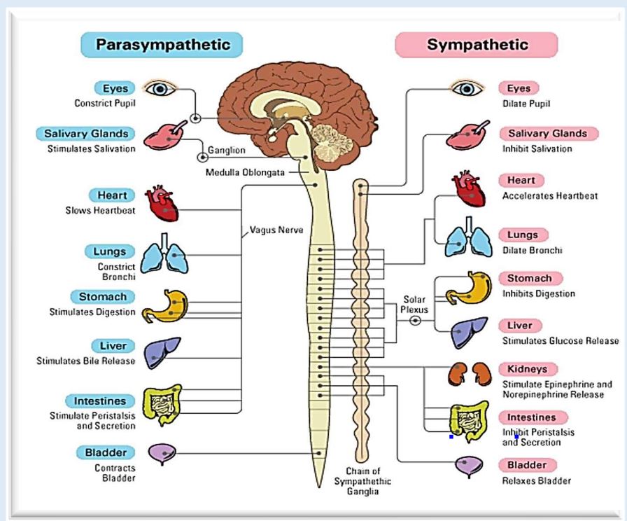
48
In the sympathetic nervous system, pre-ganglionic neurons release
that binds to in post-ganglionic neurons on target cells
that binds to in post-ganglionic neurons on target cells
In the sympathetic nervous system, pre-ganglionic neurons release acetylcholine (Ach)
that binds to nicotinic receptors in post-ganglionic neurons on target cells
that binds to nicotinic receptors in post-ganglionic neurons on target cells
49
In the sympathetic nervous system, post-ganglionic neurons then release ( ) that go on to bind receptors at target tissues.
In the sympathetic nervous system, post-ganglionic neurons then release catecholamines (adrenaline and noradrenaline) that go on to bind adrenergic receptors at target tissues.
50
Some sympathetic post-ganglionic neurons release , which binds to receptors on target cells.
Some sympathetic post-ganglionic neurons release ACh, which binds to muscarinic receptors on target cells.
51
In the parasympathetic nervous system, pre-ganglionic neurons release that binds to in postganglionic neurons
In the parasympathetic nervous system, pre-ganglionic neurons release acetylcholine (Ach) that binds to nicotinic receptors in postganglionic neurons
52
In the parasympathetic nervous system, post-ganglionic neurons then release
that go on to bind on target cells
that go on to bind on target cells
In the parasympathetic nervous system, post-ganglionic neurons then release
acetylcholine (Ach) that go on to bind muscarinic receptors on target cells
acetylcholine (Ach) that go on to bind muscarinic receptors on target cells
53
Describe what happens at the neuromuscular junction in the somatic nervous system (5 steps)
1. Ach is released following an
2. Ach binds to nicotinic receptors on , causing contraction
3. Nitchontine receptors are receptors with units
4. 2 AChs bind to alpha units, opening the channel
5. They are excitatory ligand gated ion channels that let and (causes DEPOLARIZATION and action potential)
1. Ach is released following an
2. Ach binds to nicotinic receptors on , causing contraction
3. Nitchontine receptors are receptors with units
4. 2 AChs bind to alpha units, opening the channel
5. They are excitatory ligand gated ion channels that let and (causes DEPOLARIZATION and action potential)
Describe what happens at the neuromuscular junction in the somatic nervous system (5 steps)
1. Ach is released following an action potential
2. Ach binds to nicotinic receptors on skeletal muscle cells, causing contraction
3. Nitchontine receptors are ionotropic Ach receptors with 5 units
4. 2 AChs bind to 9 alpha units, opening the channel
5. They are excitatory ligand gated ion channels that let Na+ in and K+ out (causes DEPOLARIZATION and action potential)
1. Ach is released following an action potential
2. Ach binds to nicotinic receptors on skeletal muscle cells, causing contraction
3. Nitchontine receptors are ionotropic Ach receptors with 5 units
4. 2 AChs bind to 9 alpha units, opening the channel
5. They are excitatory ligand gated ion channels that let Na+ in and K+ out (causes DEPOLARIZATION and action potential)
54
Voltage-gated are essential contributors to neuronal
, making them the most commonly targeted ion channel family by toxins found in
, making them the most commonly targeted ion channel family by toxins found in
Voltage-gated sodium channels are essential contributors to neuronal
excitability, making them the most commonly targeted ion channel family by toxins found in animal venoms
excitability, making them the most commonly targeted ion channel family by toxins found in animal venoms
55
What are two examples of acetylcholine nicotine receptor anatagonists found in nature?
1.
2.
1.
2.
What are two examples of acetylcholine nicotine receptor anatagonists found in nature?
1. Cobra toxin
2. Curare (plant used for poison darts)
1. Cobra toxin
2. Curare (plant used for poison darts)
56
A voltage gated soudium channel is also referred to as a
A voltage gated soudium channel is also referred to as a NAV
57
CLINCIAL CORRELATION: The naturally occurring marine toxinstetrodotoxin (TTX) and saxitoxin (STX) interact strongly with the region to occlude the pathway.
CLINCIAL CORRELATION: The naturally occurring marine toxinstetrodotoxin (TTX) and saxitoxin (STX) interact strongly with the Nav channel pore region to occlude the Na+ permeation pathway.
58
A g-protein coupled receptor has polypeptide chain with transmembrane (TM) domains.
A g-protein coupled receptor has a single polypeptide chain with 7 transmembrane (TM) domains.
59
In a g-protein coupled receptor, the terminal of this long protein is exposed to the outside of the cell and the terminal is exposed to the inside of the cell.
In a g-protein coupled receptor, the amino terminal of this long protein is exposed to the outside of the cell and the carboxyl terminal is exposed to the inside of the cell.
60
G-protein receptors can have one signal molecule that can interact with many different types of receptors and have
G-protein receptors can have one signal molecule that can interact with many different types of receptors and have different responses
61
are the largest family of cell-surface molecules involved in signal transmission
G-protein coupled receptors are the largest family of cell-surface molecules involved in signal transmission
62
In a trimeric receptor, there are possible outcomes based on the rank order of concentration
In a trimeric receptor, there are 6 possible outcomes based on the rank order of agonist concentration
63
GPCRs act on a second protein (enzyme or channel)
GPCRs act indirectly on a second protein (enzyme or channel)
64
A GPCR use a protein
A GPCR use a trimeric GTP-binding protein
65
A trimeric GTP-binding protein has and subunits that have domains that anchor them to the of the plasma membrane and a binding domain
A trimeric GTP-binding protein has alpha and gamma subunits that have lipid domains that anchor them to the inner layer of the plasma membrane and a GTPase binding domain
66
When a trimeric GTP-binding protein is GDP bound, it is . When it is GTP bound, it is
When a trimeric GTP-binding protein is GDP bound, it is inactive. When it is GTP bound, it is active.
67
A muscarinic receptor is also called a . They interact with cytoplasmic proteins after binding
A muscarinic receptor is also called a metabotropic acetylcholine receptor. They interact with cytoplasmic proteins after binding ACh
68
What are the three subunits of G-proteins?
1.
2.
3.
1.
2.
3.
What are the three subunits of G-proteins?
1. alpha
2. beta
3. gamma
1. alpha
2. beta
3. gamma
69
The alpha and gamma subunits of g-proteins are anchored
to and interact with the
.
to and interact with the
.
The alpha and gamma subunits of g-proteins are anchored
to the membrane and interact with the
receptor.
to the membrane and interact with the
receptor.
70
The alpha unit of a g-protein is bound to ( state)
The alpha unit of a g-protein is bound to GDP (inactive state)
71
The activated alpha of a g-protein is able to
The activated alpha of a g-protein is able to simulate and inhibit different factors
72
To act on other proteins, the alpha subunit of a g-protein uses the
energy from , which leaves bound and sends
alpha back to beta-gamma.
energy from , which leaves bound and sends
alpha back to beta-gamma.
To act on other proteins, the alpha subunit of a g-protein uses the
energy from GTP, which leaves GDP bound and sends
alpha back to beta-gamma.
energy from GTP, which leaves GDP bound and sends
alpha back to beta-gamma.
73
The cAMP-dependent pathway, also known as the adenylyl cyclase (AC) pathway, is a signaling cascade
The cAMP-dependent pathway, also known as the adenylyl cyclase (AC) pathway, is a GPCR triggered signaling cascade
74
What are the 4 steps of the Gs cAMP-dependent pathway
1. M1 activates the and interacts with on membrane
2. Activated AC uses ATP to make which
3. PKA has a catalytic subunit that other proteins to trigger a response
4. Beta 2 adrenergic receptors bind to and are coupled with GS (stimulates adenylate cylase)
1. M1 activates the and interacts with on membrane
2. Activated AC uses ATP to make which
3. PKA has a catalytic subunit that other proteins to trigger a response
4. Beta 2 adrenergic receptors bind to and are coupled with GS (stimulates adenylate cylase)
What are the 4 steps of the Gs cAMP-dependent pathway
1. M1 activates the GS-alpha subunit and interacts with adenylate cylcase on membrane
2. Activated AC uses ATP to make cAMP which dissociates protein kinase A
3. PKA has a catalytic subunit that phosphorylates other proteins to trigger a response
4. Beta 2 adrenergic receptors bind to adrenaline/epinephrine and are coupled with GS (stimulates adenylate cylase)
1. M1 activates the GS-alpha subunit and interacts with adenylate cylcase on membrane
2. Activated AC uses ATP to make cAMP which dissociates protein kinase A
3. PKA has a catalytic subunit that phosphorylates other proteins to trigger a response
4. Beta 2 adrenergic receptors bind to adrenaline/epinephrine and are coupled with GS (stimulates adenylate cylase)
75
Describe the 5 steps of the activation of a Gq alpha subunit
1. , , and activate the Gq-alpha subunit, leading to an interation with
2. PLC clevaes into and
3. IP3 makes channel open, causing
4. DAG binds to which needs to activate
5. PKC other protiens and triggers cell response
1. , , and activate the Gq-alpha subunit, leading to an interation with
2. PLC clevaes into and
3. IP3 makes channel open, causing
4. DAG binds to which needs to activate
5. PKC other protiens and triggers cell response
Describe the 5 steps of the activation of a Gq alpha subunit
1. M1, M3, and M5 activate the Gq-alpha subunit, leading to an interation with phospholipase C
2. PLC clevaes PIP2 into IP3 and DAG
3. IP3 makes Ca2+ channel open, causing DEPOLARIZATION
4. DAG binds to PKC which needs Ca2+ to activate
5. PKC phorphorylates other protiens and triggers cell response
1. M1, M3, and M5 activate the Gq-alpha subunit, leading to an interation with phospholipase C
2. PLC clevaes PIP2 into IP3 and DAG
3. IP3 makes Ca2+ channel open, causing DEPOLARIZATION
4. DAG binds to PKC which needs Ca2+ to activate
5. PKC phorphorylates other protiens and triggers cell response
76
M1 or M4: Activation of Gi- alpha subunit also interacts with
and it
and it
M1 or M4: Activation of Gi- alpha subunit also interacts with adenylate cyclase (AC)
and inhibits it
and inhibits it
77
For a receptor interacting with a Gi subtype, inhibition of AC causes a
feedback on effects of Gs.
feedback on effects of Gs.
For a receptor interacting with a Gi subtype, inhibition of AC causes a
negative feedback on effects of Gs.
negative feedback on effects of Gs.
78
Clinical Correlation: Histamine GPCRs are involved in allergies and asthma because histamine stumulates . Treatment is a that brochodilates.
Clinical Correlation: Histamine GPCRs are involved in allergies and asthma because histamine stumulates H1-GPCR. Treatment is a Beta 2 adrenergic receptor agonist that brochodilates.
79
In asthma treatment, after the physiologic effect is
accomplished (ex: vasodilation),
receptor desensitization occurs
via
accomplished (ex: vasodilation),
receptor desensitization occurs
via
In asthma treatment, after the physiologic effect is
accomplished (ex: vasodilation),
receptor desensitization occurs
via receptor phosphorylation.
accomplished (ex: vasodilation),
receptor desensitization occurs
via receptor phosphorylation.
80
In the treatment of asthma, the number of the airway smooth
muscle cell surface is by endocytosis (internalization) or degradation (downregulation)
muscle cell surface is by endocytosis (internalization) or degradation (downregulation)
In the treatment of asthma, the Phosphorylated beta-2-R number of the airway smooth
muscle cell surface is decreased by endocytosis (internalization) or degradation (downregulation)
muscle cell surface is decreased by endocytosis (internalization) or degradation (downregulation)
81
In asthma treatment, also affect the regulation of histamine GPCRs through heterologous regulation, leading to a towards vasodilation.
In asthma treatment, phosphorylated beta-2-Rs also affect the regulation of histamine GPCRs through heterologous regulation, leading to a synergistic effect towards vasodilation.
82
Protease-activated receptors (PARs) are a
unique family of GPCRs that are following of
their extracellular .
unique family of GPCRs that are following of
their extracellular .
Protease-activated receptors (PARs) are a
unique family of GPCRs that are irreversibly
activated following proteolytic cleavage of
their extracellular N- terminus.
unique family of GPCRs that are irreversibly
activated following proteolytic cleavage of
their extracellular N- terminus.
83
What are the 5 important functions of protease activated receptors?
1. Hemostasis
2. Thrombosis
3. Inflammation
4. Embryonic deverlopment
5. Cancer progression
2. Thrombosis
3. Inflammation
4. Embryonic deverlopment
5. Cancer progression
84
What are the 4 ways that enzyme linked receptor signaling is tightly regulated?
1. post-translational modification
2. desensitization
3. Internalization
4. Lysosomal degradation
2. desensitization
3. Internalization
4. Lysosomal degradation
85
Protease Activated Receptors (PARs) was first described in and
associated with disease.
associated with disease.
Protease Activated Receptors (PARs) was first described in platelets and
associated with coronary artery disease.
associated with coronary artery disease.
86
During injury, the activated platelets aggregate and release a cascade of , which increase concentration. Thrombin promotes the formation of a
in an attempt to fix the lesion.
in an attempt to fix the lesion.
During injury, the activated platelets aggregate and release a cascade of coagulation factors, which increase thrombin concentration. Thrombin promotes the formation of a
fibrin net in an attempt to fix the lesion.
fibrin net in an attempt to fix the lesion.
87
The part of the GPCR that is
(anchored in the membrane) now has
an exposed site, so it
activates itself and causes
(anchored in the membrane) now has
an exposed site, so it
activates itself and causes
The part of the GPCR that is
(anchored in the membrane) now has
an exposed “self-binding” site, so it
activates itself and causes intracellular
events.
(anchored in the membrane) now has
an exposed “self-binding” site, so it
activates itself and causes intracellular
events.
88
One thrombin molecule can activate and lead to ampolification
One thrombin molecule can activate many PARs and lead to ampolification
89
Receptor tyrosine kinases (RTK2) phosphorylate Tyr
residues on the of the receptor.
residues on the of the receptor.
Receptor tyrosine kinases (RTK2) phosphorylate Tyr
residues on the intracellular portion of the receptor.
residues on the intracellular portion of the receptor.
90
Before binding ligands, the receptor tyrosine kinases are found as
individual units on the
individual units on the
Before binding ligands, the receptor tyrosine kinases are found as
individual units on the surface of cells.
individual units on the surface of cells.
91
After binding ligand (hormone), the receptor tyrosine kinases dimerize, which causes the cytoplasmic domains to be and
After binding ligand (hormone), the receptor tyrosine kinases dimerize, which causes the cytoplasmic domains to be near each other and interact.
92
In a receptor tyrosine kinase, the cytoplasmic domains each other
In a receptor tyrosine kinase, the cytoplasmic domains phosphorylates each other
93
Receptor Tyrosine Kinase (RTK)s = genes
Receptor Tyrosine Kinase (RTK)s = 58 genes
94
What is the largest of the enzyme coupled receptor class:?
Tyrosine kinase
95
A tyrosine kinase has transmembrane segment. Can activate signaling pathways.
A tyrosine kinase has one transmembrane segment. Can activate multiple signaling pathways.
96
What are the 6 shared steps of tyrosine kinase phosphates?
1. ligand binds the receptor
2. receptor joins and
3. intrinsic enzyme, the , becomes active
4. Receptor the opposite tyrosine residues
(= ).
5. The phosphorylated tyrosine residues activate the signaling
proteins.
6. Activated intracellular signal proteins trigger an intracellular
signaling pathway
1. ligand binds the receptor
2. receptor joins and
3. intrinsic enzyme, the , becomes active
4. Receptor the opposite tyrosine residues
(= ).
5. The phosphorylated tyrosine residues activate the signaling
proteins.
6. Activated intracellular signal proteins trigger an intracellular
signaling pathway
What are the 6 shared steps of tyrosine kinase phosphates?
1. ligand binds the receptor
2. receptor joins and dimerizes
3. intrinsic enzyme, the tyrosine kinase domain, becomes active
4. Receptor phosphorylates the opposite tyrosine residues
(=autophosphorylation).
5. The phosphorylated tyrosine residues activate the intracellular signaling
proteins.
6. Activated intracellular signal proteins trigger an intracellular
signaling pathway
1. ligand binds the receptor
2. receptor joins and dimerizes
3. intrinsic enzyme, the tyrosine kinase domain, becomes active
4. Receptor phosphorylates the opposite tyrosine residues
(=autophosphorylation).
5. The phosphorylated tyrosine residues activate the intracellular signaling
proteins.
6. Activated intracellular signal proteins trigger an intracellular
signaling pathway
97
What are 3 features of receptor tyrosine kinases?
(1) High affinity
(2) activity that causes autophosphorylation.
(3) Activation of effectors to generate a
(1) High affinity
(2) activity that causes autophosphorylation.
(3) Activation of effectors to generate a
What are 3 features of receptor tyrosine kinases?
(1) High ligand binding affinity
(2) intrinsic kinase activity that causes autophosphorylation.
(3) Activation of effectors to generate a signaling cascade
(1) High ligand binding affinity
(2) intrinsic kinase activity that causes autophosphorylation.
(3) Activation of effectors to generate a signaling cascade
98
What are the four steps of insulin action?
1. Food intake
blood glucose
2. stimulates
pancreas to insulin
into the
bloodstream
3. Insulin finds
expressing IR
4. IR kinase
enzyme is
activated which
1. Food intake
blood glucose
2. stimulates
pancreas to insulin
into the
bloodstream
3. Insulin finds
expressing IR
4. IR kinase
enzyme is
activated which
What are the four steps of insulin action?
1. Food intake
increases
blood glucose
2. stimulates
pancreas to secrete insulin
into the
bloodstream
3. Insulin finds
target cell
expressing IR
4. IR kinase
enzyme is
activated which initiates
cascade
1. Food intake
increases
blood glucose
2. stimulates
pancreas to secrete insulin
into the
bloodstream
3. Insulin finds
target cell
expressing IR
4. IR kinase
enzyme is
activated which initiates
cascade
99
Unlike other
RTKs, the
insulin receptor
is a , but
it’s just not
active before
binding insulin.
RTKs, the
insulin receptor
is a , but
it’s just not
active before
binding insulin.
Unlike other
RTKs, the
insulin receptor
is a dimer, but
it’s just not
active before
binding insulin.
RTKs, the
insulin receptor
is a dimer, but
it’s just not
active before
binding insulin.
100
When blood glucose levels are high, the pancreas releases insulin which is a /
When blood glucose levels are high, the pancreas releases insulin which is a peptide hormone/ligand
101
Binding of inulin in target tissues receptors activates the of their cytoplasmic tails.
Binding of inulin in target tissues receptors activates the autophosphorylation of their cytoplasmic tails.
102
Insulin binds to the extracellular
and the auto-phosphorylate.
and the auto-phosphorylate.
Insulin binds to the extracellular
alpha subunits and the beta
subunits auto-phosphorylate.
alpha subunits and the beta
subunits auto-phosphorylate.
103
Insulin uses energy from ATP to
transfer to the of the receptor.
transfer to the of the receptor.
Insulin uses energy from ATP to
transfer phosphates to the beta
subunits of the receptor.
transfer phosphates to the beta
subunits of the receptor.
104
Phosphorylated insulin receptor sends
signal to causes mobilization of
transporters.
signal to causes mobilization of
transporters.
Phosphorylated insulin receptor sends
signal to causes mobilization of
Glut4 transporters.
signal to causes mobilization of
Glut4 transporters.
105
Vesicles with transporters fuse to the membrane, allowing glucose toenter the target cells.
Vesicles with Glut4 transporters fuse to the membrane, allowing glucose toenter the target cells.
106
What are the four insulin effectors in order?
MEK, MAPK, AP-1, CBP p300
107
Binding of insulin is followed by
internalization of the
internalization of the
Binding of insulin is followed by
internalization of the hormonereceptor complex.
internalization of the hormonereceptor complex.
108
Once inside the cell, the insulin is degraded in the
Once inside the cell, the insulin is degraded in the lysosomes.
109
Some of the insulin receptor gets degraded as well, but most are back to the cell surface.
Some of the insulin receptor gets degraded as well, but most are recycled back to the cell surface.
110
Interferon receptor with Janus kinase involving the signaling pathway. Interferon is the first line of defense against
Interferon receptor with Janus kinase involving the JAK/STAT signaling pathway. Interferon is the first line of defense against viruses
111
During chronic stress, IFN signaling is and the individual becomes more prone
to developing .
to developing .
During chronic stress, IFN signaling is downregulated and the individual becomes more prone
to developing illnesses.
to developing illnesses.
112
Intracellular receptors are only available to that diffuse through the
membrane
membrane
Intracellular receptors are only available to lipophilic ligands that diffuse through the
membrane
membrane
113
All intracellular receptors act as ligand-activated
All intracellular receptors act as ligand-activated transcription
factors.
factors.
114
Steroid receptors are located in the .When steroid binds, the complex is translocated to the nucleus (via ). In the nucleus, they act on the DNA as enhancers of
Steroid receptors are located in the cytoplasm.When steroid binds, the complex is translocated to the nucleus (via nuclear pore complexes). In the nucleus, they act on the DNA as enhancers of gene expression
115
What are the two groups of intraceullar receptors?
corticosteroids and sex
hormones
hormones
116
Thyroid hormones receptors normally sit on
the DNA, gene expression.
the DNA, gene expression.
Thyroid hormones receptors normally sit on
the DNA, inactivating gene expression.
the DNA, inactivating gene expression.
117
When binds, it displaces
a co- repressor from the receptor and
allows binding of co- activator protein,
which gene expression.
a co- repressor from the receptor and
allows binding of co- activator protein,
which gene expression.
When thyroid hormone binds, it displaces
a co- repressor from the receptor and
allows binding of co- activator protein,
which promotes gene expression.
a co- repressor from the receptor and
allows binding of co- activator protein,
which promotes gene expression.
118
The tyroid hormone receptors have a that binds ( ) and a region that interacts with the hormone.
The tyroid hormone receptors have a N-terminal region that binds DNA (zinc fingers domain) and a C-terminal region that interacts with the hormone.
119
The DNA sequences that are bound by
these thyroid hormone receptors are called
these thyroid hormone receptors are called
The DNA sequences that are bound by
these thyroid hormone receptors are called Hormone
Response Elements (HREs).
these thyroid hormone receptors are called Hormone
Response Elements (HREs).