Lecture 4
Study these flashcardsFree · spaced repetition · iPhone, Android, Mac & web
All cards
1
produces dark images on a light background
Light microscopy produces dark images on a light background
2
What type of microscopy produced this image? 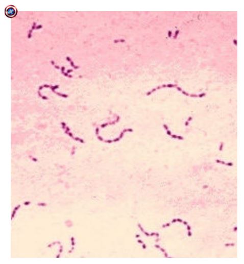
3
Calls are small and colorless so they require
Calls are small and colorless so they require stain
4
In microscopy an opaque disk is added so that
only scattered light from the
specimen are viewed. Increases compared to
brightfield microscopy.
only scattered light from the
specimen are viewed. Increases compared to
brightfield microscopy.
In dark field microscopy an opaque disk is added so that
only scattered light from the
specimen are viewed. Increases resolution compared to
brightfield microscopy.
only scattered light from the
specimen are viewed. Increases resolution compared to
brightfield microscopy.
5
What type of microscopy produced the image on the left? Right? 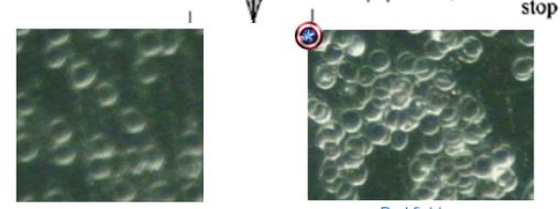
Brightfeild, darkfield (respectively)
6
What type of microscopy produced the image on the left? Right? 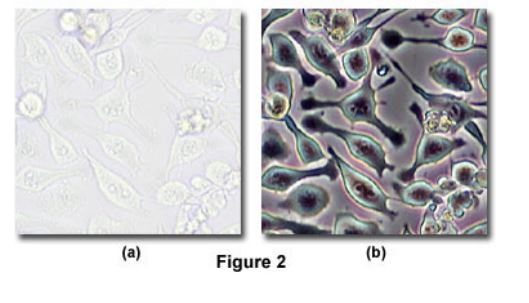
Bright field, phase contrast
7
In light that passes through the central part of the light path is recombined with light that travels around
the periphery of the specimen
the periphery of the specimen
In phase constrast microscopy, light that passes through the central part of the light path is recombined with light that travels around
the periphery of the specimen
the periphery of the specimen
8
In phase contrast microscopy, produced by these two paths produce images in which dense structures appear darker
than the background
than the background
In phase contrast microscopy, interference produced by these two paths produce images in which dense structures appear darker
than the background
than the background
9
The main benefit of phase constrast is that it gives clear visualization of
The main benefit of phase constrast is that it gives clear visualization of internal structures
10
What type of microscopy was used to generate this image? 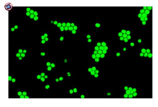
Fluorescent microscopy
11
In floursent microscopy, specimens labeled with dyes or
with attached
with attached
In floursent microscopy, specimens labeled with dyes or
antibodies with attached dyes
antibodies with attached dyes
12
Fluorescence observed upon
illuminating “coated” specimen with
of light. When using antibodies, floursent microscopy is AKA .
illuminating “coated” specimen with
of light. When using antibodies, floursent microscopy is AKA .
Fluorescence observed upon
illuminating “coated” specimen with
short wavelengths of light. When using antibodies, floursent microscopy is AKA immunofluoresence.
illuminating “coated” specimen with
short wavelengths of light. When using antibodies, floursent microscopy is AKA immunofluoresence.
13
is variation of fluorescent microscopy
producing 3D images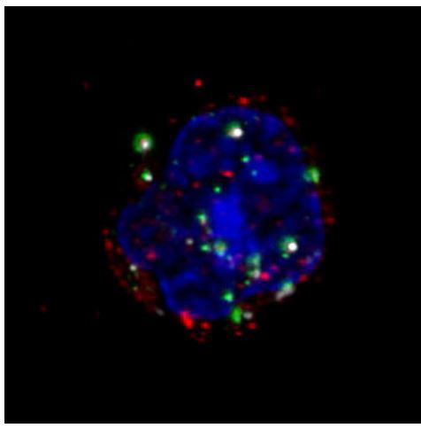
producing 3D images
Confocal micrscopy is variation of fluorescent microscopy
producing 3D images
producing 3D images
14
What type of microscopy produced this image? 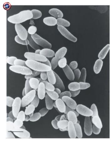
Scanning electron microscopy
15
What type of microscopy produced this image? 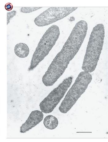
Transmission electron microscopes
16
: Beam of electrons passes through or scans the surface of a specimen producing an image (electron micrograph)
Electron microscopy: Beam of electrons passes through or scans the surface of a specimen producing an image (electron micrograph)
17
Heat fixation
(1) bacteria
(2) bacteria to slide
(3) Alters bacteria
(1) bacteria
(2) bacteria to slide
(3) Alters bacteria
Heat fixation
(1) Kills bacteria
(2) Adheres bacteria to slide
(3) Alters bacteria morphology
(1) Kills bacteria
(2) Adheres bacteria to slide
(3) Alters bacteria morphology
18
Basic stains because these dyes are -charged (
chromogen)
chromogen)
Basic stains because these dyes are positively-charged (positive
chromogen)
chromogen)
19
Positively-charged simple stains bind to cellular structures such as the
cell membrane and DNA (negative due to the phosphates, PO42-, in their structure)
cell membrane and DNA (negative due to the phosphates, PO42-, in their structure)
Positively-charged simple stains bind to negatively-charged cellular structures such as the
cell membrane and DNA (negative due to the phosphates, PO42-, in their structure)
cell membrane and DNA (negative due to the phosphates, PO42-, in their structure)
20
Simple stains are applied only to bacterial smears that have been
• Basic Stain Examples – , ,
• Basic Stain Examples – , ,
Simple stains are applied only to bacterial smears that have been heat-fixed
• Basic Stain Examples – Methylene Blue, Crystal Violet, Safranin
• Basic Stain Examples – Methylene Blue, Crystal Violet, Safranin
21
Acidic stains because these dyes are ( chromogen)
Acidic stains because these dyes are negatively-charged (negative chromogen)
22
Negatively-charged acidic stains are by the negatively-charged
such as the cell membrane and DNA
such as the cell membrane and DNA
Negatively-charged acidic stains are repelled by the negatively-charged cellular structures
such as the cell membrane and DNA
such as the cell membrane and DNA
23
In negative stains, interaction results in a background and specimen which appears as a brilliant white against a dark background upon viewing in the microscope. Negative stains aren't
In negative stains, interaction results in a darkly-stained background and unstained specimen which appears as a brilliant white against a dark background upon viewing in the microscope. Negative stains aren't heat-fixed. 
24
Examples of negative stains are and
Examples of negative stains are eosin and nigrosin
25
What type of stain produced this image? 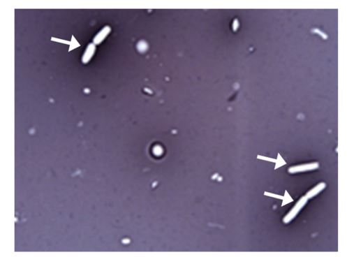
Negative stain
26
Differential stains
• Stains designed to distinguish bacterial specimens with similar and
• Stains designed to distinguish bacterial specimens with similar and
Differential stains
• Stains designed to distinguish bacterial specimens with similar morphology and
arrangement
• Stains designed to distinguish bacterial specimens with similar morphology and
arrangement
27
bacteria – thick layers of peptidoglycan
exposed to the environment
exposed to the environment
Gram (+) bacteria – thick layers of peptidoglycan
exposed to the environment
exposed to the environment
28
bacteria - thin layers of peptidoglycan
hidden from the environment by the outer
membrane
hidden from the environment by the outer
membrane
Gram (-) bacteria - thin layers of peptidoglycan
hidden from the environment by the outer
membrane
hidden from the environment by the outer
membrane
29
Gram positive or gram negative? Morphology? 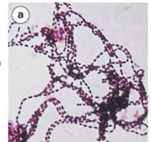
Gram positive bacillus
30
Gram positive or gram negative? Morphology? 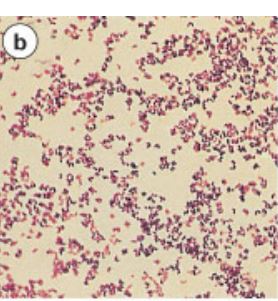
Gram (+)
coccus
and
Gram (-) coccus
coccus
and
Gram (-) coccus
31
Gram positive or gram negative? Morphology? 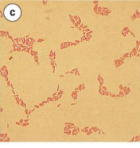
Gram (-)
bacillus
bacillus
32
Gram positive or gram negative? Morphology? 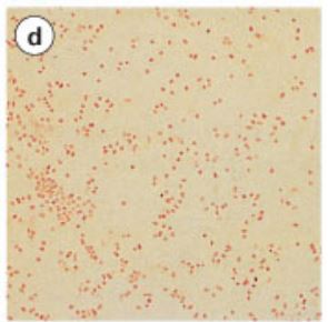
Gram (-)
coccus
coccus
33
What is the purpose of a capsule?
Prevents phagocytosis
34
Capsule stain
Stains the (some bacteria have this structure that is made of )
Stains the (some bacteria have this structure that is made of )
Capsule stain
Stains the capsule (some bacteria have this structure that is made of polysaccharides)
Stains the capsule (some bacteria have this structure that is made of polysaccharides)
35
The capsule allows the bacteria to adhere to surface and its sugar components are stick, which allows for adhesion to mucosal tissue surface, which allows and
The capsule allows the bacteria to adhere to surface and its sugar components are stick, which allows for adhesion to mucosal tissue surface, which allows colonization and infection
36
What type of stain is depicted? 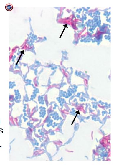
Acid-fast stain
37
Acid-fast stain
• A carbolfuschin dye used to detect the presence of
acid in the cell wall of some bacteria
( )
• A carbolfuschin dye used to detect the presence of
acid in the cell wall of some bacteria
( )
Acid-fast stain
• A carbolfuschin dye used to detect the presence of
mycolic acid in the cell wall of some bacteria
(Mycobacterium)
• A carbolfuschin dye used to detect the presence of
mycolic acid in the cell wall of some bacteria
(Mycobacterium)
38
is a waxy substance that repels most
stains
stains
Mycolic acid is a waxy substance that repels most
stains
stains
39
and binding survives a
decolorization step (alcohol step) in the staining
procedure. The acid-fast bacteria are stained, and the others become
decolorization step (alcohol step) in the staining
procedure. The acid-fast bacteria are stained, and the others become
Carbolfuschin and mycolic acid binding survives a
decolorization step (alcohol step) in the staining
procedure. The acid-fast bacteria are stained, and the others become decolorized
decolorization step (alcohol step) in the staining
procedure. The acid-fast bacteria are stained, and the others become decolorized
40
Flagella stain
• Motile structures of some bacteria
• Bacterial flagella are composed of a unique protein
called (which can initiate an immune
response)
• Motile structures of some bacteria
• Bacterial flagella are composed of a unique protein
called (which can initiate an immune
response)
Flagella stain
• Motile structures of some bacteria
• Bacterial flagella are composed of a unique protein
called flagellin (which can initiate an immune
response)
• Motile structures of some bacteria
• Bacterial flagella are composed of a unique protein
called flagellin (which can initiate an immune
response)
41
Bacterial flagella move the cell by rotating in a
motion
• Allows bacteria to escape immune responses as well
as through tissues
motion
• Allows bacteria to escape immune responses as well
as through tissues
Bacterial flagella move the cell by rotating in a
propellar-like motion
• Allows bacteria to escape immune responses as well
as spread faster through tissues
propellar-like motion
• Allows bacteria to escape immune responses as well
as spread faster through tissues
42
Bacteria with flagella are characterized based upon the
r of flagella and how they are on the
cell – this can help identify the specific bacteria in
patient samples (also their motility on a slide)
r of flagella and how they are on the
cell – this can help identify the specific bacteria in
patient samples (also their motility on a slide)
Bacteria with flagella are characterized based upon the
number of flagella and how they are arranged on the
cell – this can help identify the specific bacteria in
patient samples (also their motility on a slide)
number of flagella and how they are arranged on the
cell – this can help identify the specific bacteria in
patient samples (also their motility on a slide)
43
What type of flagella arrangement? 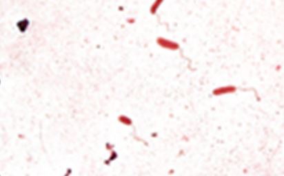
Monotrichous
44
What type of flagella arrangement? 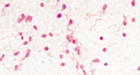
Lophotrichous
45
What type of flagella arrangement? 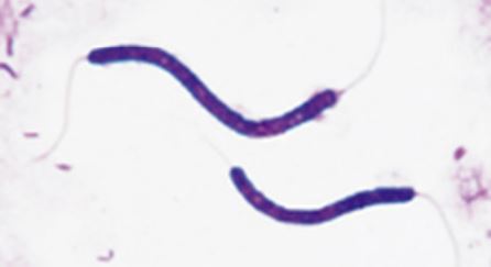
Amphitrichious 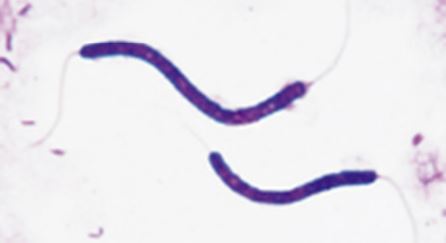
46
What type of flagella arrangement? 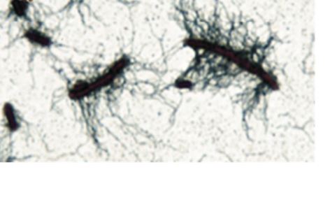
Peritrichous 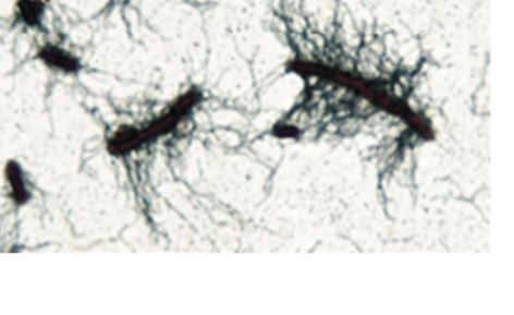
47
stain
• Dyes that are used to detect the presence
of endospores, dormant forms of some
bacteria ( and species)
• Dyes that are used to detect the presence
of endospores, dormant forms of some
bacteria ( and species)
Endospore stain
• Dyes that are used to detect the presence
of endospores, dormant forms of some
bacteria (Bacillus and Clostridium species)
• Dyes that are used to detect the presence
of endospores, dormant forms of some
bacteria (Bacillus and Clostridium species)
48
Endospores are resistant to ,
changes and
changes and
Endospores are resistant to heat, pH
changes and radiation
changes and radiation
49
Endospore covering contains
(tough coating also found in our fingernails)
(tough coating also found in our fingernails)
Endospore covering contains keratin
(tough coating also found in our fingernails)
(tough coating also found in our fingernails)
50
Endospore cytoplasm is to
prevent the conductance of heat (no water
in the cell to transfer heat from the
environment to the cell)
prevent the conductance of heat (no water
in the cell to transfer heat from the
environment to the cell)
Endospore cytoplasm is dehydrated to
prevent the conductance of heat (no water
in the cell to transfer heat from the
environment to the cell)
prevent the conductance of heat (no water
in the cell to transfer heat from the
environment to the cell)
51
Endospores have contains which
produces a impenetrable barrier
produces a impenetrable barrier
Endospores have contains calcium dipicolinate which
produces a impenetrable barrier
produces a impenetrable barrier
52
What is depicted in this image? 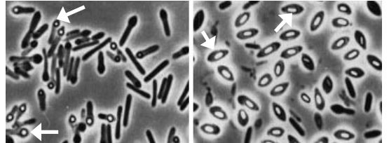
Endospore