Chapter 26
Study these flashcardsFree · spaced repetition · iPhone, Android, Mac & web
All cards
1
Draw the general schematic of how amino acids enter the TCA cycle 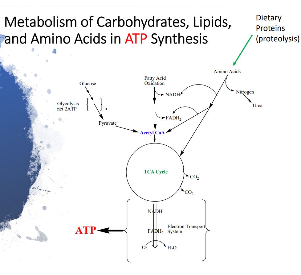
2
Unlike and whose major function is to provide energy
serve as building blocks for tissue protein synthesis.
serve as building blocks for tissue protein synthesis.
Unlike carbohydrates and triacylglycerols whose major function is to provide energy
amino acids serve as building blocks for tissue protein synthesis.
amino acids serve as building blocks for tissue protein synthesis.
3
Proteins from the diet are digested into
Proteins from the diet are digested into individual amino acids
4
Individual amino acids are then used for intracellular tissues
Individual amino acids are then used for intracellular tissues protein
biosynthesis.
biosynthesis.
5
Amino acids that cannot be synthesized
de novo are
de novo are
Amino acids that cannot be synthesized
de novo are essential amino acids.
de novo are essential amino acids.
6
The essential amino acids are
Phenylalanine, valine, Threonine,
Tryptophan, Isoleucine, Methionine,
Histidine, Arginine, Leucine, Lysine
PVT TIM HALL (mnemonic)
Tryptophan, Isoleucine, Methionine,
Histidine, Arginine, Leucine, Lysine
PVT TIM HALL (mnemonic)
7
Negative nitrogen status occurs in what 3 processes?
1. Dietary deficience of an essential amino acid
2. Extreme conditions of diabetes
3. Pathogenic infections in general starvation
2. Extreme conditions of diabetes
3. Pathogenic infections in general starvation
8
Positive nitrogen status occurs in wwhat 4 processes?
1. Fetal development (pregnancy)
2. Human development (growth)
3. Post injury or surgery (recovery phase)
4. Recovery from fasting/starvation
2. Human development (growth)
3. Post injury or surgery (recovery phase)
4. Recovery from fasting/starvation
9
Deficiency of selective amino acids
(protein malnutrition alone) i.e.
negative nitrogen balance leads to
whereas severe
nutritional deficiency (including Fats
and Carbs) leads to
(decay, extensive muscle wasting and
tissue damage etc.).
(protein malnutrition alone) i.e.
negative nitrogen balance leads to
whereas severe
nutritional deficiency (including Fats
and Carbs) leads to
(decay, extensive muscle wasting and
tissue damage etc.).
Deficiency of selective amino acids
(protein malnutrition alone) i.e.
negative nitrogen balance leads to
Kwashiorkor whereas severe
nutritional deficiency (including Fats
and Carbs) leads to Marasmus
(decay, extensive muscle wasting and
tissue damage etc.).
(protein malnutrition alone) i.e.
negative nitrogen balance leads to
Kwashiorkor whereas severe
nutritional deficiency (including Fats
and Carbs) leads to Marasmus
(decay, extensive muscle wasting and
tissue damage etc.).
10
In syndrome there is serum
albumin synthesis which causes in the blood and generalized edema
of tissues.
albumin synthesis which causes in the blood and generalized edema
of tissues.
In Kwashiorkor syndrome there is less serum
albumin synthesis which causes osmotic
imbalance in the blood and generalized edema
of tissues.
albumin synthesis which causes osmotic
imbalance in the blood and generalized edema
of tissues.
11
Sketch the schematic for amino acid metabolism (dietary proteolysis) 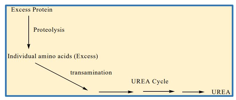
12
What is the proteosome pathway?
It is an intracellular protein degradation pathway
13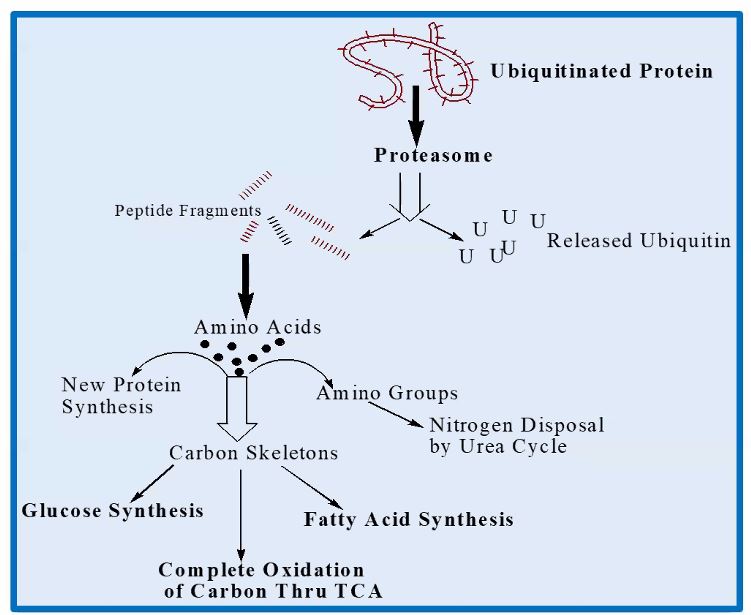
Draw out the schematic for the proteosome pathway
14
Ketogenic amino acids upon
degradation produce only or .
degradation produce only or .
Ketogenic amino acids upon
degradation produce only acetyl
CoA or acetoacetyl CoA.
degradation produce only acetyl
CoA or acetoacetyl CoA.
15
Glucogenic amino acids upon
metabolism produce or ,
, or
( cycle
intermediates).
metabolism produce or ,
, or
( cycle
intermediates).
Glucogenic amino acids upon
metabolism produce pyruvate,
3‐phosphoglycerate (glycolytic
intermediate) or oxalo acetate,
a‐ketoglutarate, succinyl CoA or
fumarate (TCA cycle
intermediates).
metabolism produce pyruvate,
3‐phosphoglycerate (glycolytic
intermediate) or oxalo acetate,
a‐ketoglutarate, succinyl CoA or
fumarate (TCA cycle
intermediates).
16
What is this structure? 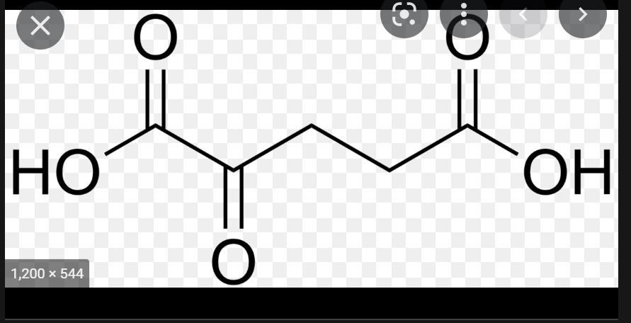
alpha ketogluterate 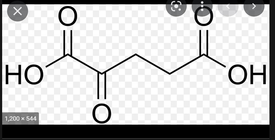
17
What is this structure? 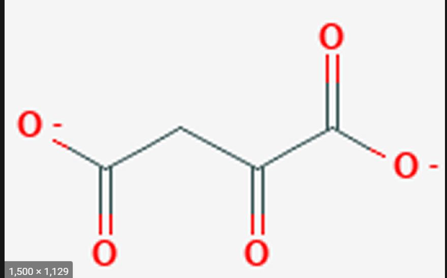
Oxaloactetate 
18
Aromatic amino acids are
Glycogenic and gluconeogenic
19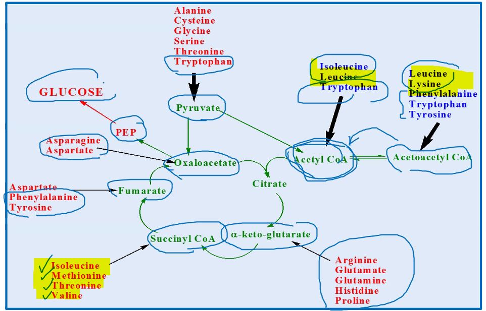
Know this
20
Transaminases (amino
transferases) use an
amino acid to donate
an to a
transferases) use an
amino acid to donate
an to a
Transaminases (amino
transferases) use an
amino acid to donate
an amine group to a
keto acid
transferases) use an
amino acid to donate
an amine group to a
keto acid
21
Keto acid becomes
and the
becomes
and the
becomes
Keto acid becomes
amino acid and the
amino acid becomes
keto acid
amino acid and the
amino acid becomes
keto acid
22
The mandatory amino acid/keto acid
pair is . serves as a coenzyme in the
reaction.
pair is . serves as a coenzyme in the
reaction.
The mandatory amino acid/keto acid
pair is glutamate/a‐ketoglutarate.. Pyridoxal phosphate (Vitamin B6
derivative) serves as a coenzyme in the
reaction.
pair is glutamate/a‐ketoglutarate.. Pyridoxal phosphate (Vitamin B6
derivative) serves as a coenzyme in the
reaction.
23
What are 3 important aminotransferases B6 and coenzyme derivatives?
1. Vitamin B6‐Pyridoxine
2. Coenzyme‐Pyridoxal‐Phosphate
3. Coenzyme‐Pyridoxamine
2. Coenzyme‐Pyridoxal‐Phosphate
3. Coenzyme‐Pyridoxamine
24
In liver diseases, that causes (for
e.g., viral hepatitis, toxic injury) the two
enzymes
are released into blood
e.g., viral hepatitis, toxic injury) the two
enzymes
are released into blood
In liver diseases, that causes cell lysis (for
e.g., viral hepatitis, toxic injury) the two
enzymes
AST and ALT are released into blood
e.g., viral hepatitis, toxic injury) the two
enzymes
AST and ALT are released into blood
25
High concentrations of AST and ALT are two
enzymes in plasma are diagnostic
markers of
enzymes in plasma are diagnostic
markers of
High concentrations of AST and ALT are two
enzymes in plasma are diagnostic
markers of liver cell damage
enzymes in plasma are diagnostic
markers of liver cell damage
26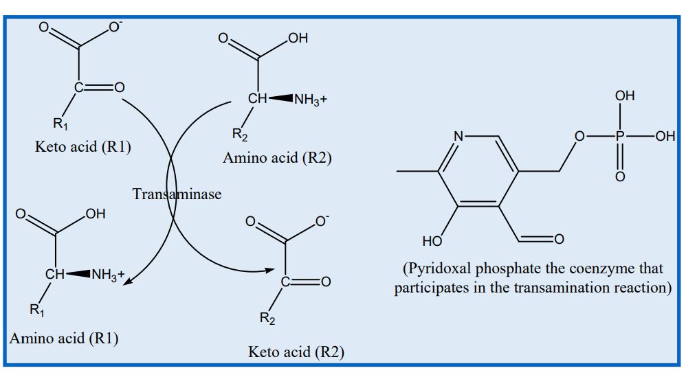
Draw the schematic for a trasnaminase reaction
27
Many tissues lack complete sets of to dispose excess
nitrogen.
nitrogen.
Many tissues lack complete sets of urea
cycle enzymes to dispose excess
nitrogen.
cycle enzymes to dispose excess
nitrogen.
28
is the main organ that operates
urea cycle.
urea cycle.
Liver is the main organ that operates
urea cycle.
urea cycle.
29
NH3 is toxic to cells, Excess
nitrogen in the form of is sent
to liver, where its can be metabolized.
nitrogen in the form of is sent
to liver, where its can be metabolized.
NH3 is toxic to cells, Excess
nitrogen in the form of glutamine is sent
to liver, where its nitrogen can be metabolized.
nitrogen in the form of glutamine is sent
to liver, where its nitrogen can be metabolized.
30
NH3 distrupts pH balance, causing
NH3 distrupts pH balance, causing alkalosis 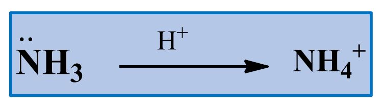
31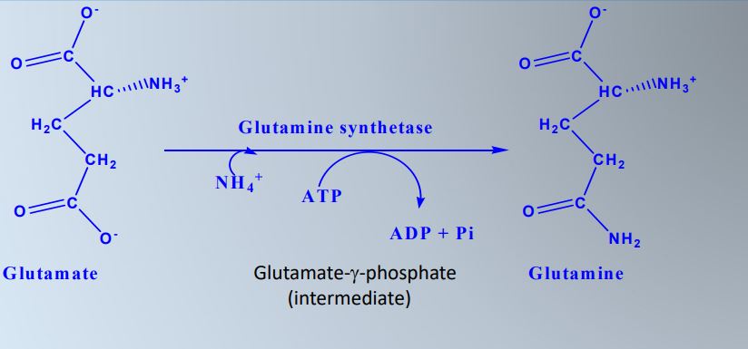
How is glutamine is formed from glutamate? Draw schematic
32
Know the processes where alpha-ketoglutarate, glutamate, and glutamine enter other metabolic cycles. Know the interconversion of a ketoglutarate, glutamate, and glutamine. 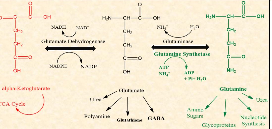
33
When glutamine reaches liver,
converts glutamine
into and . Now ammonium ion can enter the .
converts glutamine
into and . Now ammonium ion can enter the .
When glutamine reaches liver,
glutaminase converts glutamine
into glutamate and ammonia. . Now ammonium ion can enter the urea cycle.
glutaminase converts glutamine
into glutamate and ammonia. . Now ammonium ion can enter the urea cycle.
34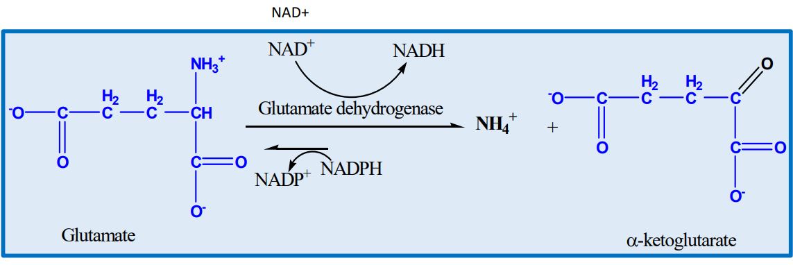
Draw the schematic for the glutamate dehydrogenase reaction
35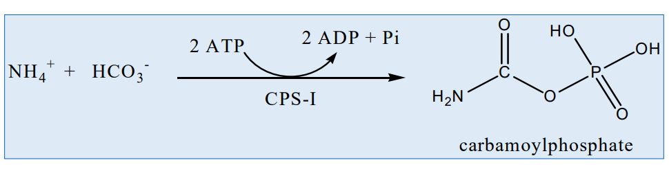
Draw the schematic for the synthesis of carbamoylphosphate
36
CPS‐I is located in the
CPS‐I is located in the
mitochondrial
matrix.
mitochondrial
matrix.
37
CPS‐I is activated by
an
N‐acetyl glutamate.
an
N‐acetyl glutamate.
CPS‐I is activated by
an allosteric activator
N‐acetyl glutamate.
an allosteric activator
N‐acetyl glutamate.
38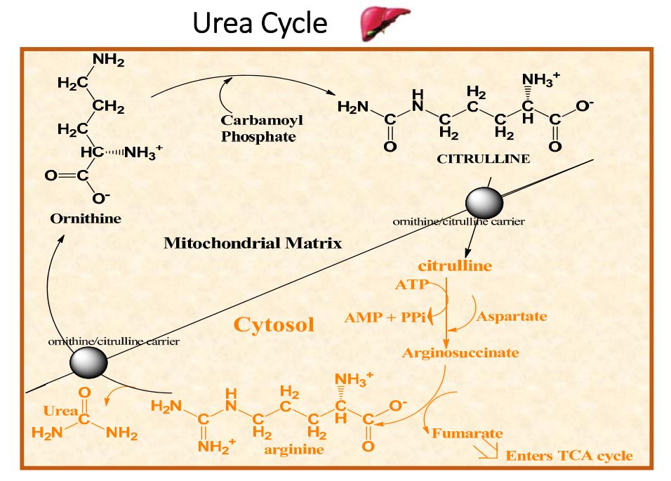
Draw out schematic for urea cycle
39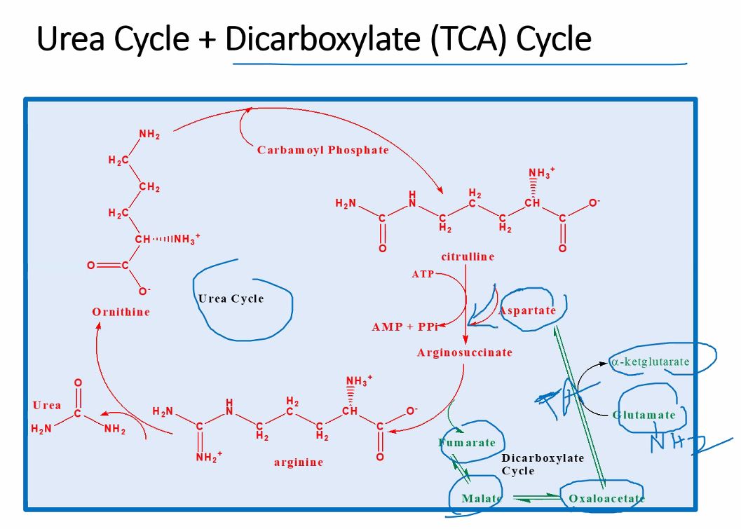
Draw out the schematic for Urea Cycle + Dicarboxylate (TCA) Cycle
40
Blood Urea Nitrogen (BUN)
is ~ Normally
~ is excreted.
is ~ Normally
~ is excreted.
Blood Urea Nitrogen (BUN)
is ~10-20 mg/dl. Normally
~10 g of urea/day is excreted.
is ~10-20 mg/dl. Normally
~10 g of urea/day is excreted.
41
Hyperammonemia, elevated
blood glutamine, and
indicates a
defect in urea cycle.
blood glutamine, and
indicates a
defect in urea cycle.
Hyperammonemia, elevated
blood glutamine, and
decreased BUN indicates a
defect in urea cycle.
blood glutamine, and
decreased BUN indicates a
defect in urea cycle.
42
Excretion= – +
Excretion=Filtration – Reabsorption + Secretion