Bio 25 Exam #2
Study these flashcardsFree · spaced repetition · iPhone, Android, Mac & web
All cards
1
4 main tissue types
epithelium
connective tissue
muscle
nervous tissue
connective tissue
muscle
nervous tissue
2
function of the epithelium
protection, secretion, absorption, excretion, filtration, diffusion, and sensory reception
3
what does polarity of the epithelial refer to
organization of different domains within the cell
4
apex
at top of cell
has microvilli or cilia
increases the surface area of a cell
involved with sensory perception, protien distribution
has microvilli or cilia
increases the surface area of a cell
involved with sensory perception, protien distribution
5
lateral domain
contains cell junctions
maintain integrity of the layers
crucial for structure
maintain integrity of the layers
crucial for structure
6
basal domain
serves as a basement
responsible for nutrient uptake and waste removal
helps with overall organization and function of the epithelial tissue
responsible for nutrient uptake and waste removal
helps with overall organization and function of the epithelial tissue
7
Simple squamous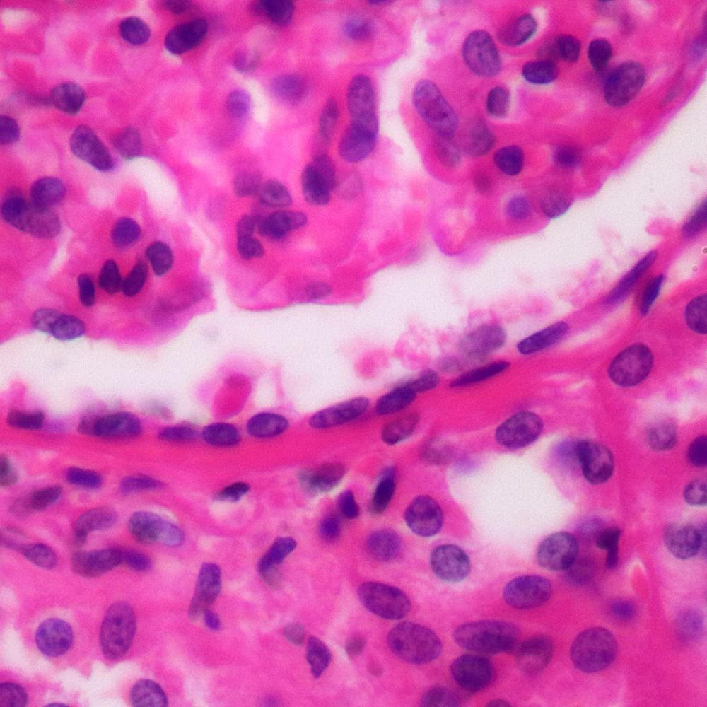
this type of epithelium typically lines blood vessels and body cavities and regulates the passage of substances into underlying tissue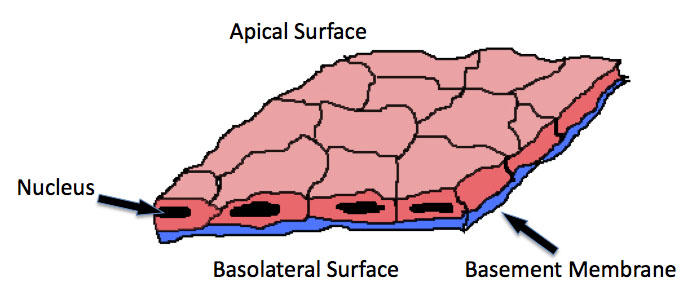
8
Stratified squamous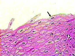
This type of epithelium usually has protective functions, including protection against microorganisms from invading underlying tissue and/or protection against water loss. The outer layer of your skin (the epidermis) is made of stratified squamous epithelial cells.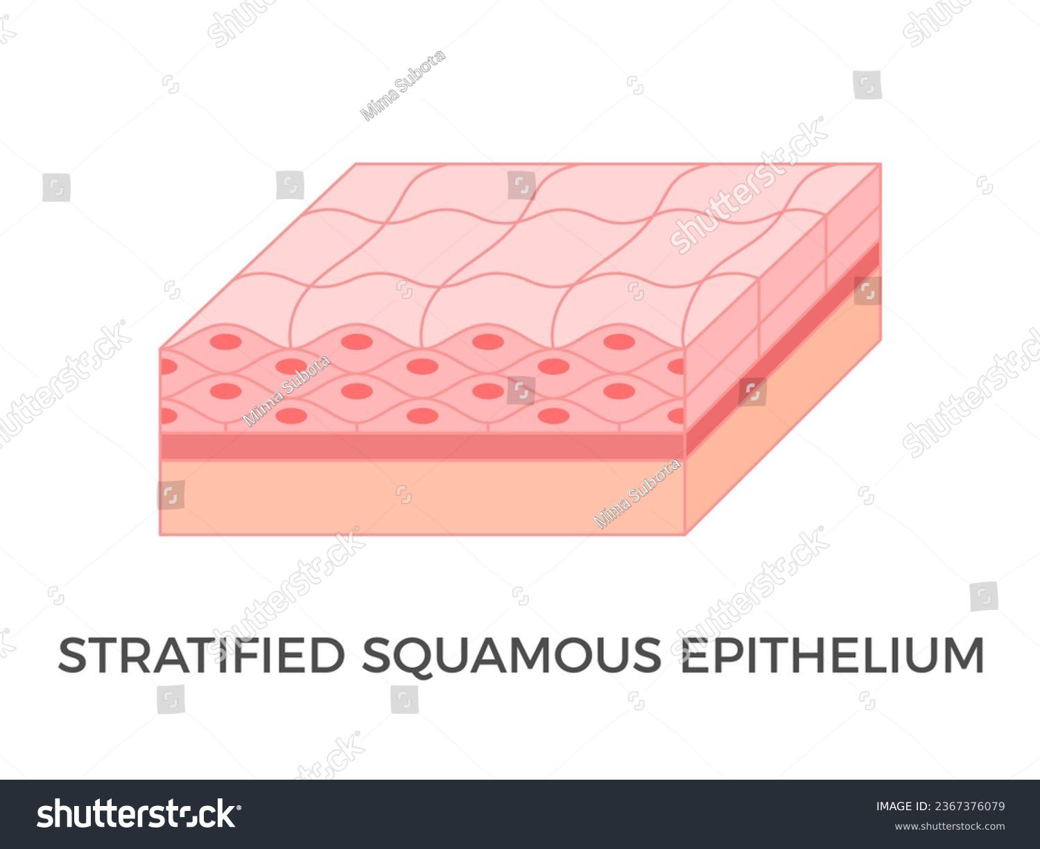
9
simple cuboidal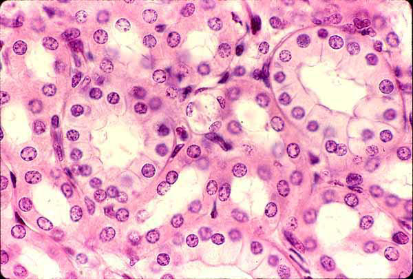
This type of epithelium is typically found in glandular (secreting) tissue and kidney tubules.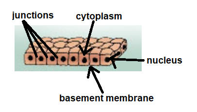
10
stratified cuboidal 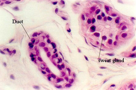
This type of epithelium is not as common and is found in the excretory ducts of your salivary and sweat glands.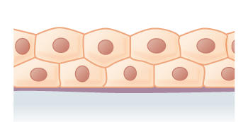
11
transitional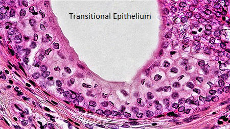
A transitional epithelium (also known as urothelium) is made up of several layers of cells that become flattened when stretched. It lines most of your urinary tract and allows your bladder to expand.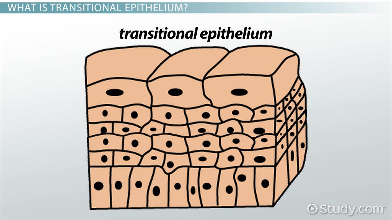
12
simple columnar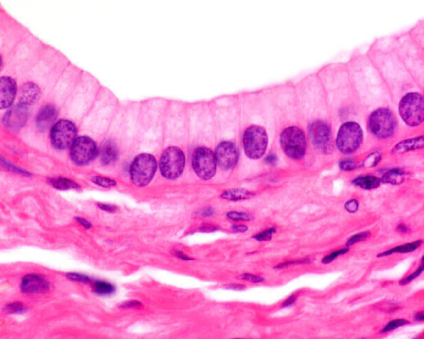
This type of epithelium is often specialized for absorption and usually has apical cilia or microvilli. These cells line your stomach and intestines.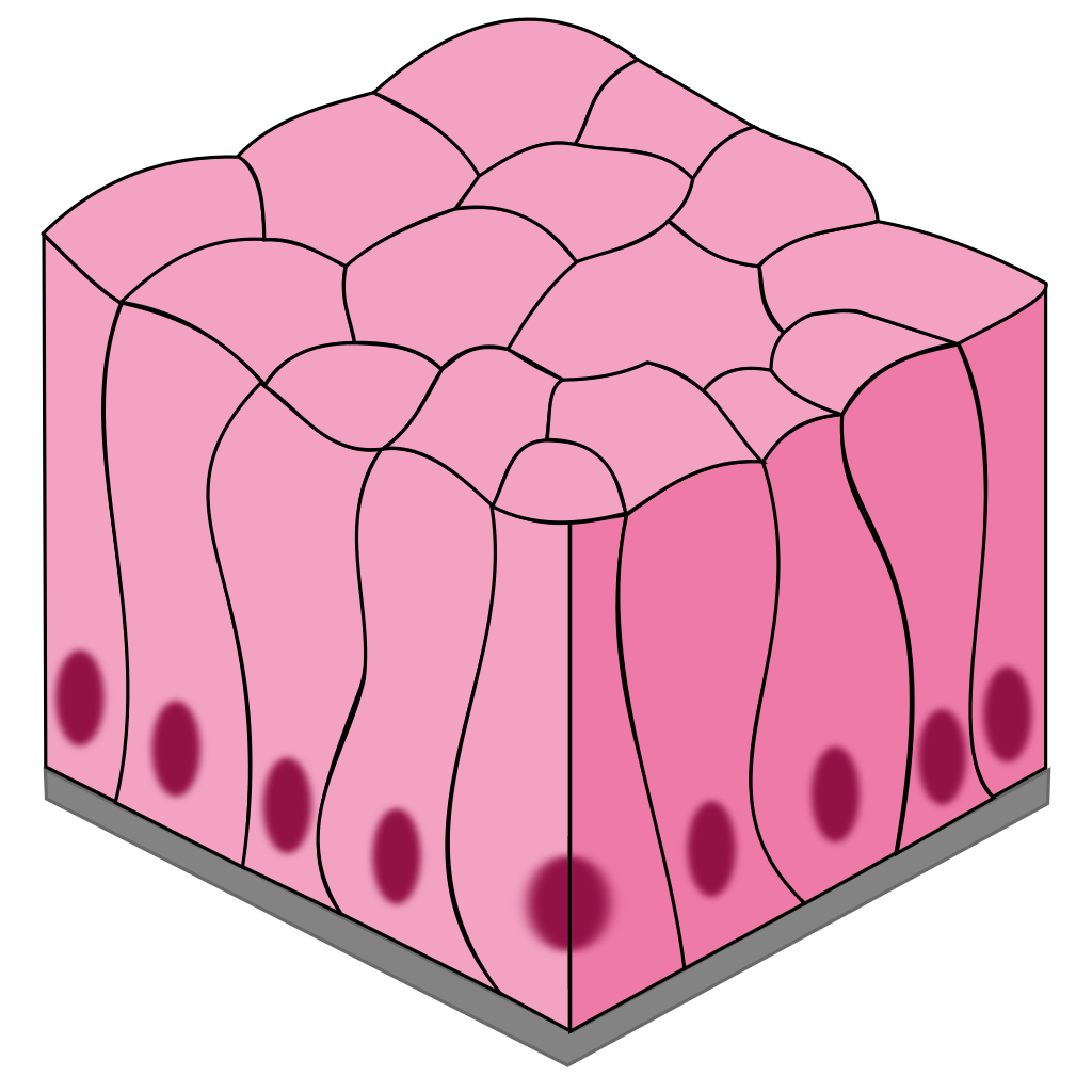
13
stratified columnar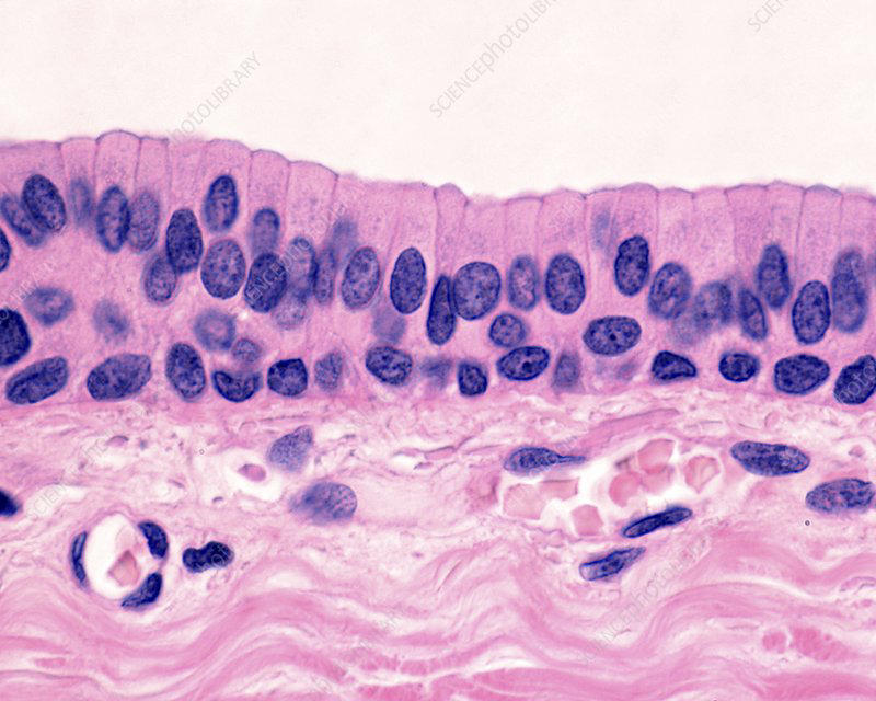
This type of epithelium is not as common and is seen in the mucous membrane (conjunctiva) lining your eyelids, where it’s both protective and mucus-secreting.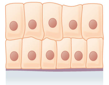
14
psuedostratified columnar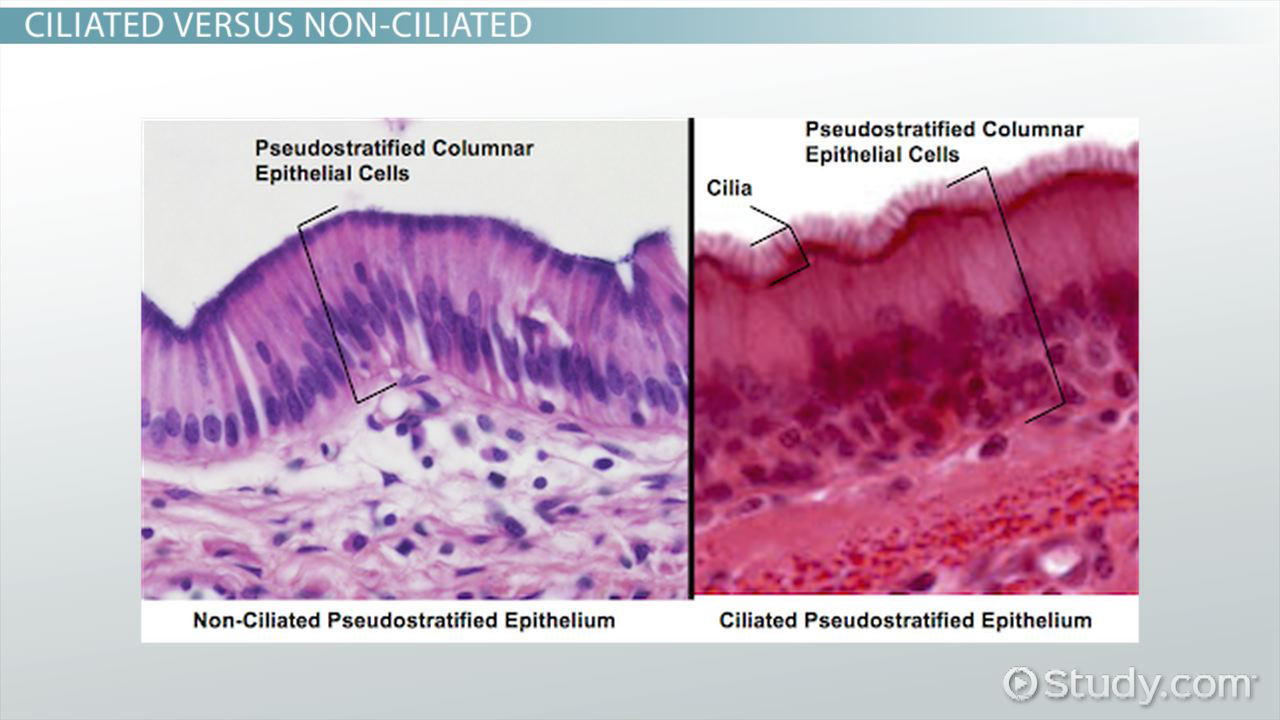
This type of epithelium lines your upper respiratory tract and usually has a lot of cilia. Found in the nasal passage of the upper respiratory tract, trachea, bronchi of the lower respiratory tract, inner ear, vas deferens, prostate gland, epididymis, and endometrium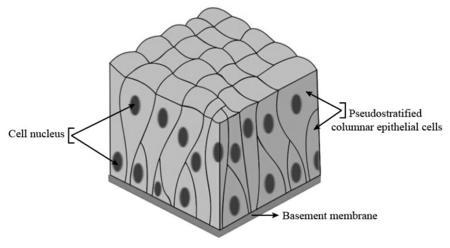
15
Microvilli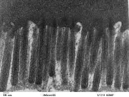
finger-like membrane protrusions, supported by the actin cytoskeleton, and found on almost all cell types. , play an important role in signal transduction leading to immune responses.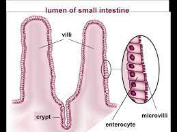
16
stereocilia 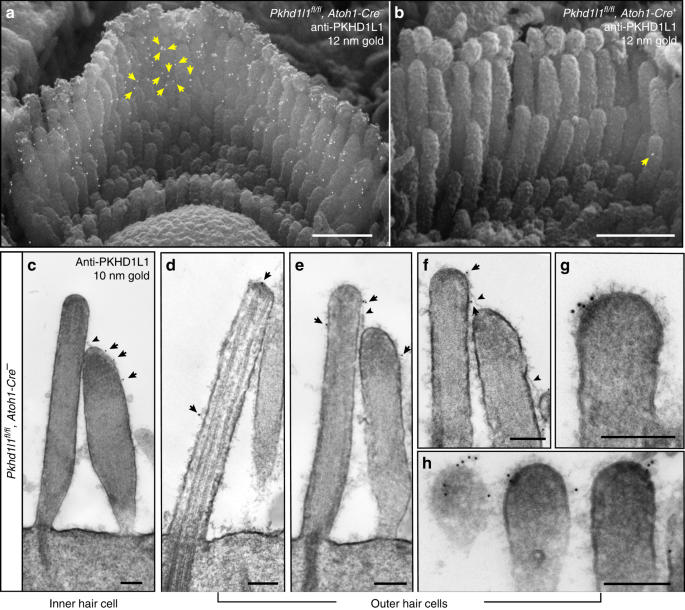
miniscule hair-like protrusions on the surface of sensory cells (also called hair cells) found deep within the cochlear and labyrinth structures of the inner ear. They serve as the key mechanosensors, responding to fluid motion for various functions, including hearing and balance.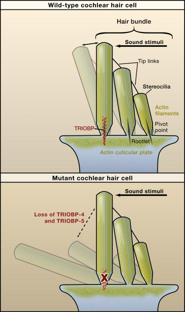
17
cilia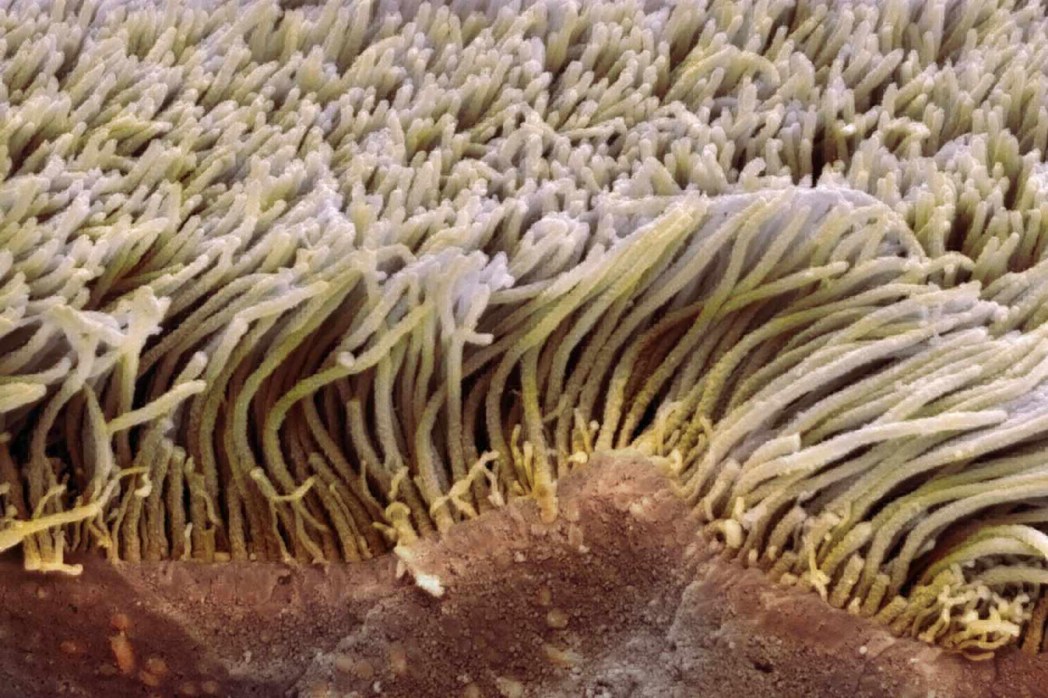
a short microscopic hairlike vibrating structure found in large numbers on the surface of certain cells, either causing currents in the surrounding fluid, or, in some protozoans and other small organisms, providing propulsion.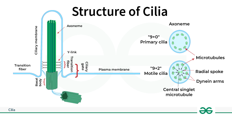
18
transepithelial absorption of glucose
Glucose is absorbed through the intestine by a transepithelial transport system initiated at the apical membrane by the cotransporter SGLT-1; intracellular glucose is then assumed to diffuse across the basolateral membrane through GLUT2.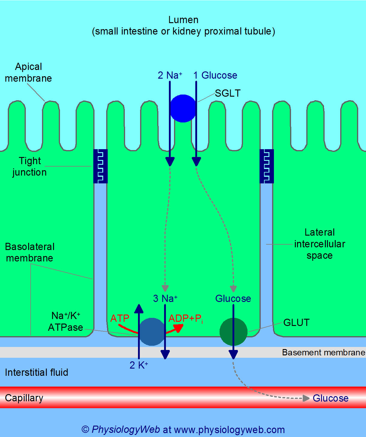
19
diffusion secondary active transport
Secondary active transport is defined as the transport of a solute in the direction of its increasing electrochemical potential coupled to the facilitated diffusion of a second solute (usually an ion) in the direction of its decreasing electrochemical potential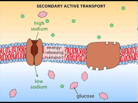
20
Active Transport of epithelium (diffusion)
For a substance to cross the epithelium, it must be transported across the cell's plasma membranes by membrane transporters. Not only do tight junctions limit the flow of substances between cells, they also define compartments in the plasma membrane. The apical plasma membrane faces the lumen.
21
components of skin
epidermis
dermis
hypodermis
dermis
hypodermis
22
epidermis layers
stratum basale (the deepest portion of the epidermis), stratum spinosum, stratum granulosum, stratum lucidum, and stratum corneum (the most superficial portion of the epidermis).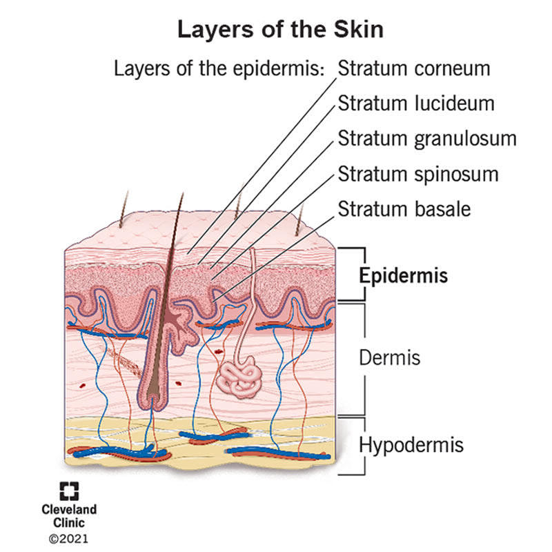
23
epidermis function
protecting your body from the outside world, keeping your skin hydrated, producing new skin cells and determining your skin color.
24
dermis layers
the papillary dermis and the reticular dermis. The papillary dermis is the superficial layer, lying deep to the epidermis. The papillary dermis is composed of loose connective tissue that is highly vascular.
25
dermis layer function
support and protect the skin and deeper layers, assist in thermoregulation, and aid in sensation.
26
hypodermis function
The hypodermis is the bottom layer of skin in your body. It has many important functions, including storing energy, connecting the dermis layer of your skin to your muscles and bones, insulating your body and protecting your body from harm. As you age, your hypodermis decreases in size, and your skin starts to sag.
27
eccrine
Eccrine sweat glands are distributed almost all over the human body, in varying densities, with the highest density in palms and soles, then on the head, but much less on the trunk and the extremities. Their water-based secretion represents a primary form of cooling in humans.
28
apocrine
Apocrine glands in the skin and eyelid are sweat glands. Most apocrine glands in the skin are in the armpits, the groin, and the area around the nipples of the breast. Apocrine glands in the skin are scent glands, and their secretions usually have an odor.
29
Holocrine
This type of secretion is rare and these glands are found in the breast and constitute some sweat glands. Holocrine glands release whole secretory cells, which later disintegrate to release the secretory products. This type of secretion is seen in sebaceous glands associated with hair follicles.
30
merocrine
The merocrine sweat glands are tubular glands and are found all over the body. They secrete watery perspiration that is carried directly to the surface of the skin. The highest number of merocrine glands are found in the palms and soles
31
Sudoriferous vs Sebaceous glands
Sudoriferous glands are sweat producing glands. These are important to help maintain body temperature. Sebaceous glands are oil producing glands which help inhibit bacteria, keep us waterproof and prevent our hair and skin from drying out.
32
hair functions
head - uv protection, insulation
nasal, ear canal, eyelashes - prevents entry of foreign material
body - sensory
nasal, ear canal, eyelashes - prevents entry of foreign material
body - sensory
33
nail function
protect fingers and toes, made of dead skin cells
eating disorders effect nail struct and production
eating disorders effect nail struct and production
34
cells of the connective tissue layer (CT)
fibroblasts, mast cells, plasma cells, macrophages, adipocytes, and leukocytes
35
fibroblasts
contributes to the formation of connective tissue, a fibrous cellular material that supports and connects other tissues or organs in the body. Fibroblasts secrete collagen proteins that help maintain the structural framework of tissues.
36
mast cells
play an important role in how the immune system responds to certain bacteria and parasites and they help control other types of immune responses. They contain chemicals such as histamine, heparin, cytokines, and growth factors
37
plasma cells
Plasma cells are differentiated B-lymphocyte white blood cells capable of secreting immunoglobulin or antibodies.
38
macropages
ingest and degrade dead cells, debris, tumor cells, and foreign materials. They promote homeostasis by responding to internal and external changes within the body, not only as phagocytes, but also through trophic, regulatory, and repair functions.
39
adipocytes
They provide energy storage, insulation from extreme temperatures and cushioning around soft organs.
40
leukocytes
Leukocytes function in body defenses. They squeeze out of the walls of blood vessels through emigration or diapedesis, then may move through tissue fluid or become attached to various organs where they fight against pathogenic organisms, diseased cells, or other threats to health.
41
matrix
The extracellular matrix of both loose and dense connective tissue proper is composed of amorphous ground substance, extracellular fluid, and collagen and elastic fibers. The amorphous ground substance is a gelatinous material surrounding the fibers and permitting extracellular fluid to diffuse through.
42
function of connective tissue
Connective tissue contributes to numerous body functions, including supporting organs and cells, transporting nutrients and wastes, defending against pathogens, storing fat, and repairing damaged tissues. Connective tissue is composed primarily of an extracellular matrix and a limited number of cells.
43
loose CT
Loose CT (or areolar tissue) is the most widespread CT of the body. It is characterized by an abundance of ground substance, plus thin and relatively few fibres and cells (Fig. 1.7). The main cellular elements are fibroblasts and a smaller amount of adipocytes.
44
how is membrane potential created
The cell membrane will use passive diffusion, facilitated diffusion,
active transport, secondary active transport, Na/K ATPase pump,
leaky channels and voltage gated channels to create a membrane
potential. This creates an electrochemical gradient. It creates an
energy potential. This potential can be converted to a form of kinetic
energy to be used for all sorts of metabolic functions
active transport, secondary active transport, Na/K ATPase pump,
leaky channels and voltage gated channels to create a membrane
potential. This creates an electrochemical gradient. It creates an
energy potential. This potential can be converted to a form of kinetic
energy to be used for all sorts of metabolic functions
45
neurons of the membrane potential
For the neuron, the main players are the Na and K voltage gated
potentials, leaky channels and the NA/K ATPase pump
potentials, leaky channels and the NA/K ATPase pump
46
Charge of the MP
Typically the membrane potential is – 70 mv with the ICF being
negative. Remember large protein anions do not pass the cell
membrane.
negative. Remember large protein anions do not pass the cell
membrane.
47
purpose of the Grade Potential
Purpose is to send messages to the neuron’s cell body. The neuron
receiving these messages will “sum up” all the messages from many
synapses and will either 1. fire, 2. be inhibited or 3. be modified.
receiving these messages will “sum up” all the messages from many
synapses and will either 1. fire, 2. be inhibited or 3. be modified.
48
size of the GP
Graded potentials can VARY ACCORDING TO THE SIZE OF THE STIMULUS –
DIFFERENT THAN THE ALL OR NONE ACTION POTENTIAL. These stimuli are
created in the dendrites
If the collection of graded potentials is strong enough as it travels down to
the axon hillock and be at -55mV or above, it will trigger an action
potential. Action potentials will degrade or lessen as they travel unlike an
action potential.
DIFFERENT THAN THE ALL OR NONE ACTION POTENTIAL. These stimuli are
created in the dendrites
If the collection of graded potentials is strong enough as it travels down to
the axon hillock and be at -55mV or above, it will trigger an action
potential. Action potentials will degrade or lessen as they travel unlike an
action potential.
49
GP simmulation
Graded potentials can accumulate by temporal summation – one
synapse firing repeatly or by spatial summation with many synapses
firing at the same time. Some synapses can be excitatory and some
inhibitory. The neuron receiving the stimuli will sum up the stimuli.
synapse firing repeatly or by spatial summation with many synapses
firing at the same time. Some synapses can be excitatory and some
inhibitory. The neuron receiving the stimuli will sum up the stimuli.
50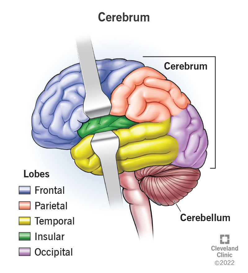
Image occlusion: name the hidden labels
51
Image occlusion: name the hidden labels
52
Image occlusion: name the hidden labels
53
Image occlusion: name the hidden labels
54
Image occlusion: name the hidden labels
55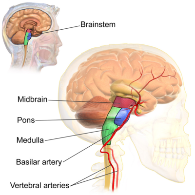
Image occlusion: name the hidden labels
56
Image occlusion: name the hidden labels
57
Image occlusion: name the hidden labels
58
Image occlusion: name the hidden labels
59
Image occlusion: name the hidden labels
60
Image occlusion: name the hidden labels
61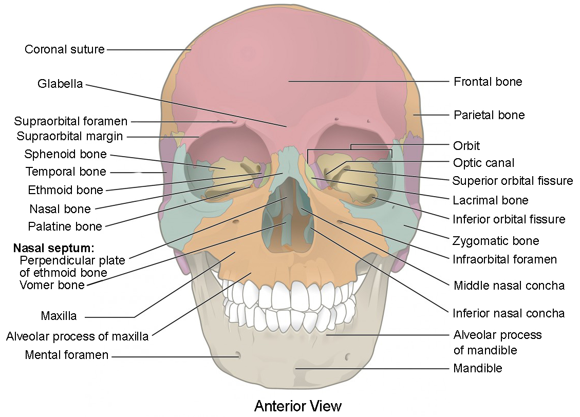
Image occlusion: name the hidden labels
62
Image occlusion: name the hidden labels
63
Image occlusion: name the hidden labels
64
Image occlusion: name the hidden labels
65
Image occlusion: name the hidden labels
66
Image occlusion: name the hidden labels
67
Image occlusion: name the hidden labels
68
Image occlusion: name the hidden labels
69
Image occlusion: name the hidden labels
70
Image occlusion: name the hidden labels
71
Image occlusion: name the hidden labels
72
Image occlusion: name the hidden labels
73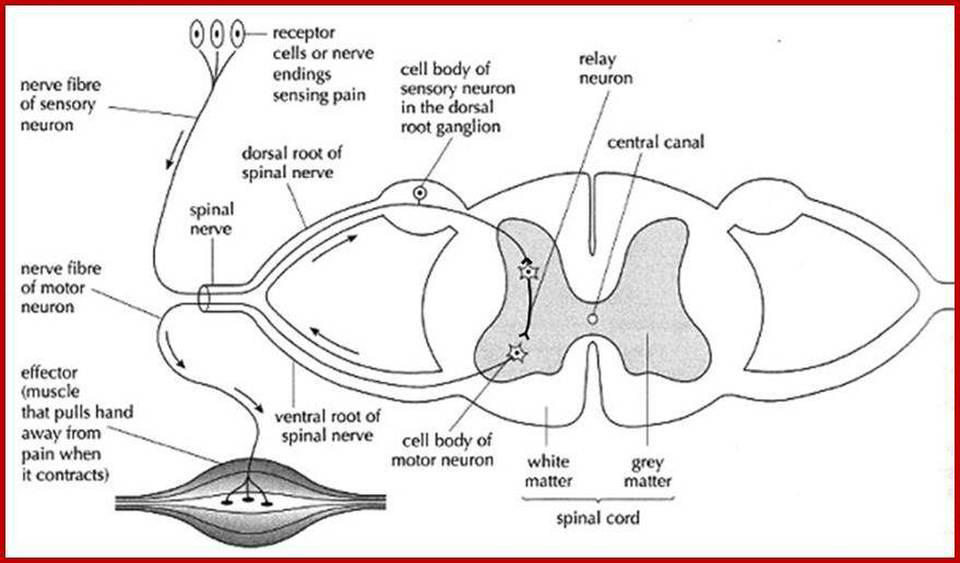
Spinal Cord Reflex
74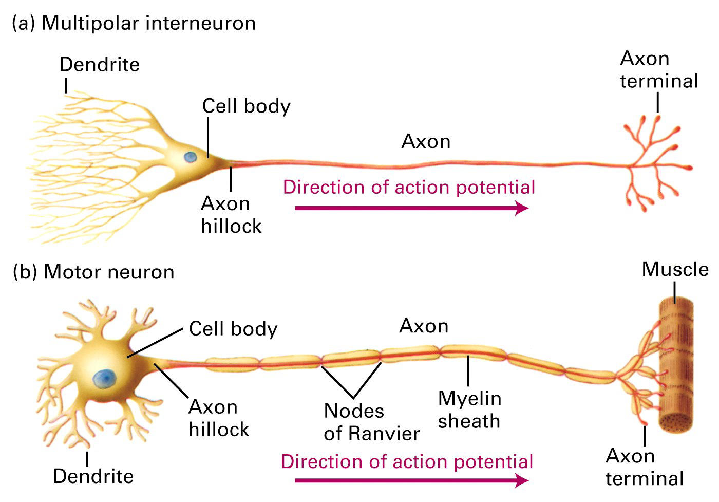
Action Potential
75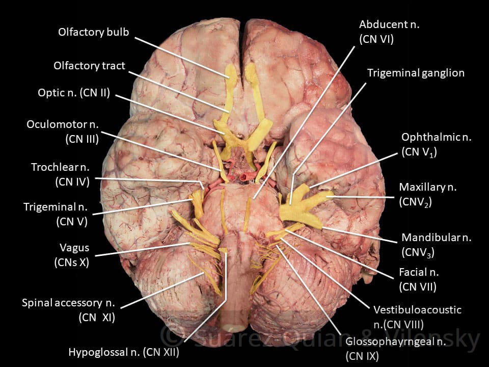
Cranial nerves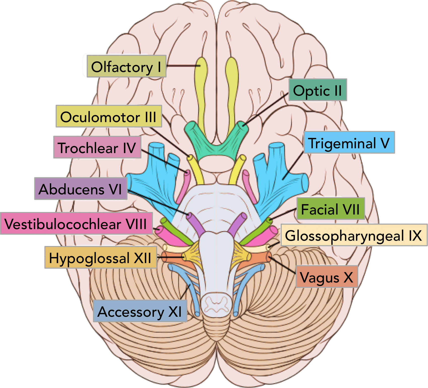
76
Image occlusion: name the hidden labels
77
Image occlusion: name the hidden labels
78
Image occlusion: name the hidden labels
79
Image occlusion: name the hidden labels
80
Image occlusion: name the hidden labels
81
precentral gyrus
It is responsible for initiating and controlling voluntary movements in the body by generating neural signals.
82
dorsolateral frontal cortex
task switching and task-set reconfiguration, prevention of interference, inhibition, planning, and working memory
83
parietal lobes function
The parietal lobes are primarily responsible for receiving and processing sensory input such as touch, pressure, heat, cold, and pain. The parietal lobes are also involved in the perception of body awareness and the construction of a spatial coordinate system (mental map) to represent the world around us.
84
frontal lobe function
The frontal lobes are important for voluntary movement, expressive language and for managing higher level executive functions. Executive functions refer to a collection of cognitive skills including the capacity to plan, organise, initiate, self-monitor and control one's responses in order to achieve a goal.
85
occipital lobe function
The occipital lobe is the visual processing area of the brain. It is associated with visuospatial processing, distance and depth perception, color determination, object and face recognition, and memory formation.
86
temporal lobe function
They are most commonly associated with processing auditory information and with the encoding of memory. The temporal lobes are also believed to play an important role in processing affect/emotions, language, and certain aspects of visual perception.
87
receptor D1
memory, attention, impulse control, regulation of renal function, locomotion.
88
receptor D2
locomotion, attention, sleep, memory, learning.
89
receptor D3
cognition, impulse control, attention, sleep.
90
receptor D4
cognition, memory, fear, impulse control, attention, sleep.
91
muscle spindles
stretch receptors within the body of a skeletal muscle that primarily detect changes in the length of the muscle. They convey length information to the central nervous system via afferent nerve fibers. This information can be processed by the brain as proprioception.
92
Golgi tendon organs (GTOs)
proprioceptors that are located in the tendon adjacent to the myotendinous junction. The Golgi tendon organ is a tree-like sensory ending enclosed in a spindle-like connective tissue capsule, that lies near the junction of a tendon with a muscle.
93
spinal cord reflexes
simple behaviors produced by central nervous system (CNS) pathways that lie entirely within the spinal cord.
94
CN 1 - olfactory
transmits information regarding a person’s sense of smell to the brain.
95
CN 2 - optic
transmits information to the brain regarding a person’s vision.
96
CN 3/4/6- oculomotor/trochlear/abducens
helps control muscle movements of the eyes.
97
CN 5 - Trigeminal
facial sensation, muscle movement
98
CN 7 - facial
facial expression
99
CN 8 - vestibulocochlear
helps with a person’s hearing and balance.
100
CN 9 - Glossopharyngeal
posterior tongue control, produces saliva
101
CN 10 - Vagas
digestion, heart rate, etc.
102
CN 11 - acessory
provides motor function to some muscles in the neck.
103
CN 12 - hypoglossal
motor nerve that supplies the tongue muscles. It originates in the medulla.
104
parasympathetic vs sympathetic
parasympathetic- rest and digest
sympathetic - fight or flight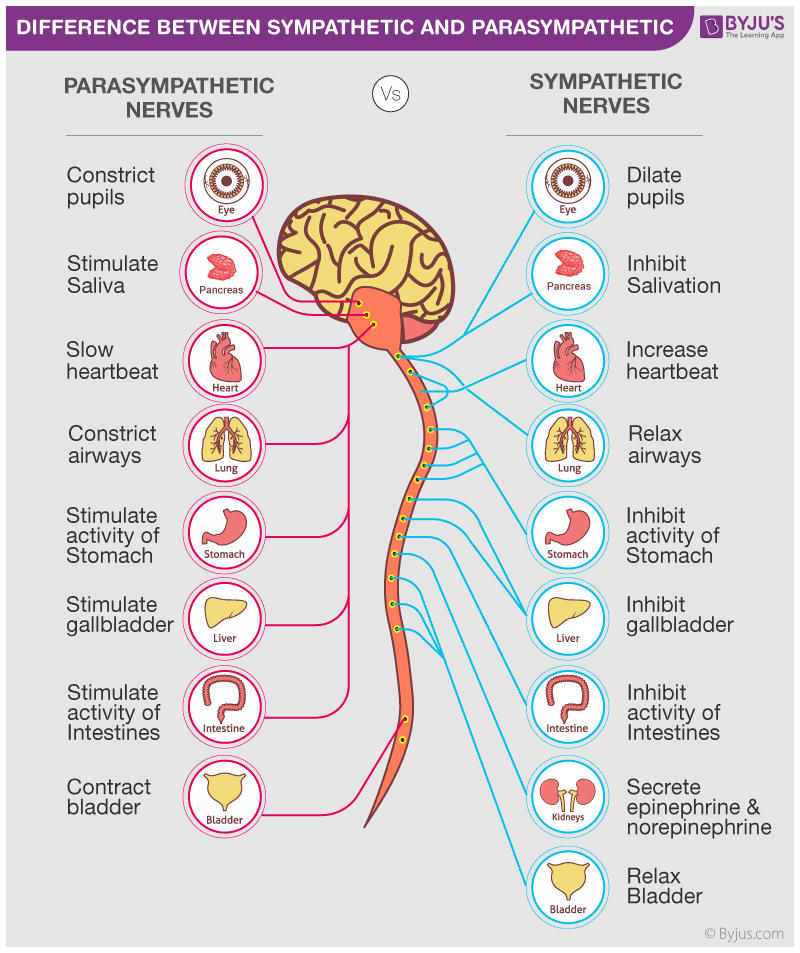
sympathetic - fight or flight
105
adrenaline
fight ir flight; increased heart rate and blood flow, heightened awareness
106
GABA
calms firing nerves in the CNS, high levels increase focus, low causes anxiety
107
noradrenaline
affects attention; contracts blood flow
108
acetylcholine
affects learning; activates muscles in the body
109
dopamine
pleasure, addiction, movement/ motivation
110
glutamate
most common; also involved in learning and memory
111
serotonin
effects mood; well being and happiness
112
endorphins
euphoria feeling from excercise, excitment, and sex