All cards
1
Normal pH range?
7.35-7.45
2
Normal sodium range?
135-145
3
Normal potassium range
3.5-5
4
what is potassium
potassium is an irritant at the neuromuscular junction
5
what does potassium allow for
it is allows for normal muscle function: cardiac, skeletal
6
what is calcium at the nerve level?
a sedative
7
calcium is inversely proportional to what?
phosphate
8
what is calcium necessary for?
blood coagulation and muscle contraction
9
if you have too much K+ what should you get?
a cardiac monitor
10
what's the function of magnesium?
function of nueromuscular junction
11
What is phosphate necessary for?
production of ATP
12
Q: List the symptoms of hyponatremia.
Decreased LOC, lethargy, confusion, seizures, coma, muscle twitching and weakness, diminished DTRs, oliguria or anuria, nausea, vomiting, and abdominal cramps.
13
What are the common neurological signs of hypernatremia?
Agitation, delirium, coma, seizures, and fever.
14
How does hypernatremia affect the skin? Why?
causes dry flushed skin, too much water loss
15
What is a common IV solution to treat sodium loss in hyponatremia?
Replace Na, usually with normal saline (NS).
16
How should fluid intake be managed for a patient with water gain in hyponatremia?
Restrict fluid.
17
Which IV solution is recommended for severe neurological signs in hyponatremia to prevent fluid overload?
3% or 5% NS IV slowly.
18
What IV fluid is advised for water loss in hypernatremia?
IV Fluid: D5W and oral glucose-electrolyte solution.
19
In the context of hypernatremia, why might 0.45 NS be used?
To prevent a swing to hyponatremia and avoid cerebral edema.
20
What is the meaning of "oliguria"?
Scant amounts of urine.
21
Define "anuria" and its relevance in renal patients.
nuria means no urine production, and it often comes up with renal patients.
22
How might a high protein tube feed relate to hypernatremia?
High protein tube feeds could contribute to excess sodium.
23
don't just treat the symptoms treat the _______
cause
24
What are the life-threatening cardiac disturbances seen in hypokalemia?
Dysrhythmias, with ECG changes including elevated U waves (specific to hypokalemia), flattened T waves, also muscle weakness, abdominal distention, and constipation.
25
Why is hyperkalemia considered the most dangerous electrolyte disorder?
Due to its association with cardiac dysrhythmias, which can lead to cardiac arrest.
26
Q: What are the gastrointestinal symptoms of hyperkalemia?
A: Nausea, diarrhea, and abdominal cramps.
27
What is crucial to monitor after potassium replacement in hypokalemia?
A: It's essential to repeat the K+ level post-replacement and monitor urine output.
28
Q: What are the primary interventions for hyperkalemia?
A: Restrict K intake, administer 10% Calcium Gluconate, IV Insulin & Glucose, Loop Diuretics, and Dialysis.
29
Q: Why is insulin given in the case of hyperkalemia?
A: Insulin is used to push potassium into the cell. This is why glucose is also given, to compensate for the sugar drop due to insulin.
30
what interventions do you take when administering parenteral k additives?
Administer slowly, urine output, phlebitis, monitor for decreased NA levels, potassium can't be administered to fast or too slow. If K is low, it can lead to fast heart rate and overload, if k is too high, your heart will slow down to the point where you will die.
31
Q2: Which two signs are indicative of hypocalcemia?
A2: Chvostek's sign and Trousseau sign.
32
Q3: What cardiac symptom can be associated with hypocalcemia?
A3: Cardiac dysrhythmias.
33
Q4: Describe the reflex response in hypocalcemia.
A4: Hyperactive reflexes.
34
Q5: What sensations might a patient with hypocalcemia experience?
A5: Numbness & tingling in fingers. (paresthesia)
35
Q6: What gastrointestinal symptoms can hypocalcemia present?
A6: Hyperactive bowel sounds, cramping, and diarrhea.
36
Q7: What is the primary intervention for hypocalcemia?
A7: Replace calcium - IV calcium gluconate, administer slowly via pump, and observe for infiltration.
37
Q9: What are the respiratory and cardiac complications of hypercalcemia?
A9: Respiratory depression/arrest and cardiac dysrhythmias.
38
Q10: Describe the musculoskeletal and mood changes in hypercalcemia.
A10: Weakness and muscle flaccidity; and CNS depression.
39
Q11: What are some symptoms related to bone health in hypercalcemia?
A11: Bone pain, pathological fractures, renal calculi due to calcium loss from bones.
40
Q12: What gastrointestinal symptoms are associated with hypercalcemia?
A12: Decreased bowel sounds, anorexia, nausea, and vomiting, constipation.
41
Q13: What are the primary interventions for hypercalcemia?
A13: IV therapy with normal saline, IV phosphate, limiting dietary calcium intake, administering loop diuretics, monitoring for renal calculi in urine, and assessing for signs of digoxin toxicity.
42
Q14: Why might a woman with osteopenic weakened bones and a high calcium level be a concern?
A14: It could indicate a hyperparathyroid problem as it's not a natural occurrence, because they should have low calcium due to weakned bones as they age
43
Q15: What specific condition is often checked for in women with hypercalcemia?
A15: Metastatic cancer.
44
what is something to look out for if someone has hypocalcemia?
bleeding, because if they have low calcium, their blood clotting ability is weakened
45
Q2: What happens to reflexes when magnesium is too low?
A2: They become hyperactive.
46
Q3: Name a cardiac problem with low magnesium.
A3: Cardiac dysrhythmias.
47
Q4: How can the bowel sound in hypomagnesaemia?
A4: Slower or quiet.
48
Q5: If someone has low magnesium, how might their appetite be?
A5: Reduced (anorexia).
49
Q7: What breathing problem can occur with high magnesium?
A7: Respiratory depression/arrest.
50
Q8: How does the heart react to too much magnesium?
A8: It might have irregular beats (dysrhythmias).
51
Q9: Describe muscles when magnesium is high.
A9: They can be weak or flaccid.
52
Q10: What might someone feel with very high magnesium levels?
A10: Warm and flushed.
53
3. Q: What are common symptoms of low phosphate levels?
A: Pins and needles (paresthesias), muscle weakness, bone pain, chest pain, confusion, cardiomyopathy, respiratory failure, seizures, coma, tissue hypoxia, and increased susceptibility to infection.
54
Q: Which eye movement is associated with hypophosphatemia?
Nystagmus (involuntary side to side movement)
55
what does hypomagenesaemia look like? and why?
hypocalcemia, because you have a small amount of sedative
56
what is hypophosphatemia similar to?
hyperkalcemia
57
5. Q: What are some symptoms of high phosphate levels?
A: Tetany, tachycardia, anorexia, nausea, vomiting, symptoms of hypocalcemia, hyperactive reflexes, and soft tissue calcifications in organs like the lungs, heart, kidneys, and cornea.
58
Q: What is tetany in the context of hyperphosphatemia?
A: Involuntary muscle contractions.
59
Q: Which form of treatment is used for severe hypophosphatemia?
A: IV phosphate.
monitor for signs of inflitrations
monitor for signs of inflitrations
60
7. Q: What's the main goal when treating hypophosphatemia?
prevention
61
10. Q: What is the primary approach in treating hyperphosphatemia?
A: Treating the underlying cause.
62
Q: What dietary restriction is recommended for hyperphosphatemia?
A: Restricting dietary phosphate.
63
Q: What medications are used for treating hyperphosphatemia?
A: Phosphate binders and oral and parenteral calcitriol.
64
range for chloride?
97-107
65
range for calcium?
8.6-10.2
66
range for phosphate?
2.5-4.5
67
range for magnesium?
1.3-2.3
68
3. Q: What might someone with hypochloremia experience?
A: Hyperexcitability of muscles, tetany, hyperactive DTRs (deep tendon reflexes), muscle weakness, muscle cramps, agitation, irritability, tremors, slow shallow respirations, dysrhythmias, seizures, and coma.
69
4. Q: What are symptoms of hyperchloremia?
A: Tachypnea (fast breathing), deep and rapid respirations (Kussmaul’s respirations), weakness, lethargy, diminished cognitive ability, hypertension, decreased cardiac output, dysrhythmias, and coma.
70
Q: What's meant by "Kussmaul’s respirations"?
A: Really deep and rapid breathing.
71
6. Q: What's the main approach to treat hypochloremia?
A: Correct the underlying cause, encourage dietary intake of foods rich in chloride content, and administer IV normal saline.
72
7. Q: How is hyperchloremia generally treated?
A: Correct the underlying cause, restore the electrolyte, fluid, and acid-base balance, restrict sodium and chloride intake, and may administer hypotonic IV solutions, LR solution, or IV sodium bicarbonate. Additionally, diuretics may be used.
73
Q: What forms when carbon dioxide combines with water?
A: Carbonic acid (H₂CO₃).
74
Q: How does carbonic acid (H₂CO₃) dissociate?
A: Into a hydrogen ion (H⁺) and bicarbonate (HCO₃⁻).
75
Q: What are metabolic acids?
A: Any acids that are not carbonic acid.
76
4. Q: What is the purpose of buffers in the body?
A: Buffers are pairs of chemicals that work together to maintain the normal pH of body fluids.
77
Q: How does a base work in terms of trapping ions?
A: A base traps H⁺ ions, and the bicarbonate base (HCO₃⁻) combines with the hydrogen ion to form carbonic acid (H₂CO₃).
78
Q: What happens when an acid releases ions?
A: Carbonic acid (H₂CO₃) releases a hydrogen ion (H⁺) to form bicarbonate (HCO₃⁻).
79
Q: What do you excrete when you exhale?
A: Carbonic acid in the form of CO₂ and water.
80
Q: What does PaCO₂ reflect?
A: The level of CO₂ in the blood, with a normal range being 35 - 45 mmHg.
81
Q: How does an increased PaCO₂ affect pH and respiratory rate?
A: If PaCO₂ rises, pH falls (acidosis) and the respiratory rate & depth increase to exhale acids.
82
Q: How does a decreased PaCO₂ influence pH and respiratory rate?
A: If PaCO₂ decreases, pH rises (alkalosis) and the respiratory rate & depth decrease to retain acids.
83
Q: Which organ is the primary controller of carbonic acid levels?
A: The lungs.
84
6. Q: What is the role of the kidneys in acid excretion?
A: Kidneys excrete all acids except carbonic acid.
85
Q: What is the normal blood level of bicarbonate (HCO₃⁻)?
A: 22 - 26 mEq/L.
86
Q: How does an increase in HCO₃⁻ levels affect pH?
A: If HCO₃⁻ levels rise, pH rises (alkalosis).
87
Q: How does a decrease in HCO₃⁻ levels affect pH?
A: If HCO₃⁻ levels drop, pH drops (acidosis).
88
Q: How do kidneys regulate acid balance?
A: Through the absorption or excretion of acids & bases, and by producing HCO₃⁻ to restore losses.
89
Q: Which organ is the primary controller of bicarbonate levels?
A: The kidneys.
90
13. Q: How does potassium compensate for hydrogen ion level changes during acidosis?
A: In acidosis, potassium moves out of cells to make room for hydrogen ions, leading to an increase in serum potassium levels.
91
Q: How does potassium compensate for hydrogen ion level changes during alkalosis?
A: In alkalosis, potassium moves into cells, leading to a decrease in serum potassium levels.
(think K is low, al K low sis)
(think K is low, al K low sis)
92
ROME?
respiratory opposite, metabolic equal
93
ph (normal)
co2 (off)
bicarb (off)
what's the compensation?
co2 (off)
bicarb (off)
what's the compensation?
fully compensated
94
ph (abnormal)
co2 (abnormal)
bicard (abnormal)
what's the compensation?
co2 (abnormal)
bicard (abnormal)
what's the compensation?
partially compensated
95
ph abnormal
co2 abnormal
bicarb normal
OR
ph (abnormal)
co2 (normal)
bicarb abnormal
whats the compensation?
co2 abnormal
bicarb normal
OR
ph (abnormal)
co2 (normal)
bicarb abnormal
whats the compensation?
uncompensated
96
normal bicarb range?
22-26
97
fill in the blanks 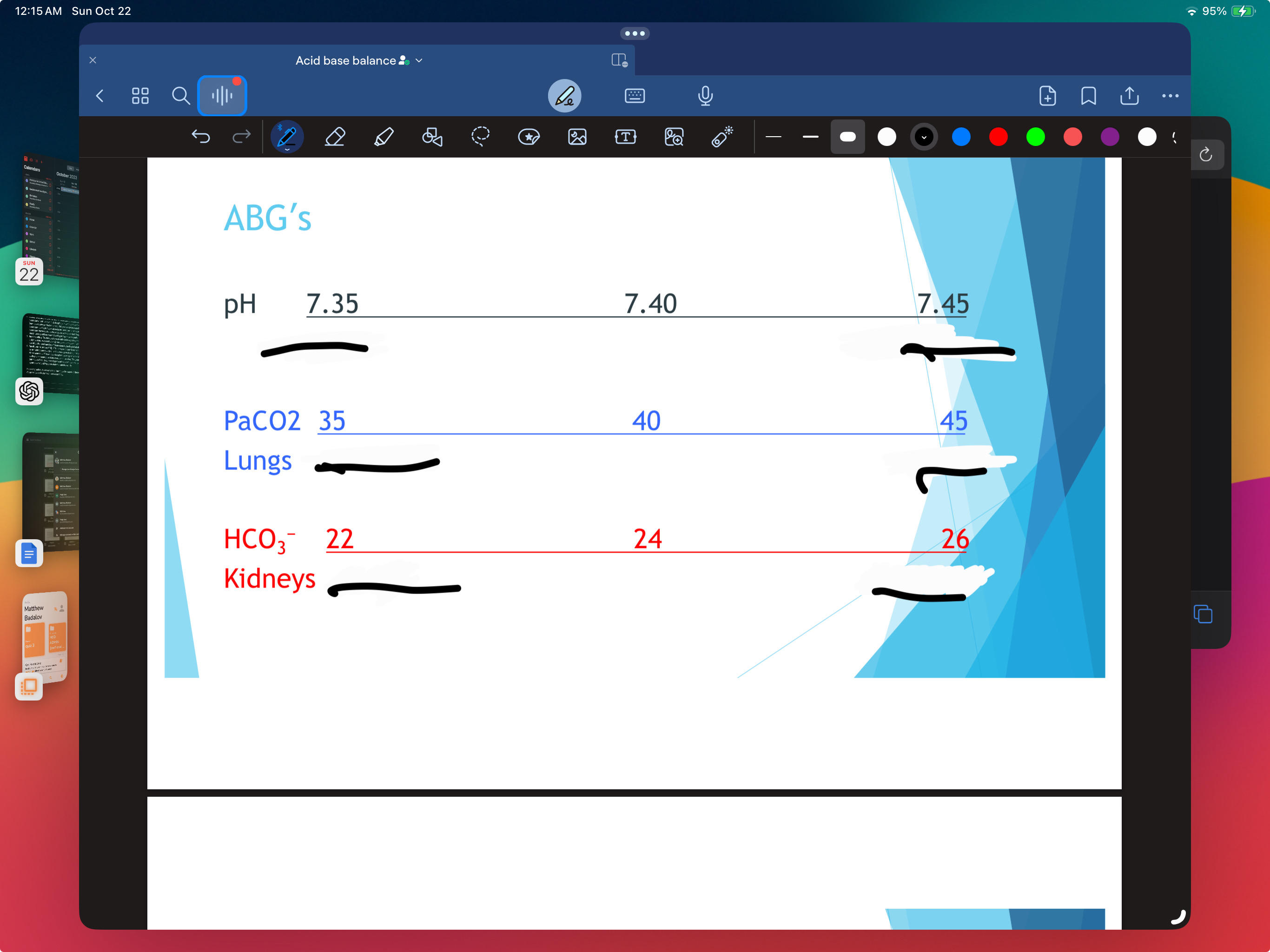
remember ROAM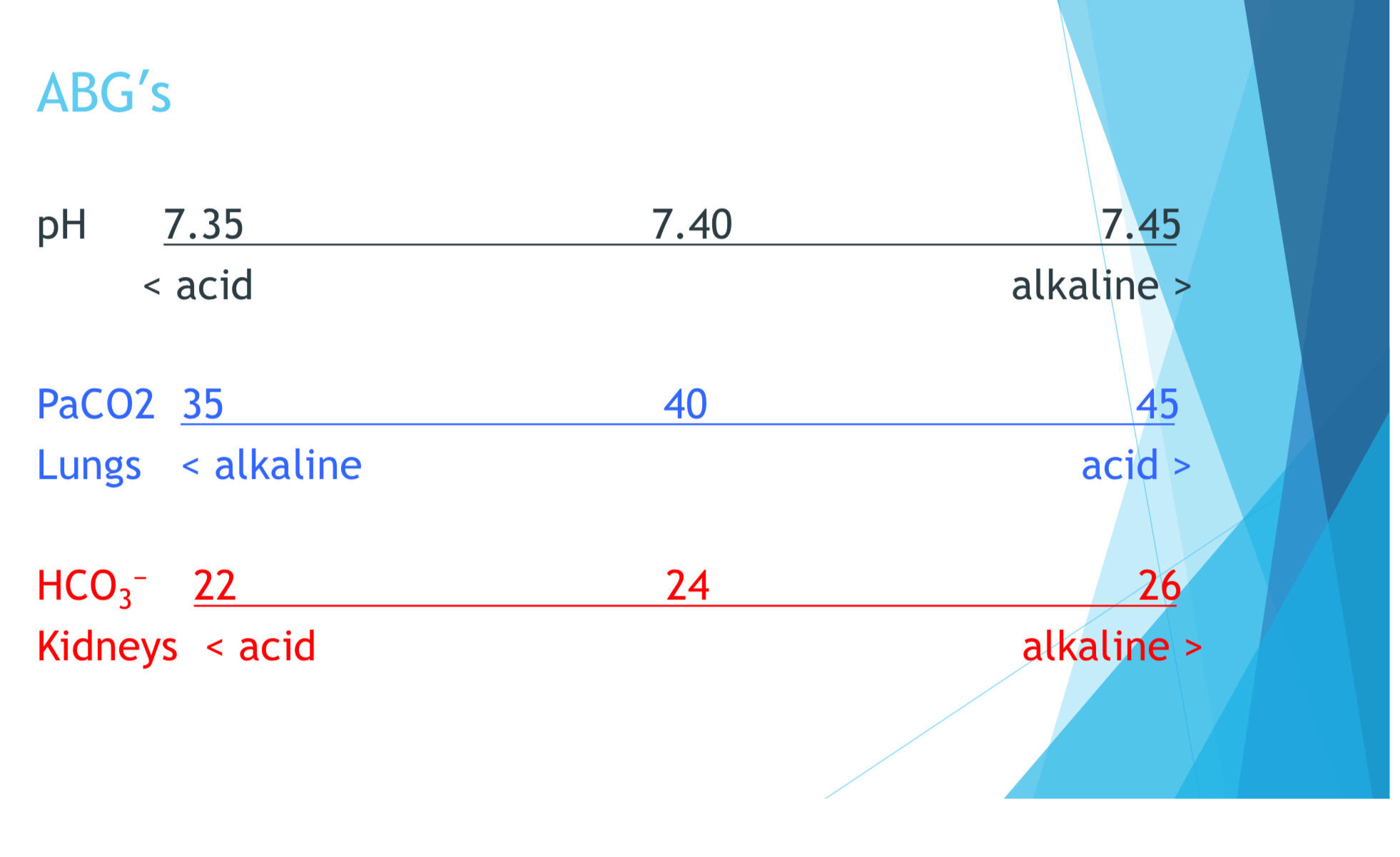
98
Q: Name a few causes of respiratory acidosis?
A: Hypoventilation (main), hypercapnia, pneumonia, atelectasis, pulmonary edema, bronchial asthma, COPD, respiratory failure, airway obstruction, and more.
99
Q: Which drugs or conditions affecting the respiratory center can lead to respiratory acidosis?
A: Narcotics, benzodiazepines, barbiturates, anesthesia, head trauma.
100
Q: What mental changes might be observed in a patient with respiratory acidosis?
A: Apprehension, confusion, and decreasing level of consciousness leading to unresponsiveness.
101
Q: Describe some physical manifestations seen in respiratory acidosis.
A: Ashen color, rapid and shallow respirations, tachycardia, low BP, diminished deep tendon reflexes (DTRs), and dysrhythmias. There's also an increase in potassium levels, resulting in hyperkalemia.
102
Q: What is a priority nursing intervention for a patient with respiratory acidosis?
A: Protecting the airway and ensuring adequate ventilation.
103
Q: Which medical interventions can assist in breathing for patients with respiratory acidosis?
A: Respiratory assistance, bronchodilators, supplemental O2.
104
Q: If a patient has COPD and is in respiratory acidosis, what oxygen flow is recommended?
A: Low flow O2.
105
Q: What might a nurse encourage a patient to do to clear the airways in respiratory acidosis?
A: Cough and deep breathe. The use of a chest tube might also be relevant.
106
Q: What should a nurse be alert for when caring for a patient with respiratory acidosis?
A: Respiratory failure.
107
Q: Main breathing pattern causing respiratory alkalosis?
A: Hyperventilation
108
Q: Name a few potential causes of respiratory alkalosis?
A: Anxiety, fever, sepsis, heart failure, liver failure, hypoxia, salicylate poisoning.
109
Q: Common symptoms of respiratory alkalosis?
A: Lightheadedness, dizziness, anxiety, confusion, restlessness, blurred vision, dry mouth.
110
Q: How can respiratory alkalosis affect extremities?
A: Spasms, tetany, and paresthesia.
111
Q: Which electrolyte imbalance can accompany respiratory alkalosis?
Hypokalemia (fluid into cells) low serum K
112
Q: Primary goal in treating respiratory alkalosis?
A: Raise CO2.
113
Q: Other than treating the root cause, name two interventions for respiratory alkalosis?
A: Supplemental O2 and sedation.
114
Q: What precautions are taken in respiratory alkalosis?
A: Seizure precautions.
115
Q: What should nurses monitor for in patients with respiratory alkalosis?
A: Respiratory fatigue/failure.
116
Q: Main causes of metabolic acidosis?
A: Base deficit, excess acids, diarrhea, renal failure, DKA.
117
Q: Name a symptom of metabolic acidosis related to respiration?
A: Kussmaul breathing (rapid deep breathing)
118
Q: What complications can arise if metabolic acidosis is untreated?
A: Dysrhythmias, coma, cardiac arrest.
119
Q: Name a few symptoms of metabolic acidosis?
A: Lethargy, warm skin, nausea, muscle weakness.
120
Q: Main treatments for metabolic acidosis?
A: Treat underlying cause, support breathing and BP, monitor neurological status.
121
Q: Primary reasons for metabolic alkalosis?
A: Excess base ingestion, loss of acids, prolonged vomiting.
122
Q: Name a diuretic causing metabolic alkalosis?
A: Potassium wasting diuretics.
123
Q: Common symptoms of metabolic alkalosis?
A: Hypokalemia, confusion, nausea, shallow breathing, tetany.
124
Q: What can prolonged metabolic alkalosis lead to?
A: Seizures, dysrhythmias, coma.
125
Q: Key interventions for metabolic alkalosis?
A: Administer KCL, monitor potassium, provide supplemental O2, address underlying causes.
126
Q: What happens during respiratory acidosis regarding CO2?
A: Hypoventilation leads to retention of CO2.
127
Q: In respiratory alkalosis, what's the initial respiratory response?
A: Hyperventilation to exhale more CO2.
128
Q: Which electrolyte abnormality is often seen with acidosis?
A: Hyperkalemia (increased potassium)
129
Q: What electrolyte change is commonly associated with alkalosis?
A: Hypokalemia (decreased potassium)
130
Q: Can the kidney or lung compensate for its own acid-base imbalances?
A: No, kidney or lung cannot compensate for itself.
131
Q: Which tissue anchors the skin layers to the underlying tissues?
A: Subcutaneous tissue
132
Q: What type of cells predominantly make up the epidermis?
A: Epithelia cells
133
Q: What are the primary functions of the epidermis?
A: Protection and waterproofing
134
Q: What type of tissue is the dermis mainly composed of?
A: Connective tissue
135
Q: Name two structures found in the dermis.
A: Nerves and blood vessels (Other answers like hair follicles, glands are also correct.)
136
Q: What does adipose tissue in the subcutaneous layer contain?
A: Blood & lymph vessels and fat cells
137
what vitamin does the skin help to synthesize when exposed to sunlight?
vit. D
138
impaired skin integrity =
risk for infections
139
fill blanks 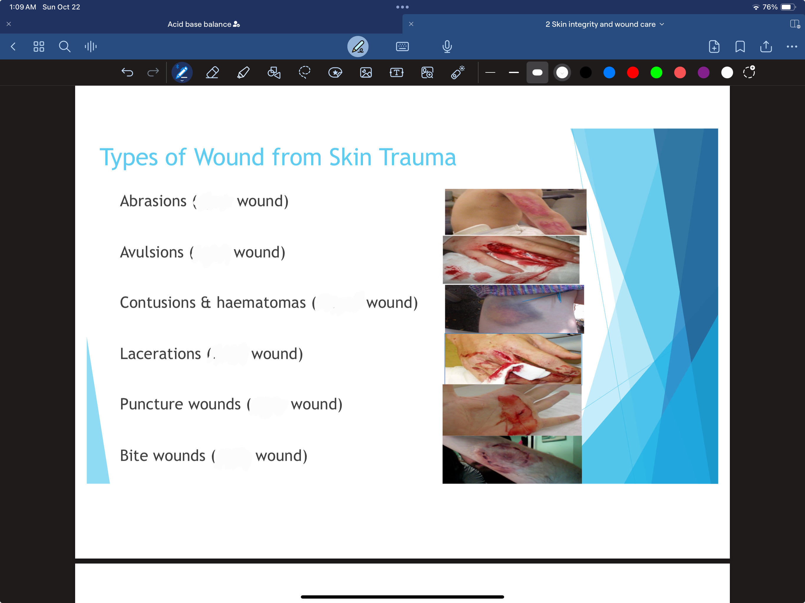
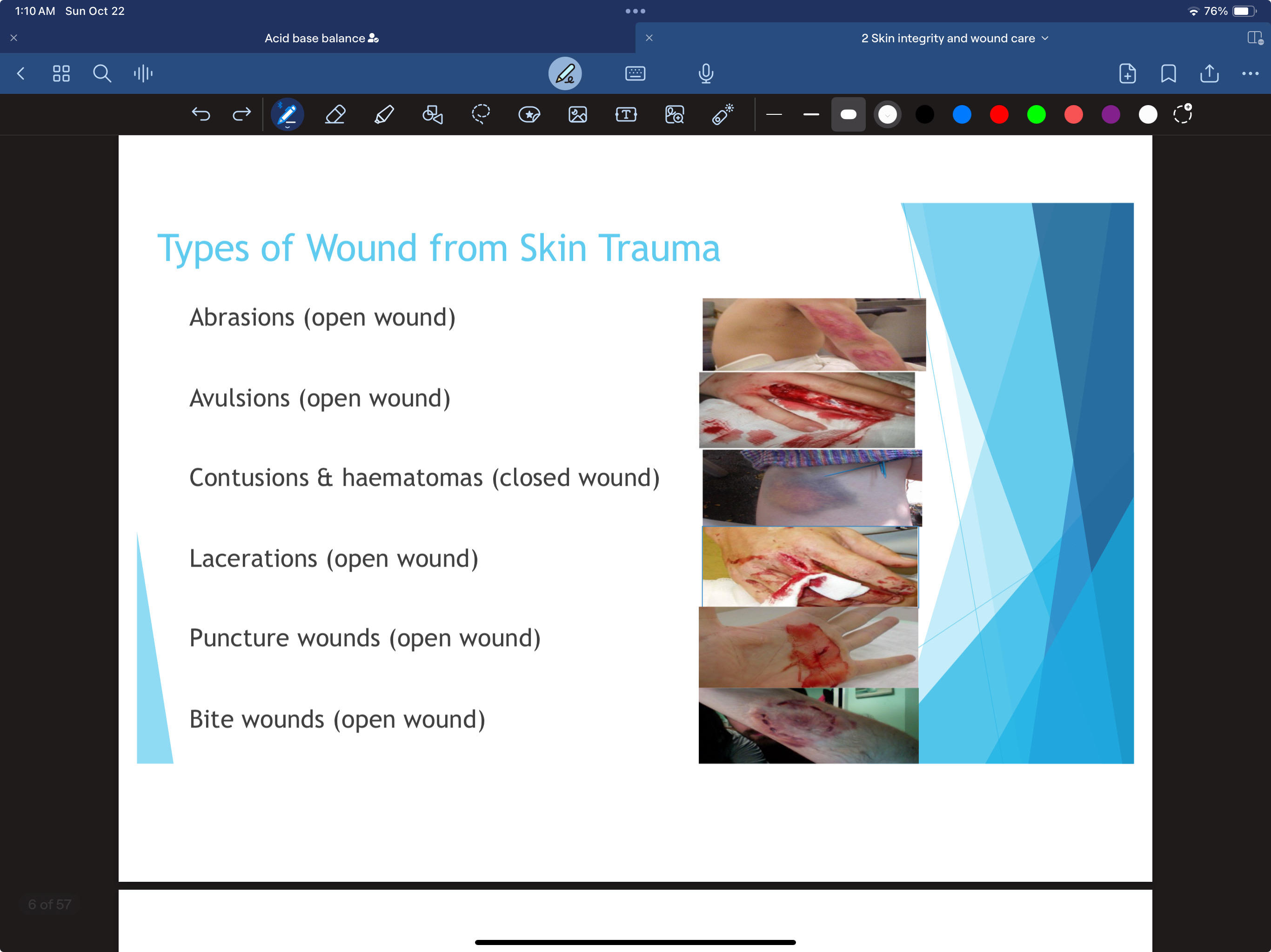
. 
140
Q: What is the primary focus when dealing with skin trauma?
A: Haemostasis - stopping the bleeding.
141
Q: How can haemostasis be achieved?
A: Haemostasis can be achieved spontaneously or through first-aid measures.
142
Q: If haemostasis is difficult to achieve, what are the recommended steps to take?
Apply direct pressure to the wound.
Elevate the wound above the level of the heart.
Consider using a short-term tourniquet for limb injuries.
Administer a local anaesthetic containing adrenaline if necessary.
Elevate the wound above the level of the heart.
Consider using a short-term tourniquet for limb injuries.
Administer a local anaesthetic containing adrenaline if necessary.
143
Q: What is the main characteristic of primary intention wound healing?
A: It has minimal tissue loss and edges are closed, typically leading to minimal scarring.
144
Q: How are the edges of a wound closed in primary intention healing?
A: Using sutures, clips, or tape.
145
Q: What happens during secondary intention wound healing?
A: The wound edges aren't closely approximated, leading to healing through granulation and contraction. This can result in scarring.
146
Q: What are some examples of wounds that heal by secondary intention?
A: Burns and major traumas, such as pressure injuries.
147
Q: What distinguishes tertiary intention healing from primary and secondary?
A: It's used when a wound is infected or has foreign materials, and closure happens after initial cleaning, typically 3-5 days later.
148
Q: Give an example of when tertiary intention might be used.
A: For severe bites that need cleaning before closing.
149
Q: What characterizes a partial thickness wound?
A: It's shallow and involves the loss of the epidermis and part of the dermis.
150
Q: How can you recognize a full thickness wound?
A: It's deeper, and subcutaneous fat is visible.
151
if a wound is red (healthy granulation tissue) you wanna ______ it
protect
152
if a wound is yellow (slough) you want to _______ it
cleanse
153
if a wound is black (nonviable necrotic tissue) you want to _______ it
debride (remove) it.
154
Q: What are the primary local factors that can impact wound healing?
A: Pressure, dehydration, maceration, trauma, edema, infection, excessive bleeding, necrosis, and biofilm.
155
Q: List the main systemic factors influencing how a wound heals.
A: Age, circulation, oxygenation of tissues, nutrition status, wound etiology, medications, health status, immunosuppression, and adherence to treatment plan.
156
Q: What is the term for normal bleeding after an injury?
A: Hemorrhage.
157
Q: What could cause prolonged hemorrhage in wounds?
A: Issues with coagulation.
158
Q: Which wound complication is a localized collection of blood under the tissue?
Hematoma
159
Q: What indicates a wound infection?
A: Presence of purulent drainage.
160
Q: What's the term for the separation of wound layers?
Dehiscence
161
Q: When might dehiscence (wound opening again) occur?
A: After strains like vigorous coughing or sitting up unsupported.
(usually after abdominal surgery)
(usually after abdominal surgery)
162
Q: What's a complete wound layer separation exposing organs?
Evisceration
163
Q: What's the immediate action for evisceration?
A: Call for help, keep the patient calm, and cover the wound with sterile saline-soaked towels. low fowlers with knee bent, no ice.
164
Q: What's an abnormal passage between organs or the body's exterior?
A: Fistula formation.
165
Q: How long before a dressing change should pain medication be administered?
A: 30-45 minutes.
166
Q: How should one refer to a previous stage 3 pressure ulcer that has healed?
A: History of stage 3 pressure ulcer (healed), not as stage 3, 2, or 1 (backstaging)
167
Q: Stage 1 characteristic?
A: Nonblanchable redness, intact skin.
168
Q: Stage 2 description?
A: Partial-thickness loss; might have fluid-filled blister.
169
Q: Features of Stage 3?
A: Full-thickness loss, visible fat, possible undermining.
170
Q: Key traits of Stage 4?
A: Full-thickness, exposed bone/tendon, possible osteomyelitis.
171
Q: Treatment difference between Stage 1 and 2?
A: Stage 1 uses transparent dressing. stage 1 will never break
172
Q: Unique trait of Stage 2 vs. Stage 3?
A: Stage 2 has no slough.
173
Q: Characteristics of unstageable injury?
A: Full thickness loss with slough (yellow, tan, gray, green, brown) or eschar (tan, brown, black) covering ulcer base.
174
Q: Describe suspected deep tissue injury?
A: Purple/maroon discoloration or blister; painful, firm, mushy, boggy; temp variation compared to adjacent tissue.
175
Q: When is assessment done in Acute Care?
A: On admission and every 24 hours; more often in critical care.
176
Q: Frequency in Long term care?
A: On admission, weekly for 4 weeks, then quarterly or with condition change.
177
Q: Home Care assessment guidelines?
A: At every visit; also check bony prominences, blanch test reddened areas, and inspect & palpate risk areas.
178
Q: What's the Braden scale cutoff indicating severe risk?
A: Score < 11.
179
Q: Mobility considerations in assessment?
A: Determine if patient can change position independently in bed or chair and their activity tolerance.
180
Q: Importance of nutritional status?
A: Assess weight, appearance, eating habits, serum albumin level, vitamins A & C, zinc & copper. Monitor fluid intake. Assessment recommended within 24 hours of admission.
181
how would you position a pateint to minimize shearing force for pressure injuries?
HOB 30 degrees or less
182
Q: What should be used for wound irrigation in pressure injuries?
A: N/S or sterile water (avoid hydrogen peroxide).
183
Q: What is "debridement" in the context of pressure injuries?
A: Removal of nonviable, necrotic tissue.
184
Q: What is the recommended diet for wound healing?
A: High in protein, fat, carbohydrates, vitamins (especially A, C, E), and minerals like zinc.
185
Q: What should you monitor in patients with pressure injuries to assess wound healing progress?
A: Protein status and hemoglobin.
186
Q: Before changing a wound dressing, what initial action should be taken regarding the patient's pain?
A: Evaluate the pain and pre-medicate the patient if indicated.
187
Q: When should you wear sterile gloves during wound care?
A: Before directly touching an open or fresh wound.
188
Q: When should you avoid or reconsider changing a dressing?
A: When the dressing is over closed wounds that are wet or infected.
189
Q: How should a wound be packed?
A: Not too tight and not over the wound edges.
190
Q: How does the Wound V.A.C. help in wound healing?
A: It uses the application of sub-atmospheric (negative) pressure to a wound through suction.
191
Q: What are the benefits of using a Wound V.A.C.?
A:
Accelerates wound healing.
Optimizes blood flow.
Removes wound fluid.
Maintains a moist environment.
Accelerates wound healing.
Optimizes blood flow.
Removes wound fluid.
Maintains a moist environment.
192
Q: What are the important signs or symptoms that should be immediately reported to the health care provider?
A: Any evidence of fresh bleeding, sharp increase in pain, retention of irrigant, and signs of infection.