Biochem Lectures 1-3
Study these flashcardsFree · spaced repetition · iPhone, Android, Mac & web
All cards
1
Basic Amino Acid Structure 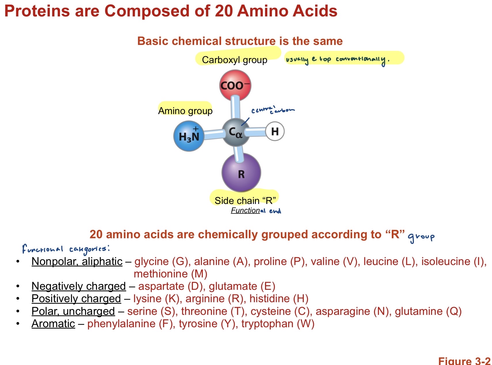
Central alpha carbon, with Hydrogen to right, Functional R group on bottom, Amino Group (NH3+) , and Carboxyl group ( COO-). These are in their Zwitterionic state so the groups are charged to produce a net 0 charge. Carboxyl is conventionally depicted at the top with the Hydrogen pointing toward?
2
Amino Acids: NonPolar, Aliphatic
Glycine (G), Alanine (A), Valine (V), Leucine (L), Isoleucine (I), Methionine (M). Generally hydrocarbons (other than Glycine)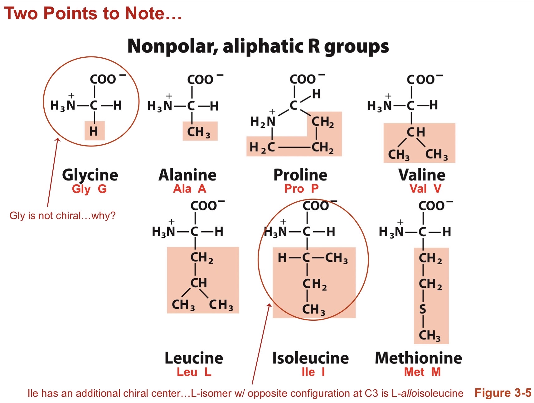
3
Amino Acids: Negatively Charged and Positively Charged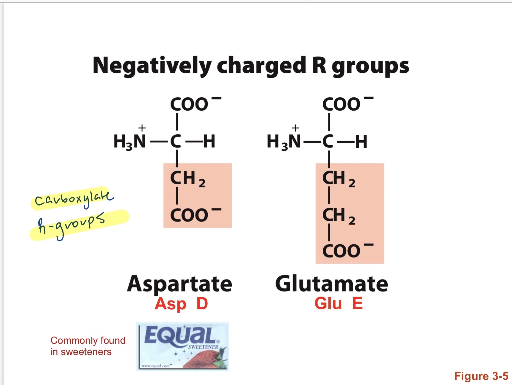
Negative: Aspartate (D), Glutamate (E). Generally carboxylate R groups Postively Charged: Lysine (K), Arginine (R), Histidine (H)Generally amino containing R groups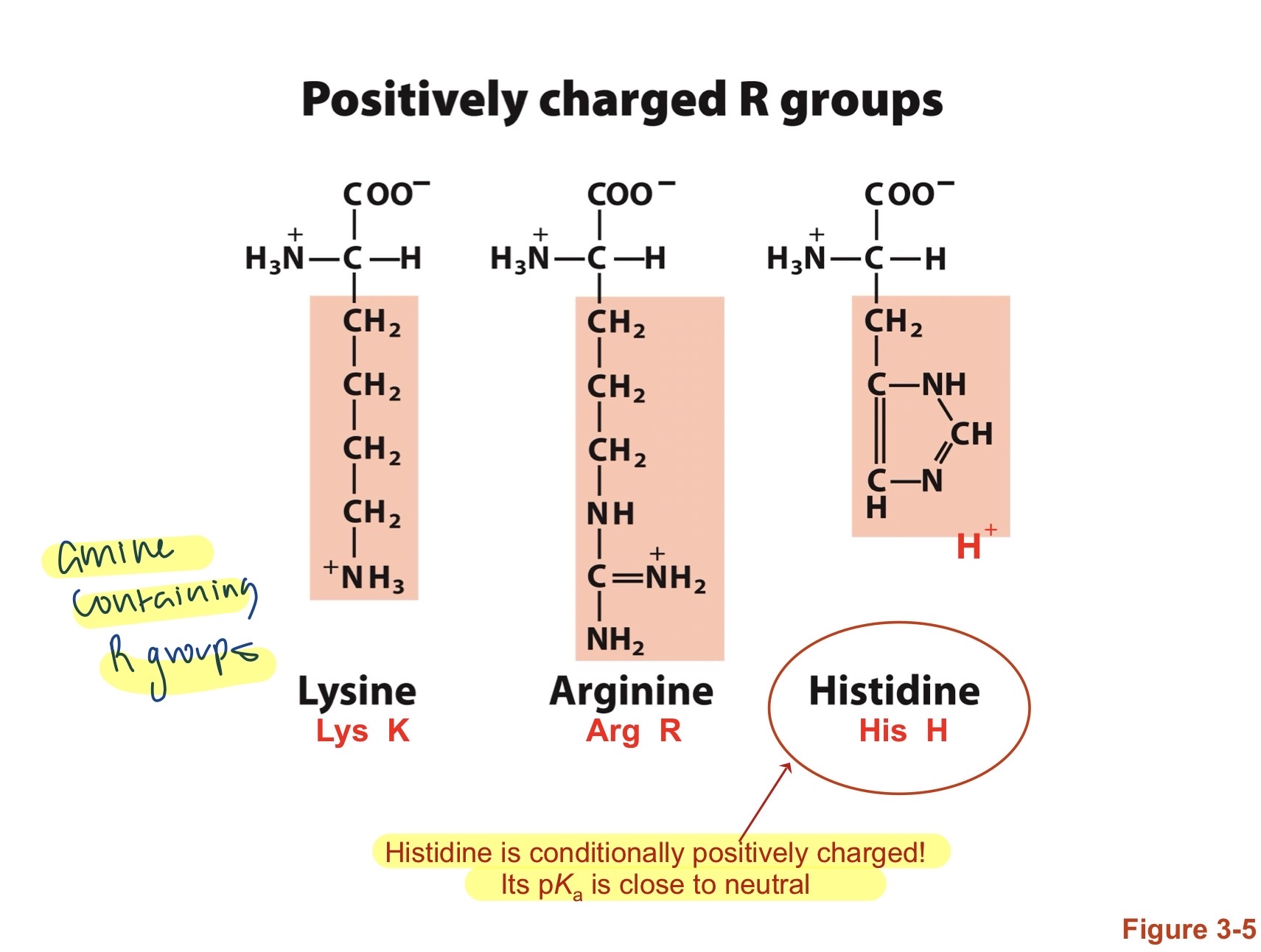
4
Amino Acids: Polar, Uncharged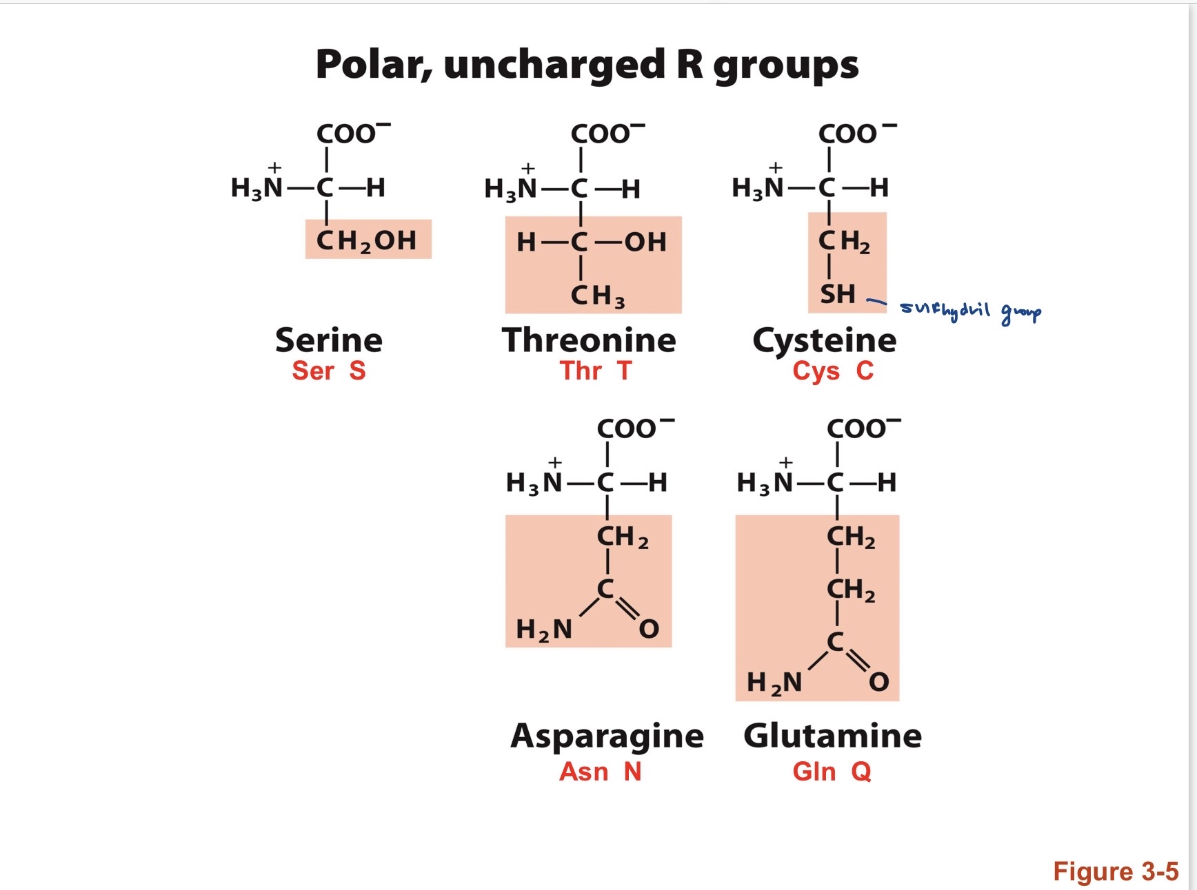
Serine (S)m Threonine (T), Cysteine (C), Asparagine (N), Glutamine (Q) cysteine has a sulfydril group in its R group so when close to anotehr cysteine oxidation can occur and create a disulfide bond. 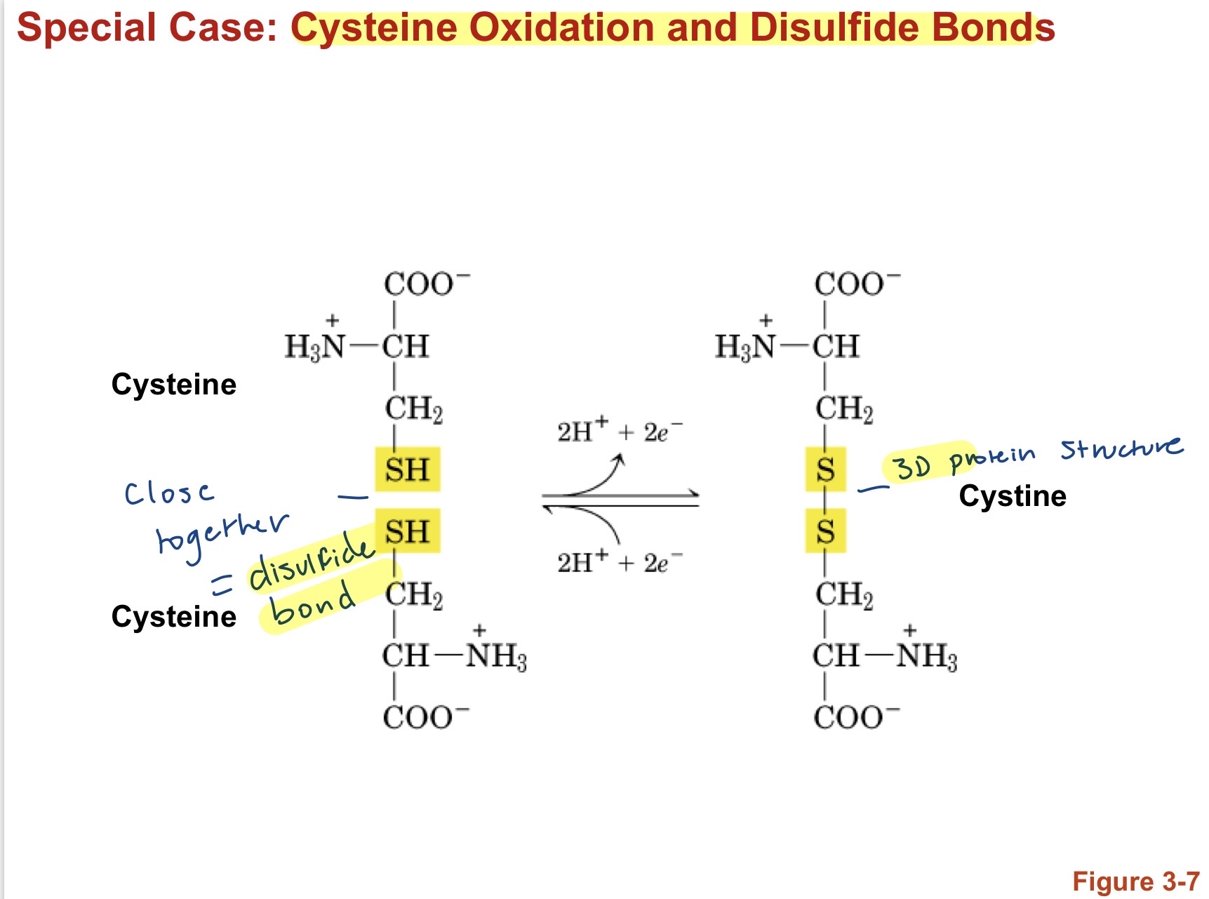
5
Amino Acids: Aromatic
Phenylalanine (F), Tyrosine (Y), Tryptophan (W)Generally ring structures and can absorb light. 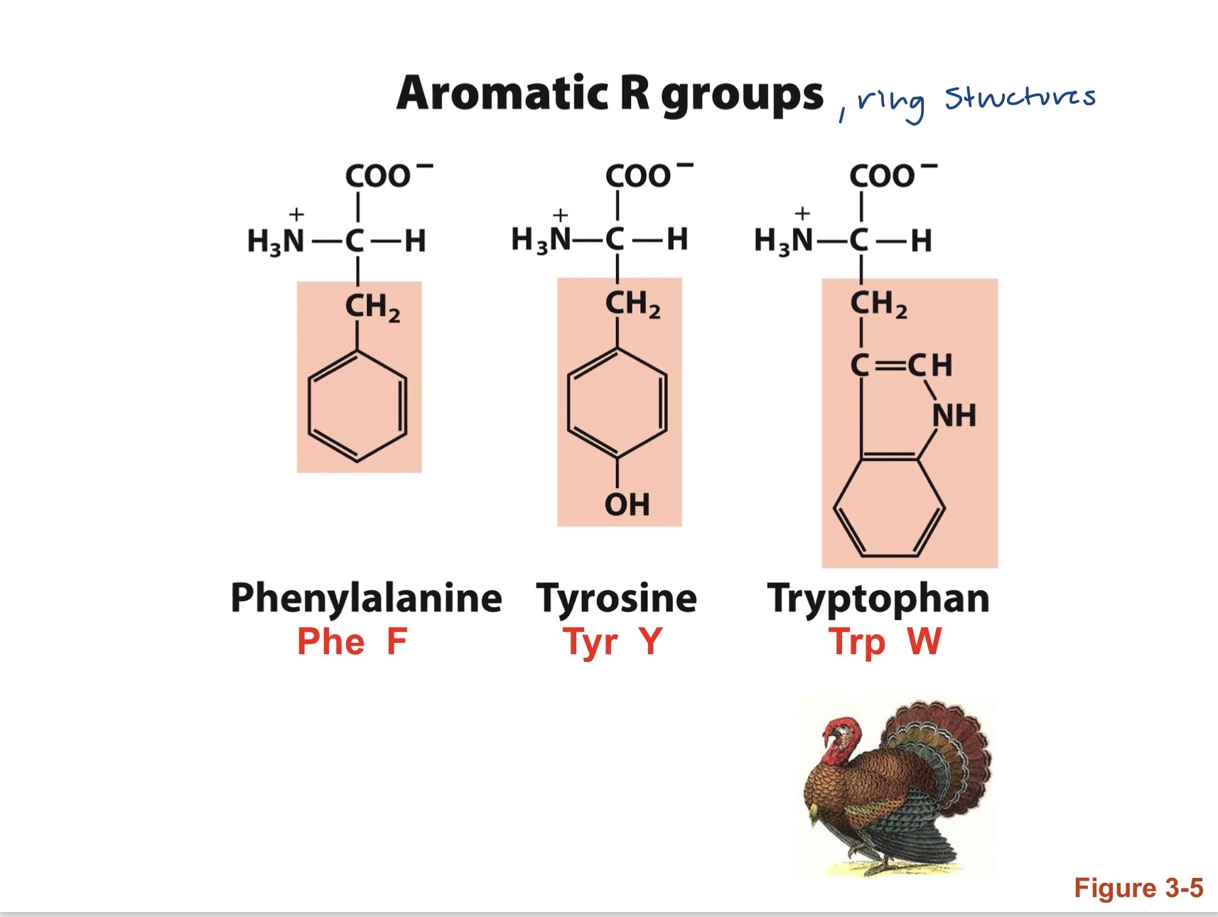
6
Amino Acids
Most AA are Chiral molecules( non-superimposable mirror image) and exist as an L enantiomer. To determine chirality spell CORN! ( Carboxyl up and H out) If Spells corn its an L configuration, if spells CONR then is a D configuration. *Glycine is achiral becuase its R group is a hydrogen. *Proline has a unique fold on R group which bends over and forms a bodn with its Amino group.* I\sroleucine has an extra chiral center
7
Chirality of AA and Optical Activities 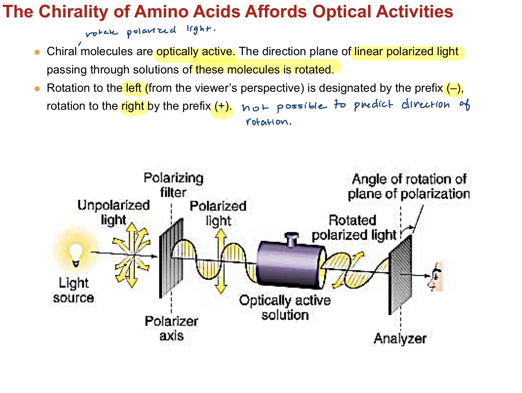
Chiral molecules are optically active in that theu can rotate the direction plane of linear polarized light when passed through their solutions. Leftwards rotation (from viewers perspective is (-) and right is (+). Cannot infer rotation from chiral molecule. L and D designate the chemical configuration not direction rotation. If Soln. has both + and - in equal forms or L and D configurations in equal forms then it is called a racemate and it is optically inactive. molecules with more than one chiral center can also be optically active..
8
Absorption of UV light by Aromatics. 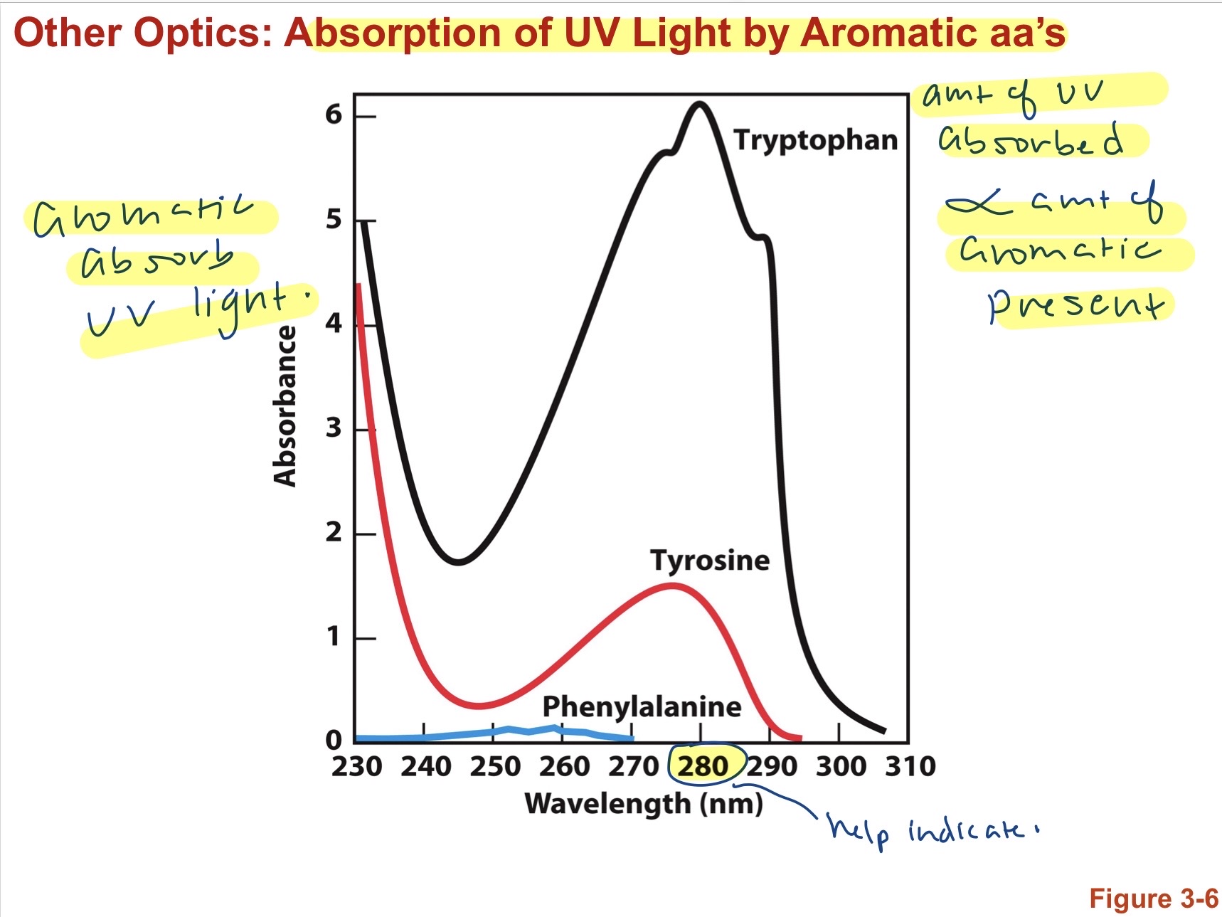
Amoutn of UV absorbed is proportional to the amount of aromatic present. wavelength 280 nm is largest abosrption. Tryptophan absorbs the most followed by Tyrosine, and Phenylalanine. $Transmission= \frac{I}{I_0}$, where Transmission is between 100% and 0 %. Whatever isnt transmitted is absorbed. $Absorbance (A)= log\frac{I_0}{I}=log \frac{1}{T}= \epsilon cL$ A is linearly proportional to C and L, $\epsilon$ is the molar absorption/ extinction coefficient with units $M^{-1} cm^{-1}$. Therefore, $c= \frac{A}{\epsilon L}$. A is typically measured at 280 nm, L is constant at 1 cm, and $\Epsilon$ is know for each aa. it is the sum of the E's of all Trp and Tyr residues assuming that the protein has an average number of Trp and Tyr residues compared to all know protein sequences. 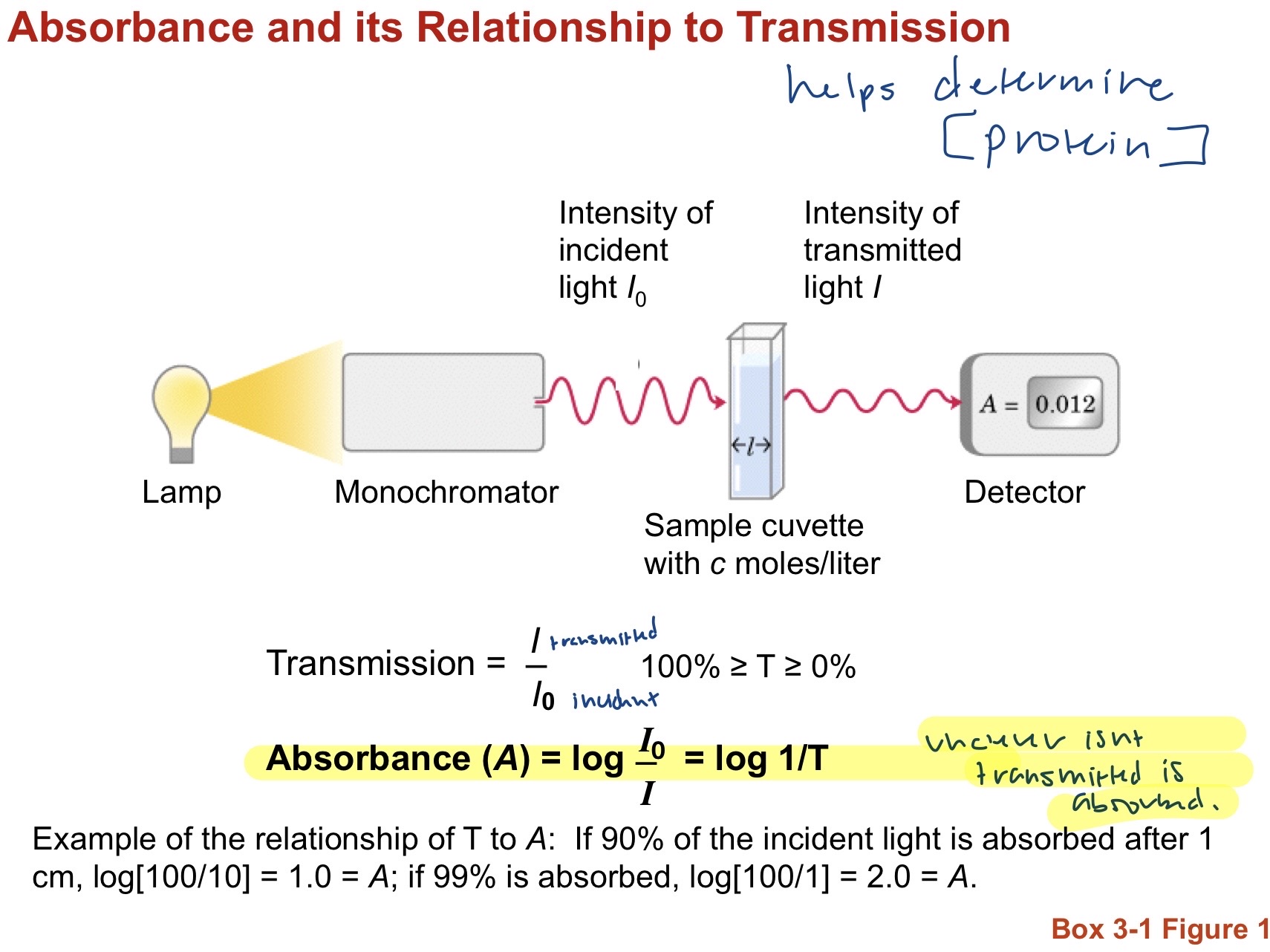
9
Nonionic and Zwitterionic forms of AA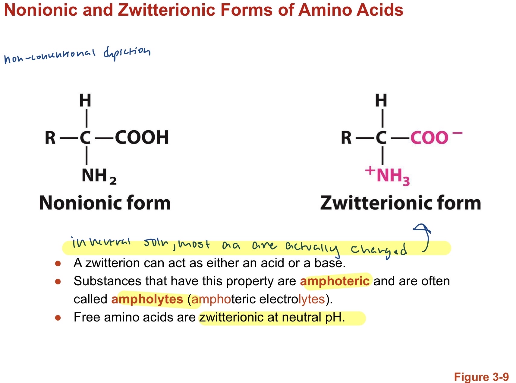
In a neutral soln. most aa are charged (zwitterionic), which can act as either an acid or base. These substances are amphoteric/ ampholytes Free amino acids are zwitterionic at neutral pH.
10
Amino Acid Titration, Ionization, and Isoelectric Point (IEP)
pK's are at the midpoitns of inflections, when the solution pH gets greater than pKa of a group then it is deprotonated. At low pH (acidic) the groups are fully protonated. Carboxyl is acidic and has a very low pKa (~2) so will be first to deprotonate. The free carboxyl will deproatante before any on the R group (~4). Amino groups have a very high pKa so will usually deprotonate last(~9), followed by amino groups on R group(>9). R group pKa's are generall y somewhere in between. $pI=\frac{1}{2}(pK_1+pK_2)$ where PI is pH at which the net electric charge of a molecule is zero. $pH=pK_a+log\frac{[A^-]}{[HA]}$Always make sure to consider current pH and how it affects protonation when asked to draw peptide bonds. 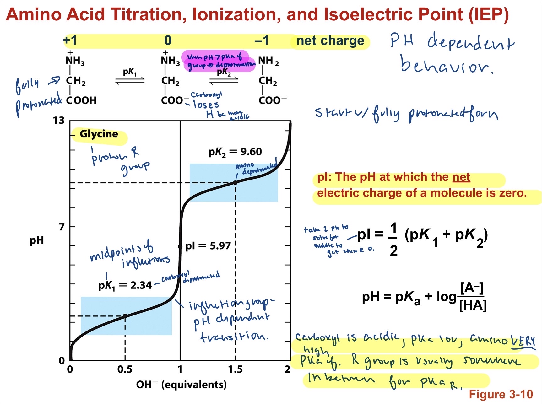
11
Peptide Bond Formation 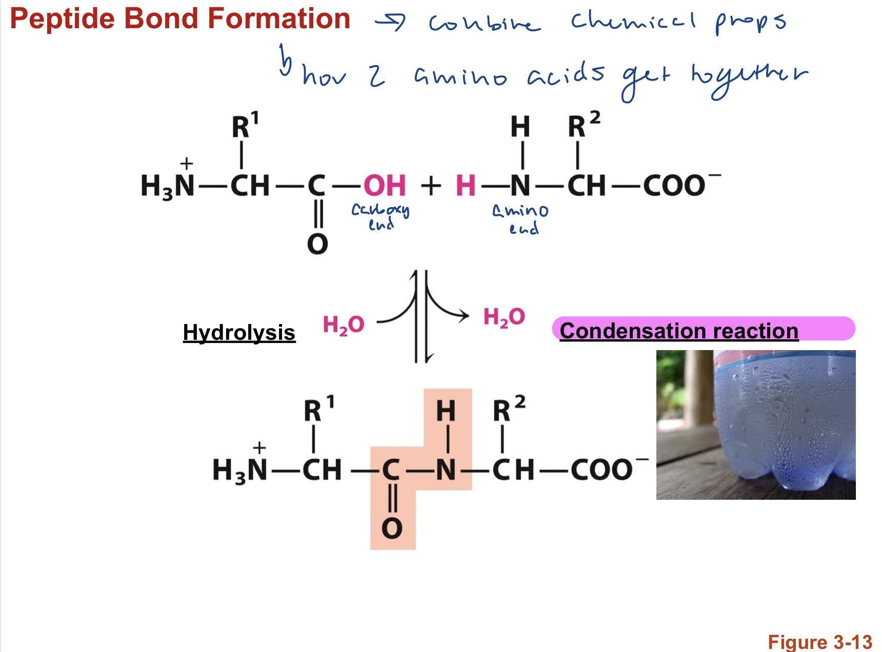
How 2 amino acids stick together. OH from Carboxy terminus and H from Amino terminus go through a condensation reaction to create a larger complex molecule producing water as a byproduct. Hydrolysis is the opposite and uses water to break down a larger molecule into smaller pieces. characterized by carboxy C=O attatched to an amine N-H. Polypeptides are written from the end with the free NH3+ (N-Terminal). Resonance causes the carbonyl oxygen to have a partial negative and the amide nitrogen to have a partial positive resulting in a small electric dipole and partial double peptide bond that forces the peptide bond to be planar. virtually all peptide bonds will be in the trans configuration s R groups dont clash.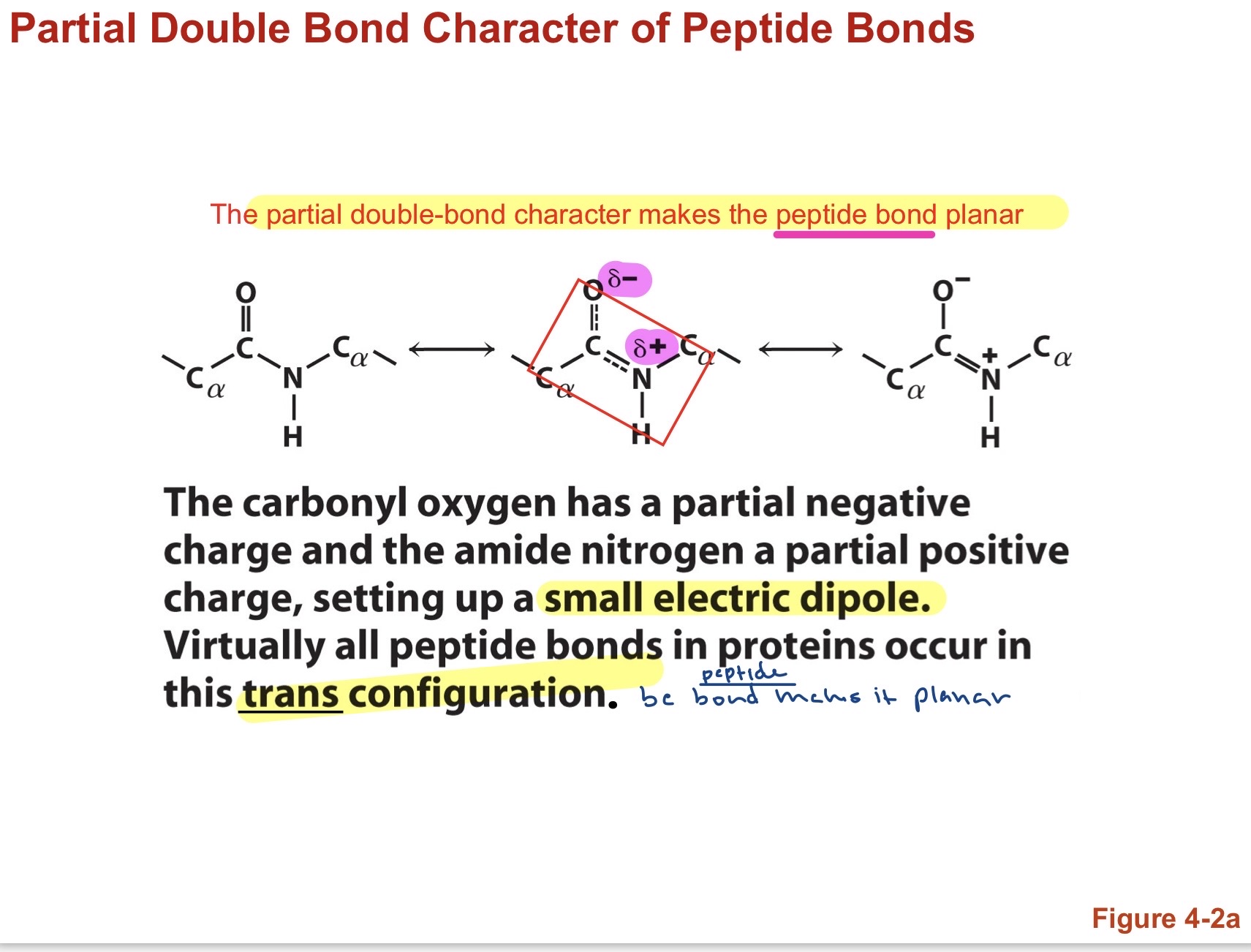
12
Oligopeptide dihedral angles $\phi$ phi and $\psi$ psi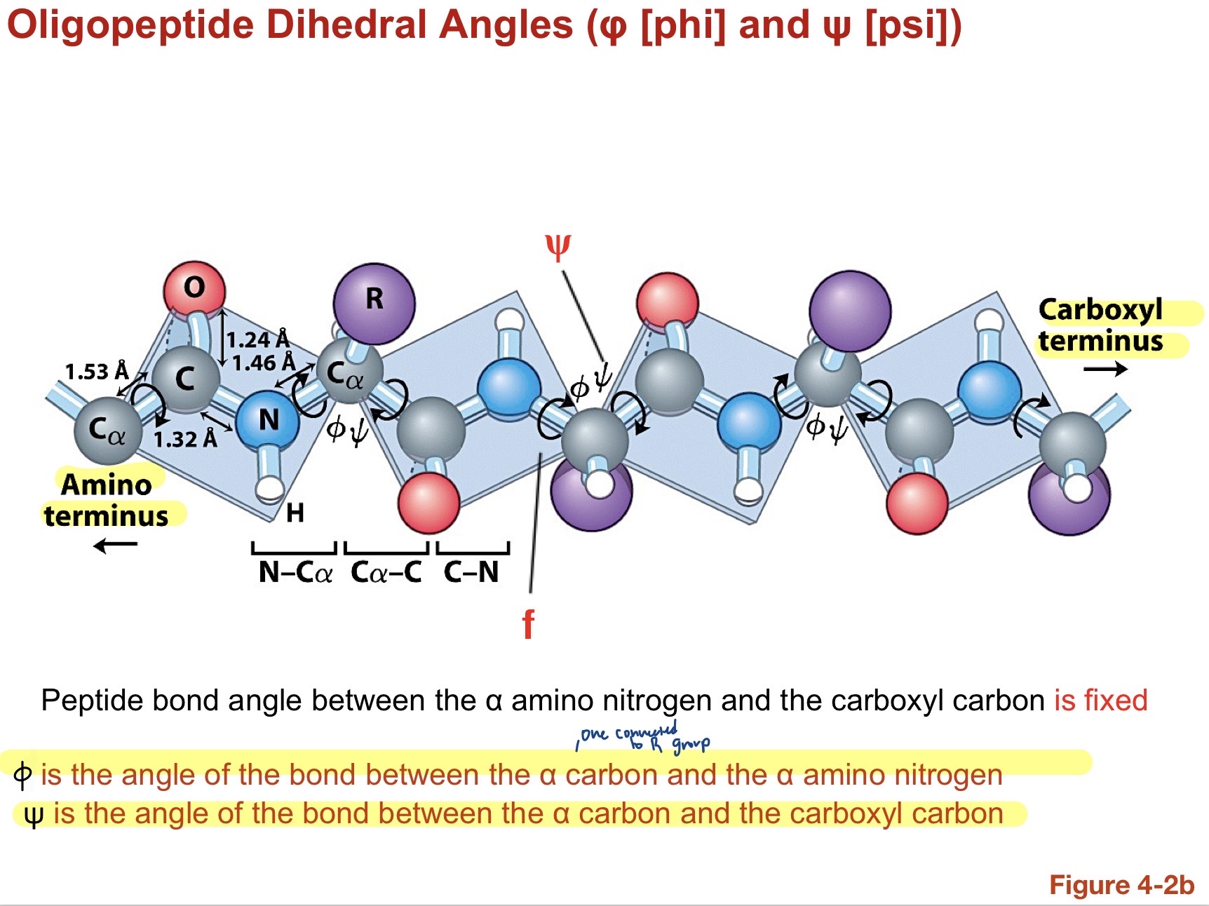
Phi is angle of bond between Alpha carbon and alpha amino nitrogen whule Psi is the angle of the bond between the alpha carbon and the carboxyl carbon. Peptide bond angle between the alpha amino nitrogen and the carboxyl carbon is fixed bc of forced planar structure from double bond. some phi and psi angles are physically impossible (like when both =0) becuase of steric clash between carboxyl O and amino H. Usually Phi and Psi= 180 if the two peptide planes are parallel and in trans. (fully extended form of the backbone).
13
Hydrogen Bonding
Stabilize secondary protein structures .HNOF. often utilizes water. R group functionality relies on this.
14
Alpha Helix 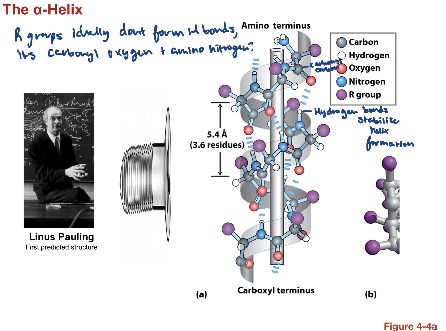
Hydrogen bonds between carbonyl oxygen and amino Hydrogen stabilize the helix formation. R group ideally dont form H bonds here. has 3.613 amino acids per turn on average with 13 atoms forming a ring closed by a hydrogen bond. For every alpha carbon Phi=-60deg and Psi=-45 to -50 deg. Both left and right handed helices are possible but mose are right handed (swirl/thumb point up). R groups project away from the helix core to make them more accessible. There is an inhernent negative dipole at the C terminal and a positive one at the N terminal so positive aa are often found at the C terminus and vice versa to neutralize the dipole moment and stabilize the alpha helix.
15
AA with Low propensity to be in an alpha helix.
- Side Chains that contain H bond donor an acceptors in close proximity to the main chain (Asp with its carboxylic (-) acid, but not Glu) compete with main chain H bonds and destabilize the alpha helix. - Gly is more flexible than other amino acids, allows for more possible angles (helpful for turns) but destabilizes amino acid because has high entropic cost to fold into alpha helix. - Pro has a fixed phi angle at ~65deg becuase of its ring structure. It is thus rigid and causes bad backbone packing (cannot put in helix formation). Also cannot form hydrogen bonds becuase it lacks the amide hydrogen. -Interactions between adjacent amino acids on turns can also affect stability, ex: if one is positive and other is negative near each other it stabilizes the alpha helix.
16
Beta Sheet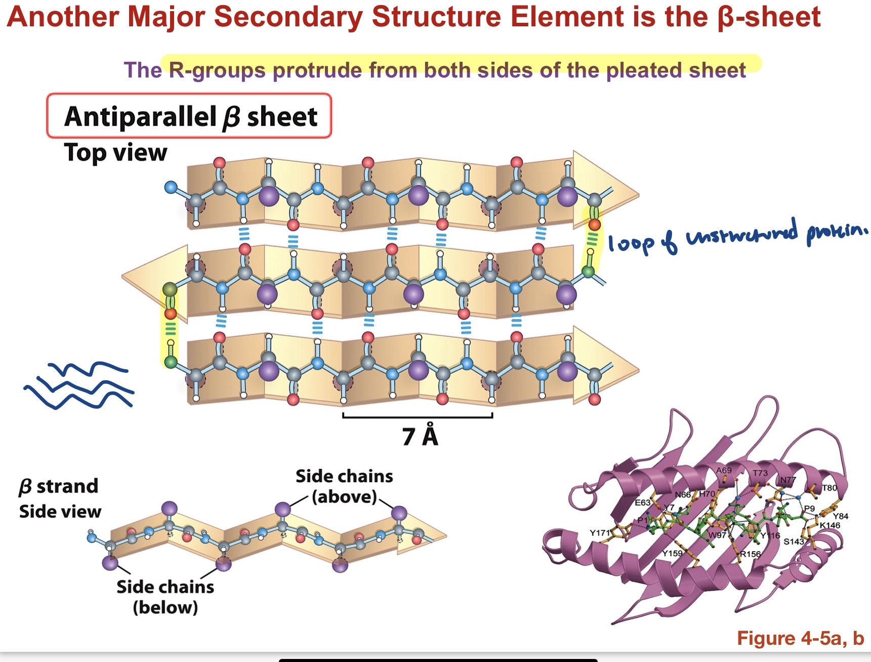
AntiParallel:Side Chains protrude above and blow the sheet, while H bonds occur from side to side between Carboxyl oxygen and amide hydrogen. Loops of unstructured proteins connect the opposite facing sheets. Parallel: same as antiparallel but now H bonds are fartehr apart and connect diagonally, weakening the bond since they dont line up directly .aa compostion determines whether Antiparallel or parallel formation occurs.
17
Beta Turn types: 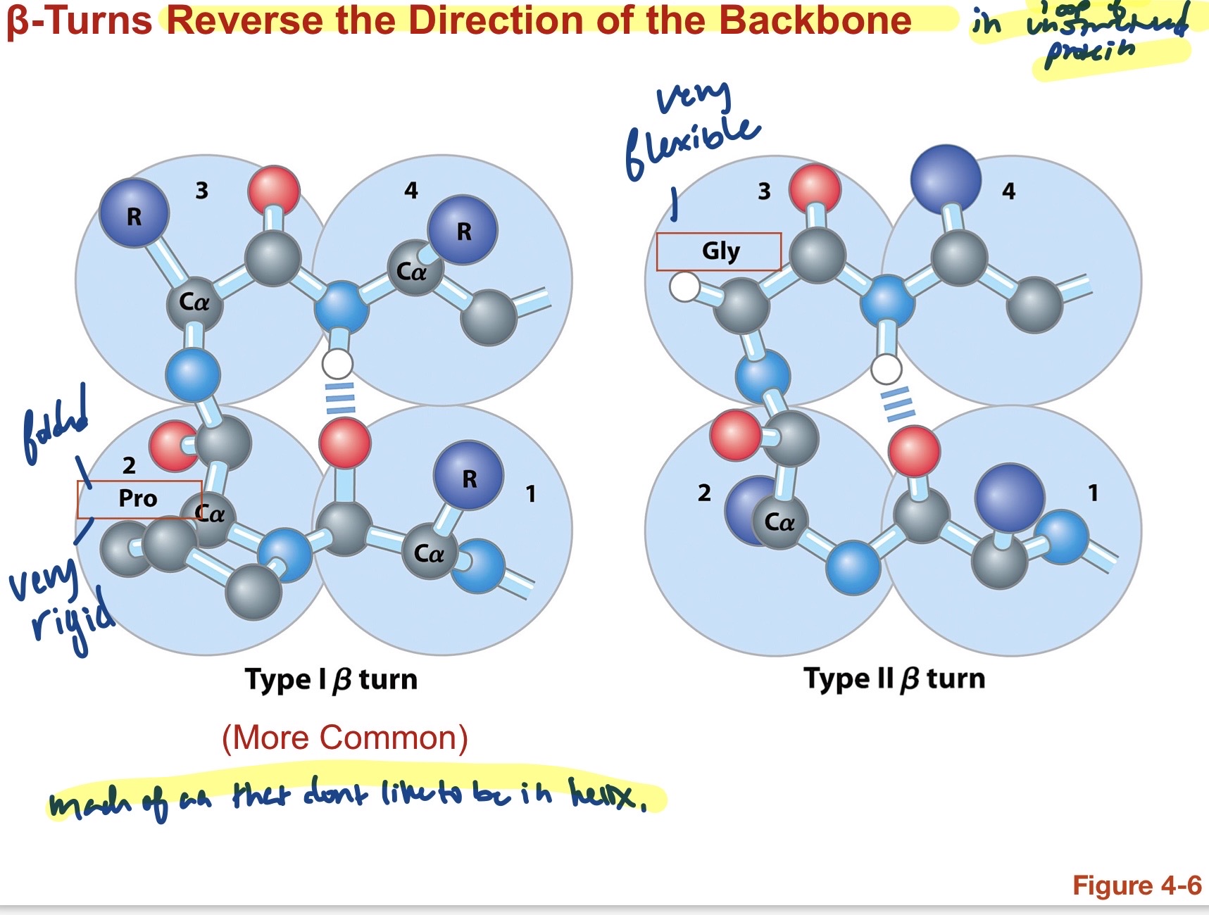
Type 1: A loop of unstructured protein that uses rigid proline since its folded in on itself to reverse the direction of the backbone. More common .Occurs in cis formation with the preceeding residue. helpful because phi is locked at -65deg . Type 2: a loop of unstructured protein that uses flexible glycine. Less common. 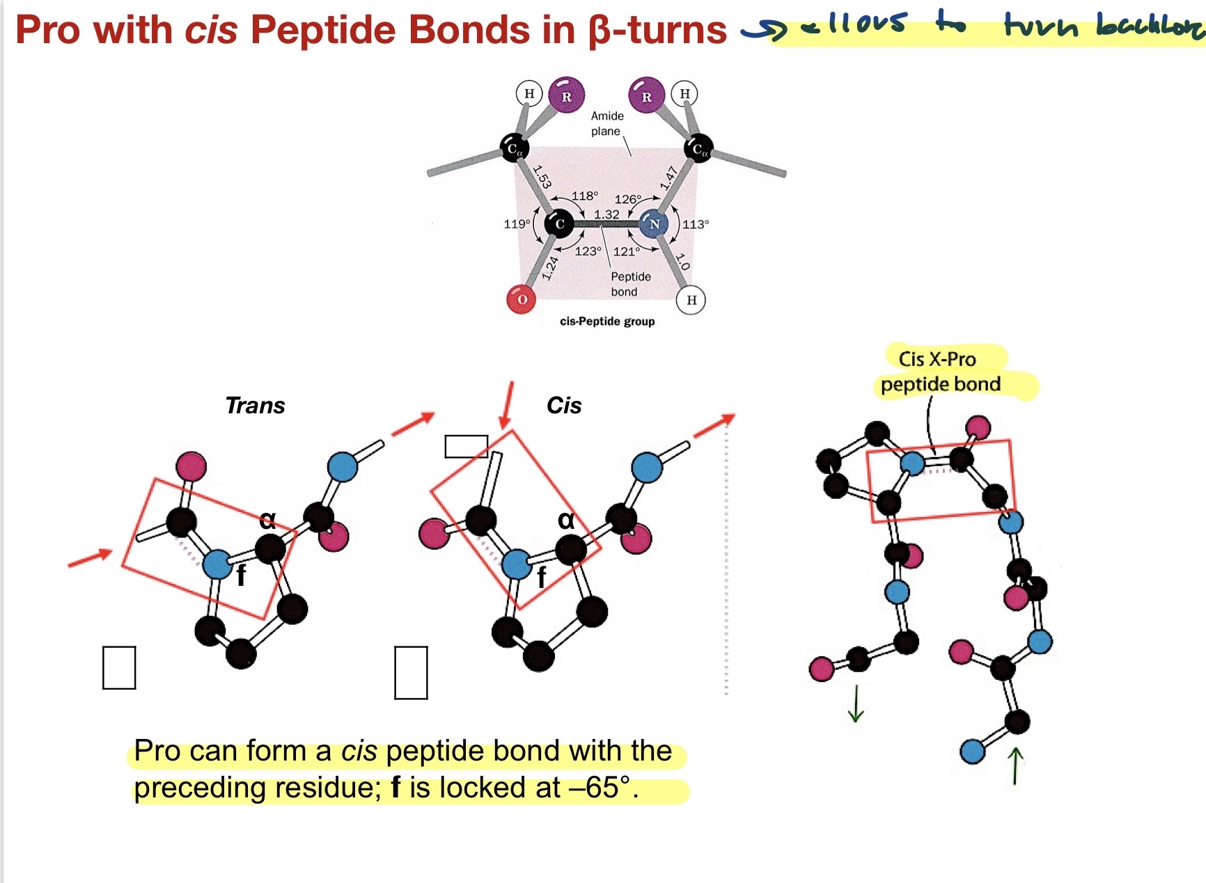
18
Secondary structures overall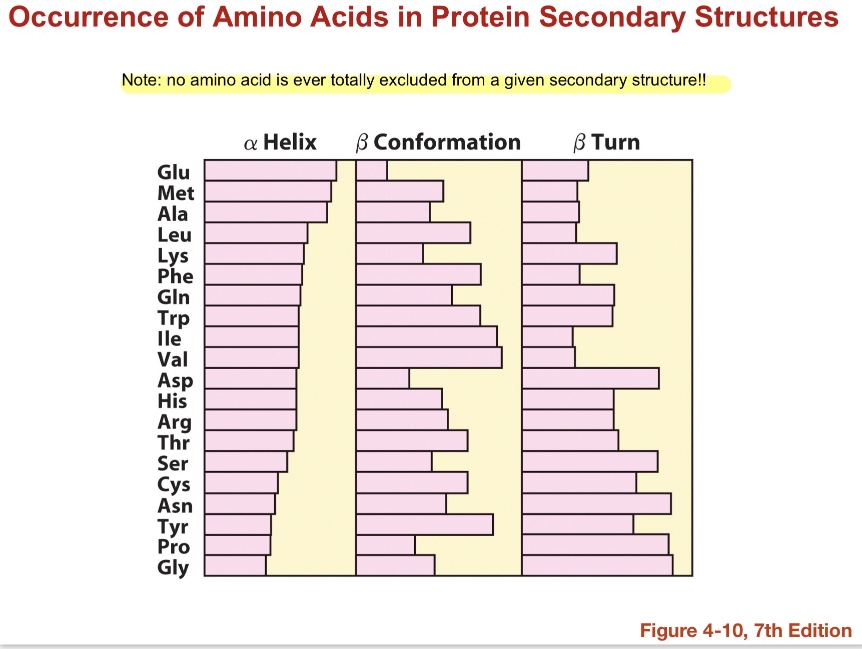
No amino acid is ever totally excluded from a given secondary structure. All can be Alpha helices, Beta sheets, and Beta turns. As the pH changes, protonation of the secondary structure changes and this can change the attraction or repulsion of its interactions, thus polypeptides generally need a certain pH range. Bends and turns are characterized by glycine and proline. Cysteines close together could form disulfide bonds. charged polar will likely be on outside while nonpolar uncharged will likely be on the inside.
19
Ramachandran plots 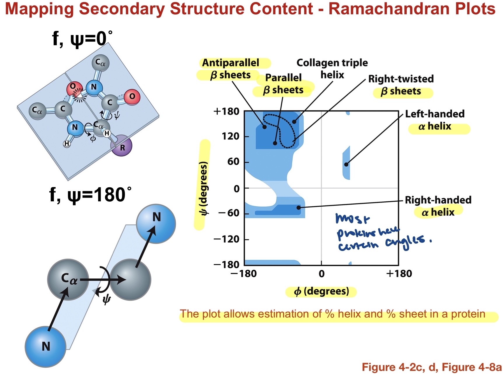
allows an estimation of the % helix and % beta sheet in a protein. based on Phi (x) and Psi angles (y)
20
Super-secondary structure and Domains
Secondary structure elements (helices, beta sheets, beta turns) that are arranged into motifs/ folds. most proteins are made up of a limited number of motifs. Larger polypeptides are often composed of Domains which are somewhat independent regions linked by less well defined connecting bridges. 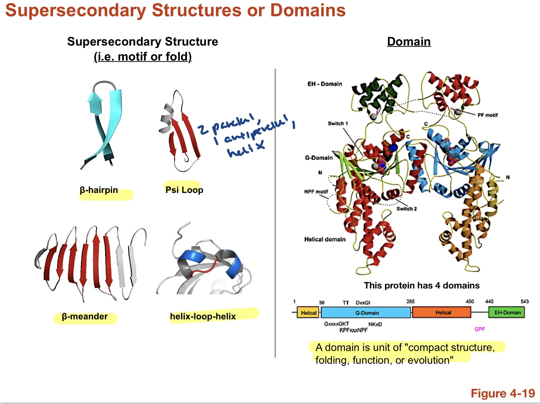
21
Tertiary structure.
Unique arrangment of secondaey structure creates highyvaried fold. The unique folds allow for unique functions.
22
Atypical fibrous protein structures: Coiled Coils
Two right handed helices twisted in a left haded superhelix, packed against each other in parallel orientation. Only 3.5 residues per turn bc helices are twisted, hydrophobic residues face inward in these amphiphilic strucutres. Very stable and strong, Ex: Myosin and Keratin. Leucine zippers? 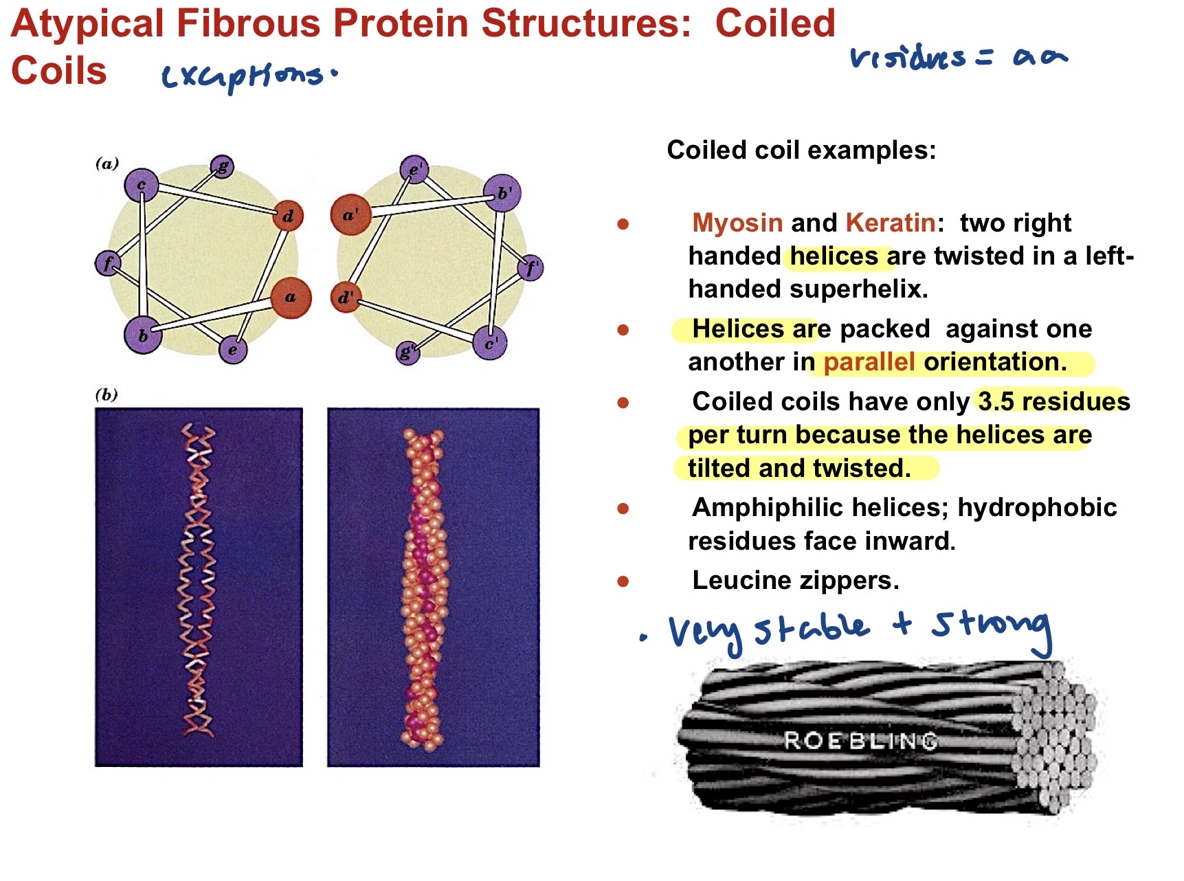
23
Atypical Fibrous protein structures: Collagen Triple Helix.
Three parallel left hand helices (3.3 residues/turn) twisted in a right hand superhelix. Every 3rd residue is glycine (flexible) near the center of the triple helix becuase it is the least bulky. Proline also there bc rigid and fixes angle of helix exterior to force others inward for stabiliziation. Contains post-transationally modified amino acids to increase H bonding modifications are catalyzed by family of dioxygenase enzymes that require ascorbate to maintain activity. Proline and hydroxypoline are 30% of collagen bc fixed phi angle is needed to lock conformation for stability. unlike alpha helices, there is a lot of proline in the collagen triple helix.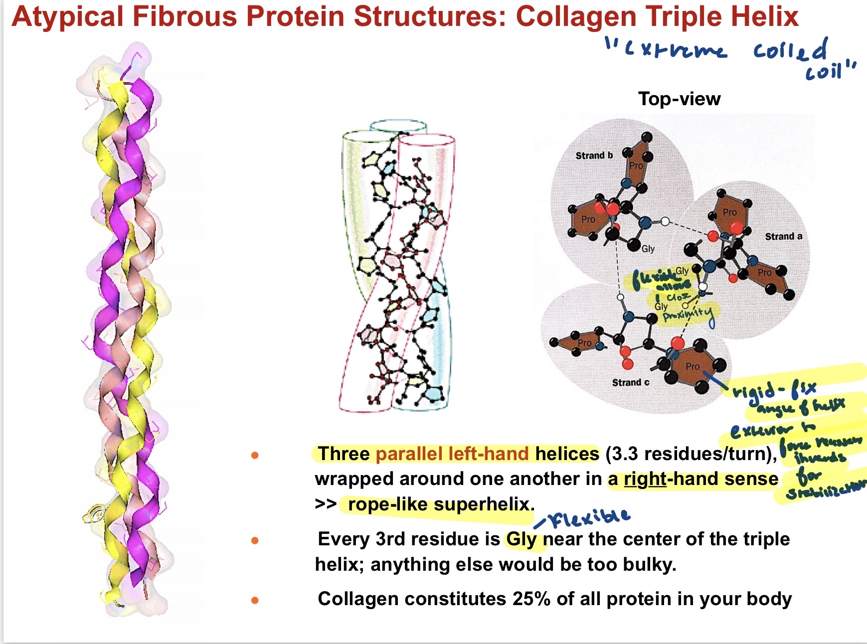
24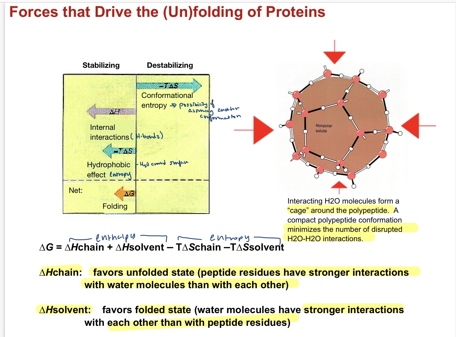
Forces that unfold proteins.
25
Arfinsens folding experiment
Added Urea and Mercapto-ethanol to native state protein and its unfolded to an inactive state. once those things weereremoved it quickly folded back to native state. Showed that all that is needed for structure is the primary sequence. Begins with local secondary structure formation and hydrophobic collapse, compaction then occurs as pieces get closer in proximity to others, disulfide bonds then form( conformational adjustment) , and then subunit assembly occurs to create the final form.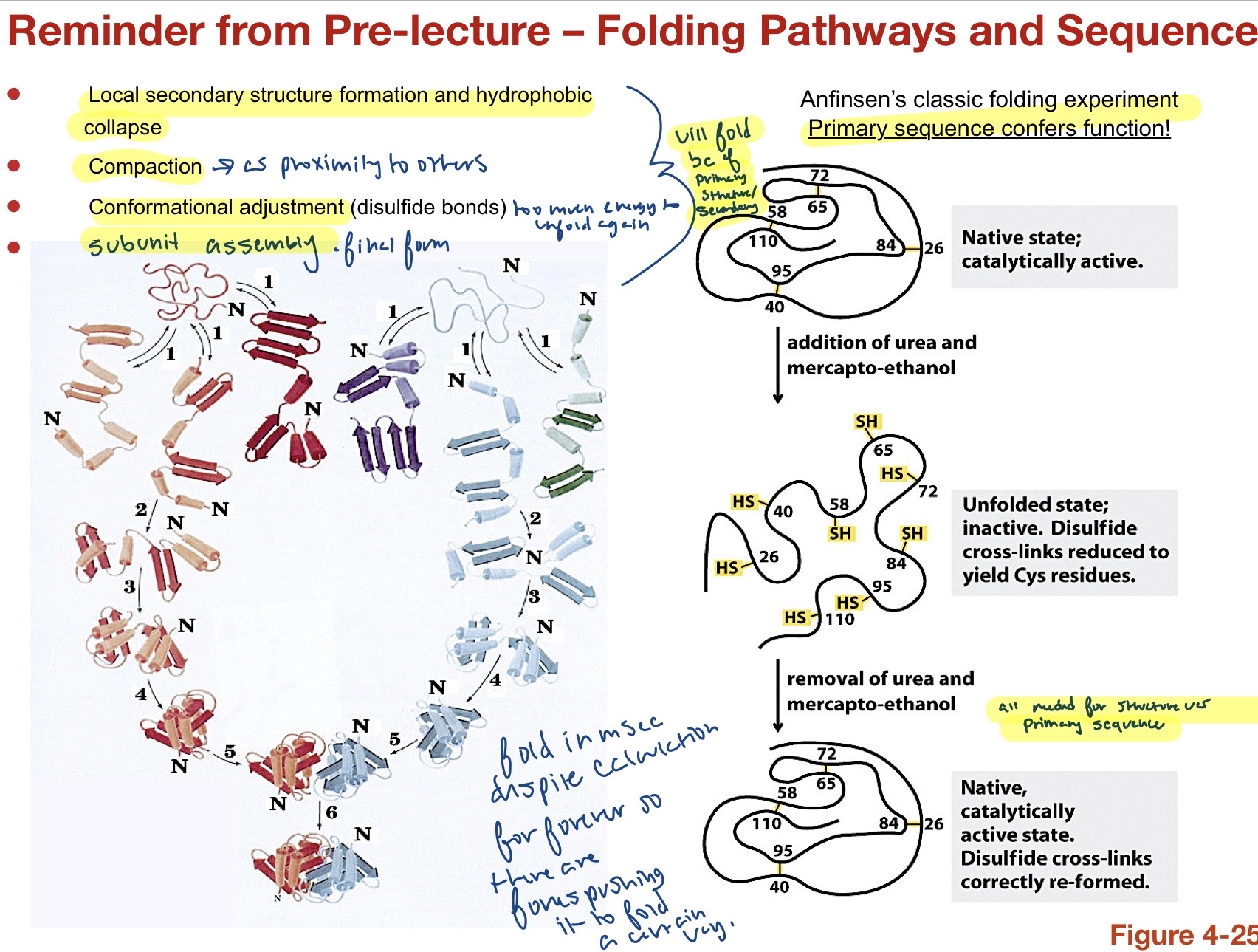
26
Denaturing a protein
- High of low ph (alters ionization state of side chains creaitng new repulsion/attraction destabilizing H bonds. Detergents (associate with nonpolar AA nd disrupt hydrophobic interactions. High Temp ( increases vibrational and rotational energy) Chaotropic agents (make interactions with polar residues more favorable than interactions with water.) the unfolded state under all these conditions has a lower free energy than in the native state hence the denaturazation.
27
SDS Page
Loading buffer contains SDS (cheap accessible things to coat proteins in soln with a negative charge.), Reducing agent (Beta ME, DTT, Mercaptoethanol)to break up the protein disulfide bonds and put in simple form), Bromophenol Blue (to visualize the readout), Glycerol, and Buffer( conducts current allowing protein to run down gel). Put this in gel and do electrophoresis and bands will appear based on weight of different components. Smaller weights go farther becuase can fit through pores in gel. allows you to see how impure a protein is or how many subunits a protein has. A rough starting point. If used non reducing conditions (native gel) then protein would be left in tact to see interactions between sub units and the weight band would be larger. often compared to a molecular weight ladder to get a crude read 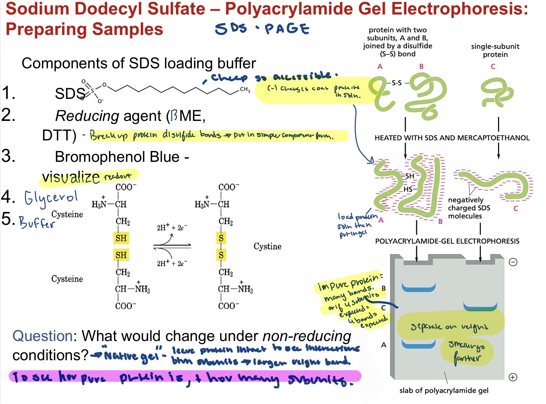
28
Western Blotting
Utilizes specific antibodies to identify a specific protein first do SDS Page to separate antigen (AA) from other molecules in a protein. Then incubate with specific primary antibodies that bind antigen, locates the exact position of the antigen. USes electric current to transfer proteins onto a blotting membrane, Dry milk, non-specific proteins are added to block antibodies from sticking all over the membrane leaving only the antigen available for binding. Labeled secondary antibody binds to the first antibody to show that anitgen/ protein of interest is present- seen as ONE band on the western blot. antigen is the target of an antibody that are made to remove foreign molecules. can cross link antigens into AGGREGATES, forming clusters that will either be eaten by cells or killed by special proteins in blood. 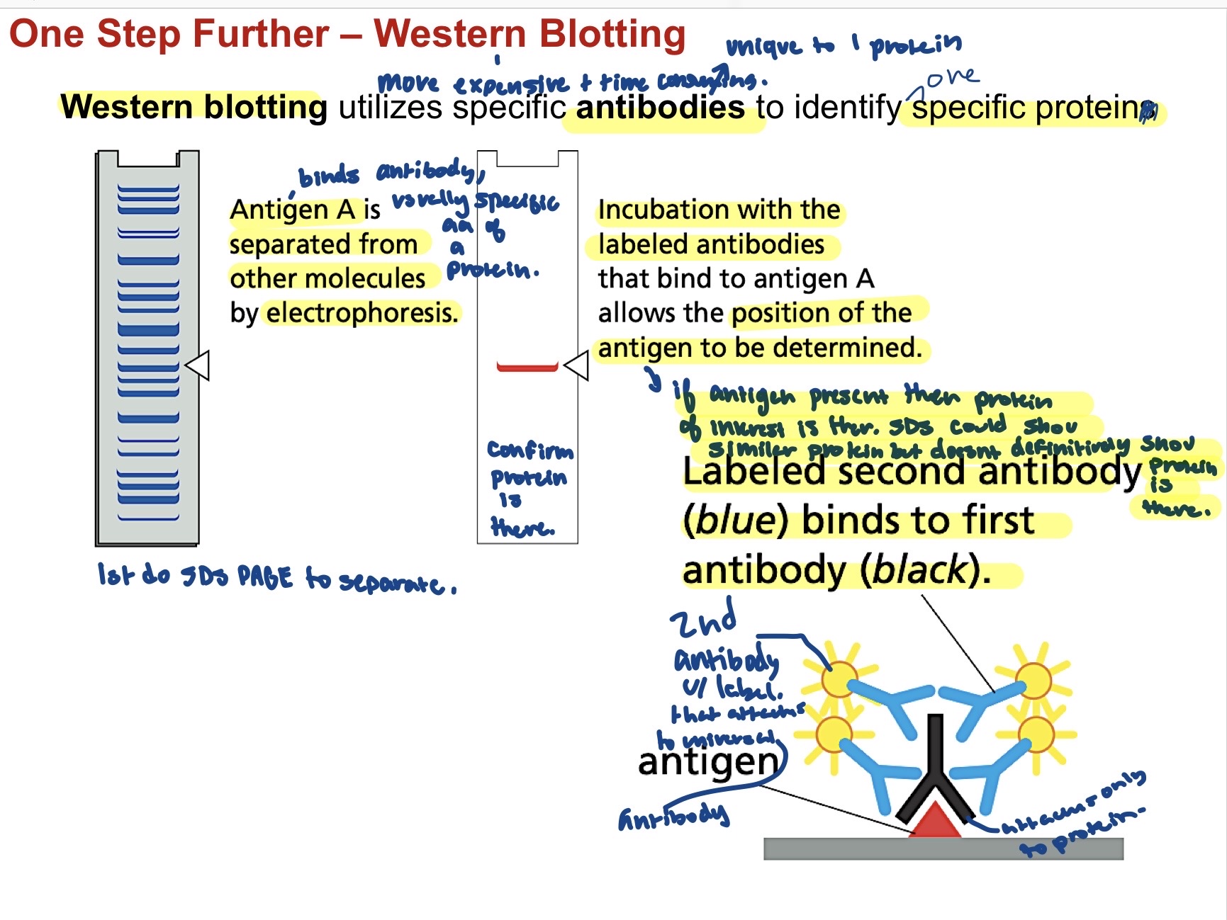
29
Antibody structure 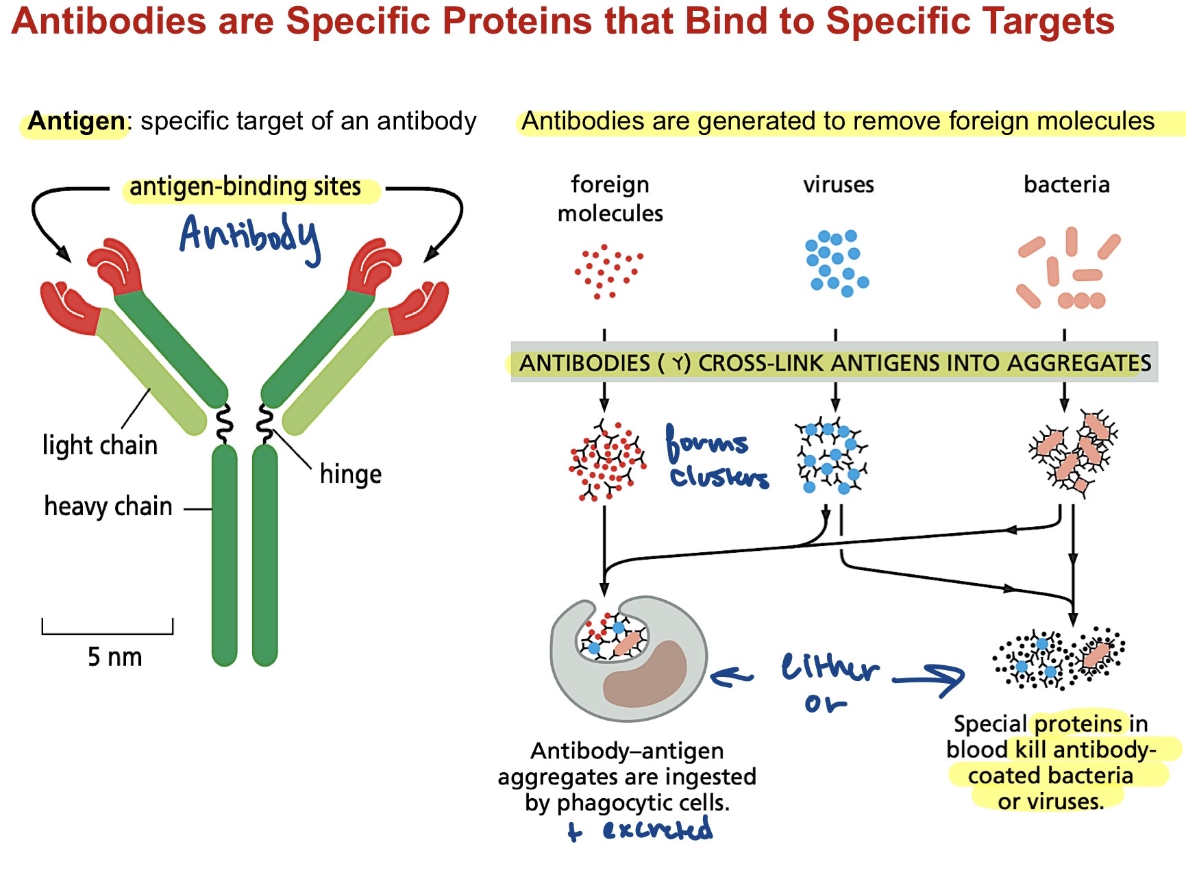
Polyclonal antibodies generate MANY antibodies to MULTIPLE binding sites on antigen, more general targetting but more protection from mutation. Monoclonal antibodies generate ONE type of antibody to a SINGLE binding site, much more specific and affective but less resilient to mutations. 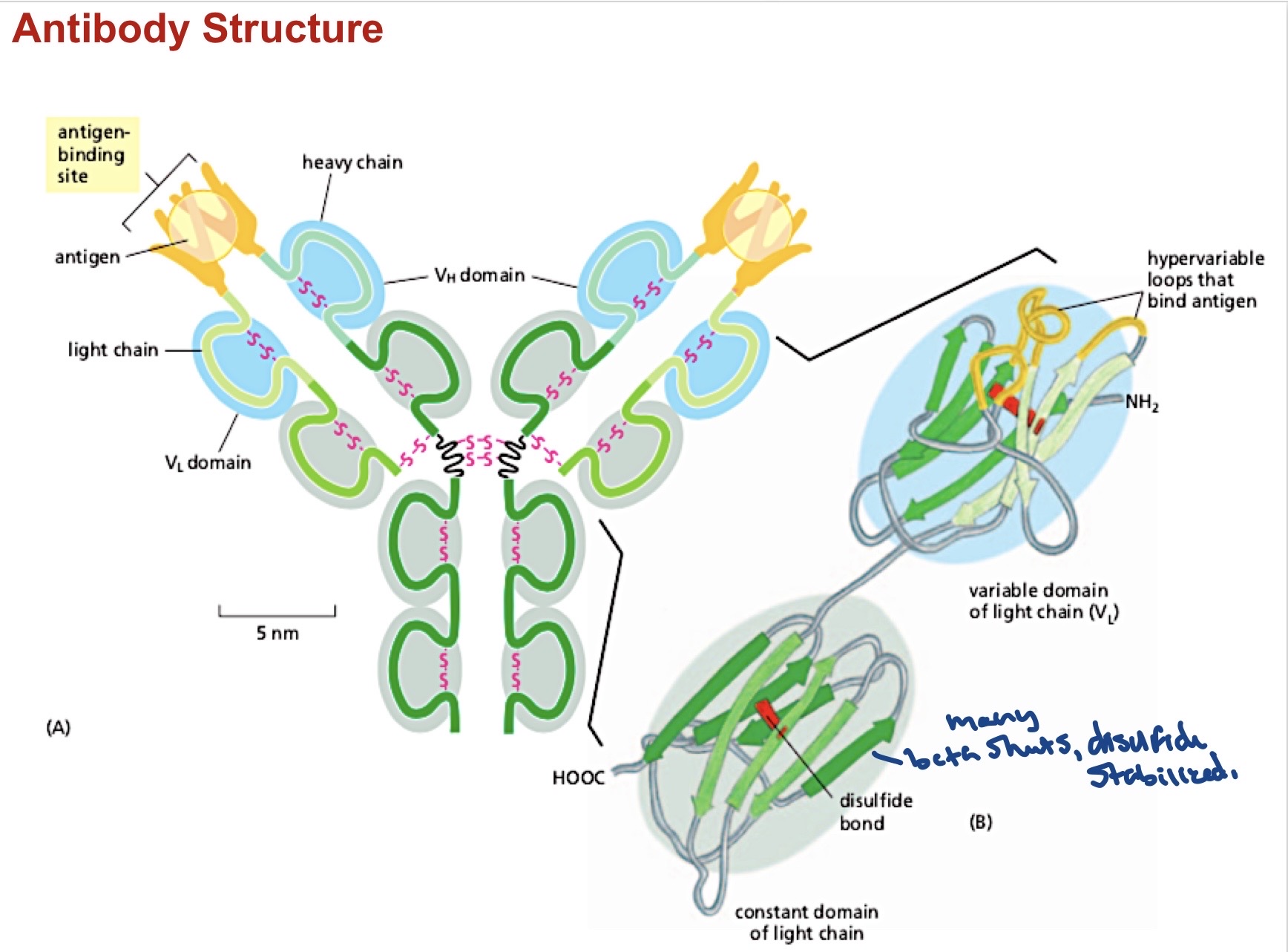
30
ELISA- Antibody based visualization of protein
utilized to quantify small amounts of protein. Less time consuming than a western, done only w/ the pure protein, unlike western which just shows if protein is present in the mix. More color=more enzyme = more protein present. Protein of interest is fixed to a surface by a capture antibody, then another anytobod(which is modified with an enzyme) attaches to the protein/ antigen. Substrate for this enzyme is added, resulting in a readout to detect it (usually a color change.). 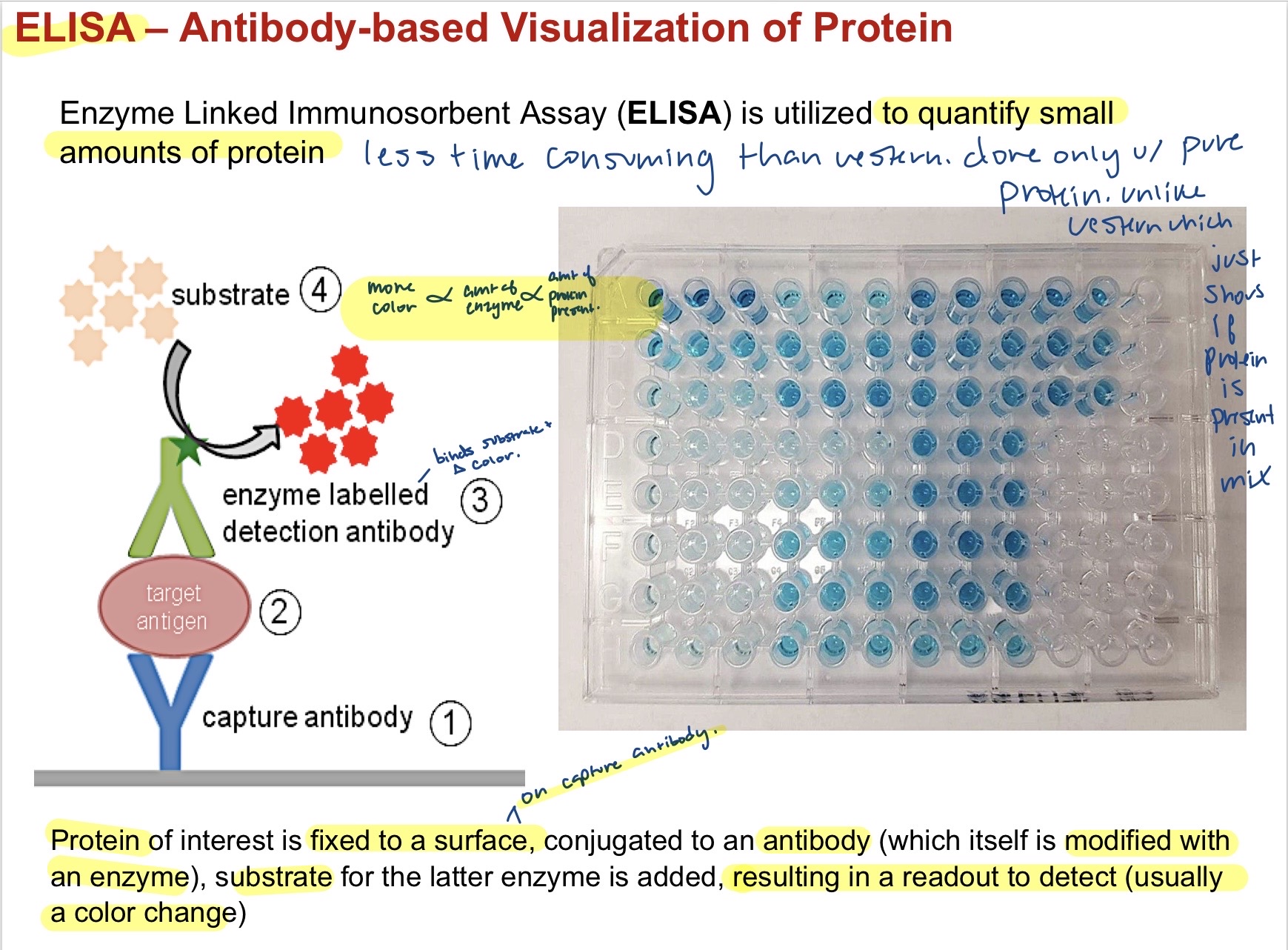
31
Absorption of UV light by Aromatic R groups in proteins- UV spectroscopy
The best way to quanitify a protein that has aromatics present is through UV absorption instead of ELISA Phe, Tyr, and Trp are aromatics, Trptophan absorbs the most due to its doubly conjugated system. amount of light absorbed is proportional to amount of protein present.
32
Circular Dichroism Spectroscopy
uses the differntial absorption of polarized light by chiral amino acids routinely used for assessing secondary structure content of proteins. this is just a bulk technique through that does not provide atomic resolution, just shows the most dominant secondary structure present. Read out will only ever show an alpha , beta, or random coil ( stuff btwn sheets) read based on most dominant. If tied then alpha helix will show most dominant over beta sheets. 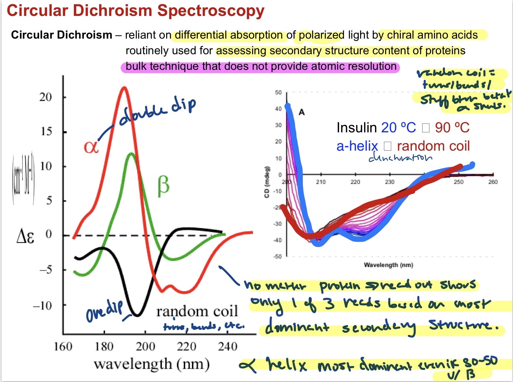
33
Chromatographic purification of protein mixtures
Column chromatogaphy separates proteins on the basis of some physical or chemical property. Multiple types:Gel Filtration/Size Exclusion, Ion Exchange, and Affinity chromatography. 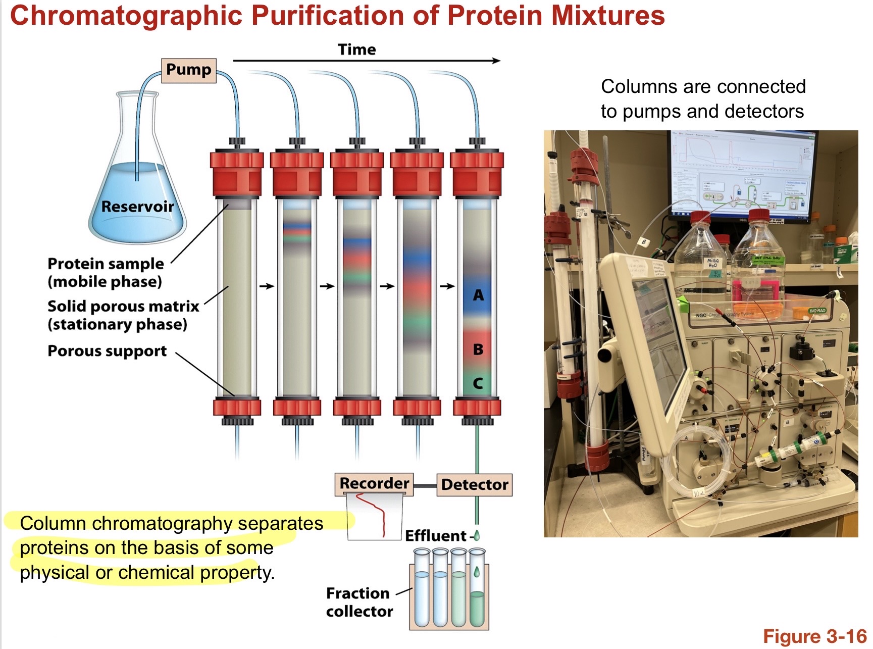
34
Get Filtration or Size Exclusion Chromatography
Separates proteins based on their weight/size Protein mixture is added to a column that has different size polymer beads in resin, separating the proteins into a size gradient. Larger molecules pass more freely coming out first. 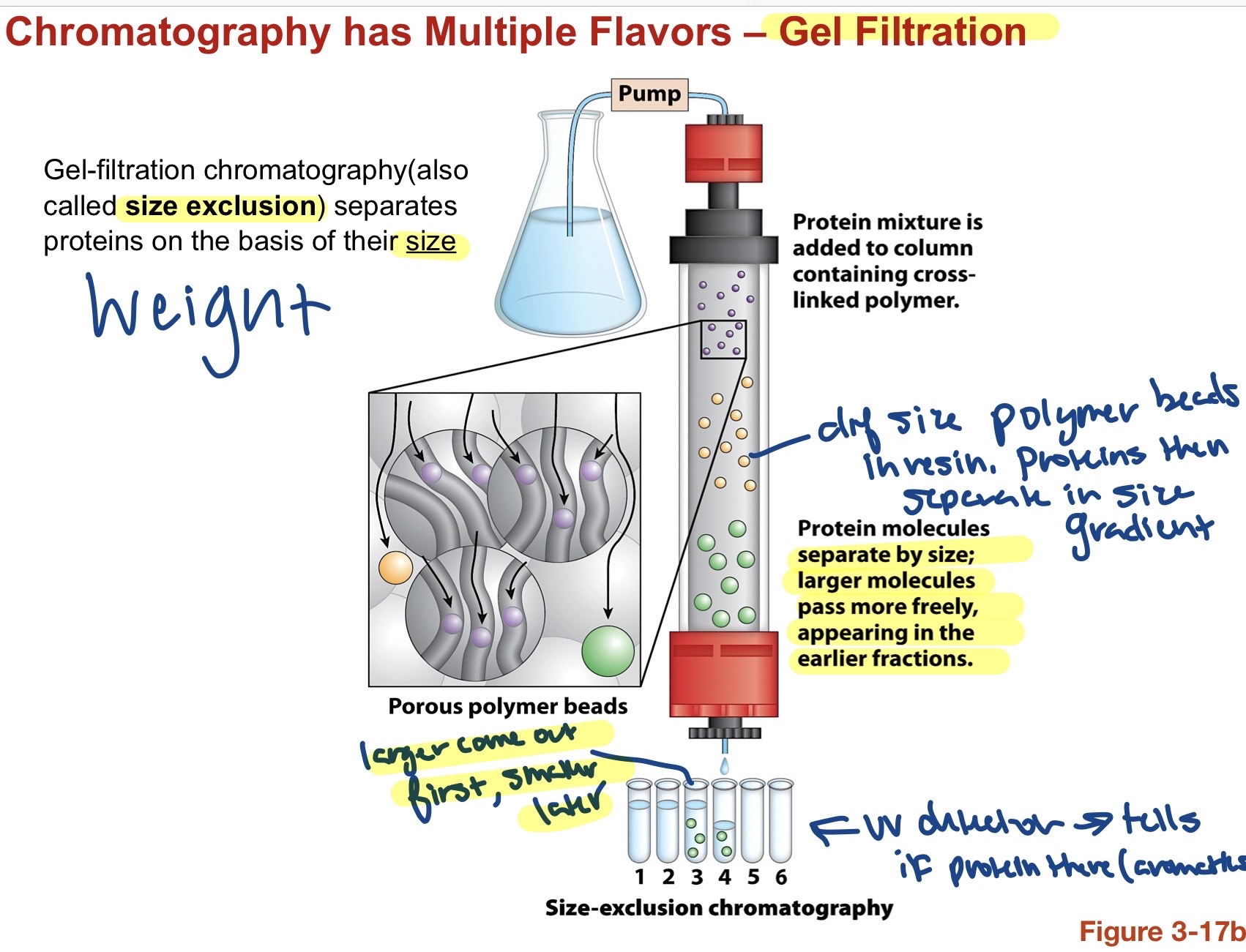
35
Ion Exchange chromatography
Separates proteins based on their charge. protein mixture is added to a column containing cation exchangers. (-) resin repels (-) charge proteins and they elute earlier. If anion exchanger then would be the oppoiste due to repulsion. Charge is dependent on the pH of the solution becuase this changes the net charge of the amino acids. 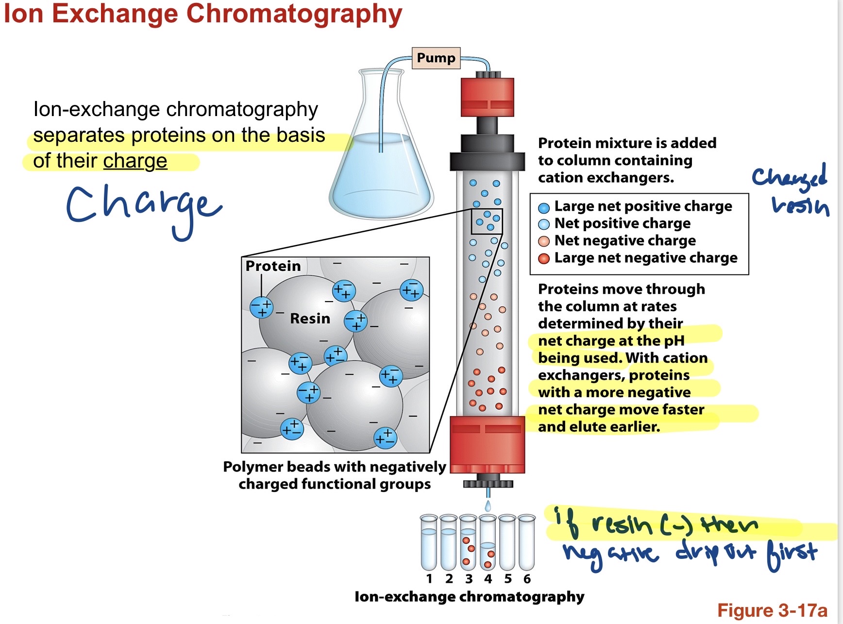
36
Affinity Chromatography
Separates proteins based on a specific binding interaction between the mobile and stationary phase(resin). Immobilized receptor is a binding site to a specific protein. flow a mixture of proteins in and only the protein that sticks to the receptor will bind. Then add a reagent and elute to unbind the protein allowing for the specific protein to come out. 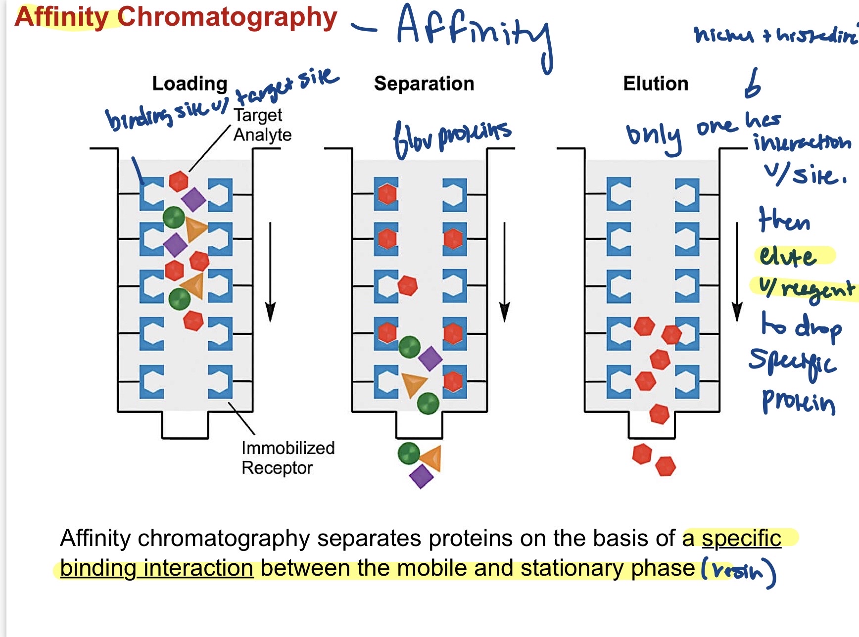
37
Purification and protein yield
The more pure you want a protein the less protein you will be left with from the overall mixture.
38
Fragmentation of peptides and Protein Sequencing.
after finding out if a protein is present and purifying it, you then need to fragment it to see what there. Proteases cleave the peptide bond AFTER specific residues. Chart details key Proteases, what they cleave, and where they cleave on the amino acid. Fragments can then be coated in charge, their mass/ charge (m/z) can be determiend, and this can be compared with experimental Ms/Ms theoretical spectra to determine the aa makeup of complex mixtures. 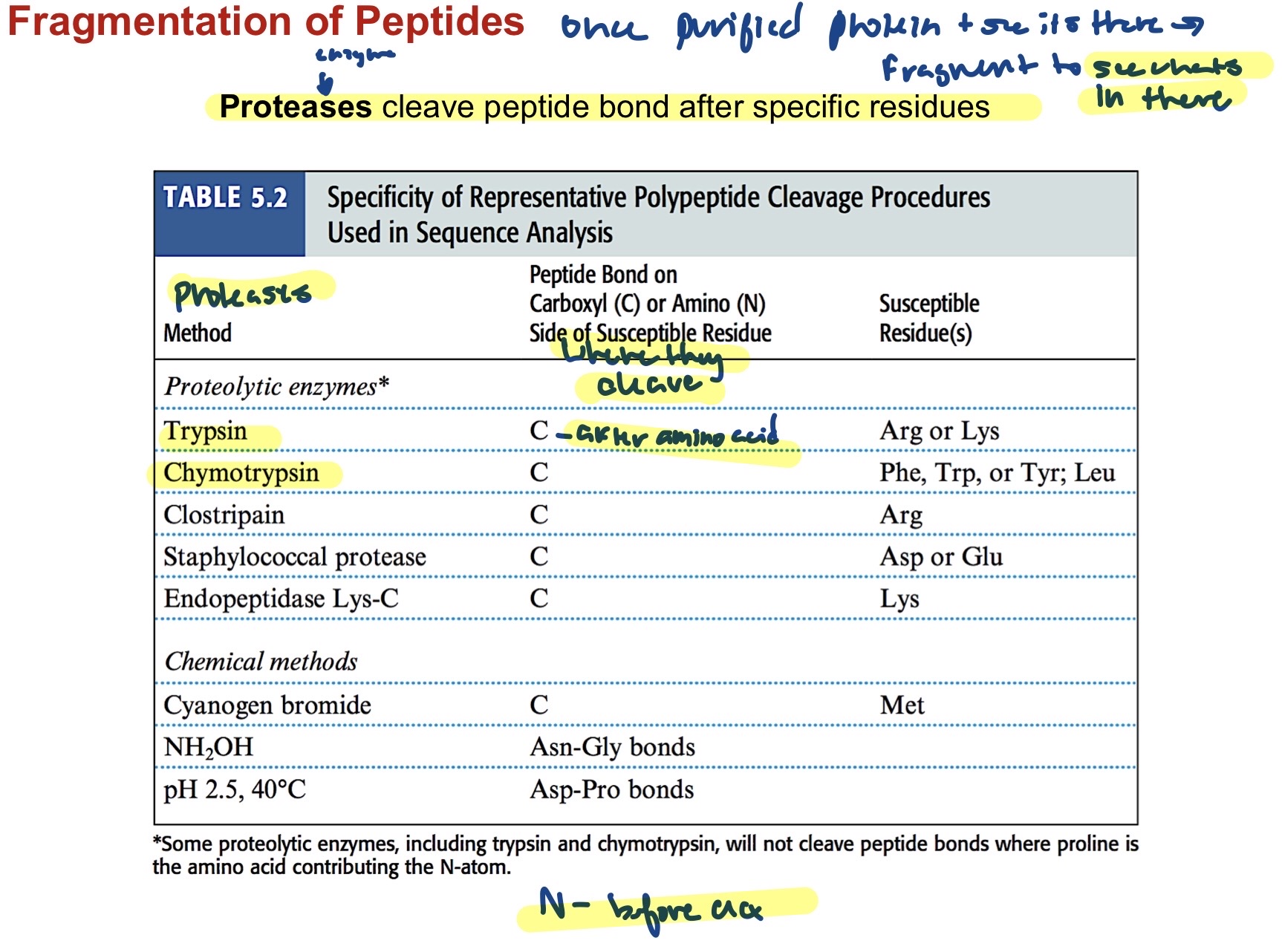
39
What can you do with sequence information?
1) Predict functional sited based on patches of conserved amino acids (if a certain sequence has been maintained across a lot of organisms then you can infer its important for something) 2) Analyze variability of a single protein across multiple organisms. 3) Establish evolutionary trends in protein sequences of different organisms (helps with phylogeny bc more similar = more closely related). 4) Predict Protein structural variation based on amino acid sequence. (motifs and stuff?)
40
Protein Dynamics- FRET
Forster Resonance Energy Transfer- reliant on conjugation of donor-acceptor fluorophores to protein. Fluoresence to see how felexible a protein is. When donor/acceptor are greater than 10nm away then only slight read but when less than 10 nm and in correct orientation there is a larger read. Flickering helps monitor protein moving in time. Donor acceptor intensity ratios relate to molecular distance.. 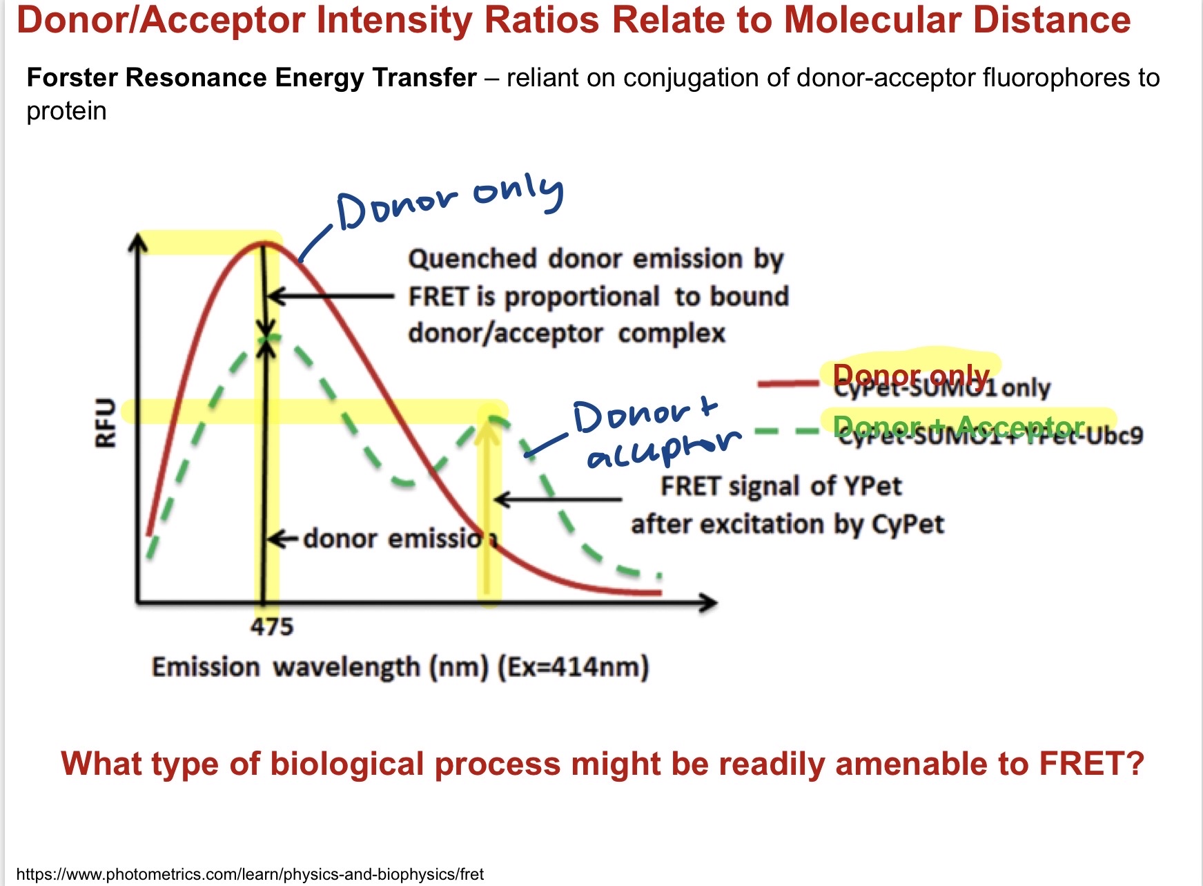
41
Protein Ligand interactions- Isothermal Titration Calorimetry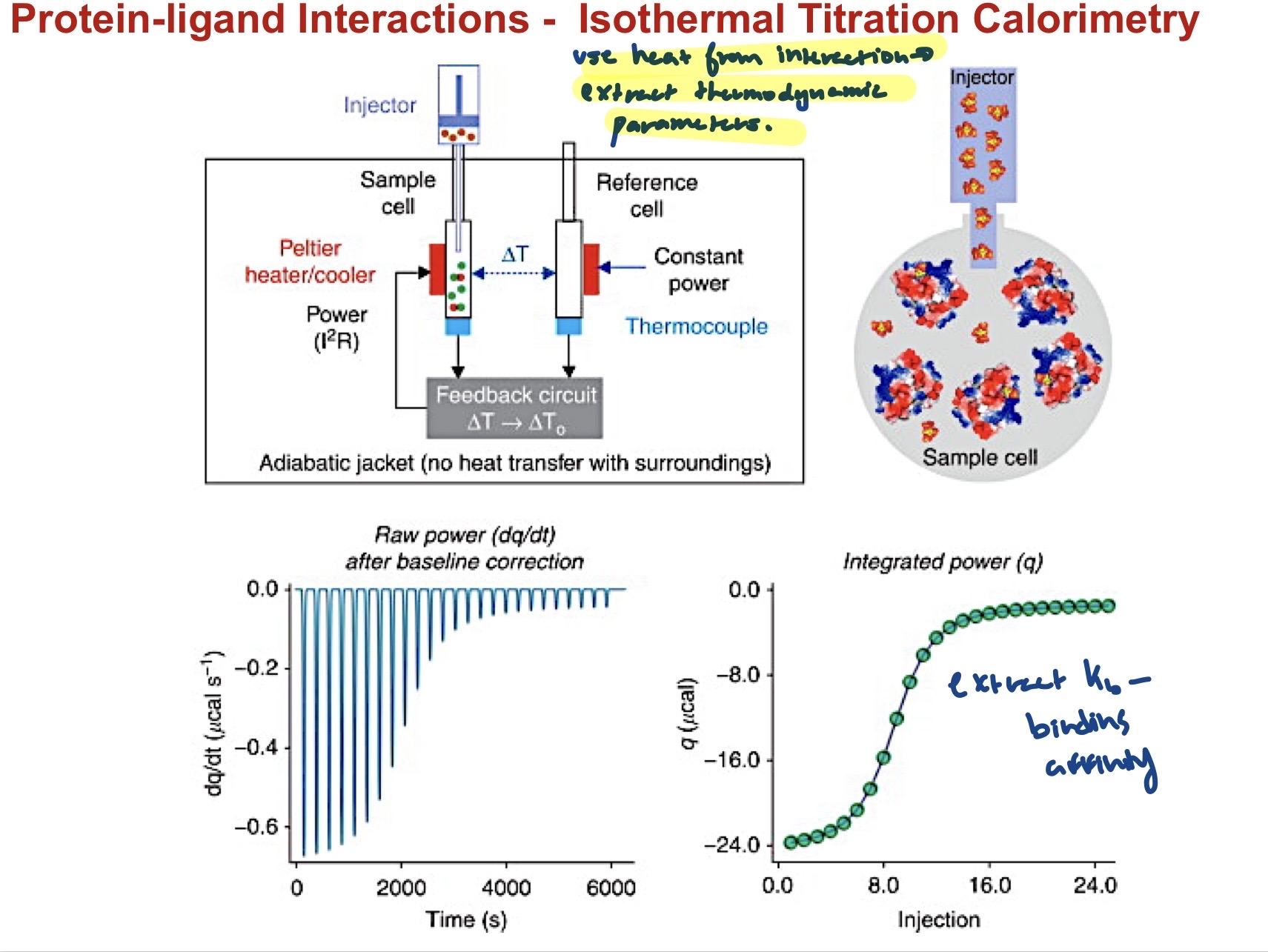
Use heat from an interaction to extract thermodynamic paramters. kb=binding affinity= slope, height is enthalps. From read you can tell the affinity between a proteink and a ligand from the slope. 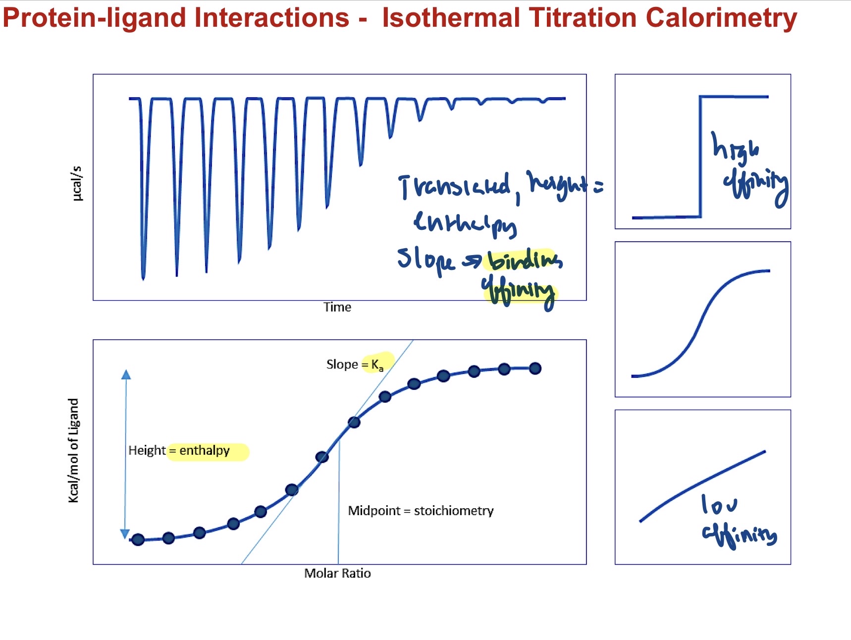
42
Protein Stability- Differential Scanning Calorimetry
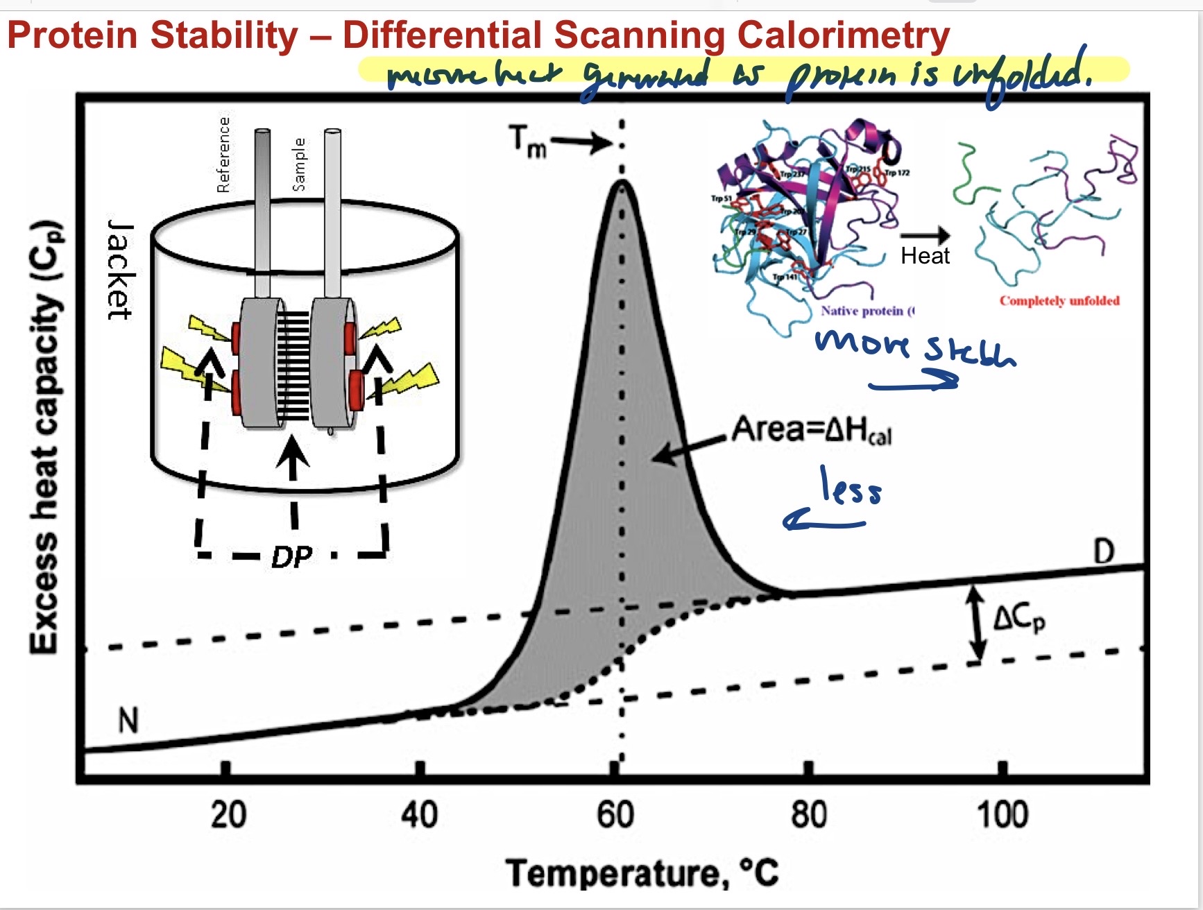
Measure the heat generated as the protein is unfolded. Higher temperature needed means more stable than a lower temperature.