2.1 Cell Structure ✓
Study these flashcardsFree · spaced repetition · iPhone, Android, Mac & web
All cards
1
Which type of cell doesnt have membrane bound organelles?
prokaryote
2
What is an electron micrograph?
photograph of image seen using electron microscope
3
What is magnification?
number of times larger image appears compared to size of object
4
What is a photomicrograph?
photograph of image seen using optical microscope
5
What is resolution?
clarity of image (higher resolution= clearer image)
6
What type of magnification do microscopes produce?
linear
7
What does linear magnification mean?
specimen maginfied x100 appears 100 x longer and wider than it really is
8
What are some advantages of using optical microscopes?
-relatively cheap
-easy to use
-portable (use in field + lab)
-can study whole living specimens
-easy to use
-portable (use in field + lab)
-can study whole living specimens
9
Why do structures very close together appear as 1 object when using an optical light microscope?
wavelength of visiblelight 400-700nm so structures closer than 200nm appear as 1 object
10
What equation is used to work out magnification?
magnification= image size/ actual size
11
What equation is used to work out total magnification from eyepiece and objective lens?
total mag= mag power of objective lens x mag power of eyepiece lens
12
What are the steps for using an optical microscope?
1) specimen on slide + clipped in place
2) rotate nosepiece so lowest power objective lens over specimen
3) adjust coarse focus knob until image clear in focus
4) adjust iris diaphragm for optimum light
5) rotate nosepiece bringing next objective lens over specimen and use fine focus knob to focus
6) repeat step 5 using next objective lens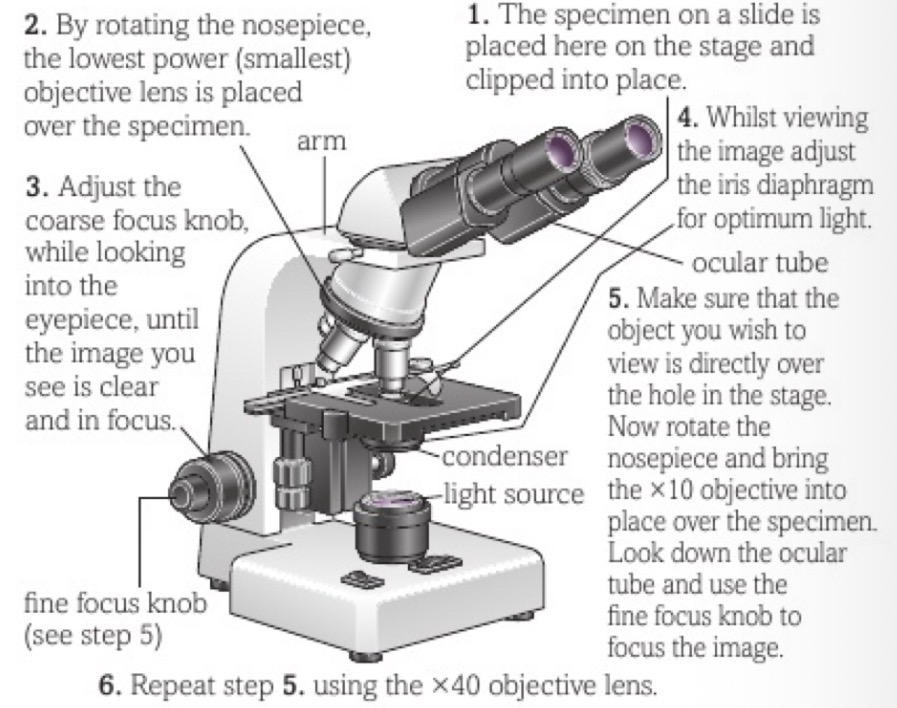
2) rotate nosepiece so lowest power objective lens over specimen
3) adjust coarse focus knob until image clear in focus
4) adjust iris diaphragm for optimum light
5) rotate nosepiece bringing next objective lens over specimen and use fine focus knob to focus
6) repeat step 5 using next objective lens
13
What are laser microscopes also called?
confocal microscope
14
What are laser microscope used for?
-in medical profession to give swift diagnosis and earlier more effective treatment
-biological research
-biological research
15
Why do electron microscopes have greater resolution + higher mag?
beam of electrons have smaller wavelength (0.004nm)
16
How must the specimen be treated for use in TEM?
chemically fixed by dehydration and stain (metal salts)
17
Describe images produced by TEM
-2D
-black + white
-very high mag
-black + white
-very high mag
18
Describe images produced by SEM
-3D
-relatively high mag (more than optical less than SEM)
-black + white (but colour can be added)
-relatively high mag (more than optical less than SEM)
-black + white (but colour can be added)
19
How must the specimen be treated for use in SEM?
-placed in vaccum
-often coated with fine film of metal
-often coated with fine film of metal
20
What are disadvantages of electron microscopes?
-large + expensive to use
-need skill + training to use
-specimens have to be dead
-stains potentially hazardous to user
-need skill + training to use
-specimens have to be dead
-stains potentially hazardous to user
21
Describe images produced by laser microscopes
-high resolution
-high contrast
-depth selectivity so can focus on structures at different depths (whole living specimens or cells)
-high contrast
-depth selectivity so can focus on structures at different depths (whole living specimens or cells)
22
How can you use light to view unstained colourless transparent specimens?
-light interference instead of light absorption
-dark background against which illuminated specimen shows up
-dark background against which illuminated specimen shows up
23
What is differential staining?
stain bonds to specific cell structures staining each structure differently
24
How are prepared and permanently fixed slides made?
-dehydrate
-embedded in wax to prevent distortion while slicing
-special instrument use to make sections (thin slices)
-stained + mounted in special chemical to preserve
-embedded in wax to prevent distortion while slicing
-special instrument use to make sections (thin slices)
-stained + mounted in special chemical to preserve
25
What is an eyepiece graticule?
measuring device placed in eyepiece and acts as ruler when viewing object
26
What is a stage graticule?
precise measuring device placed on microscope stage and used to calibrate value of eyepiece divisions at different mag
27
How long is a stage graticule?
1mm long
28
How do you use stage graticule and eyepiece graticule?
1) insert eyepiece graticule into eyepiece
2) place stage graticule on stage + bring into focus
3) align graticules and check value of 1 eyepiece division
4) use this ratio to measure images in microscope
2) place stage graticule on stage + bring into focus
3) align graticules and check value of 1 eyepiece division
4) use this ratio to measure images in microscope
29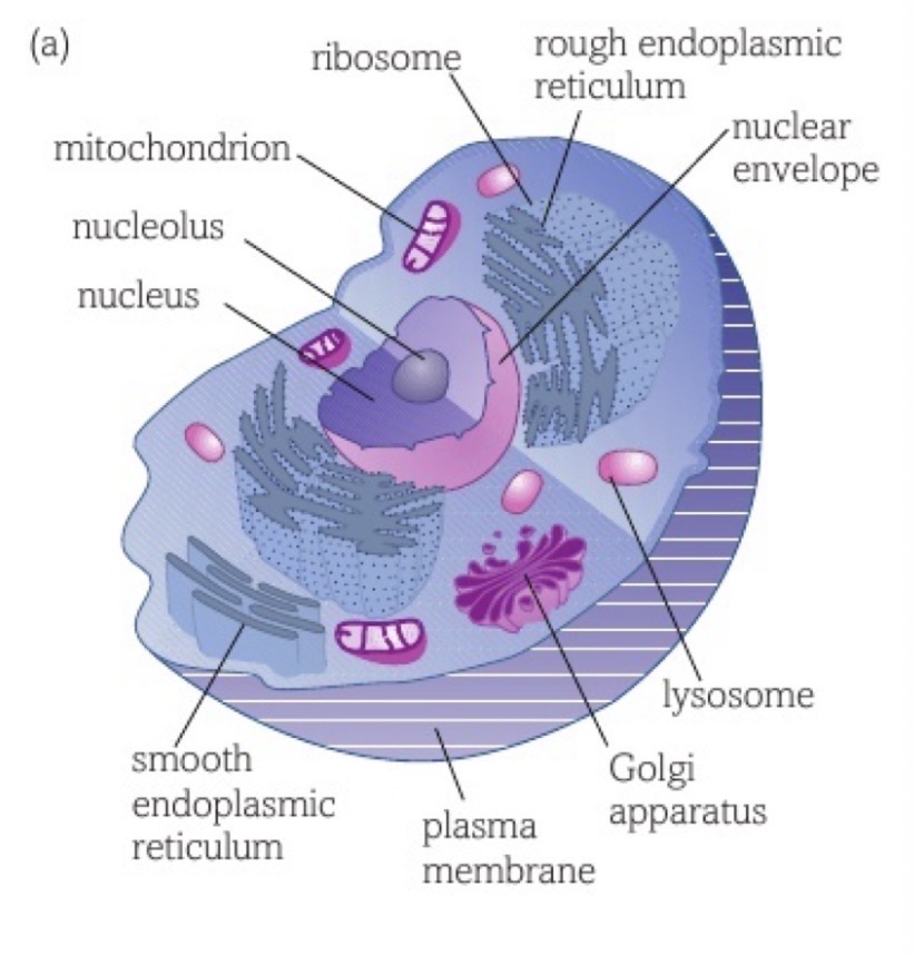
Image occlusion: name the hidden labels
30
Image occlusion: name the hidden labels
31
Image occlusion: name the hidden labels
32
Image occlusion: name the hidden labels
33
Image occlusion: name the hidden labels
34
Image occlusion: name the hidden labels
35
Image occlusion: name the hidden labels
36
Image occlusion: name the hidden labels
37
Image occlusion: name the hidden labels
38
Image occlusion: name the hidden labels
39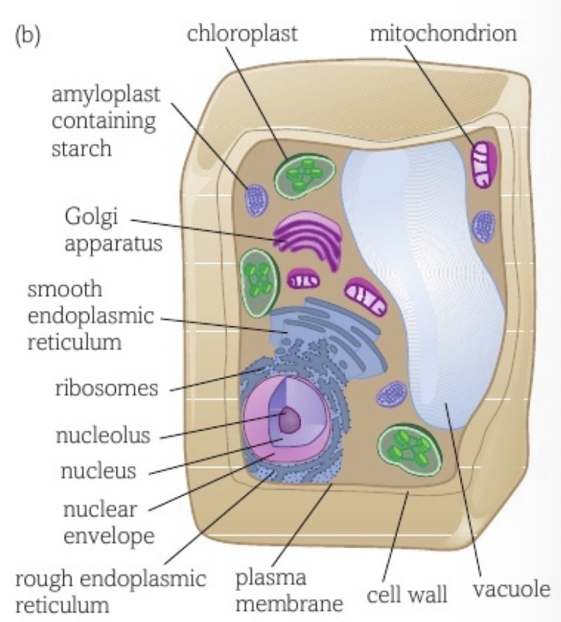
Image occlusion: name the hidden labels
40
Image occlusion: name the hidden labels
41
Image occlusion: name the hidden labels
42
Image occlusion: name the hidden labels
43
Image occlusion: name the hidden labels
44
Image occlusion: name the hidden labels
45
Image occlusion: name the hidden labels
46
Image occlusion: name the hidden labels
47
Image occlusion: name the hidden labels
48
Image occlusion: name the hidden labels
49
Image occlusion: name the hidden labels
50
Image occlusion: name the hidden labels
51
Image occlusion: name the hidden labels
52
Image occlusion: name the hidden labels
53
What is the nuclear envelope?
double membrane with pores surrounding nucleus
54
What does the nucleolus contain?
RNA
55
Does the nucleolus have a membrane?
no
56
What is chromatin?
genetic material consisting of DNA wound around histone proteins
57
What is chromatin like when cell isnt dividing?
spread out/ extended
58
What is chromatin like when cell is dividing?
condensed/ coiled tightly into chromosomes
59
What is the function of the nuclear envelope?
separate contents of nucleus and rest of cell
60
Where can dissolved substances and ribosomes pass through the nuclear envelope?
where outer and inner membrane fuse
61
What enters/ leaves the nucleus through pores in nuclear envelope?
-messenger RNA
-steroid hormones
-steroid hormones
62
Where are ribosomes made?
nucleolus
63
What is the rough endoplasmic reticulum?
system of membranes coated in ribosomes, containing cisternae that are continuous with nuclear membrane
64
What is the function of the rough endoplasmic reticulum?
-intracellular transport system: cisternae form channels used for transportation around cell
-provides large SA for ribosomes + proteins then actively pass through membrane into cisternae to be transported to golgi apparatus
-provides large SA for ribosomes + proteins then actively pass through membrane into cisternae to be transported to golgi apparatus
65
What is the smooth endoplasmic reticulum?
system of membranes, containing cisternae that are continuous with nuclear membrane (no ribosome coating)
66
What is the function of the smooth endoplasmic reticulum?
-contains enzymes for catalysing lipid metabolism reactions
-absorption, synthesis and transport of lipids
-absorption, synthesis and transport of lipids
67
What are examples of reactions that SER enzymes catalyse?
-synthesis of cholesterol
-synthesis of lipids/ phospholipids
-synthesis of steroid hormones
-synthesis of lipids/ phospholipids
-synthesis of steroid hormones
68
What is the golgi apparatus?
stack of membrane-bound flattened sacs
69
What is the function of the golgi apparatus?
-protein modification
-proteins packaged into vesicles + pinched off
-proteins packaged into vesicles + pinched off
70
What are examples of protein modifications done in golgi apparatus?
-add sugar to make glycoproteins
-add lipid molecules to make lipoproteins
-folded into their 3D shape
-add lipid molecules to make lipoproteins
-folded into their 3D shape
71
Where do proteins in vesicles go after golgi apparatus?
-stored in cell
-moved to plasma membrane to be incorporated into it or exported out cell
-moved to plasma membrane to be incorporated into it or exported out cell
72
What are cisternae?
fluid-filled cavities used for transportation
73
Which shapes + lengths can mitochondria be?
spherical, rod-shaped or branched
74
What is the structure of a mitochondrion?
-2 membranes with fluid filled space between
-inner membrane folded into cristae
-inner part is fluid-filled matrix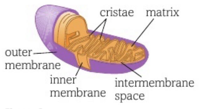
-inner membrane folded into cristae
-inner part is fluid-filled matrix
75
What is the function of mitochondria?
site of ATP production during respiration
76
What can mitochondria do if energy need increases?
self replicate and make more
77
What is the structure of chloroplast?
-double membrane
-inner membrane continuous with thylakoids (stacks of flattened membrane sacs) which contains chlorophyll
-fluid filled matrix (stroma)
-contains loops of DNA and starch grains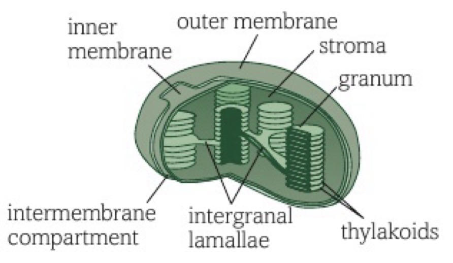
-inner membrane continuous with thylakoids (stacks of flattened membrane sacs) which contains chlorophyll
-fluid filled matrix (stroma)
-contains loops of DNA and starch grains
78
What is a granum?
stack/ pile of thylakoids
79
What is the function of chloroplast?
photosynthesis
80
What are the 2 stages of photosynthesis in the chloroplast?
1. light energy trapped by chlorophyll + used to make ATP (in grana), water also split for H ions
2. hydrogen reduces CO2 using energy from ATP to make carbs (in stroma)
2. hydrogen reduces CO2 using energy from ATP to make carbs (in stroma)
81
What is the tonoplast?
vacuole membrane
82
What is the function of the vacuole?
-filled with water + solutes
-maintains stability (turgid cells support plant)
-maintains stability (turgid cells support plant)
83
Where are lysosomes formed?
golgi apparatus
84
What is the function of lysosomes?
-keep powerful hydrolytic enzymes seperate from rest of cell
-engulf old cell organelles + foreign matter, digest and return components to be reused
-engulf old cell organelles + foreign matter, digest and return components to be reused
85
What are cilia and undulipodia?
protrusions from cell surrounded by cell surface membrane contining microtubules
86
What are cilia and undulipodia formed from?
centrioles
87
What is the function of cilia/ undulipodia?
-cilia in airways
-one cilium on cells to detect signals about environment with receptors
-undulipodium on sperm enable movement
-one cilium on cells to detect signals about environment with receptors
-undulipodium on sperm enable movement
88
What are ribosomes made out of?
ribosomal RNA
89
What is the process of ribosomes forming?
made in nucleolous as 2 subunits, pass nuclear envelope and combine in cytoplasm
90
What is the main function of ribosomes attached to RER?
synthesising proteins that are exported out of cell
91
What is the main function of ribosomes free in cytoplasm?
synthesise proteins used in cell
92
What are centrioles made from?
2 bundles of microtubules at right angles to each other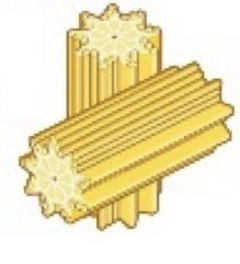
93
What are microtubules made from?
tubulin protein subunits
94
What is the function of centrioles?
-before cell division, forms spindle of tubulin threads which chromosomes attach to and motor proteins walk along pulling chromosomes to opposite ends
-form cilia + undulipodia
-form cilia + undulipodia
95
What is the cytoskeleton?
network of protein structures in cytoplasm consisting of:
-rod-like microfilaments
-intermediate filaments
-straight, cylindrical microtubules
-cytoskeleton motor proteins
-rod-like microfilaments
-intermediate filaments
-straight, cylindrical microtubules
-cytoskeleton motor proteins
96
What is the function of protein microfilaments in the cytoskeleton?
-support + mechanical strength
-keep cells shape stable
-allow cell movement
-keep cells shape stable
-allow cell movement
97
What is the function of microtubules in the cytoskeleton?
-shape and support to cells
-help substances + organelles move through cytoplasm by forming a track which motor proteins walk and drag organelles along
-form spindle before cell divides
-make up cilia, undulipodia + centrioles
-help substances + organelles move through cytoplasm by forming a track which motor proteins walk and drag organelles along
-form spindle before cell divides
-make up cilia, undulipodia + centrioles
98
What is the function of intermediate filaments in the cytoskeleton?
-anchor nucleuswithin cytoplasm
-extend between cells in some tissues enabling cell-cell signalling, stabilising tissues
-extend between cells in some tissues enabling cell-cell signalling, stabilising tissues
99
What is the function of the cell wall?
-prevent cells bursting
-strength+ support
-maintain cell shape
-permeable to allow solutions through
-strength+ support
-maintain cell shape
-permeable to allow solutions through
100
How are prokaryotic cells different to eukaryotic?
-much smaller
-less well-developed cytoskeleton with no centrioles
-no nucleus
-no membrane-bound organelles
-wall of peptidoglycan not cellulose
-smaller ribosomes
-naked DNA not wound around histone proteins but floats free in cyriplasm in loop (not linear)
-less well-developed cytoskeleton with no centrioles
-no nucleus
-no membrane-bound organelles
-wall of peptidoglycan not cellulose
-smaller ribosomes
-naked DNA not wound around histone proteins but floats free in cyriplasm in loop (not linear)
101
What extra things do some prokaryotic cells have that eukaryotic cells dont have?
-protective waxy capsule around cell wall
-small DNA loops (plasmids)
-flagella (whip like projections for movement)
-pili (small hair like projections for bacteria to adhere to host cells or eachother and allow passage for DNA transfer)
-small DNA loops (plasmids)
-flagella (whip like projections for movement)
-pili (small hair like projections for bacteria to adhere to host cells or eachother and allow passage for DNA transfer)
102
How do prokaryotic cells divide?
binary fission (DNA copied before division so each cells has large loop and plasmids)
103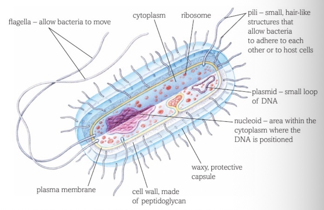
Image occlusion: name the hidden labels
104
Image occlusion: name the hidden labels
105
Image occlusion: name the hidden labels
106
Image occlusion: name the hidden labels
107
Image occlusion: name the hidden labels
108
Image occlusion: name the hidden labels
109
Image occlusion: name the hidden labels
110
Image occlusion: name the hidden labels
111
Image occlusion: name the hidden labels