3.2 Transport in Animals (✓)
Study these flashcardsFree · spaced repetition · iPhone, Android, Mac & web
All cards
1
Which 3 factors influence need for a transport system?
1) size
2) sa:v
3) level of metabolic activity
2) sa:v
3) level of metabolic activity
2
How does size influence the need for a transport system?
Diffusion pathway increases with size, so in a larger animal diffusion rate too slow + outer cells use all supplies
3
How does sa:v infuence the need for a transport system?
Large animals have low sa:v so each g of tissue has less area of body surface for exchange
4
How does level of metabolic activity influence the need for a transport system?
If active, cells need a good nutrients + oxygen supply for energy for movement (mamals need even more energy for keeping warm)
5
What are the features of an effective transport system? (3)
1) fluid/medium
2) pump
3) exchange surfaces
2) pump
3) exchange surfaces
6
What are the features of an efficient transport system? (2)
1) tubes/vessels
2) 2 circuits
2) 2 circuits
7
What is the function of the fluid/medium in a transport system?
To carry nutrients, oxygen + waste
8
What is the function of the pump in a transport system?
create pressure to push fluid around body
9
What is the function of the exchange surfaces in a transport system?
to exchange substances in and out of the body
10
What is the function of the tubes/vessels in a transport system?
to carry blood by mass flow
11
What is the function of the 2 circuits in a transport system?
1 to get oxygen and 1 to deliver oxygen
12
What is a double circulatory system?
blood flows through the heart twice for each circuit around the body
13
What are the 2 circulations in a double circulatory system?
1) pulmonary circulation
2) systemic circulation
2) systemic circulation
14
What is the function of the pulmonary circulation?
to recieve oxygen into blood from the lungs
15
What is the function of the systemic circulation?
supply oxygen to body
16
What is a single circulatory system?
where blood flows through heart once for each circuit of the body
17
What is the blood pressure like in single circulatory systems and how does this effect the rate of blood flow?
low blood pressure making blood flow slow
18
What is the blood pressure like in pulmonary circulation and why?
not too high as would otherwise damage delicate capillaries in lungs
19
What is the blood pressure like in systemic circulation and why?
high so rate of blood flow (and therefore delivery and removal of substances) is high
20
What is the difference between energy requirements in fish and mammals?
fish require less (so single circulatory system sufficient) whereas mammals require more due to higher demand and keeping themselves warm (so need double circulatory system)
21
What is the meaning of an open circulatory system?
blood is not always held in blood vessels and instead circulates through body cavity meaning tissues and cells are bathed directly in blood
22
How does circulation work in an open circulatory system?
Some animals depend on movement for circulation but others (eg insects) have a muscular pumping organ like heart and therefore their circulation works w/ out movement
23
Where is the muscular pumping organ in an insect?
lies under dorsal (upper) surface of body
24
How does blood move through the open circulatory system in an insect?
1) blood enters 'heart' through ostria
2) 'heart' pumps blood towards head by peristalsis
3) at forward end of 'heart' (by head) blood pours into body cavity
2) 'heart' pumps blood towards head by peristalsis
3) at forward end of 'heart' (by head) blood pours into body cavity
25
What are ostia?
pores which blood uses to enter heart in insects
26
What is peristalsis?
wave-like, involuntary muscle contractions that move substances along bodily tubes
27
What do some larger and more active insects also have in their circulatory system?
open ended tubes attached to the heart that direct blood (eg to wings)
28
What are the disadvantages of a open circulatory system?
-low blood pressure so slow blood flow
-circulation affected by movement (or lack of)
-circulation affected by movement (or lack of)
29
What are the advantages of an open circulatory system?
-easier heat distribution
30
Does movement affect all open circulatory systems?
yes
31
What is tissue fluid?
A seperate fluid from blood that bathes the tissues and cells
32
What are the advantages of an closed circulatory system?
-higher blood pressure so faster blood flow
-circulation independent of movement
-circulation independent of movement
33
What are the disadvantages of a closed circulatory system?
-More complex
-blood may clot
-blood may clot
34
What is the endothelium? (blood vessel)
the inner layer/lining of a blood vessel made of a single layer of cell
35
Why is the endothelium smooth? (blood vessels)
to reduce friction with blood
36
Why is an arteries wall thick?
to withstand high pressure
37
Why is an arteries lumen relatively small?
to maintain pressure
38
Why is an arteries inner wall folded?
to allow lumen to expand as blood flow increases
39
What does the tunica intima of the artery wall contain and what is it for?
thin layer of elastic tissue allowing the wall to stretch and then recoil to maintain pressure
40
What does the tunica media of the artery wall contain?
thick layer of smooth muscle
41
What does the tunica adventitia of the artery wall contain and what is it for?
relatively thick layer of collagen and elastic tissue providing strength to withstand high pressure and recoil to maintain pressure
42
What is the inner layer of an artery called?
tunica intima
43
Which layer is the tunica media (artery)?
middle
44
Which layer is the tunica adventitia (artery)?
outer
45
What are arterioles?
small blood vessels that distribute the blood from artery to capillaries
46
What does an arterioles wall contain and what does this do?
smooth muscle which contracts to constrict the diameter of the arteriole, increasing resistance to flow and reduces rate of flow
47
Why is constriction of arterioles useful?
can divert flow of blood to regions demanding more oxygen
48
What is the function of a capillary?
to allow exchange between blood and tissue fluid
49
What is a capillaries lumen like and why?
very narrow (diameter ~ same as red blood cell's) so red blood cells squeezed against walls, helping transfer oxygen as reduces diffusion pathway (also increases resistance and reduces rate of flow)
50
Why are capillary walls leaky?
to allow blood plasma and dissolved substances to leave blood
51
What does a capillary wall consist of and why?
single layer of flattened endothelial cells to reduce diffusion distance
52
What are venules?
small blood vessels that collect blood from capillaries and lead into veins
53
What do venule walls consist of?
thin layers of muscle and elastic tissue outside endothelium and thin outer layer of collagen
54
What is the lumen like in veins and why?
relatively large to ease flow of blood
55
What are the walls like in veins and why?
thinner layers of collagen, smooth muscle and elastic tissue (than arteries) as dont need stretch, recoil or constrict
56
Why do veins have valves?
to help blood flow back to heart and prevent flowing in opposite direction
57
Why can veins be flattented by action of surrounding skeletal muscle?
walls are thin
58
What is hydrostatic pressure?
pressure that a fluid exerts when pushing against sides of a vessel/ container
59
What is lymph?
fluid held in lymphatic system (similar composition to tissue fluid)
60
What does lymph contain that blood doesnt? Why?
lymphocytes as these are produced in lymph nodes
61
What is the lymphatic system?
system of tubes returning excess tissue fluid to blood system
62
What is onotic pressure?
pressure created by osmotic effects of solutes
63
What is the fluid portion of the blood called?
Plasma
64
What is another word for red blood cells?
erythrocytes
65
What is another word for white blood cells?
leucocytes
66
Which dissolved substances does the plasma contain?
-oxygen + carbon dioxide
-minerals
-glucose
-amino acids
-hormones
-plasma proteins
-minerals
-glucose
-amino acids
-hormones
-plasma proteins
67
Which cells does the plasma contain?
-erythrocytes
-leucocytes
-platelets (fragments)
-leucocytes
-platelets (fragments)
68
How is tissue fluid formed?
at arterial end of capillary, blood has relatively high hydrostatic pressure, pushing plasma out of capillary walls through the tiny gaps between cells
69
Which parts of the blood remain in the blood during exchange and why?
-red blood cells
-platelets
-most white blood cells
-plasma proteins
too large to be pushed through gaps in capillary wall
-platelets
-most white blood cells
-plasma proteins
too large to be pushed through gaps in capillary wall
70
By which procceses does exchange occur across plasma membrane between tissue fluid and cell?
-diffusion
-facilitated diffusion
-active uptake
-facilitated diffusion
-active uptake
71
Is blood plasma leaking from the capillary and carrying dissolved substances into tissue fluid mass flow or diffusion?
mass flow
72
Why can the tissue fluid return to the blood stream?
Venous end of capillary is much lower pressure allowing some tissue fluid to return to capillary
73
What are lymph nodes?
swellings found at intervals along lymphatic system which are important to immune response
74
Where else does tissue fluid go other than the blood?
lymphatic system which drains excess tissue fluid out of tissues and returns it to blood system in subclavian vein in chest
75
What is hydrostatic pressure like in the blood plasma?
high
76
What is hydrostatic pressure like in the tissue fluid?
low
77
What is hydrostatic pressure like in the lymph?
low
78
What is oncotic pressure like in the blood plasma?
more negative
79
What is oncotic pressure like in the tissue fluid?
less negative
80
What is oncotic pressure like in the lymph?
less negative
81
Which cells are in the blood plasma?
-RBC
-neutrophils
-lymphocytes
-neutrophils
-lymphocytes
82
Which cells are in the tissue fluid?
some neutrophils, especially in infected areas
83
Which cells are in the lymph?
lymphocytes
84
Which proteins are in the blood plasma?
plasma proteins
85
Are there proteins in the tissue fluid?
few proteins
86
Are there proteins in the lymph?
few proteins
87
Are there fats in the blood plasma?
yes transported in lipoproteins
88
Are there fats in the tissue fluid?
yes few fats
89
Are there fats in lymph?
More fats (esp digestive system)
90
How does oncotic pressure influence movement of fluid into and out of capillary?
-(arteriole end) hydrostatic pressure going out of capillary greater that oncotic going in
-opposite at venule end
-means net outflow at arteriole end and net inflow at venule end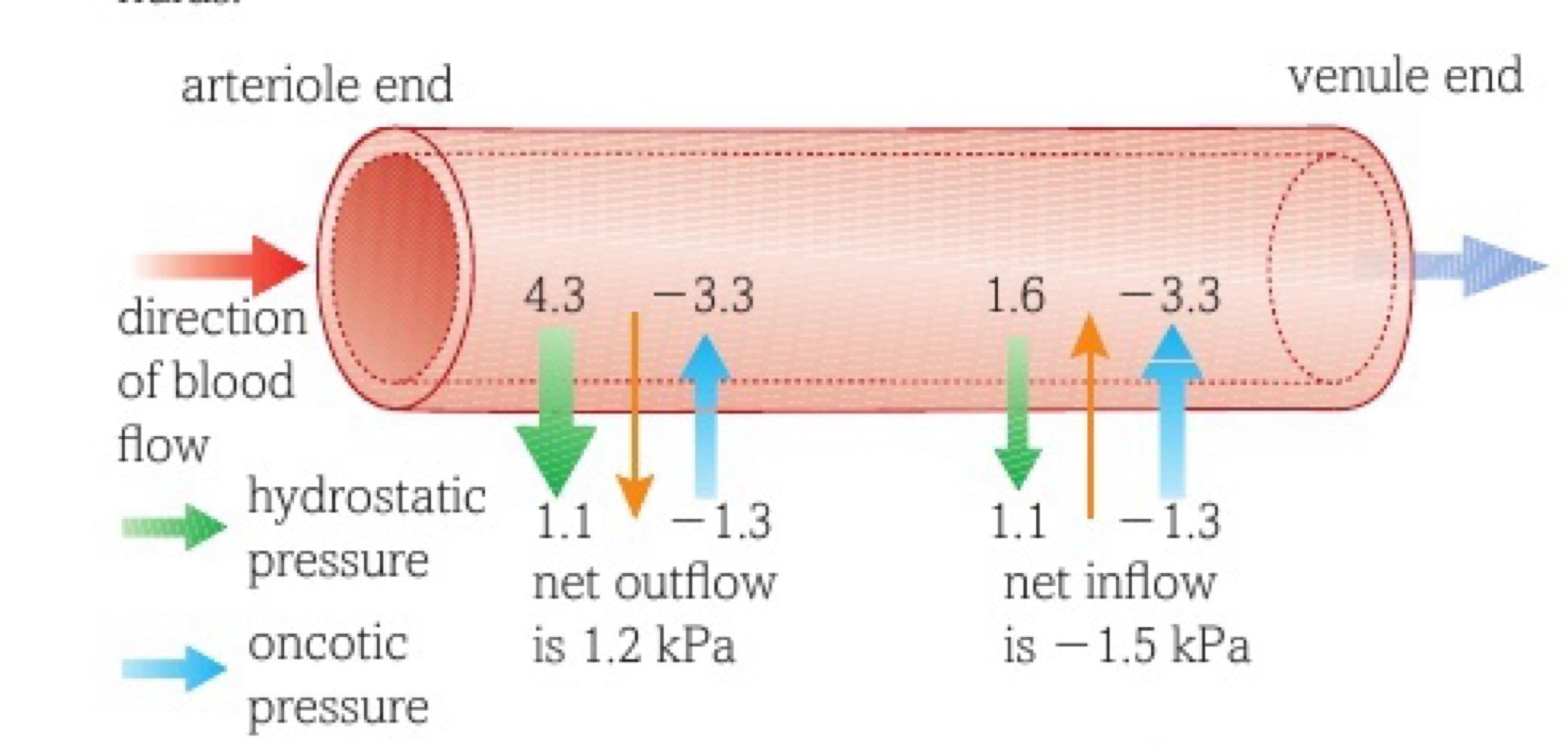
-opposite at venule end
-means net outflow at arteriole end and net inflow at venule end
91
What are the atrio-ventricular valves?
valves between atria and ventricles
92
What is the cardiac muscle?
specialised muscle found in walls of heart chambers
93
What are semilunar valves?
valves preventing blood reentering heart from arteries
94
What does the main part of the heart consist of?
cardiac muscle
95
Where are the coronary arteries and what is their role?
lie over surface of heart to supply oxygen to heart muscle
96
What is myocardial infarction?
heart attack
97
Which vessel carries deoxygenated blood from the body into the heart?
vena cava
98
What do tendinous cords do?
prevent valves from turning inside out when venticle walls contract
99
What is the wall between the ventricles called?
interventricular septum
100
What is the vessel called that carries oxygenated blood out of the heart and into the body?
aorta
101
What is the muscle wall like in the atria? Why?
very thin as do not need to create much pressure
102
What is the function of the atria?
recieve blood from veins and push into ventricles
103
What is the function of the right ventricle?
pump deoxygenated blood out of heart into lungs
104
What are the right ventricles walls like?
thicker than atria
105
Why shouldnt the pressure of blood coming out of right venticle be too high?
alveoli in lungs delicate and could be damaged by high pressure
106
What are left ventricle walls like?
a lot thicker than right ventricle
107
Why does the blood coming out of the left ventricle need to be high pressure?
need sufficient pressure to overcome resistance of systemic circulation so able to pump around body
108
What is the structure of cardiac muscle?
fibres that branch producing cross bridges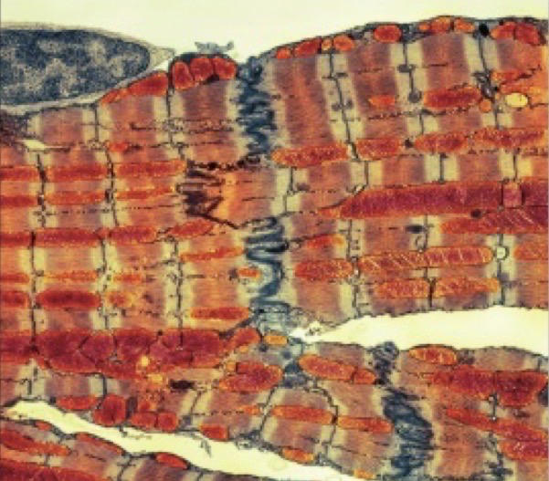
109
Why does cardiac muscle need cross-bridges?
-spread stimulus around heart
-muscle produces squeezing action rather than reduction in length
-muscle produces squeezing action rather than reduction in length
110
What is found between myofibrils?
mitochondria for energy
111
What are myofibrils?
muscle fibrils
112
What seperates muscle cells? What is its function?
intercalated discs, to facilitate syncronised contraction
113
What is a cardiac muscle cell divided into?
contractile units called sarcomeres
114
What is the cardiac cycle?
coordinated sequence of events in 1 full heart beat
115
What is the role of the heart?
create pressure to push blood around blood vessels
116
What is the role of valves?
ensure blood flows in right direction
117
What happens during ventricular systole?
left and right ventricle pump together, contraction starting at the apex pushing blood up to arteries
118
What happens during atrial systole?
left and right atria contract together, small increase in pressure created due to thin walls helping fill ventricles with blood
119
What happens during diastole?
all muscular walls relax, elastic recoil causes chambers to increase in volume allowing blood to flow in from veins
120
How do the atrio ventricular valves work?
Diastole:
1) pressure in ventricles drops below pressure in atria
2) blood in atria pushes atrio-ventricular valves open
3) blood entering goes straight through to ventricles
4) pressure in chambers rises as fill with blood
Atrial systole:
5) valves stay open during contraction
6) valves close once atria relax due to swirling action in blood when venticle full
Ventricular systole:
7) ventricular pressure rises as contracts, pushing blood upwards
8) valve pocket fills, keeping them closed
1) pressure in ventricles drops below pressure in atria
2) blood in atria pushes atrio-ventricular valves open
3) blood entering goes straight through to ventricles
4) pressure in chambers rises as fill with blood
Atrial systole:
5) valves stay open during contraction
6) valves close once atria relax due to swirling action in blood when venticle full
Ventricular systole:
7) ventricular pressure rises as contracts, pushing blood upwards
8) valve pocket fills, keeping them closed
121
What is the function of tendinous cords?
attached to valves to prevent them turning inside out
122
How do the semilunar valves work?
Before ventricular systole:
1) pressure in major arteries higher than ventricles so semilunar valves closed
Ventricular systole:
2) blood pressure increases in venticles quickly
3) ventricle pressure higher than artery so semilunar valves forced open
Diastole:
4) elastic tissue in ventricle walls recoils returning ventricle to original size
5) pressure in ventricles drops quickly
6) ventricle pressure lower than artery pressure so blood flows back towards ventricle
7) semilunar valves pushed closed by blood collecting in valve pockets
1) pressure in major arteries higher than ventricles so semilunar valves closed
Ventricular systole:
2) blood pressure increases in venticles quickly
3) ventricle pressure higher than artery so semilunar valves forced open
Diastole:
4) elastic tissue in ventricle walls recoils returning ventricle to original size
5) pressure in ventricles drops quickly
6) ventricle pressure lower than artery pressure so blood flows back towards ventricle
7) semilunar valves pushed closed by blood collecting in valve pockets
123
How does the structure of artery walls create a more even flow?
-artery wall close to heart has a lot of elastic tissue
-when blood leaves heart walls stretch
-as blood moves on/ out of aorta pressure starts dropping
-elastic recoil of walls maintain pressure
-when blood leaves heart walls stretch
-as blood moves on/ out of aorta pressure starts dropping
-elastic recoil of walls maintain pressure
124
The further blood flows along arteries the ____ pressure drops and fluctuations become ___ obvious
more, less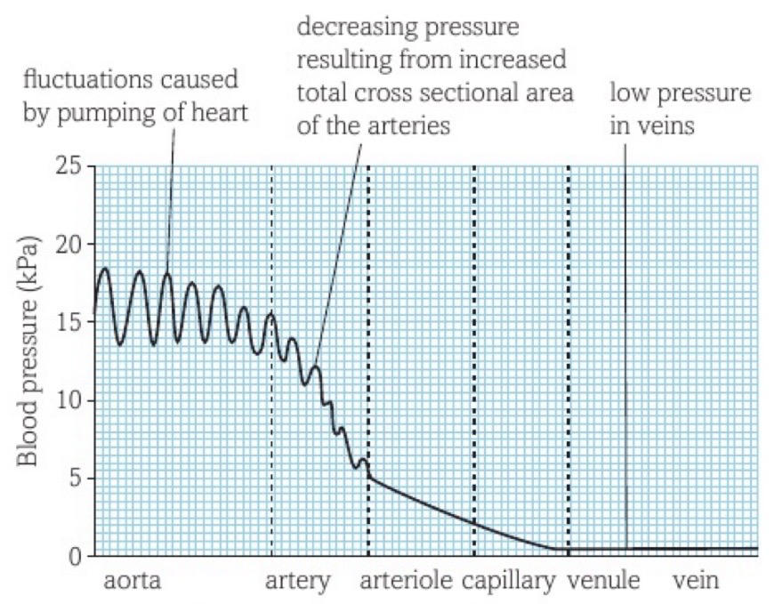
125
Why is it important to maintain pressure gradient between aorta and arterioles?
keeps blood flowing towards tissues
126
What is bradycardia?
a slow heart rhythm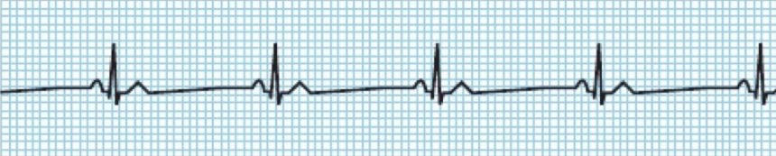
127
What is tachycardia?
a rapid heart rhythm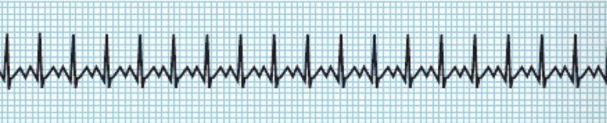
128
What is an ectopic heartbeat?
extra beat or early beat of ventricles
129
What is an electrocardiogram?
a trace that records electrical activity of heart
130
What is fibrillation?
uncoordinated contraction of atria and ventricles
131
What is myogenic muscle?
muscle that can initiate its own contraction
132
What is purkyne tissue?
consists of specially adapted muscle fibres that conduct wave of excitation from AVN down septum to ventricles
133
What is the sino-atrial node (SAN)?
hearts pacemaker: small patch of tissue which sends out waves of electrical excitation at regular intervals to initiate contractions
134
What's an example of myogenic muscle?
cardiac muscle
135
Does the atrial or venticular muscle contract at a higher frequency?
atrial
136
What's the average bpm of a human?
55-80bpm
137
Where is the SAN?
top of right atrium
138
Where is the AVN?
top of atrioventricular septum
139
How does a wave of excitation cause atria to contract?
wave of excitation spreads over walls of both atria, travelling along membranes of muscle tissue causing cardia muscle to contract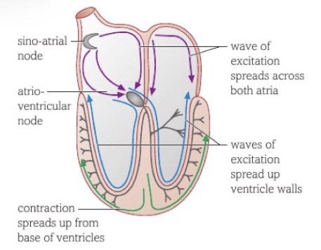
140
Why does wave of excitation not travel directly to ventricle muscle tissue?
tissue at base of atria cant conduct
141
Why is the wave of excitation delayed at the AVN?
allow time for atria to finish contracting and pushing bloodinto ventricles before they contract
142
How do the ventricles contract?
1) after delay in AVN wave of excitation carried down purkyne tissue running down interventricular septum
2) at base of septum wave spreads out over walls of ventricles
3) as excitation spreads upwards ventricles contract meaning they contract from base upwards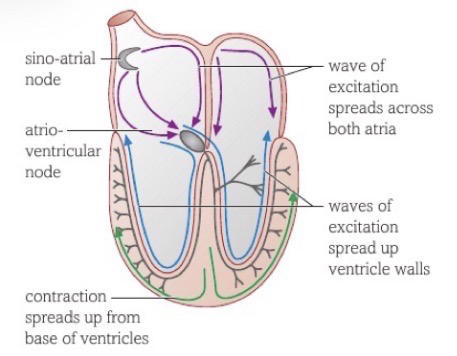
2) at base of septum wave spreads out over walls of ventricles
3) as excitation spreads upwards ventricles contract meaning they contract from base upwards
143
Why is it important that the ventricles contract from the base upwards?
push blood up towards major arteries at top of heart
144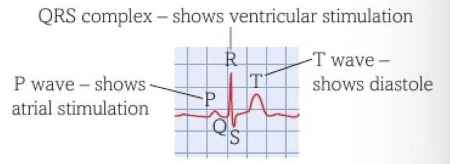
What do P, Q, R, S and T show on an electrocardiogram?
145
What does affinity mean?
a strong attraction
146
What is dissociation?
releasing oxygen from oxyhaemoglobin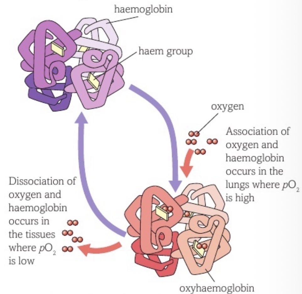
147
What is another name for red blood cells?
erythrocytes
148
What does haemoglobin become when it takes up oxygen?
oxyhaemoglobin
149
How many subunits does haemoglobin have?
4
150
What does each haemoglobin subunit consist of?
polypeptide chain and haem group (non-protein)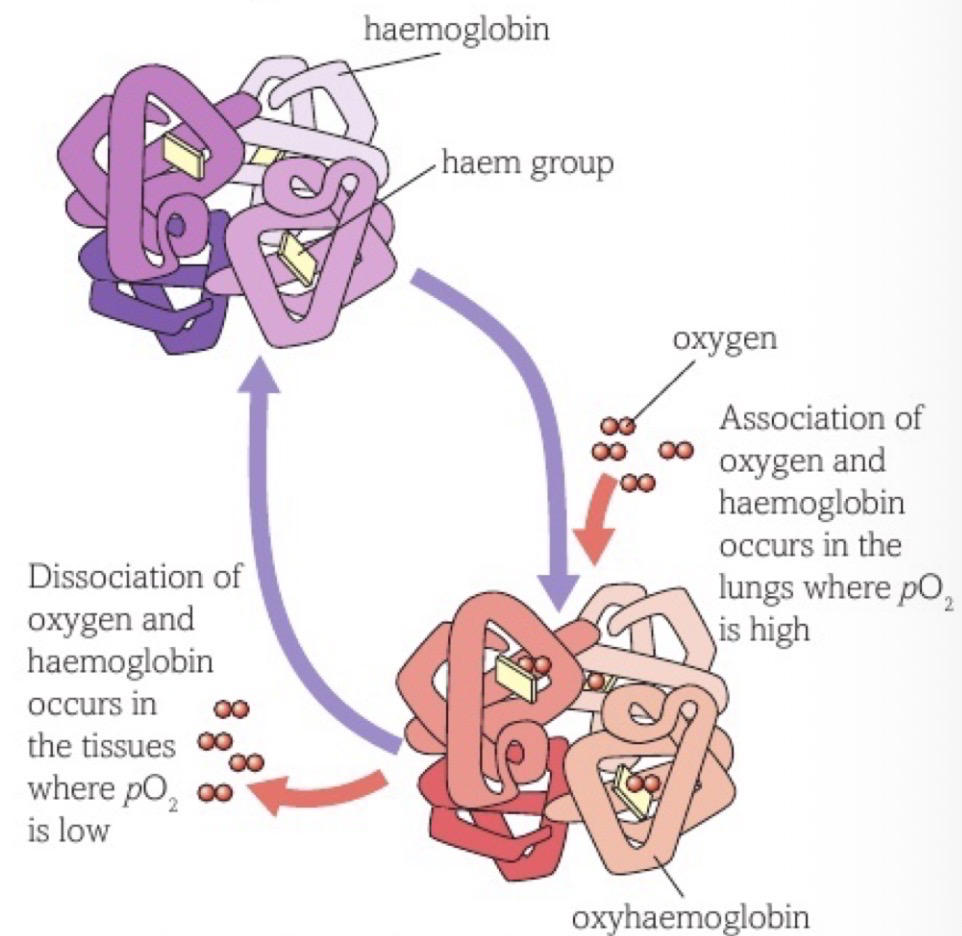
151
What in the haem group attracts and holds oxygen?
iron ion (Fe2+)
152
What has a high affinity for oxygen in haemoglobin?
haem group
153
How many oxygen molecules can each haem group hold?
1
154
How many oxygen molecules can each haemoglobin hold?
4
155
Roughly how many molecules of haemoglobin are in each red blood cell?
280 million
156
Does oxygen bind reversibly or irreversibly to haemoglobin?
reversibly
157
How does oxygen become bonded to haemoglobin?
-O absorbed into blood as passing alveoli
-O molecules diffuse into blood plasma and enter RBC
-become associated with haemoglobin
-O molecules diffuse into blood plasma and enter RBC
-become associated with haemoglobin
158
How does oxygen associating with haemoglobin help maintain a steep concentration gradient in the lungs?
oxygen molecules are out of solution
159
What is the partial pressure of oxygen (pO2)?
relative pressure that oxygen contributes to mixture of gases (way of measuring oxygen concentration)
160
What is another name for partial pressure of oxygen (pO2)?
oxygen tension
161
What is the haemoglobin dissociation curve caused by?
-at low oxygen tension HG doesnt readily associate accounting for low saturation level of HG at low oxygen tensions
-as oxygen tension rises, diffusion gradient into HG increases enventually causing one oxygen molecule to associate with a haem group
-this causes conformational change allowing more oxygen to enter HG and associate relatively easily accounting for steep part of curve
-as HG reaches 100% saturation curve levels off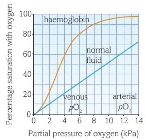
-as oxygen tension rises, diffusion gradient into HG increases enventually causing one oxygen molecule to associate with a haem group
-this causes conformational change allowing more oxygen to enter HG and associate relatively easily accounting for steep part of curve
-as HG reaches 100% saturation curve levels off
162
What is conformational change?
slight change in shape of haemoglobin after one oxygen molecule associates which makes it easier for other oxygen molecules to associate
163
Why doesnt haemoglobin readily associate with oxygen molecules at low oxygen tension?
haem groups attracting oxygen are in centre of haemoglobin making it difficult for oxygen to reach haem group
164
How does fetal haemoglobin differ from adult haemoglobin?
fetal has higher affinity for oxygen so dissociation curve is to left of adults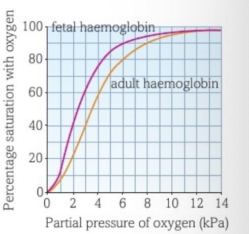
165
Why does fetal haemoglobin have a higher affinity for oxygen than adult?
must be able to associate with oxygen in environment where the oxygen tension low enough to make adult haemoglobin release oxygen
166
How does oxygen move from fetal haemoglobin to adult haemoglobin?
-in placenta where oxygen tension low fetal haemoglobin absorb oxygen from surrounding fluid, reducing oxygen tension further
-so oxygen from mothers blood diffuses into placenta, reducing oxygen tension in mothers blood making maternal haemoglobin release more oxygen
-so oxygen from mothers blood diffuses into placenta, reducing oxygen tension in mothers blood making maternal haemoglobin release more oxygen
167
What is carbonic anhydrase?
enzyme catalysing combination of CO2 and water
168
What is haemoglobinic acid?
compound formed by buffering action of haemoglobin as it combines with excess hydrogen ions
169
What is the Bohr effect?
effect extra CO2 has on haemoglobin, explaining releases of more oxygen
170
What is chloride shift?
movement of chloride ions into erythrocytes to balance charge as hydrogencarbonate ions leave cell
171
Which 3 ways is CO2 transported by in the blood?
-dissolved directly in plasma
-combined directly with haemoglobin
-transported in form of hydrogencarbonate ions (HCO3-)
-combined directly with haemoglobin
-transported in form of hydrogencarbonate ions (HCO3-)
172
What percentage of CO2 is carried by being dissolved directly in plasma?
5%
173
What percentage of CO2 is carried by being combined directly with haemoglobin?
10%
174
What percentage of CO2 is carried in form of hydrogencarbonate ions?
85%
175
What compound is formed when CO2 is directly comboned with haemoglobin?
carbaminohaemoglobin
176
How are hydrogencarbonate ions formed in the blood?
-CO2 in plasma diffuses into RBC
-combines with water to form weak carbonic acid (catalysed by carbonic hydrase)
-carbonic acid dissociates to release H+ ions and hydrogencarbonate ions
-hydrogencarbonate ions diffuse out of RBC and into plasma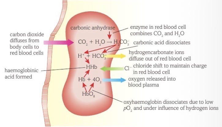
-combines with water to form weak carbonic acid (catalysed by carbonic hydrase)
-carbonic acid dissociates to release H+ ions and hydrogencarbonate ions
-hydrogencarbonate ions diffuse out of RBC and into plasma
177
How is the RBC becoming too acidic due to hydrogen ions building up prevented?
H+ ions taken out of solution by associating with haemoglobin to produce haemoglobinic acid (HHb)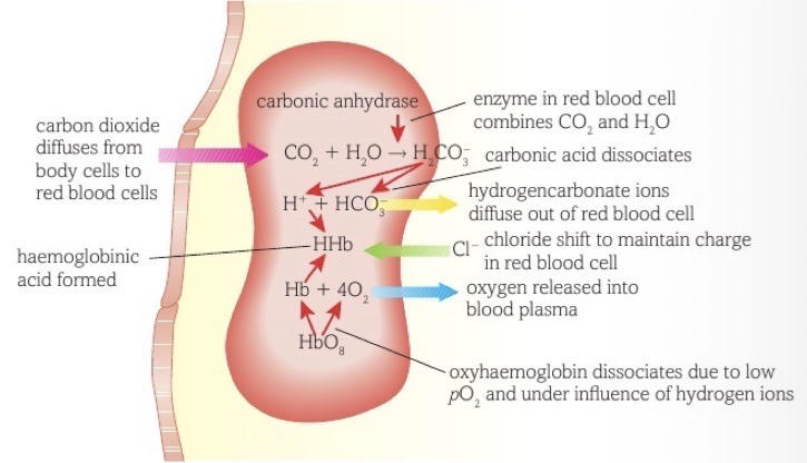
178
Why does oxyhaemoglobin dissociate?
due to low pO2 and under influence oh H+ ions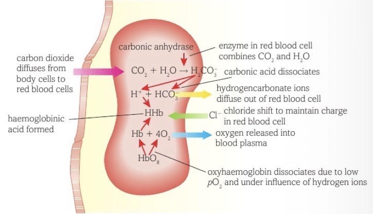
179
What is the Bohr effect?
-CO2 enters RBC forming carbonic acid which dissociates to release H+ ions
-H+ ions affect pH of cytoplasm (more acidic)
-changes in pH affect tertiary structure of haemoglobin, reducing affinity of haemoglobin for oxygen
-haemoglobin unable to hold as much oxygen so oxygen releases from oxyhaemoglobin to tissues
-H+ ions affect pH of cytoplasm (more acidic)
-changes in pH affect tertiary structure of haemoglobin, reducing affinity of haemoglobin for oxygen
-haemoglobin unable to hold as much oxygen so oxygen releases from oxyhaemoglobin to tissues
180
Why is the Bohr effect useful for respiring tissues?
respiring more means more CO2 produced and more O2 needed so Bohr effect as a result of increase in CO2 helps meet demand for O2
181
What is the Bohr shift?
-more CO2 present so haemoglobin less saturated with O2
-causes haemoglobin dissociation curve to shift downwards and the the right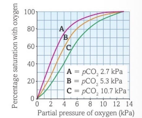
-causes haemoglobin dissociation curve to shift downwards and the the right