Bio 201 practical 3
Study these flashcardsFree · spaced repetition · iPhone, Android, Mac & web
All cards
1
The forehead. It is developed from 2 bones that usually fuse together. If the bones do not fuse you will see a Metopic Sculpture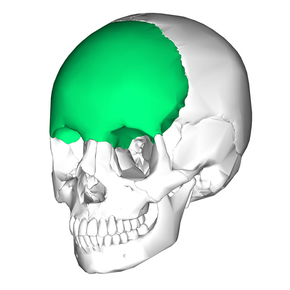
frontal bone
2
connects the frontal bone to the 2 parietal bones across the top of the skull 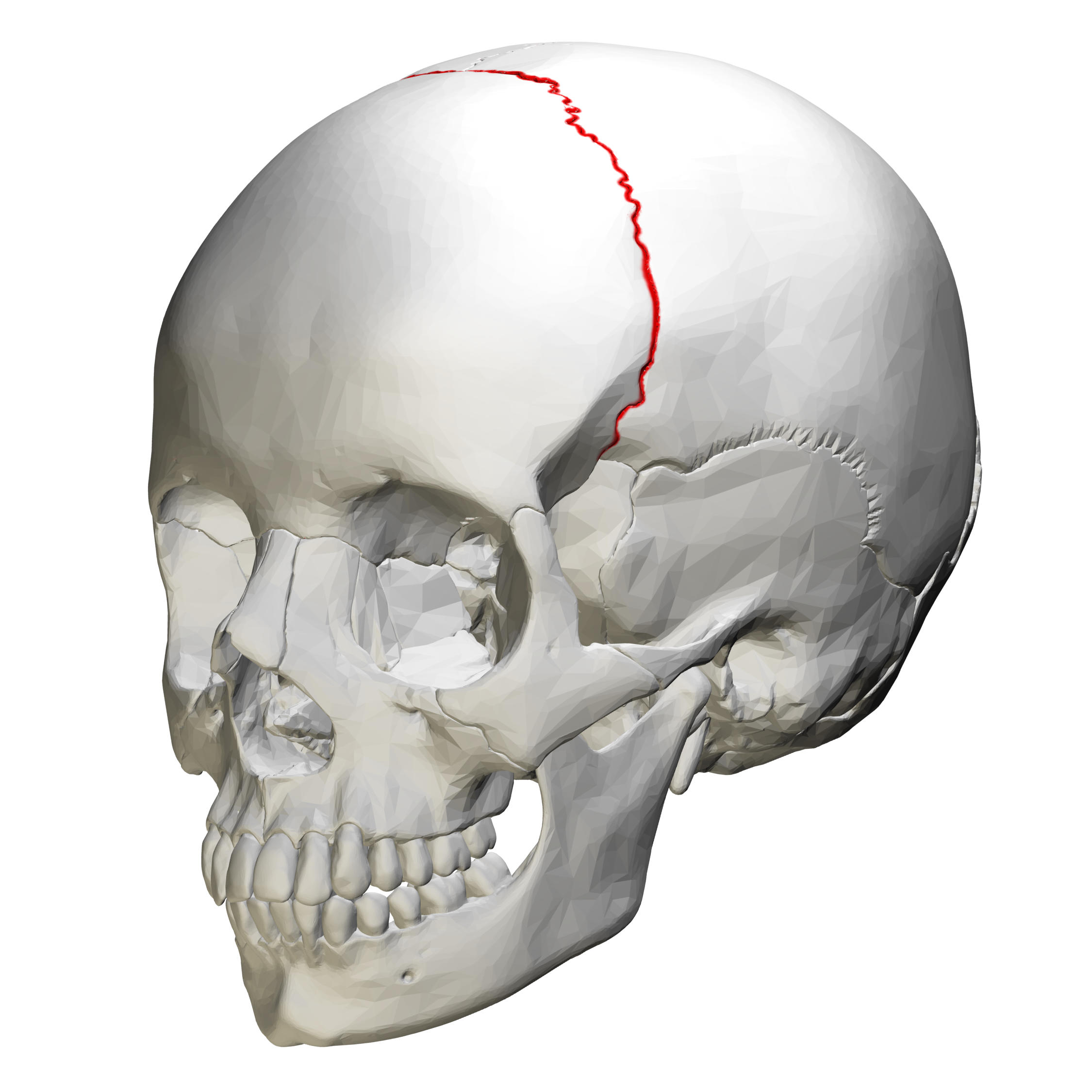
coronal suture
3
is a cavity in the frontal bone above the nose that lightens the skull and makes the tone of the voice unique 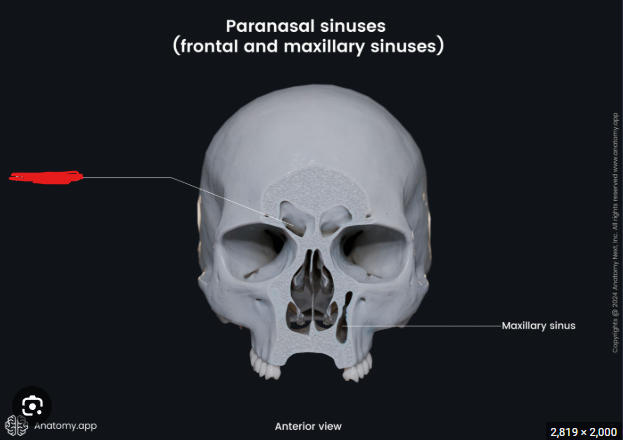
frontal sinus
4
are on the side of the skull and connects the frontal bone with the coronal suture 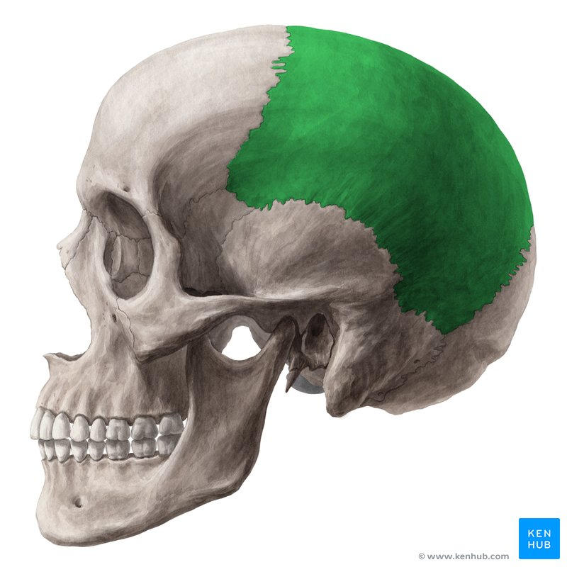
parietal bones
5
Connects the parietal bones across the top of the skull 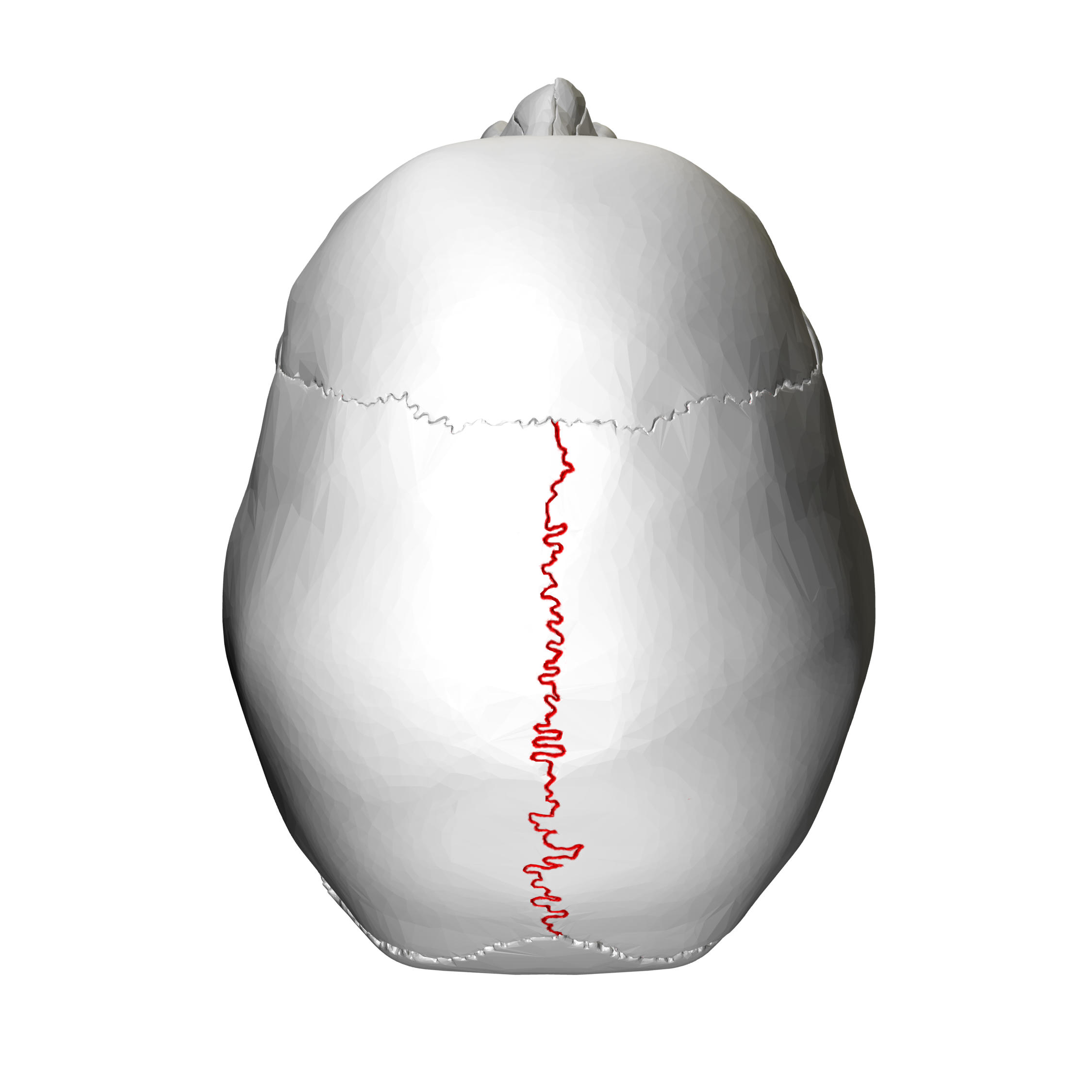
sagittal suture
6
are on the sides of the skull and contain several complex features 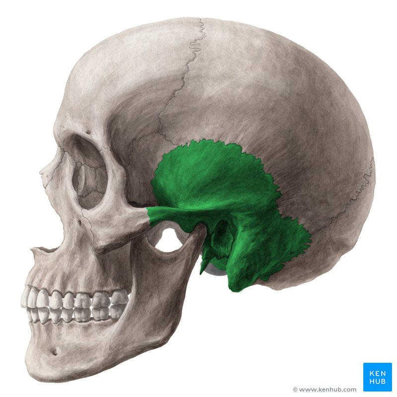
temporal bones
7
connects the squamous temporal bone with the parietal bones 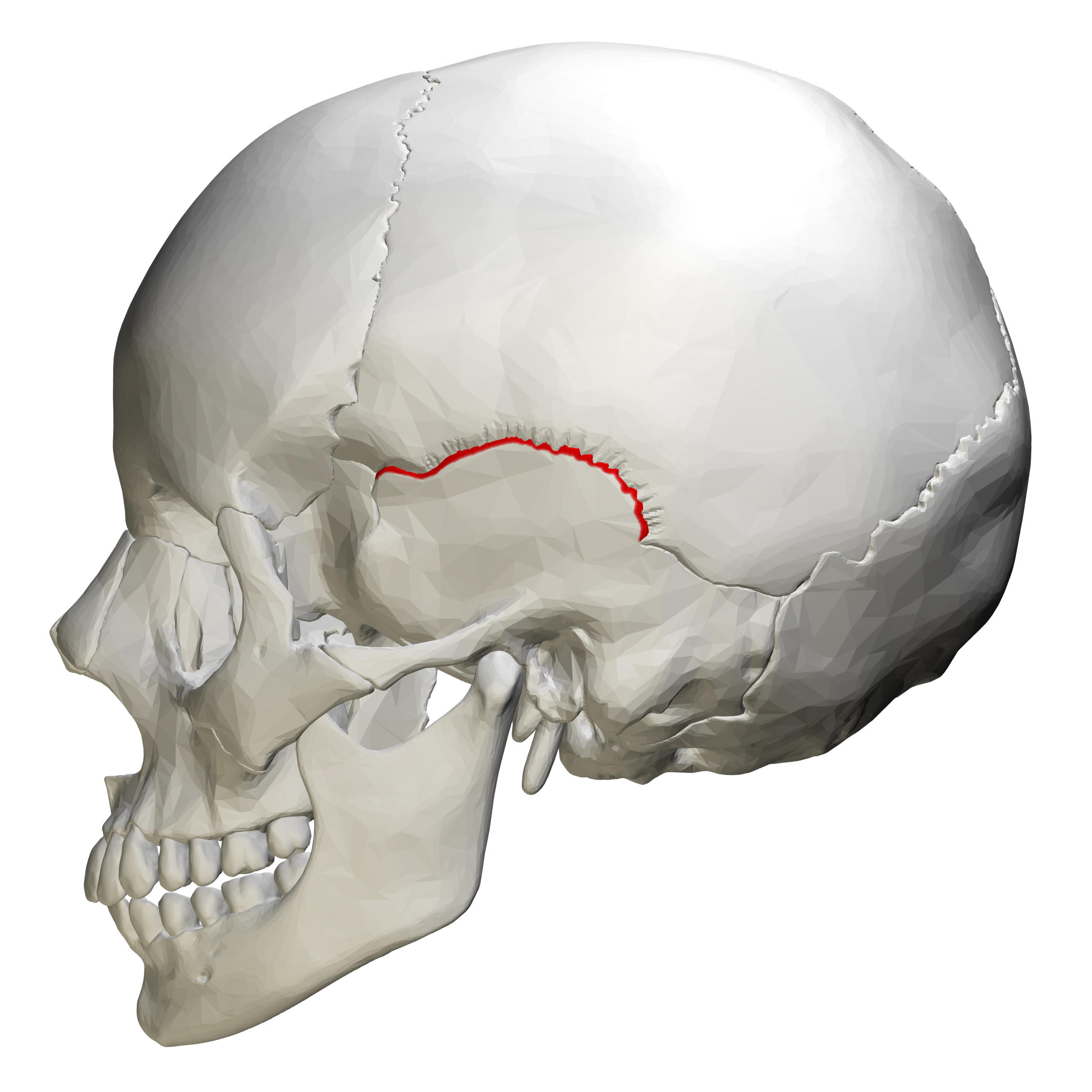
squamosal suture
8
hole in the temporal bone and the opening of the auditory canal 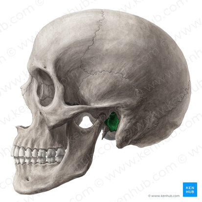
external auditory meatus
9
smooth surface on the underside of the temporal bone for articulation with the head of the mandible 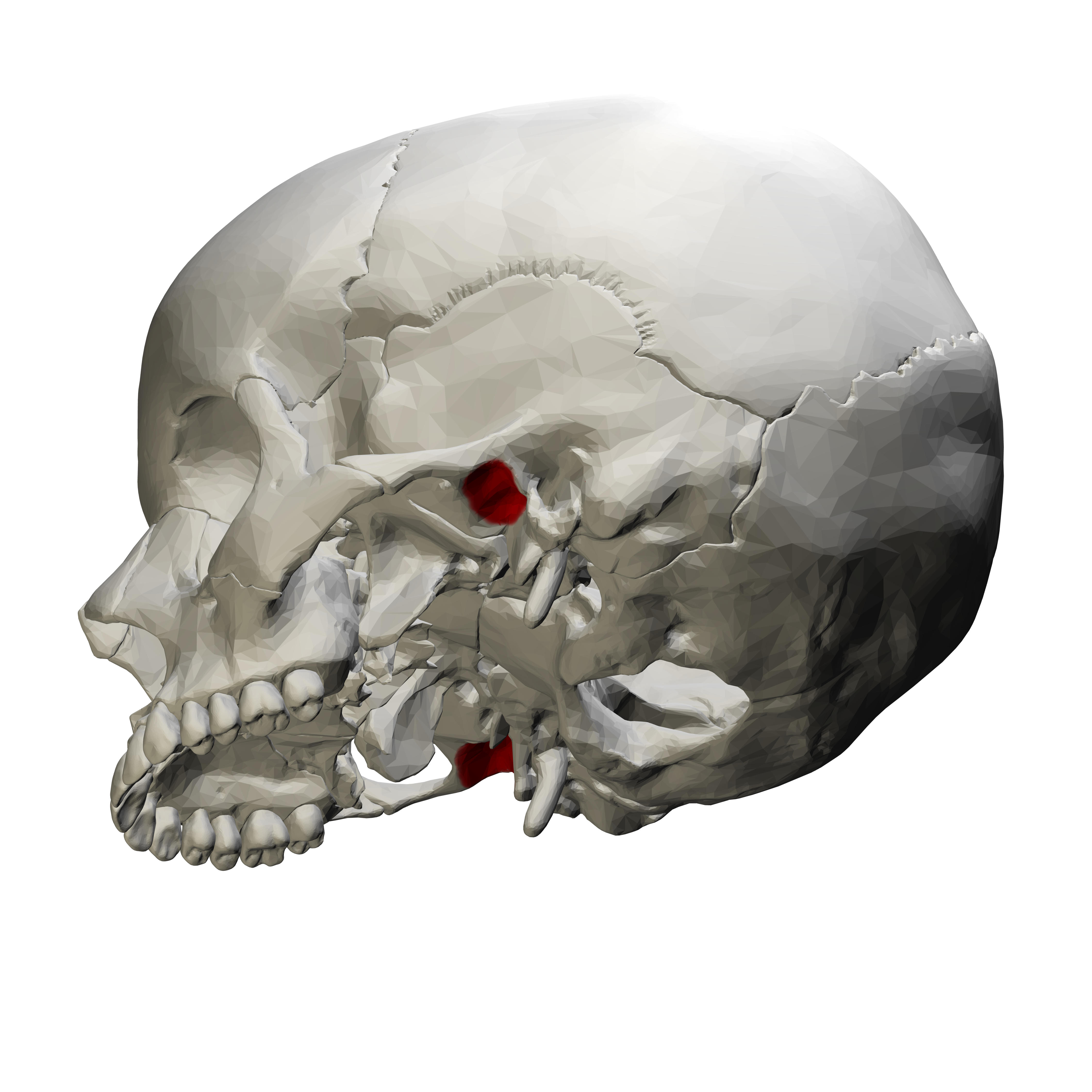
mandibular fossa
10
extension of temporal bone that fuses with the zygomatic process of the maxilla to form the zygomatic arch 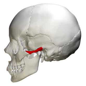
zygomatic process
11
bony projection from the base of the temporal bone 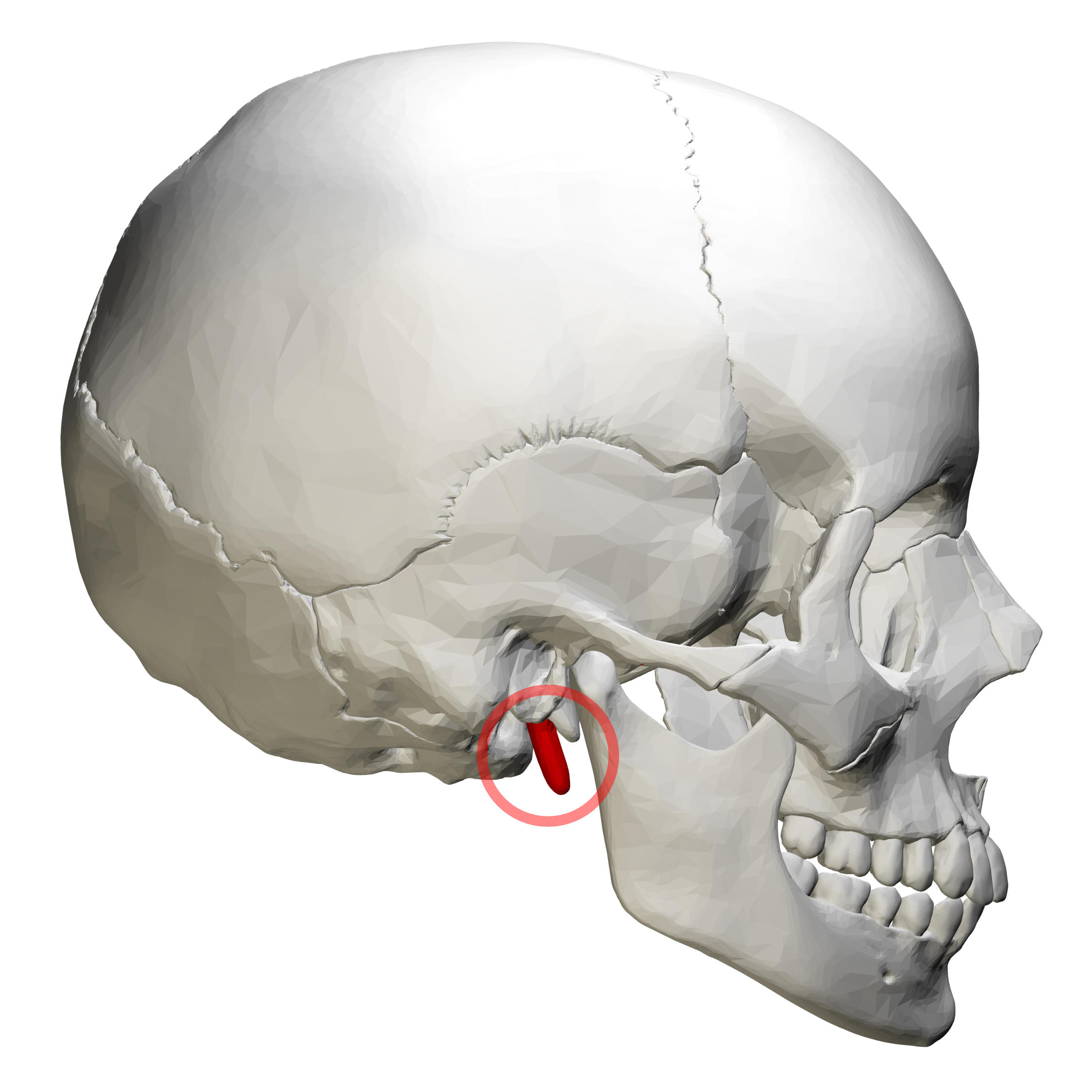
styloid process
12
rounded extenstion of temporal bone behind the external auditory meatus 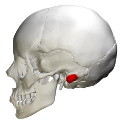
mastoid process 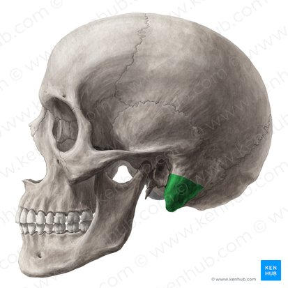
13
between temporal and occipital bones. passageway for internal jugular vein and cranial nerves 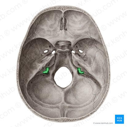
jugular foramen
14
passageway for internal cartoid 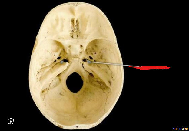
carotid canal
15
temporal bone that extends into the internal skull and protects the organs of hearing and balance 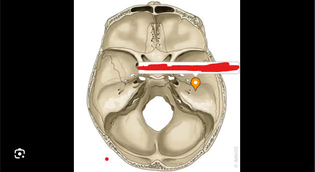
petrous part 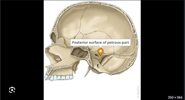
16
opening in petrous temporal bone that conveys CN VII and VII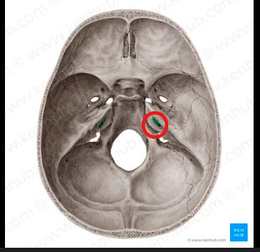
internal auditory meatus
17
is the back of the skull 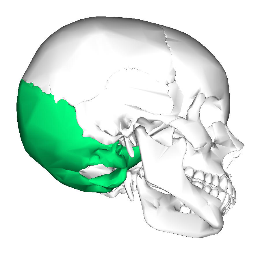
occipital bone
18
connects the occipital bone to the parietal bone 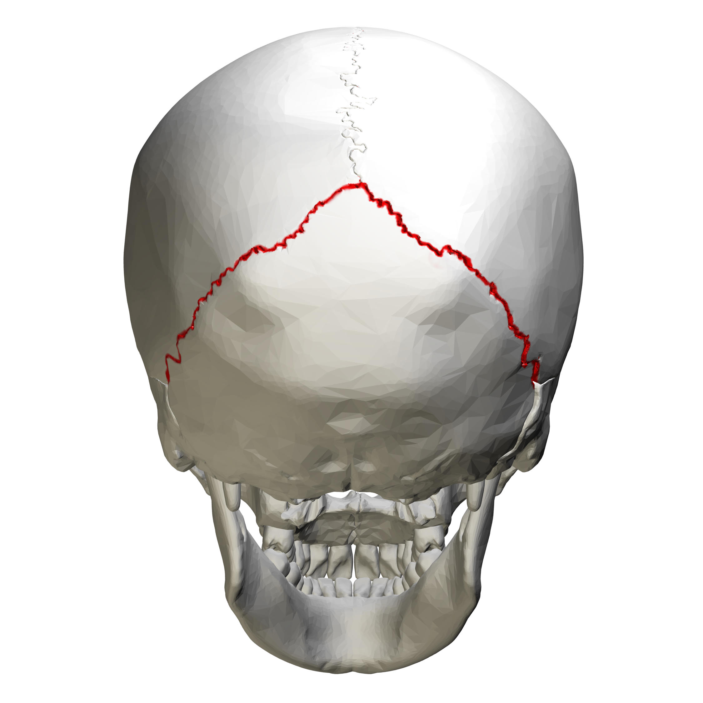
lambdoidal suture
19
large hole in the base of skull that is a passageway for the spinal cord 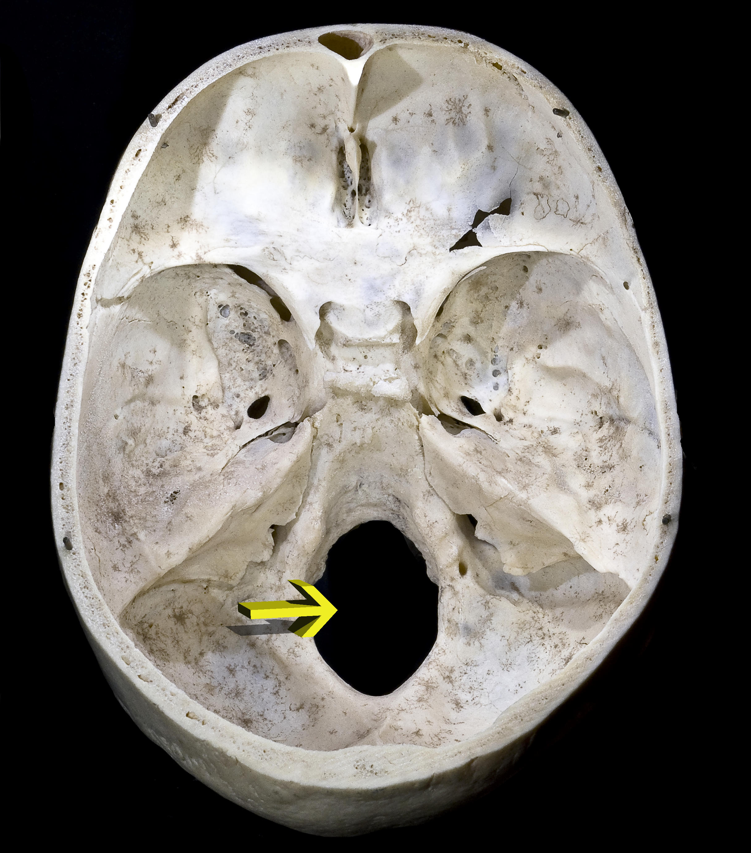
foramen magnum
20
two rounded process on either side of the foreman magnum that articulate with the first cervical vertebrae (the atlas) 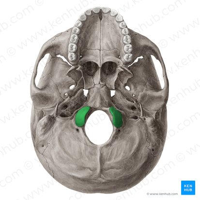
occipital condyles
21
enlargement on occipital bone for attachment of neck ligaments and muscle 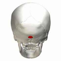
external occipital protuberance 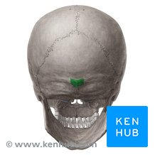
22
is in the middle of the skull and has several important features 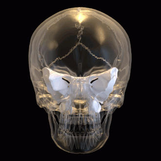
sphenoid bone 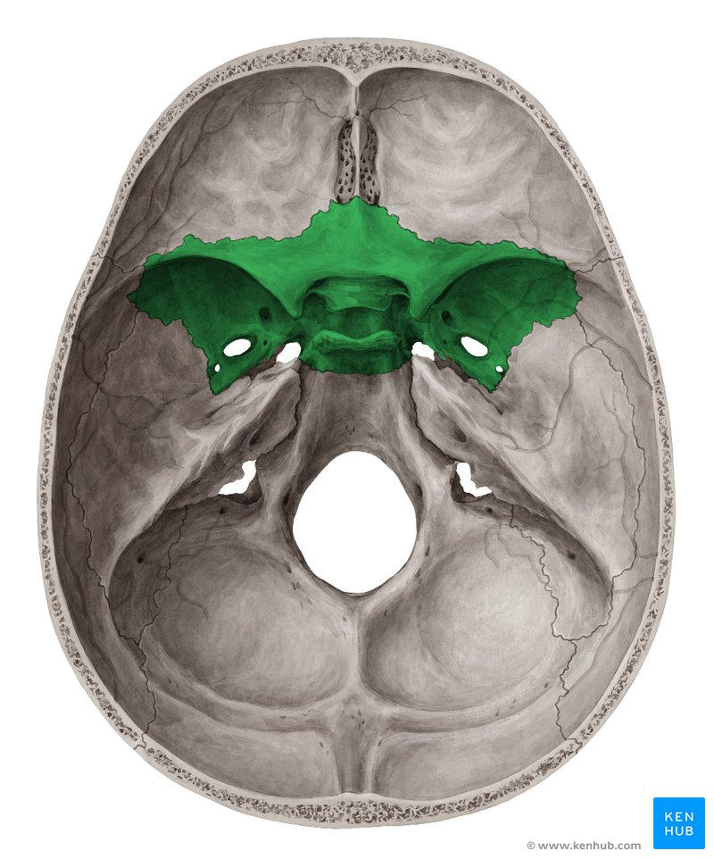
23
form part of the lateral surface of the skull and part of the orbits 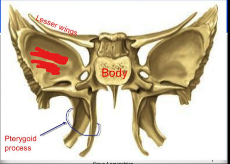
greater wings 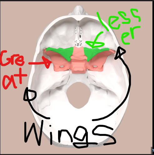
24
caintains the optic canals 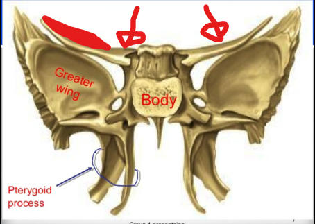
lesser wings 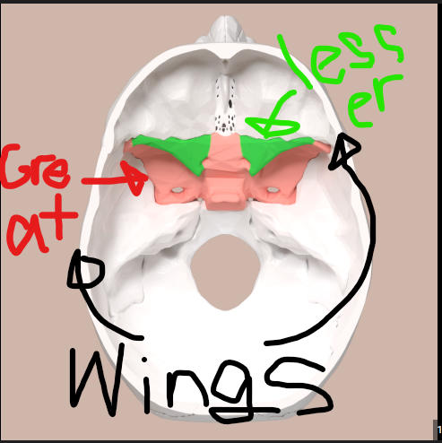
25
canal, passageway for the optic nerve 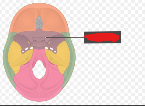
optic foramen 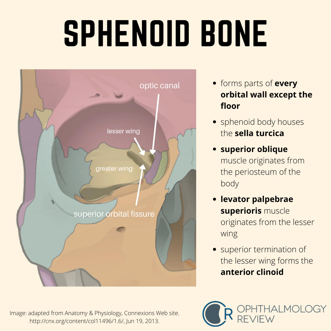
26
is the opening of between the greater and lesser wings and is a passageway fro blood vessels and nerves to the orbit 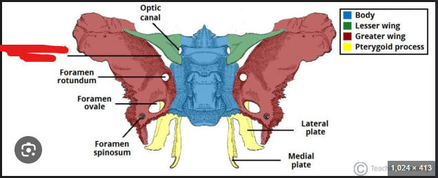
superior orbital fissure 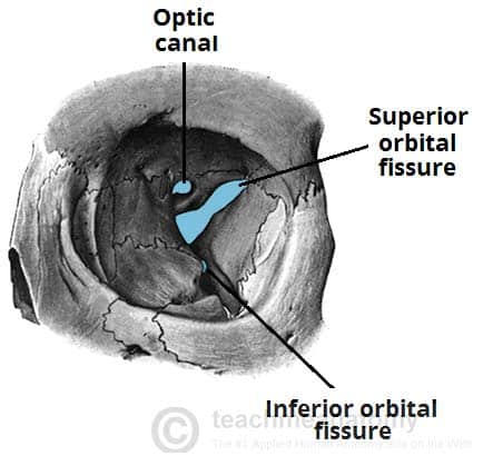
27
gap between the sphenoid bone and the maxilla that is a passsageway for blood vessels and nerves to the orbit 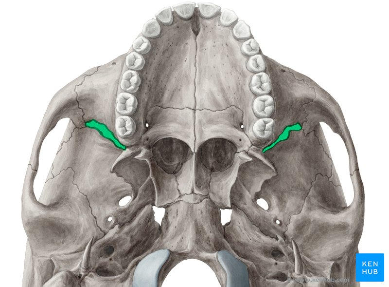
inferior orbital fissure 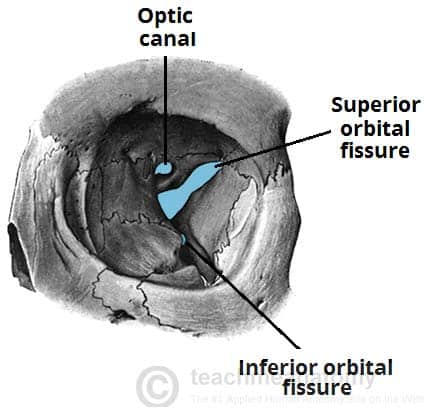
28
saddle shaped depression that surrounds the pituitary gland 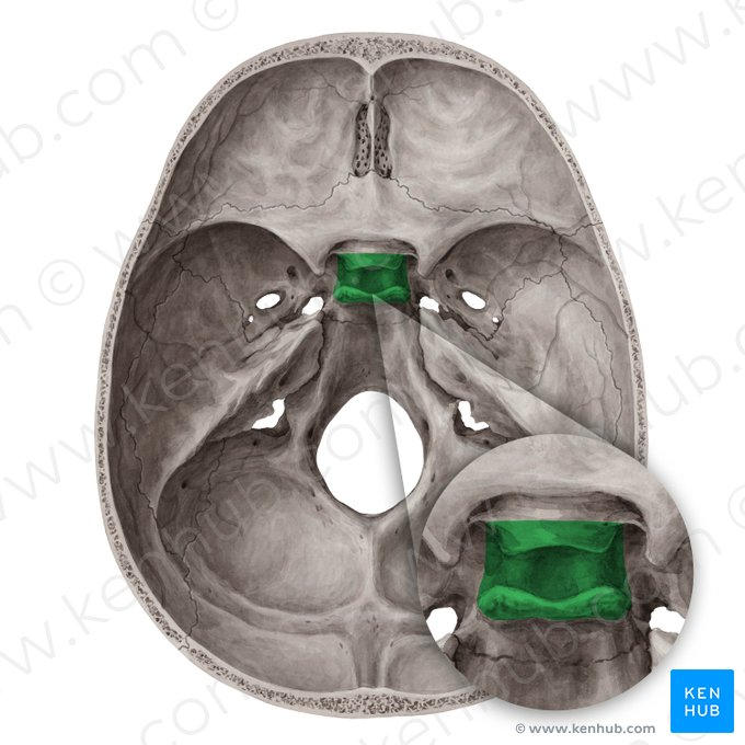
sella turcica 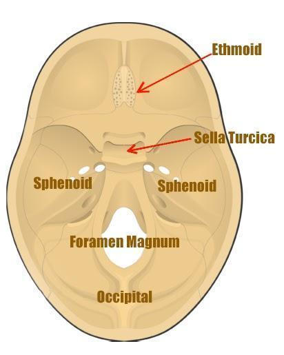
29
hole in greater wing that conveys the maxillary nerve into the orbit 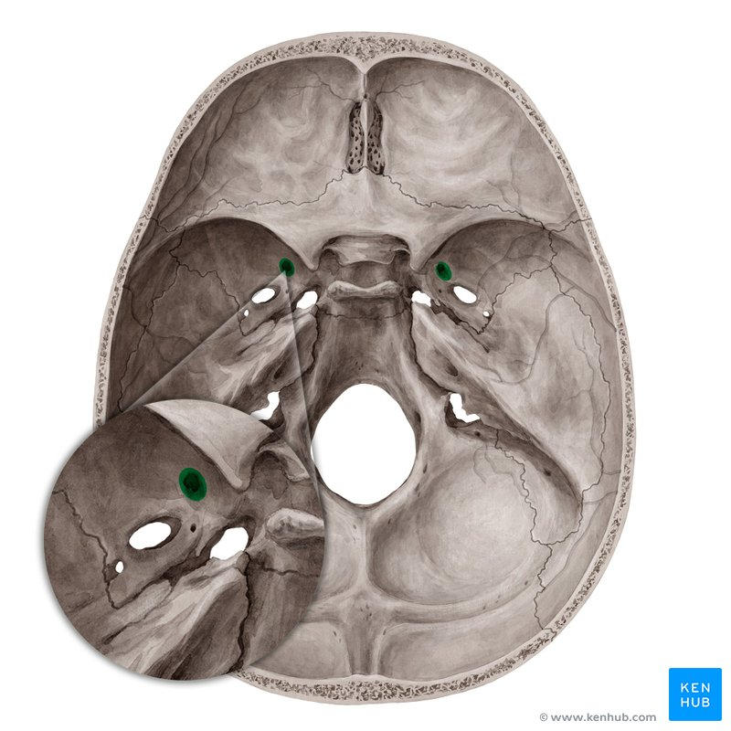
foramen rotundum 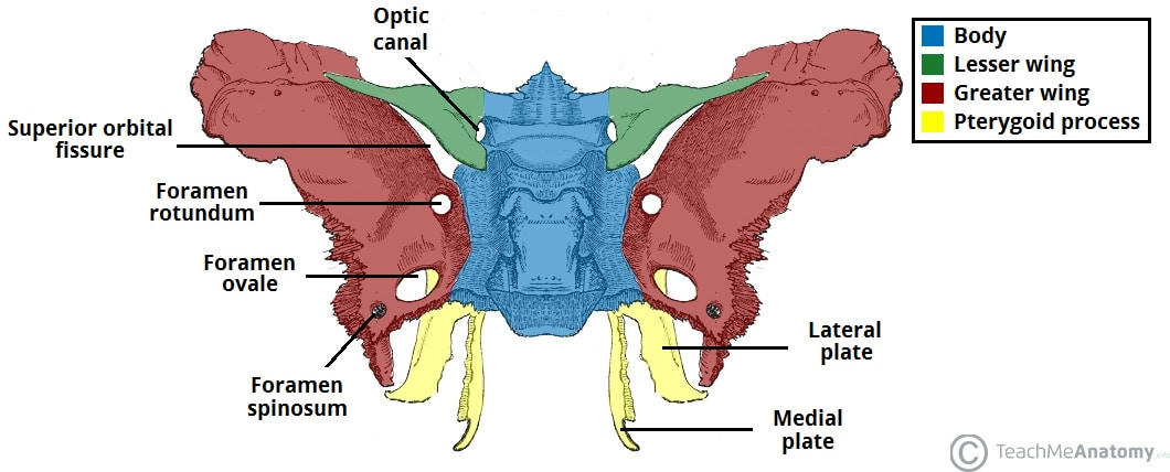
30
hole in greater wing that conveys the middle meningeal artery into the skull 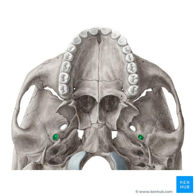
foramen spinosum 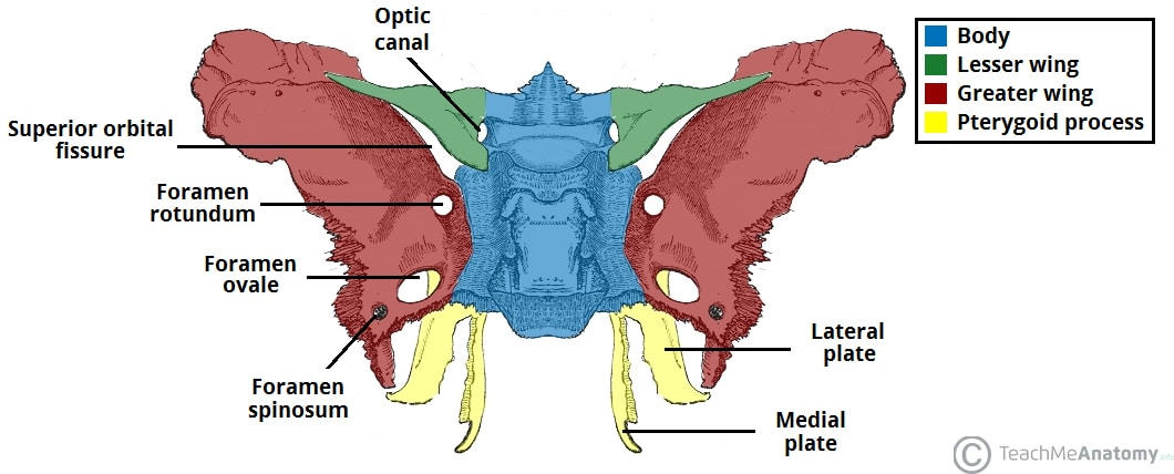
31
hole in greater wing that conveys the mandibular nerve out of the skull 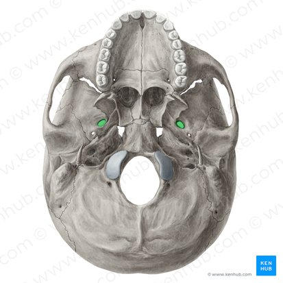
foramen ovale 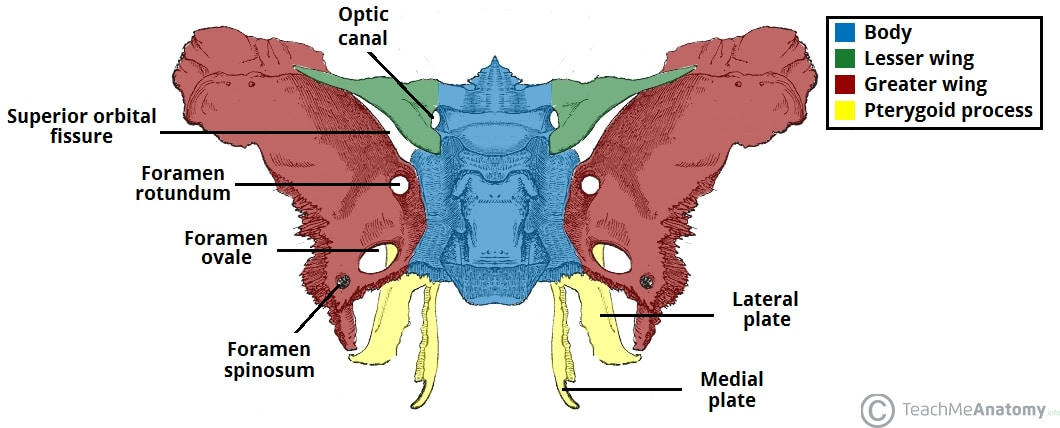
32
an irregularly shaped space of the base of the skull at the junction of the spehnoid and petrous temporal bone. Does not convey structures as is covered with cartilage during life 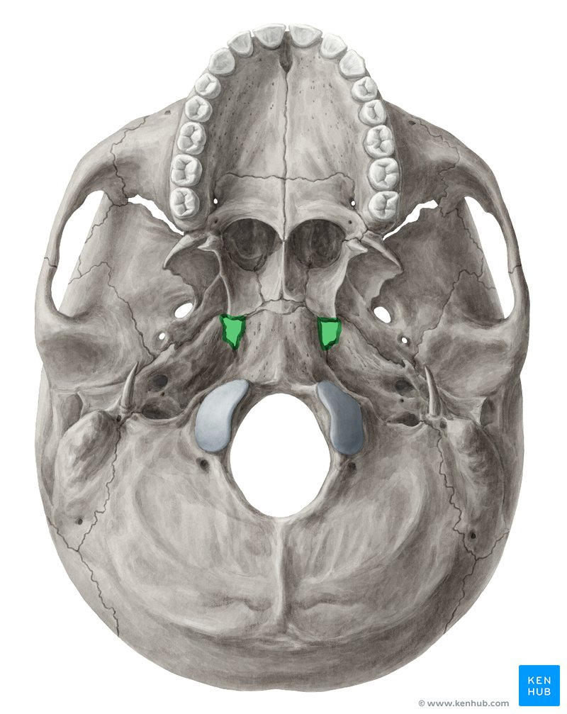
foramen lacerum 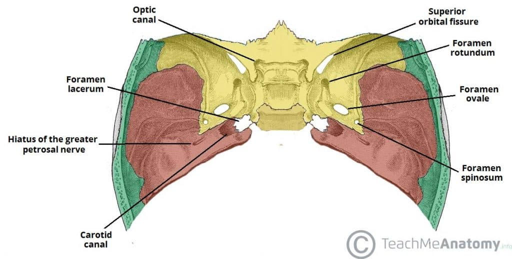
33
plates of bone that extend from the greater wing and serve as the attachment of muscles (it groups all three of those) 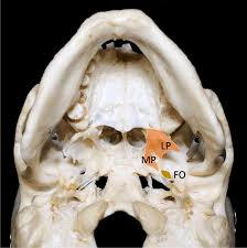
pterygoid processes 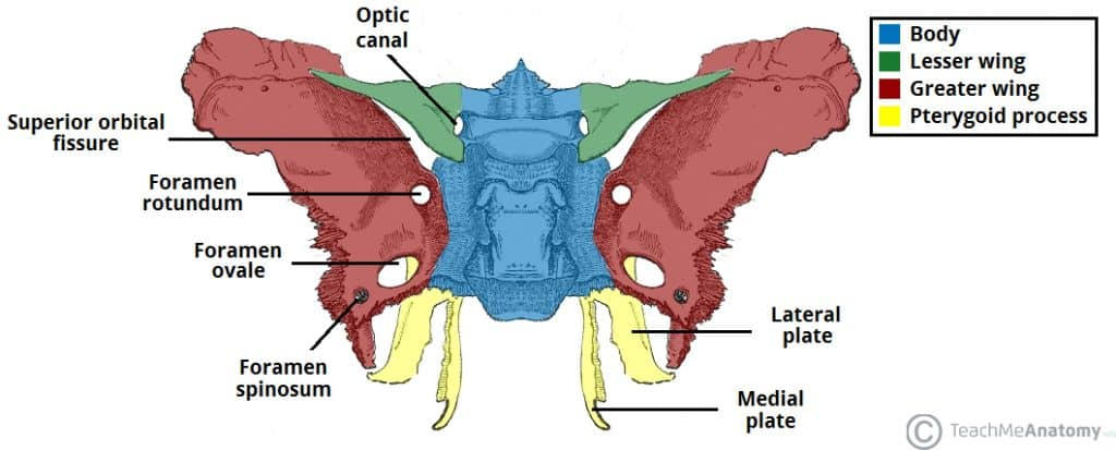
34
a delicate bone between the orbits that contains the ethmoid air sinuses 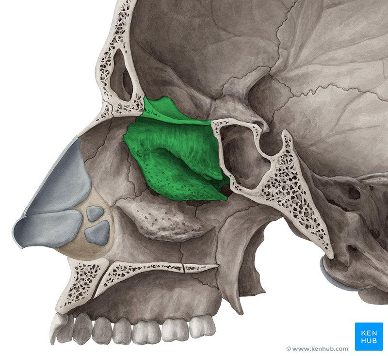
ethmoid bone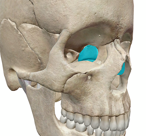
35
crest of bone between the cribiform plates 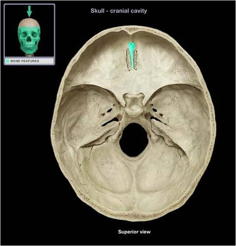
crista galli 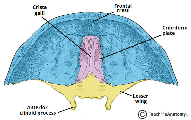
36
extension of ethmoid bone that forms the upper half of the nasal septum 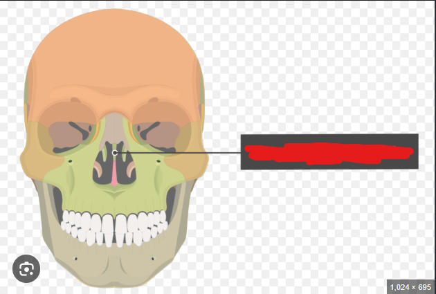
perpendicular plate 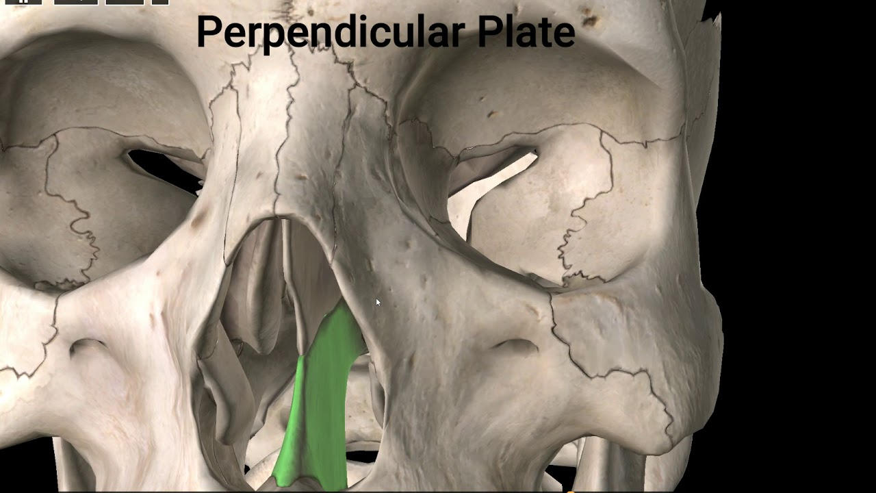
37
a thin perforated piece of bone that convey the olfactory nerves from the nasal cavity into the skull 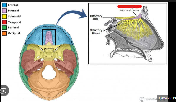
cribriform plate 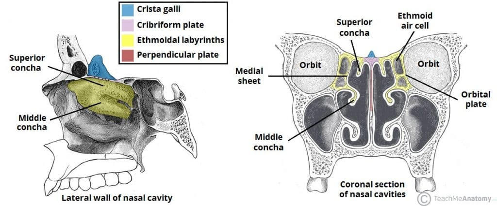
38
ridges of bone that support nasal folds and increases the surface area of the nasal mucosa 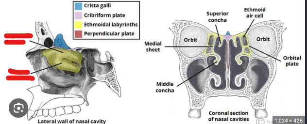
superior and middle nasal concha 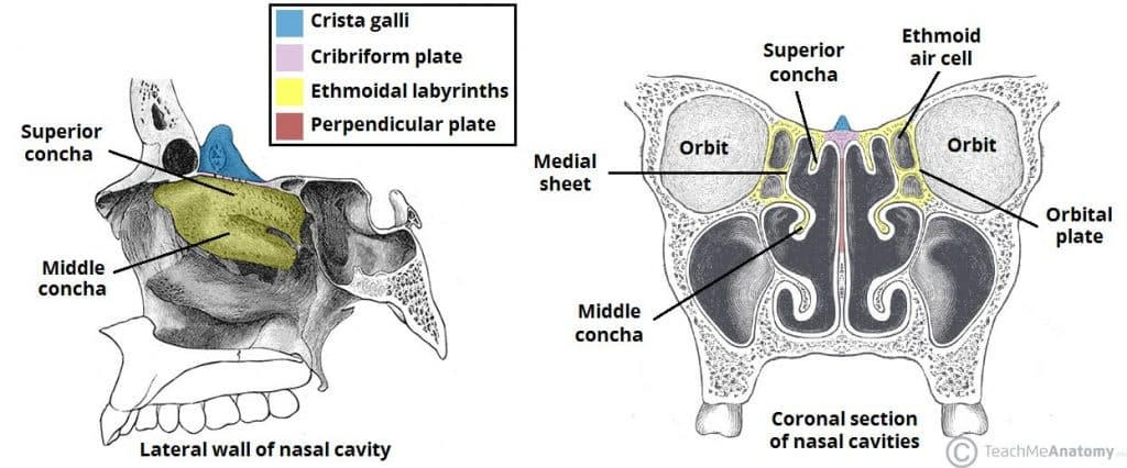
39
the lower jaw 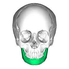
mandible
40
The vertical portion of the mandible that has a connection to the temporal bone 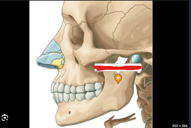
ramus 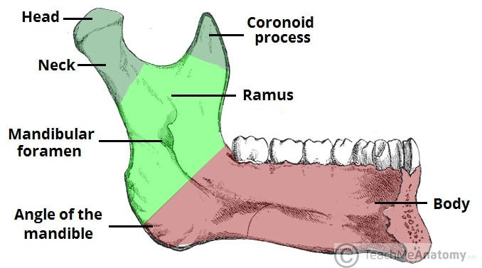
41
extension of bone off the ramus that serves as attachment for muscle 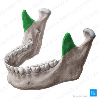
coronoid process 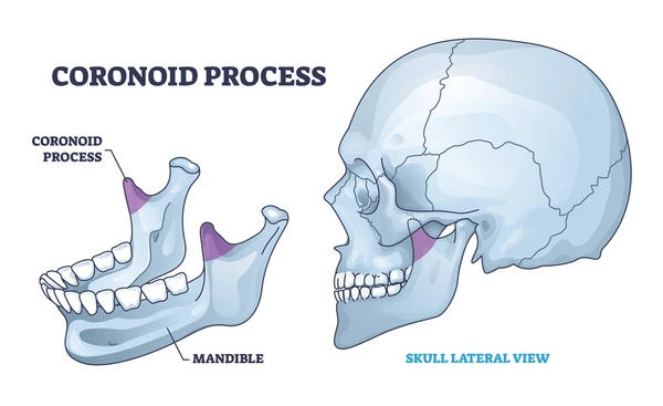
42
area between the ramus and coronoid process 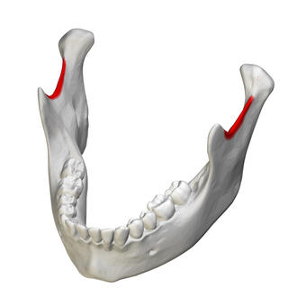
mandibular notch 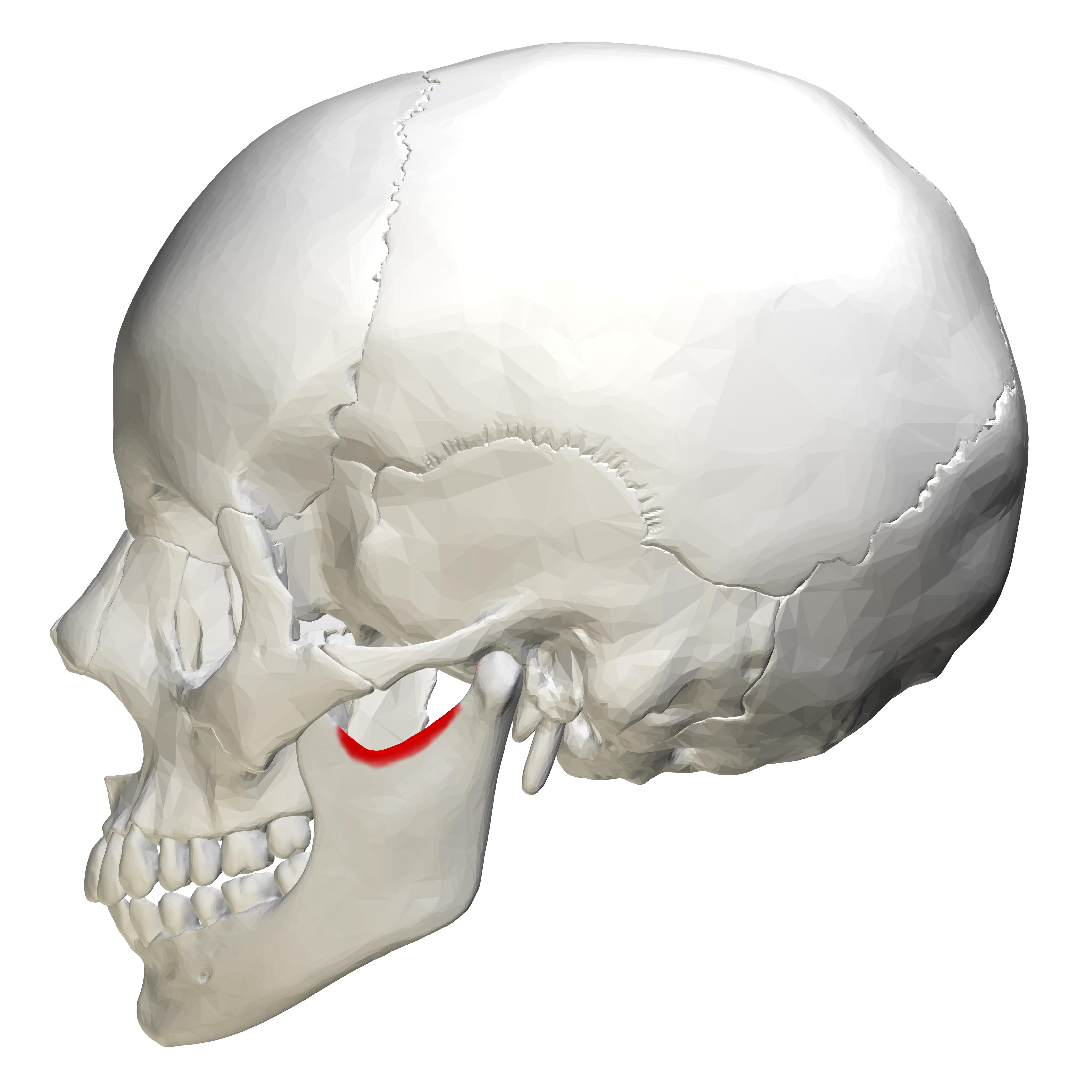
43
extension of ramus that contains the mandibular condyle that attaches to the temporal bone at the mandibular fossa 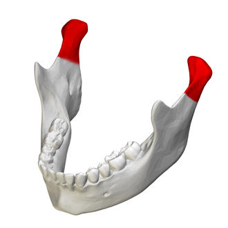
condylar process 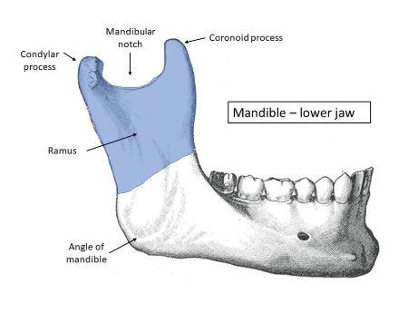
44
hole in mandible that conveys the inferior alveolar nerve and artery 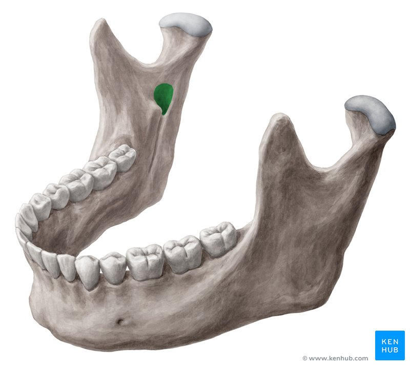
mandibular foramen 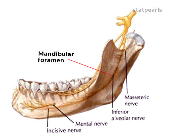
45
contain lower teeth 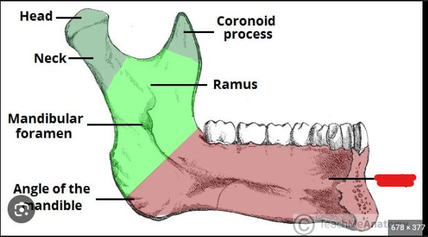
Body 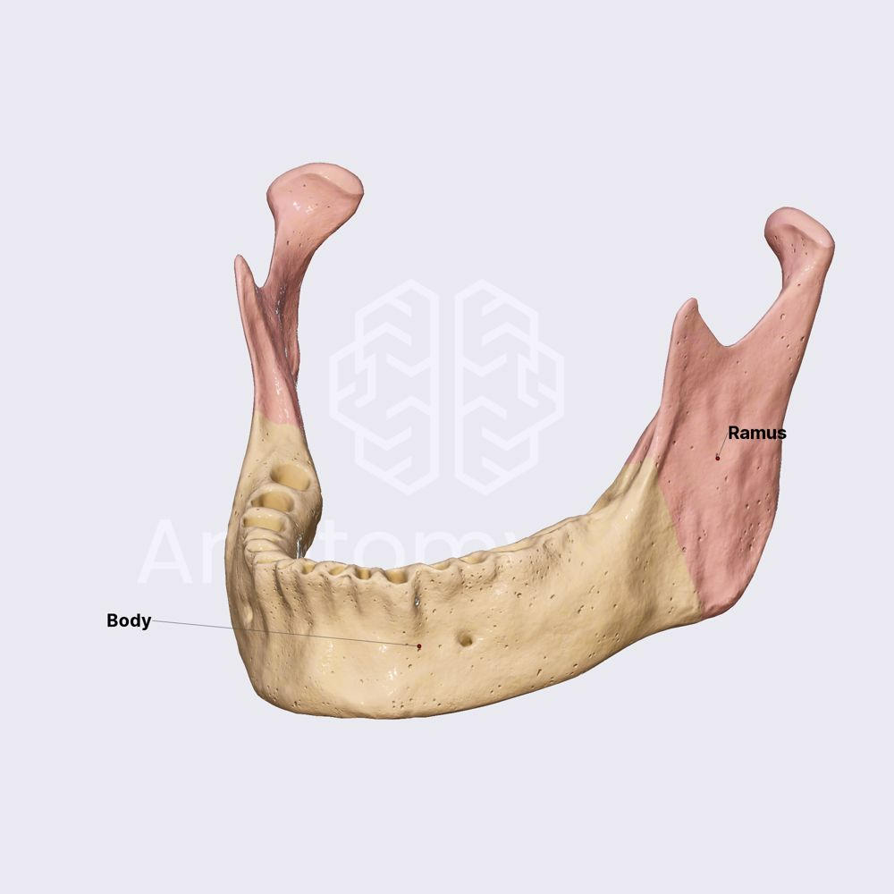
46
hole in mandible that conveys nerve onto the surface 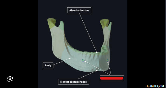
mental foramen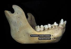
47
socket for a tooth in the alveolar process of the mandible 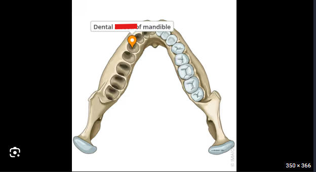
alveolus 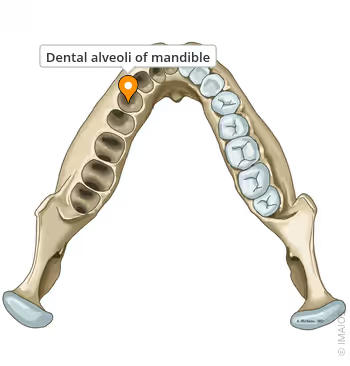
48
this part 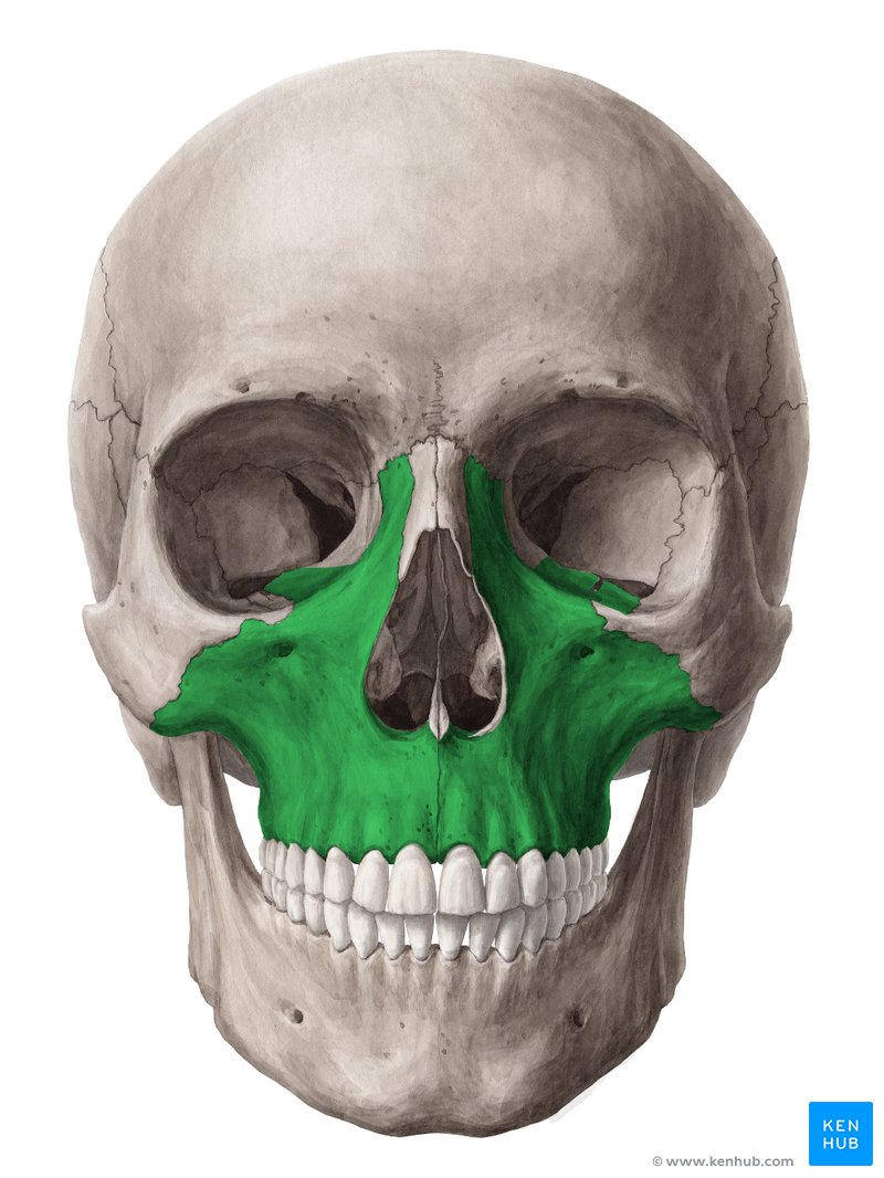
maxillae 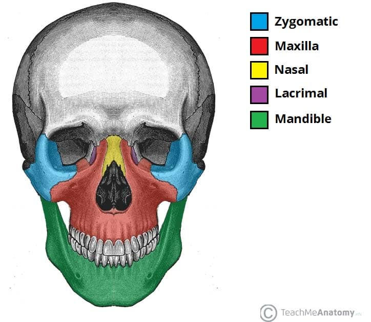
49
hole in maxillae 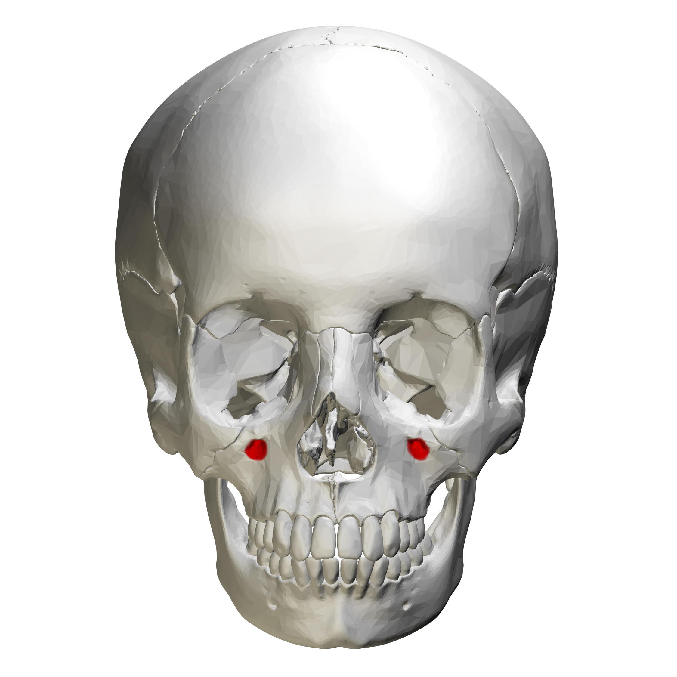
infraorbital foramen 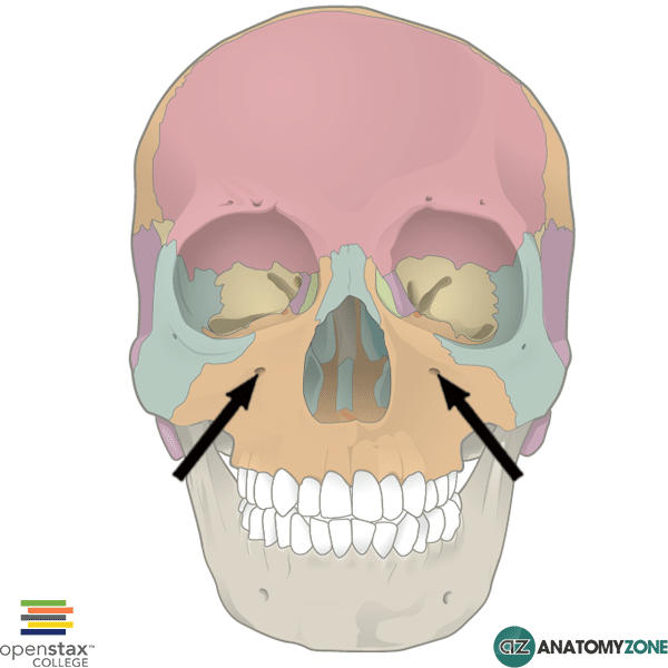
50
air sinus in the maxilla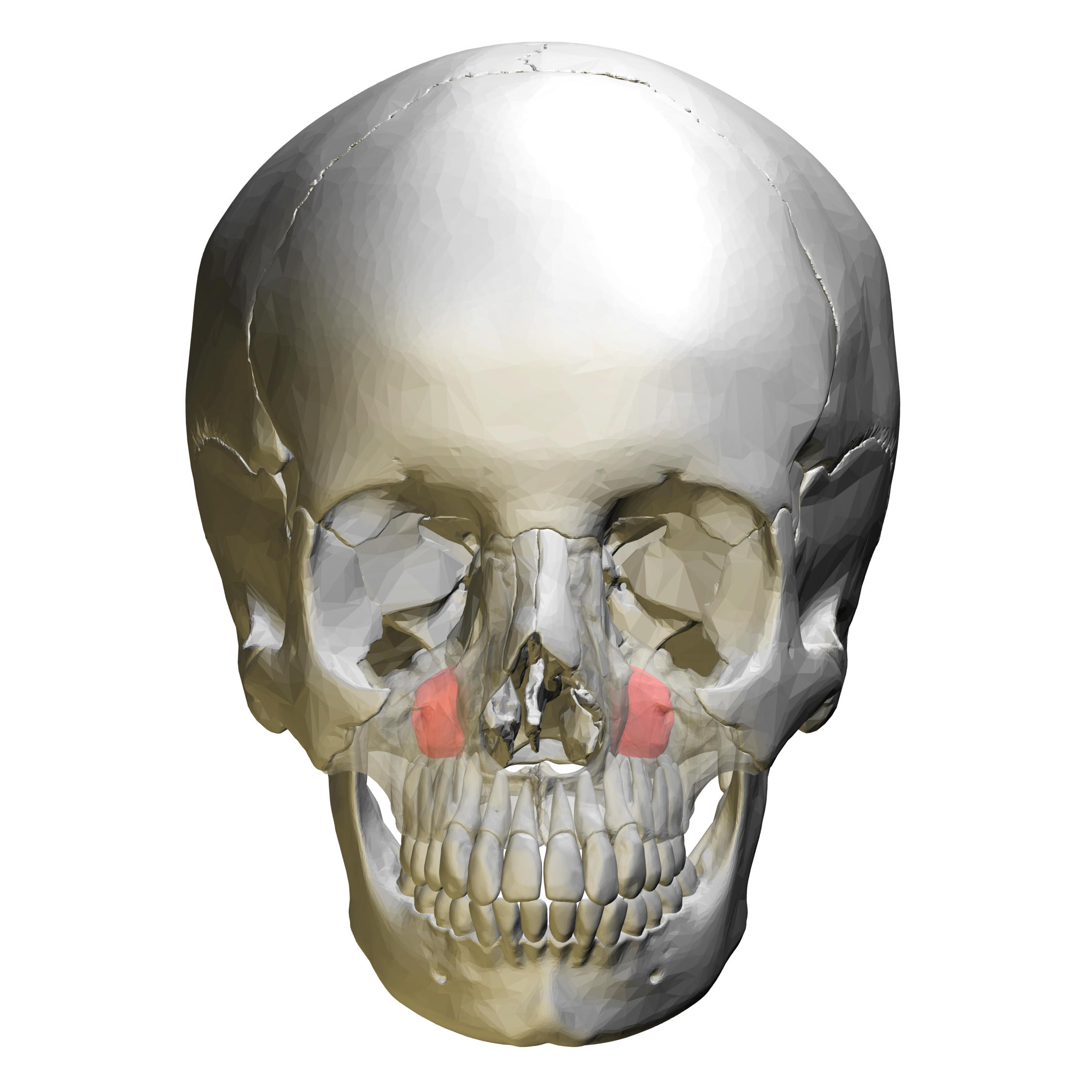
maxillary sinus 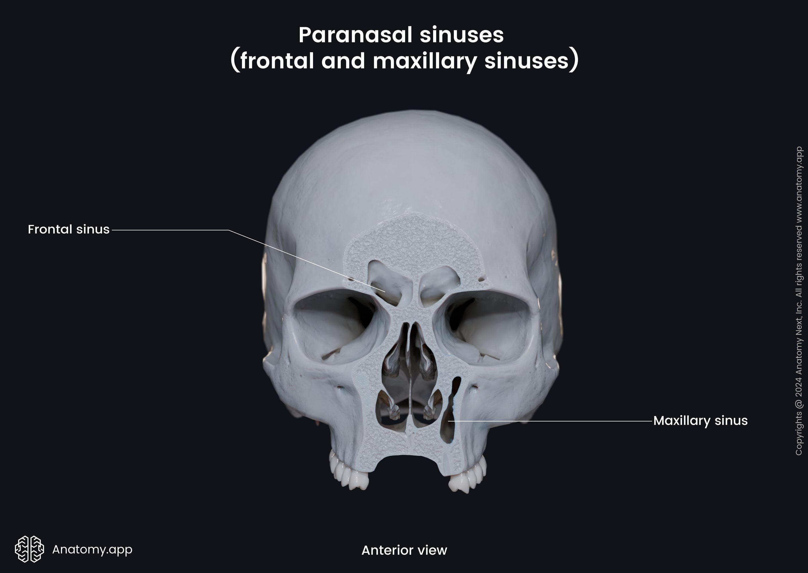
51
anterior part of the hard palate 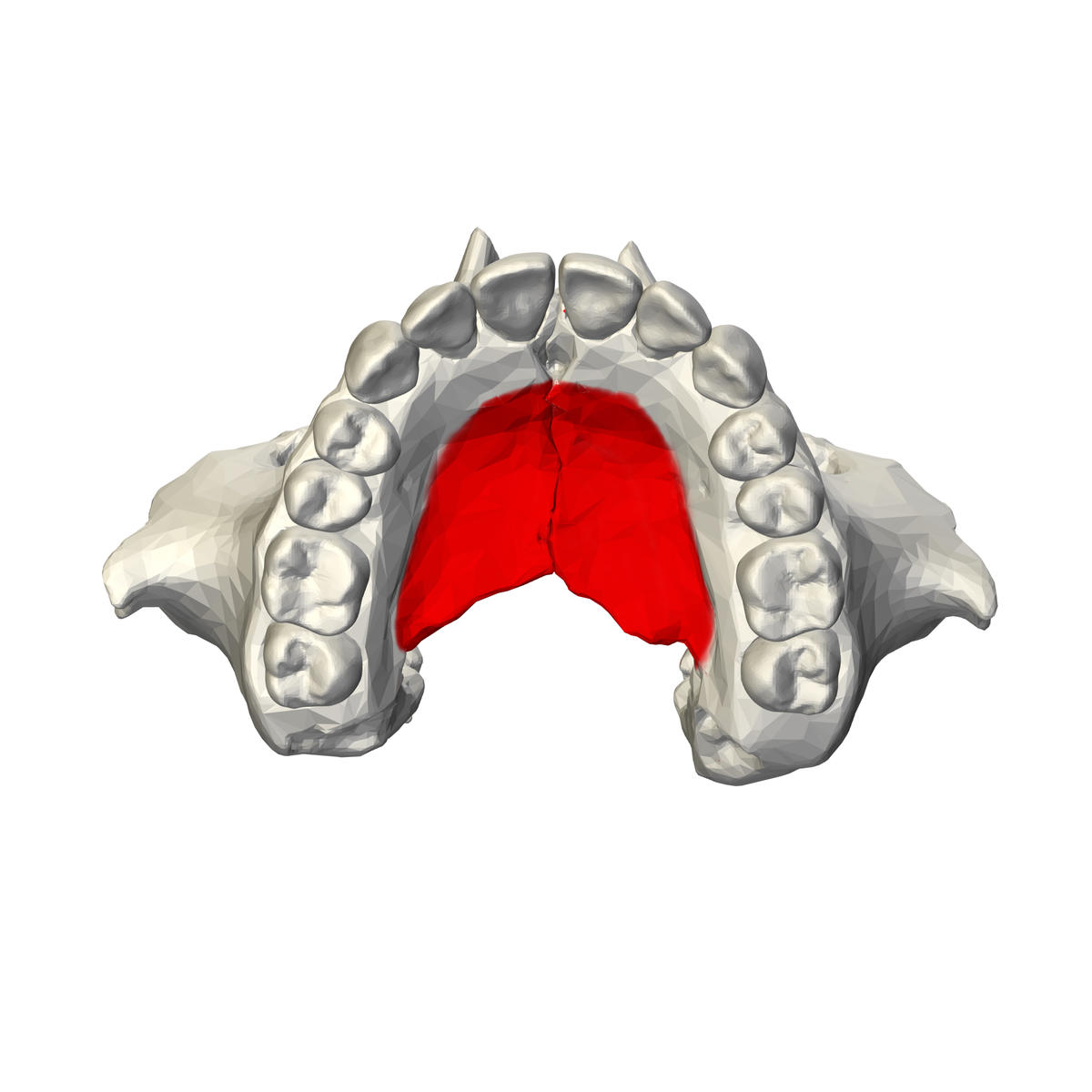
palatine process 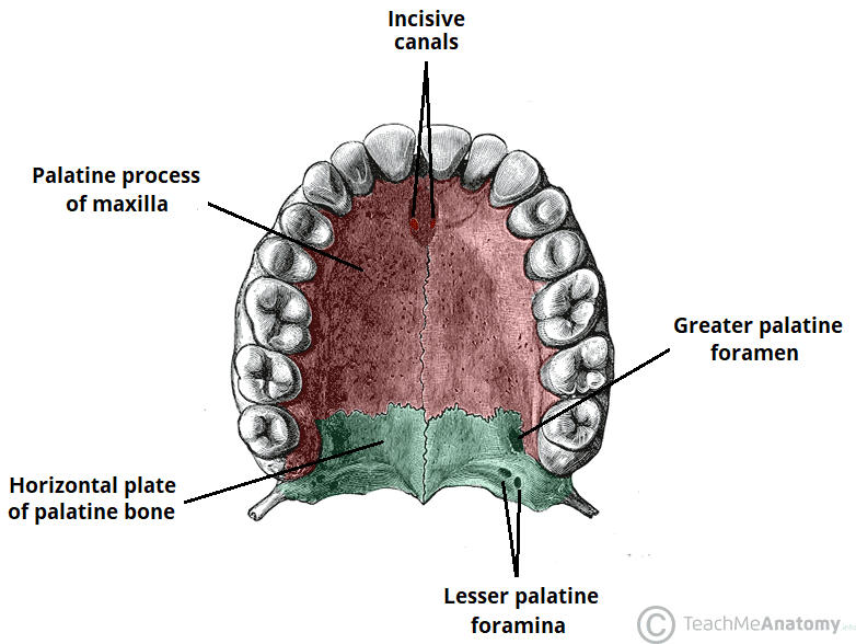
52
socket for a tooth in the alveolar process of maxilla 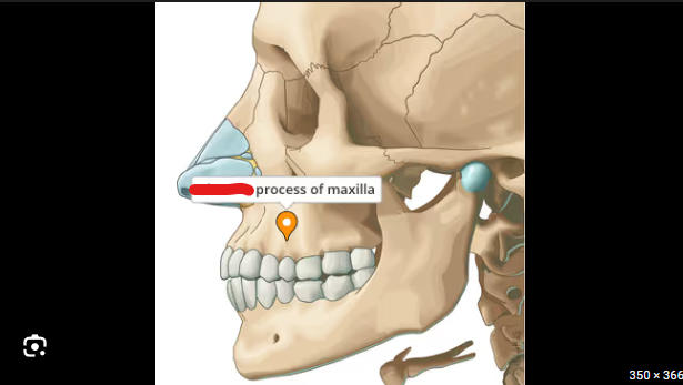
alveolus 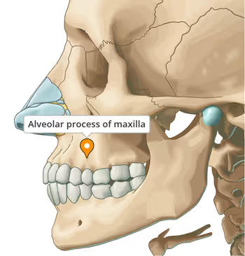
53
small bones found within the sutures 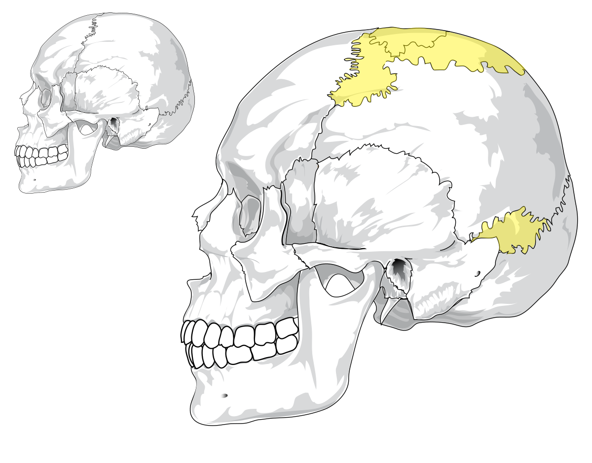
sutural bones 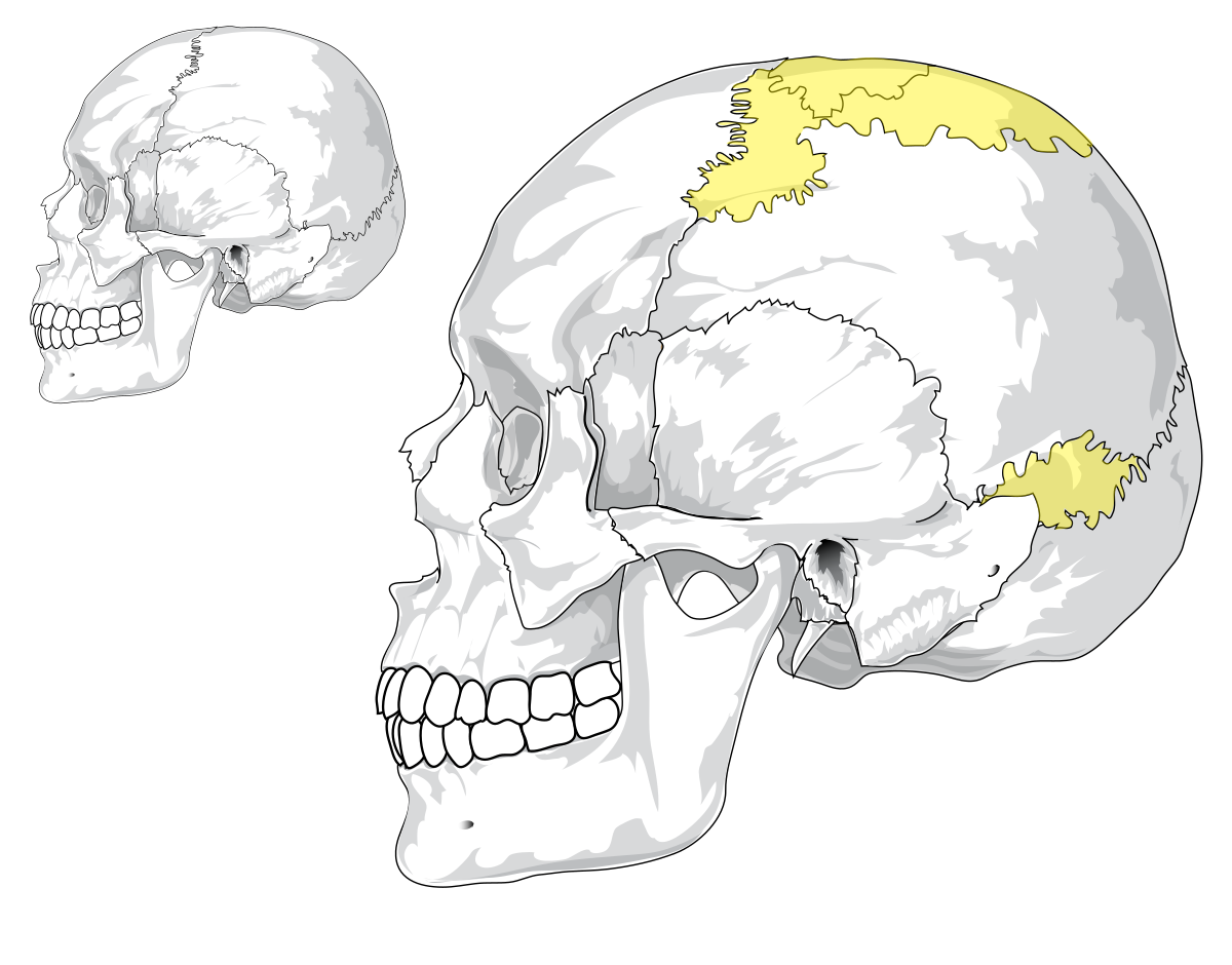
54
form the posterior part of the hard plate 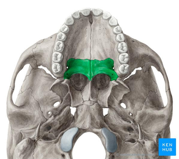
palatine bones 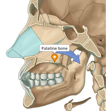
55
small bones that help the zygomatic arch 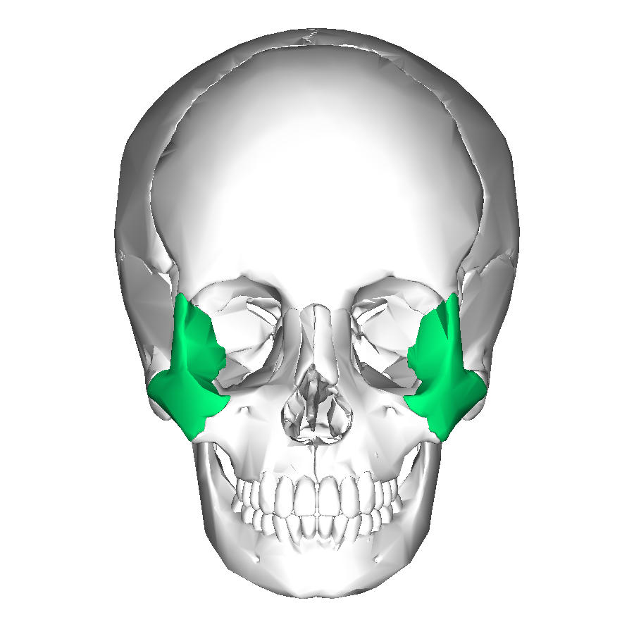
zygomatic bones
56
bones that form the ridge of the nose 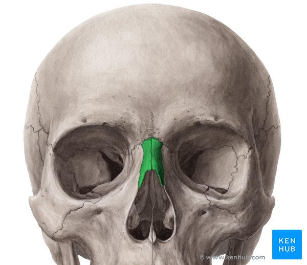
nasal bones
57
forms part of the medial wall and curves around the lacrimal duct 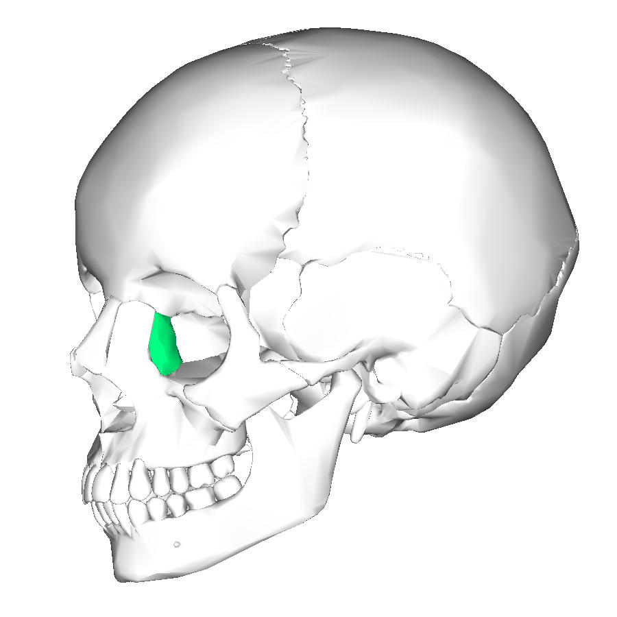
lacrimal bones 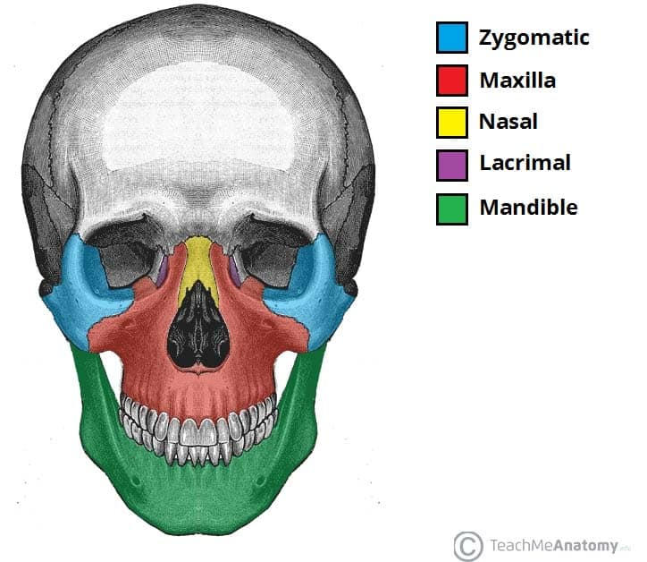
58
thin plate of bone that resembles a plow share and forms the inferior part of the nasal
septum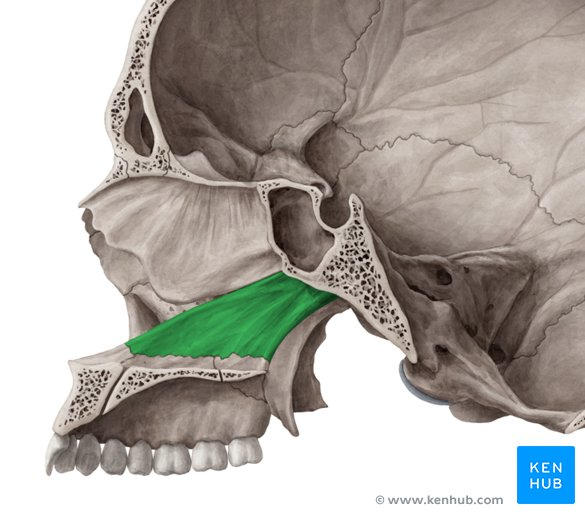
septum
vomer 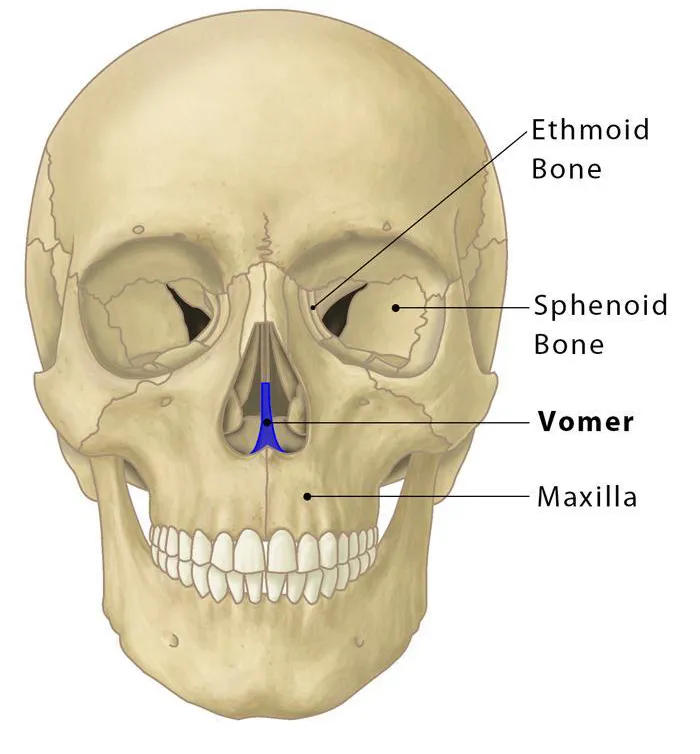
59
small bone the projects medially from the lateral wall of the nasal
cavity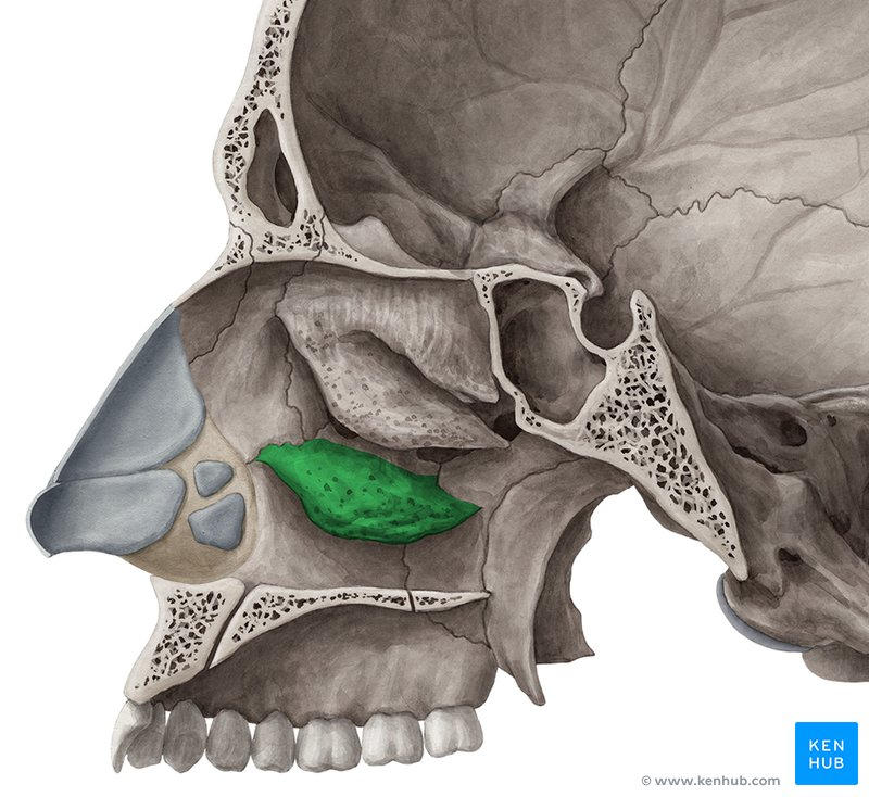
cavity
inferior nasal conchae 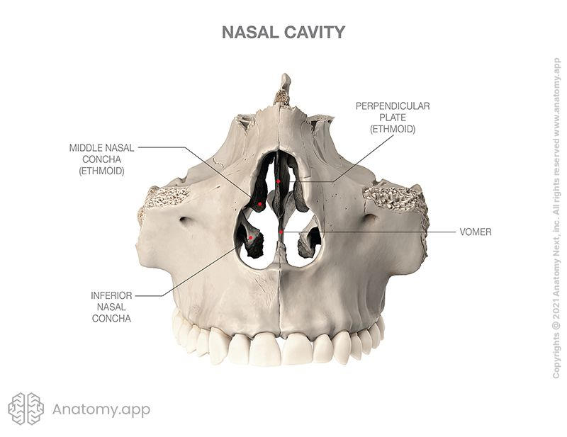
60
U-shaped bone in the neck that does not articulate with other bones located between
the larynx and the mandible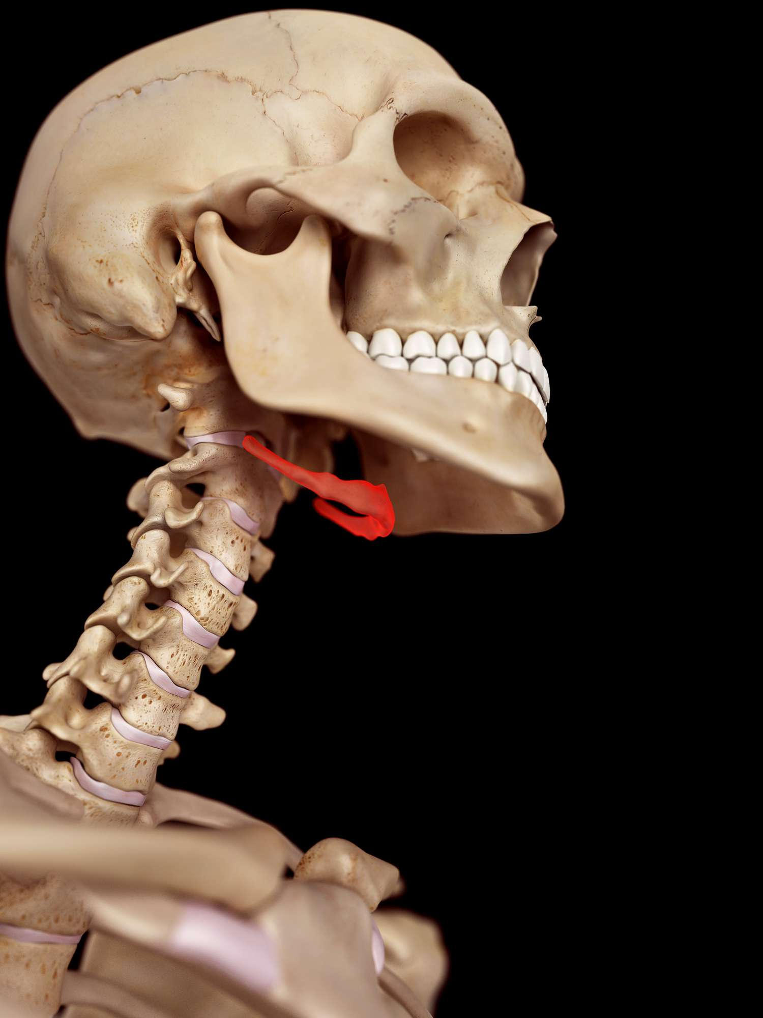
the larynx and the mandible
Hyoid 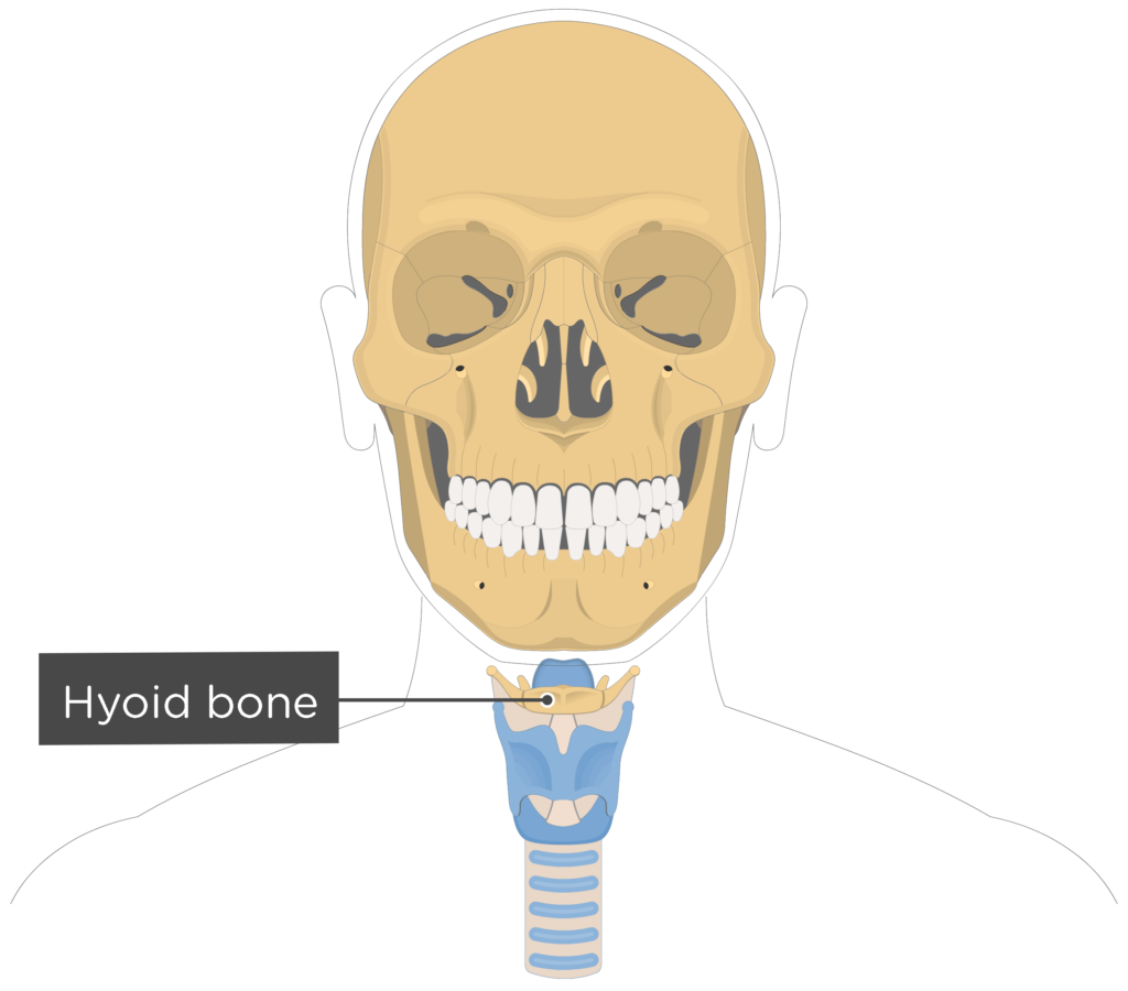
61
3 bone boned 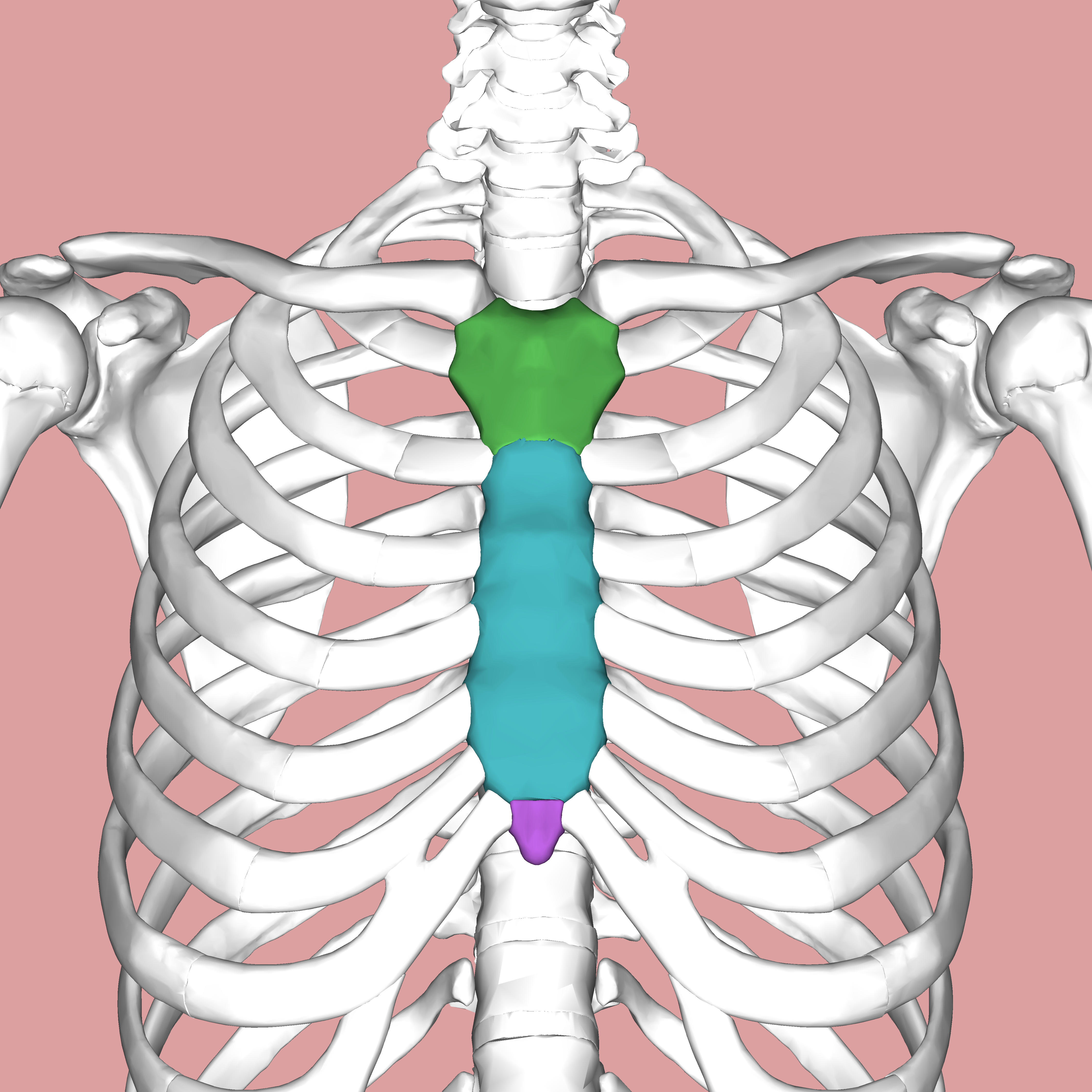
sternum
62
upper part of the sternum that articulates with the clavicle and first two
ribs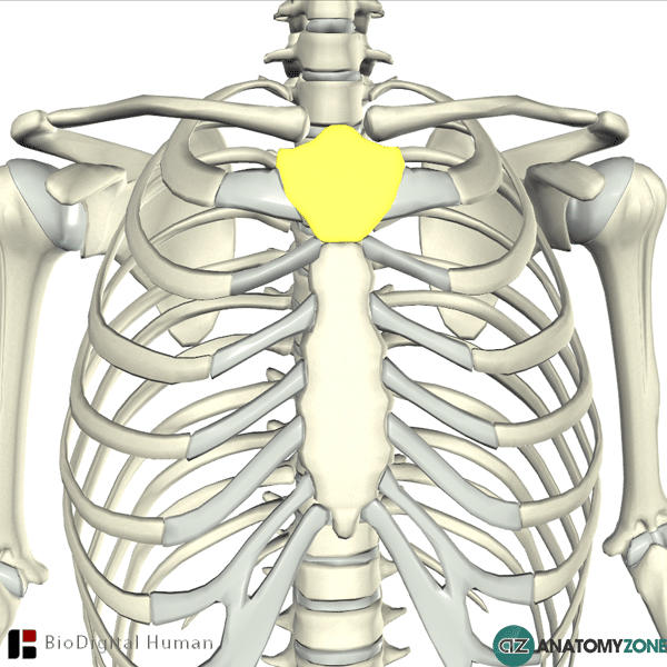
ribs
manubrium 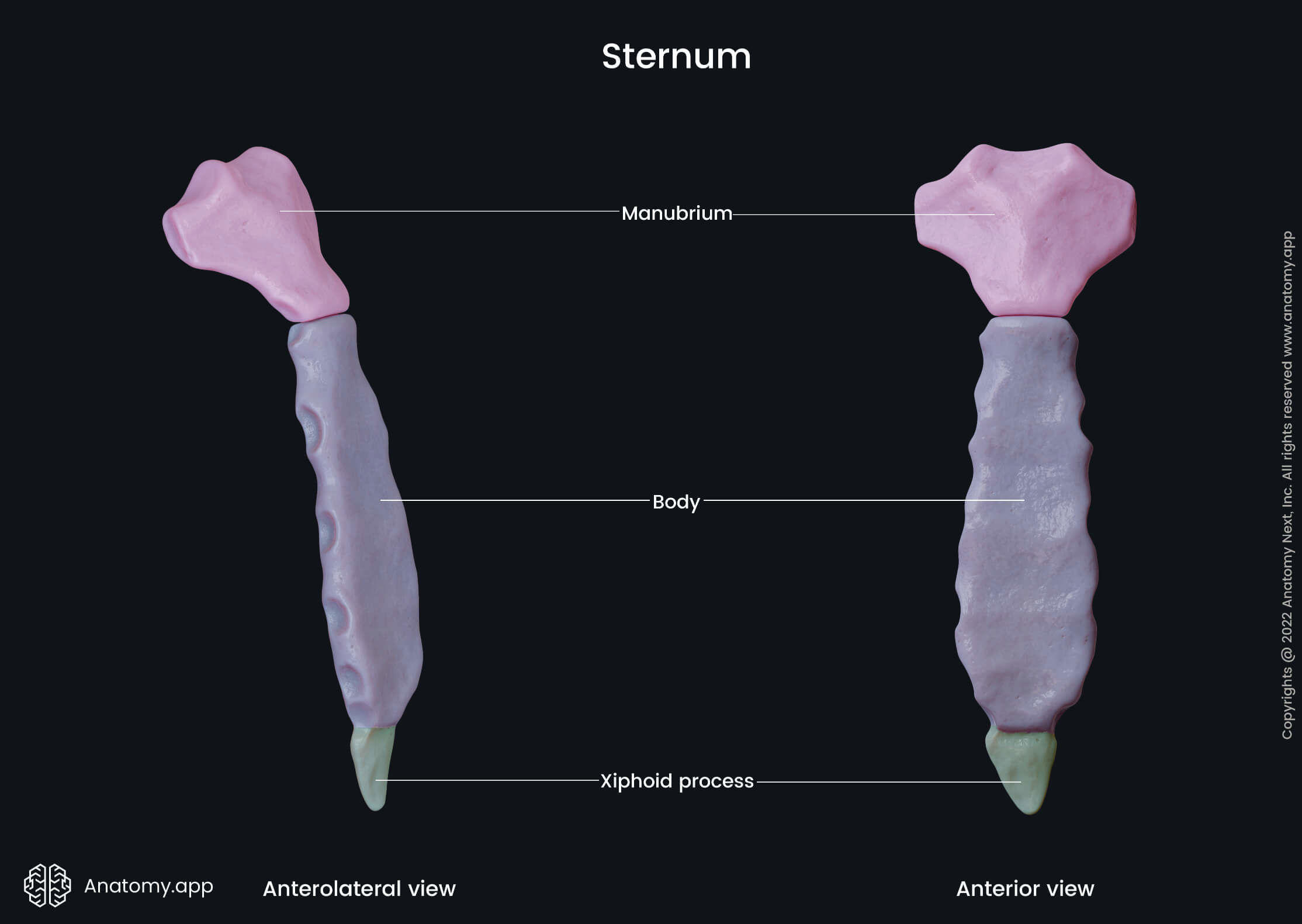
63
long middle part of the sternum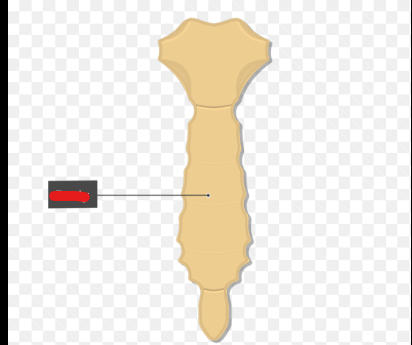
body 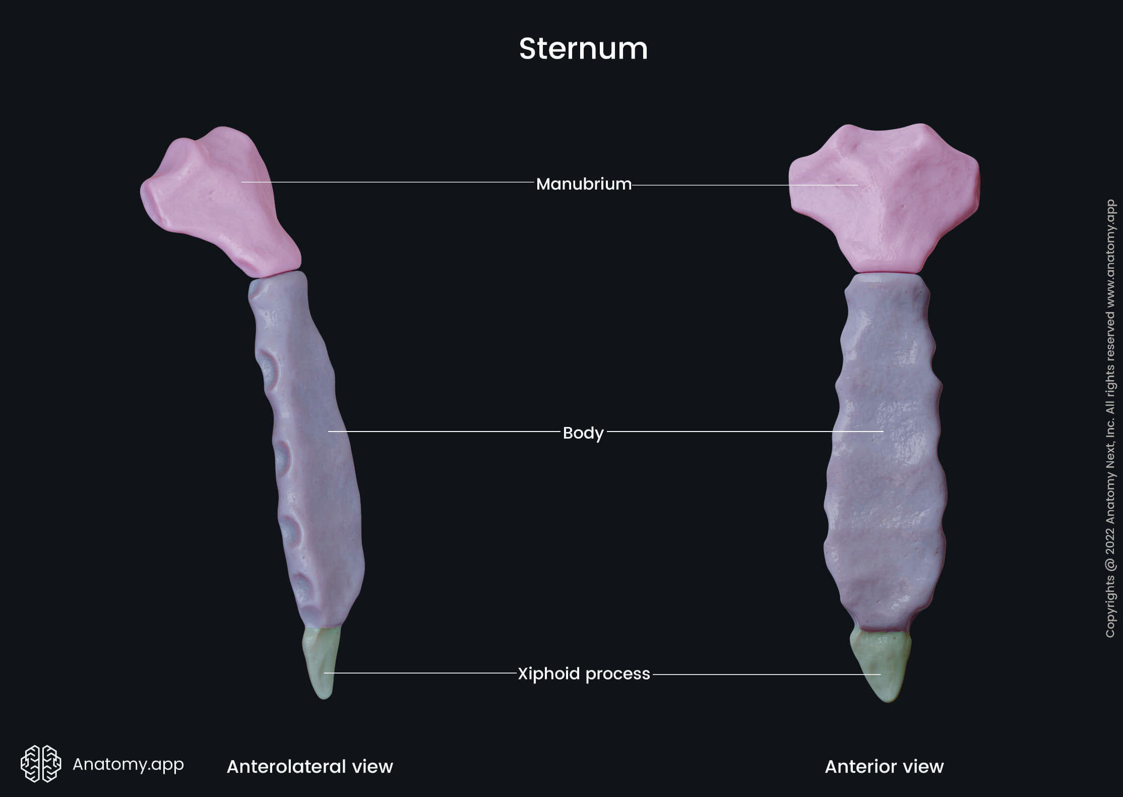
64
pointed tip of the sternum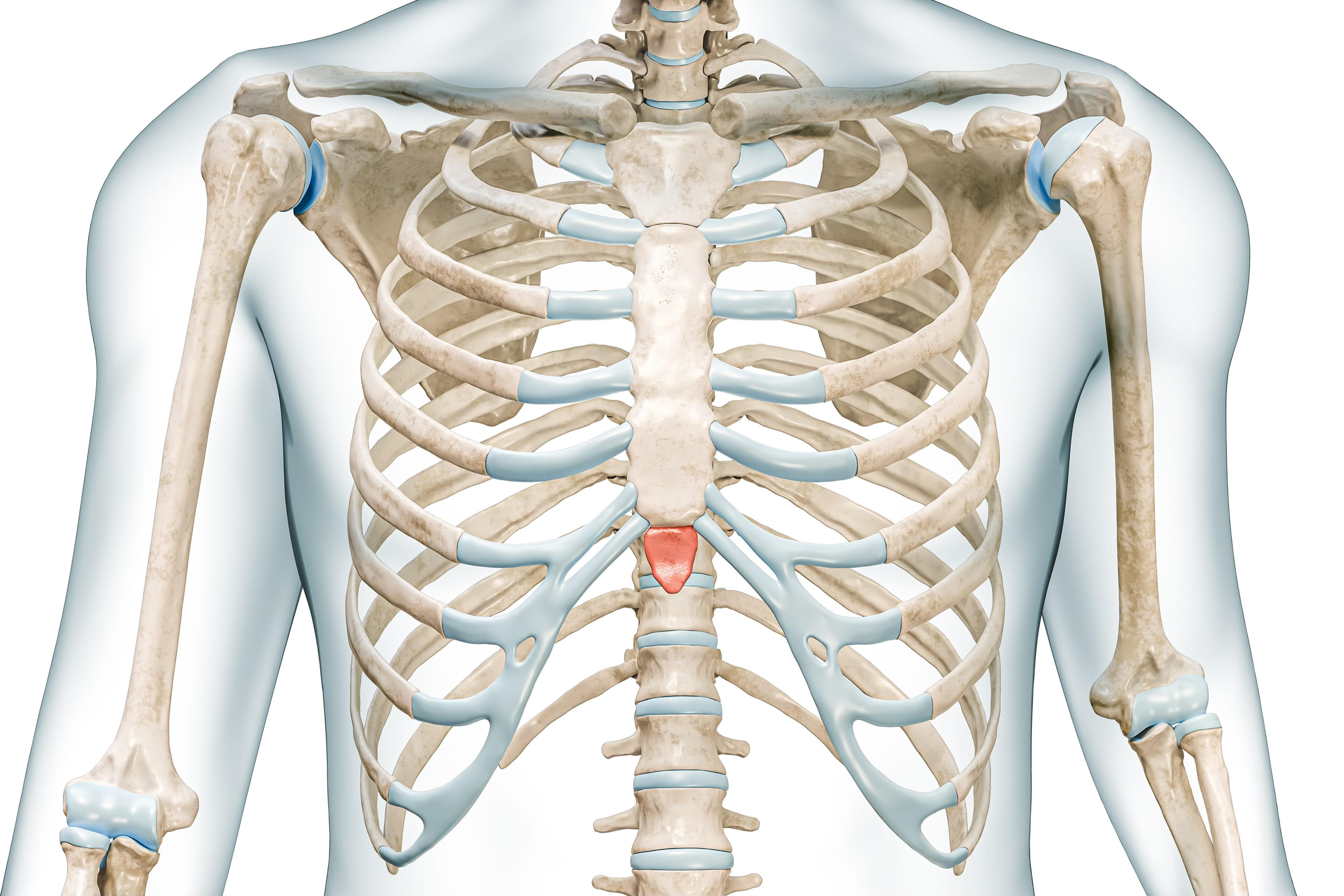
xiphoid process 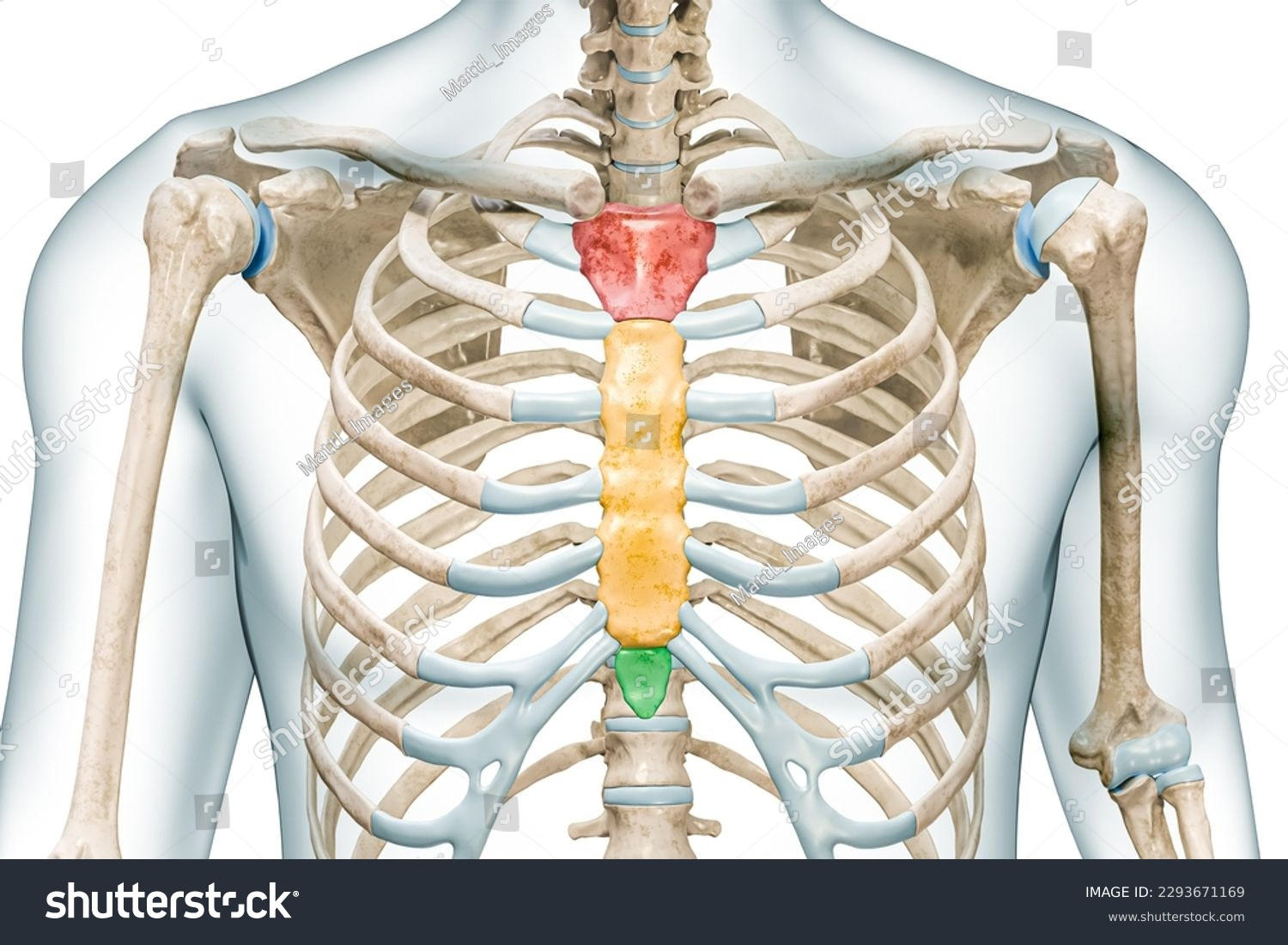
65
1-7 attach directly to the sternum 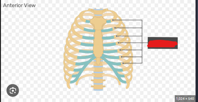
true ribs 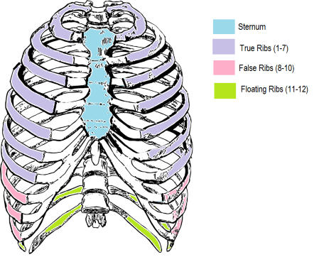
66
attach to the sternum via a costal cartilages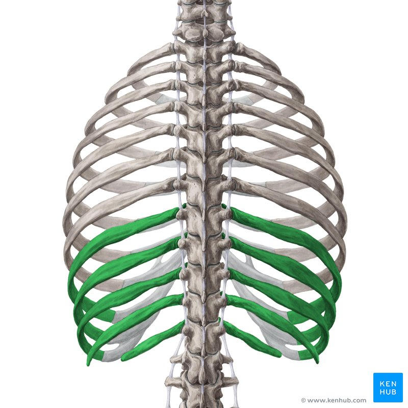
false ribs 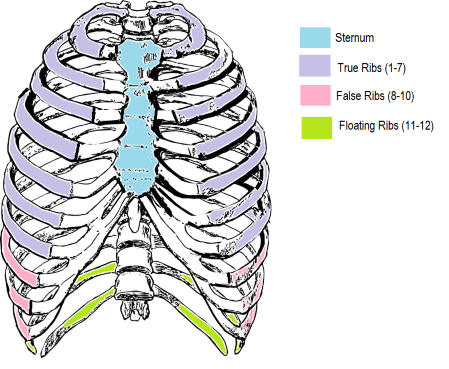
67
do not attach to sternum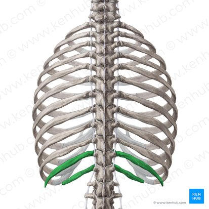
floating ribs 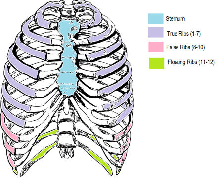
68
on each rib. articulates with the body of a vertebra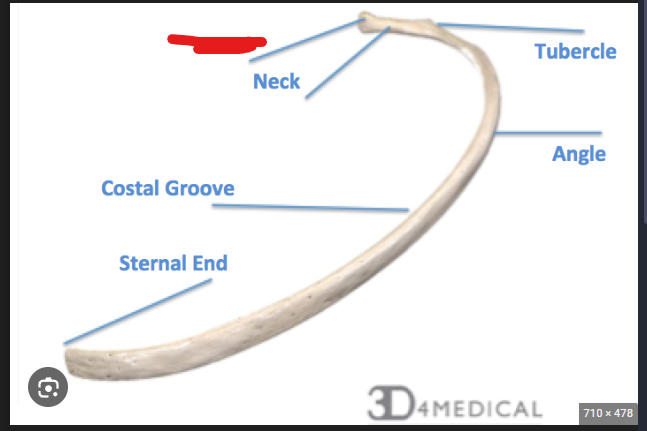
head 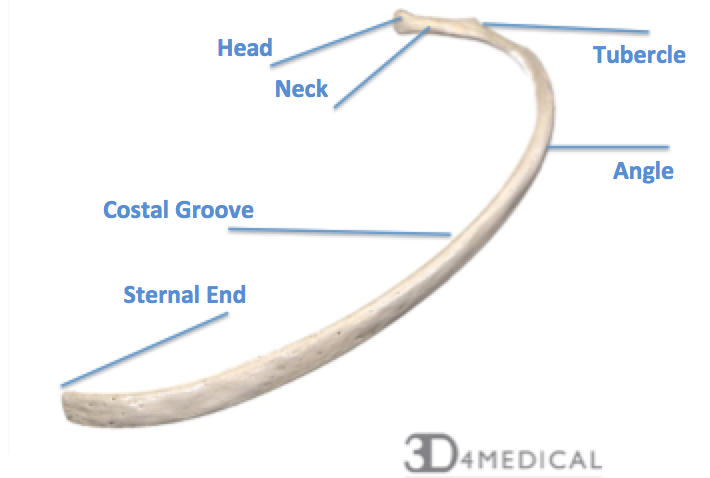
69
on each rib. below head 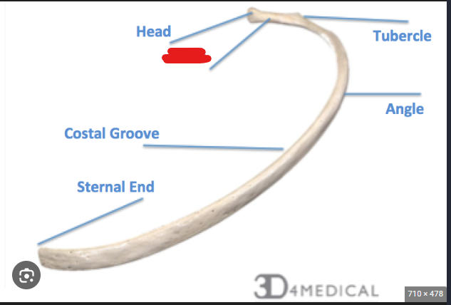
neck 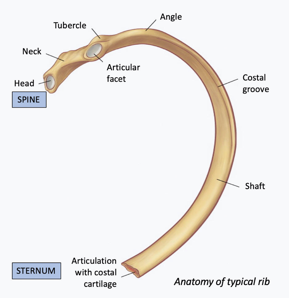
70
On each rib. raised area that articulates with the transverse process of a
vertebra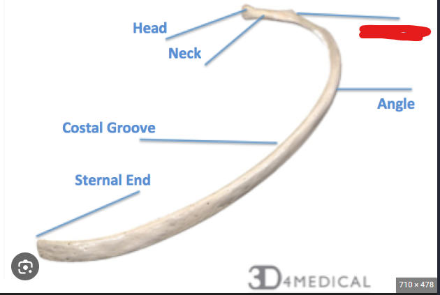
vertebra
tubercle 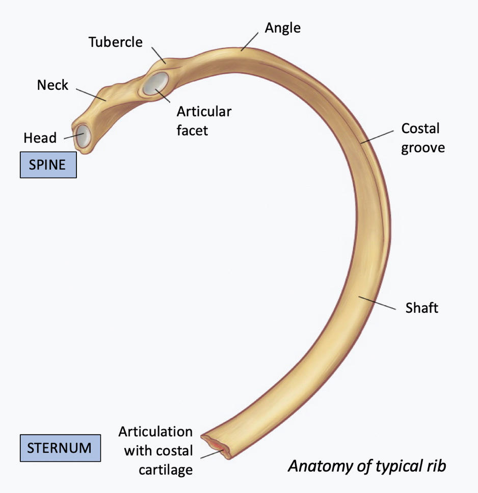
71
On each rib. Long flattened part behind the tubercle 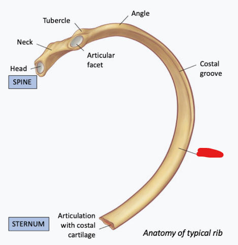
shaft 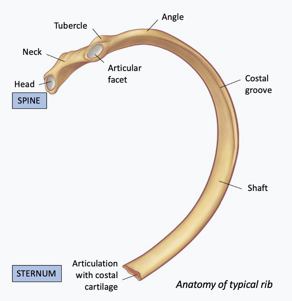
72
connects ribs to sternum 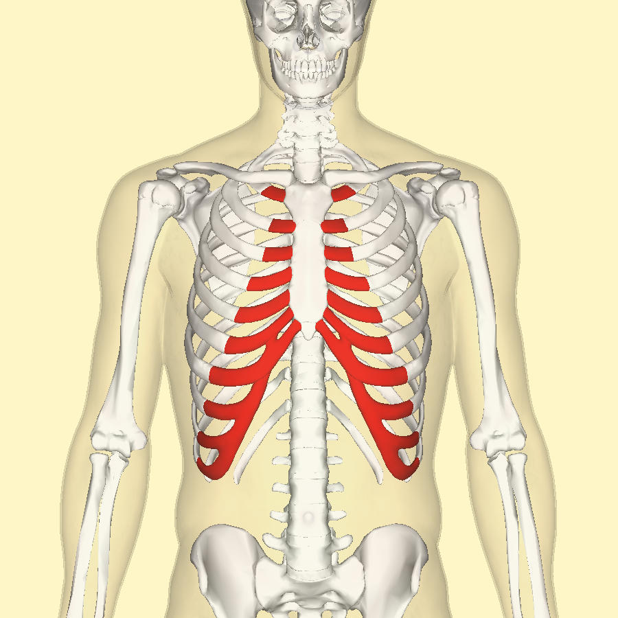
costal cartilages
73
the spine but sciency name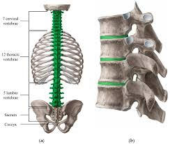
vertebral column
74
largest part of vertebrae 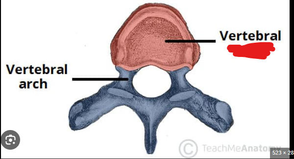
body
75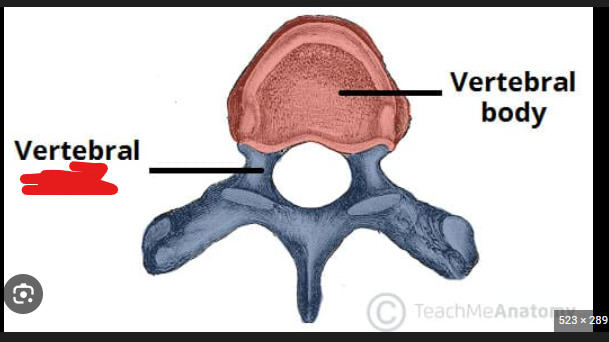
vertebral arch 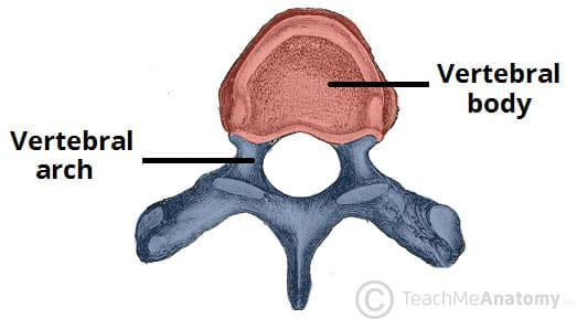
76
superior part that contains the spinous process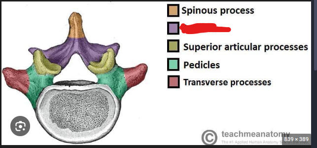
lamina 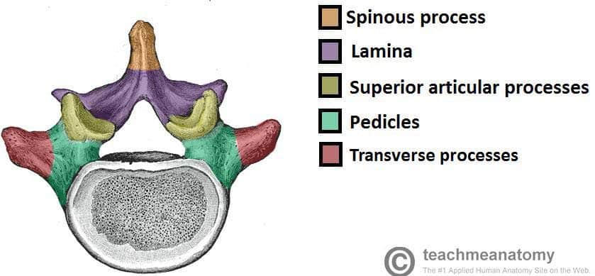
77
vertical part that connects the lamina to the body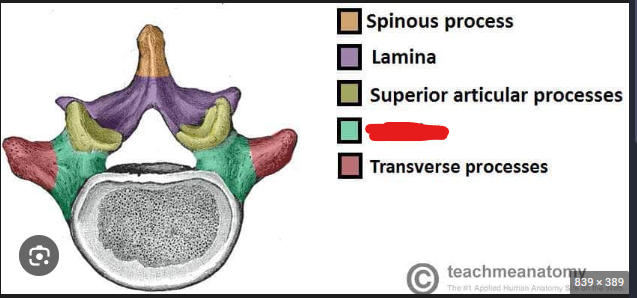
pedicle 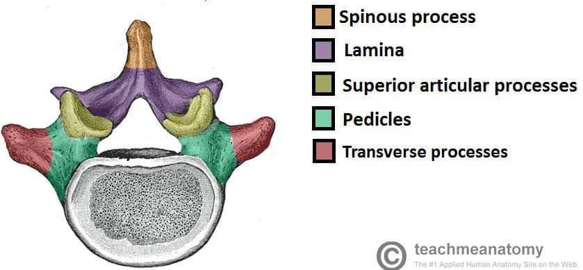
78
opening that conveys the spinal cord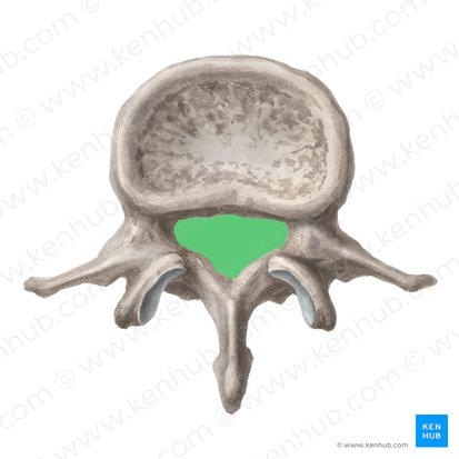
vertebral foramen 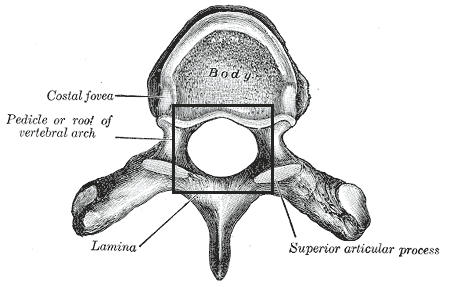
79
extension of lamina that extends off the dorsal surface of the
vertebra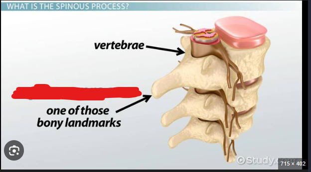
vertebra
spinous process 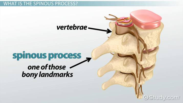
80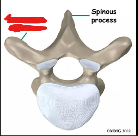
transverse process
81
extension of the vertebra that serve as
attachments for vertebrae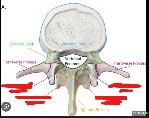
attachments for vertebrae
superior and inferior articulate processes 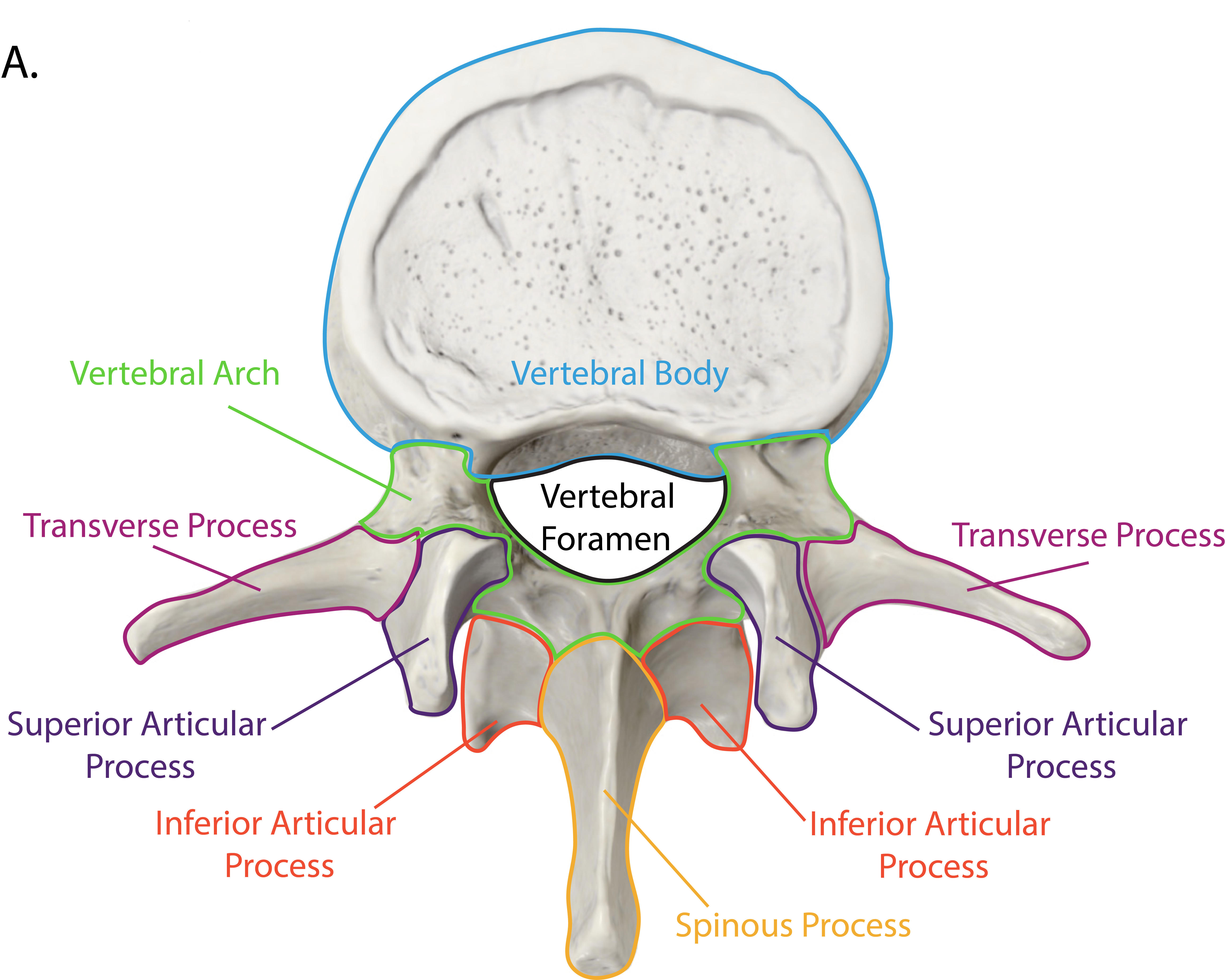
82
smooth, flat surface covered with hyaline
cartilage on the superior articular process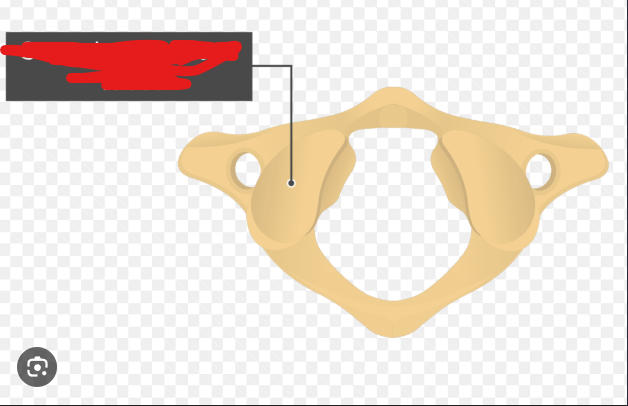
cartilage on the superior articular process
superior articular facet 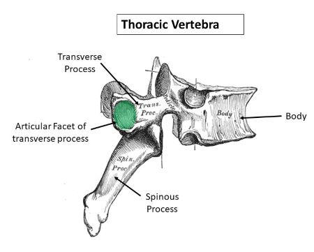
83
smooth flat surface covered with hyaline cartilage
on the inferior articular process.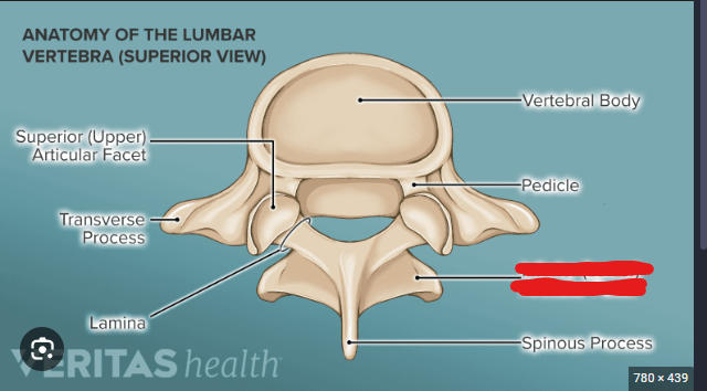
on the inferior articular process.
inferior articular facet 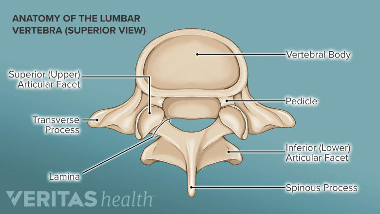
84
space for spinal nerve formed by the intervertebral
notches of adjacent vertebra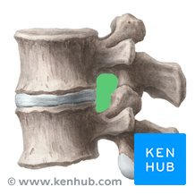
notches of adjacent vertebra
intervertebral foramen 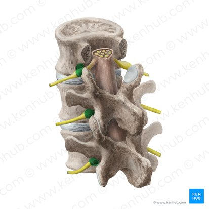
85
pads of fibrocartilage between bodies of adjacent vertebra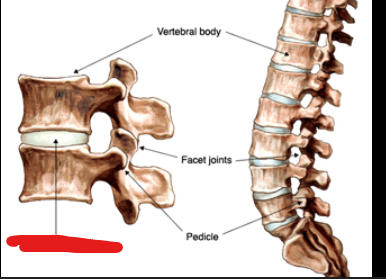
intervertebral discs 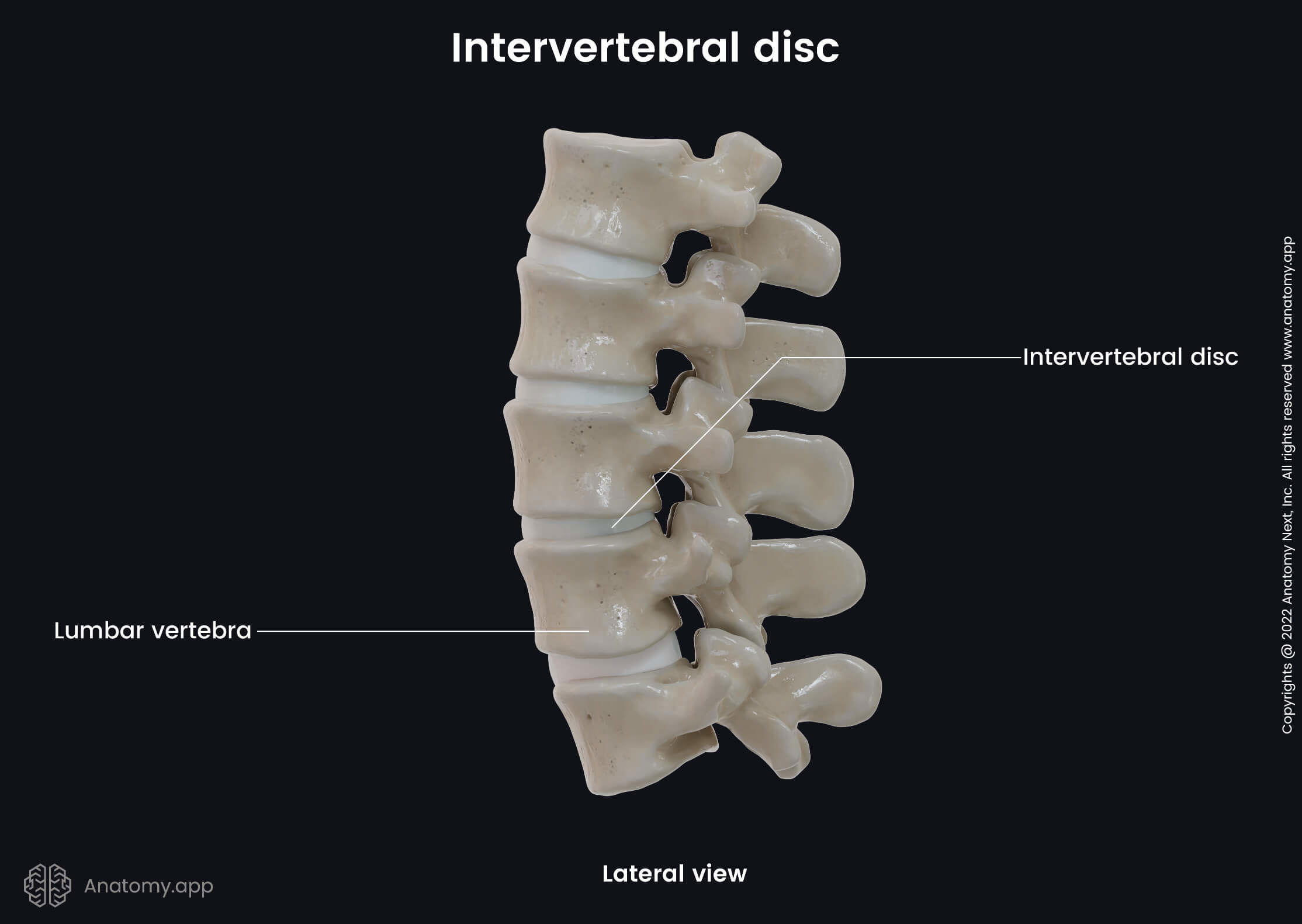
86
no body or spinous process 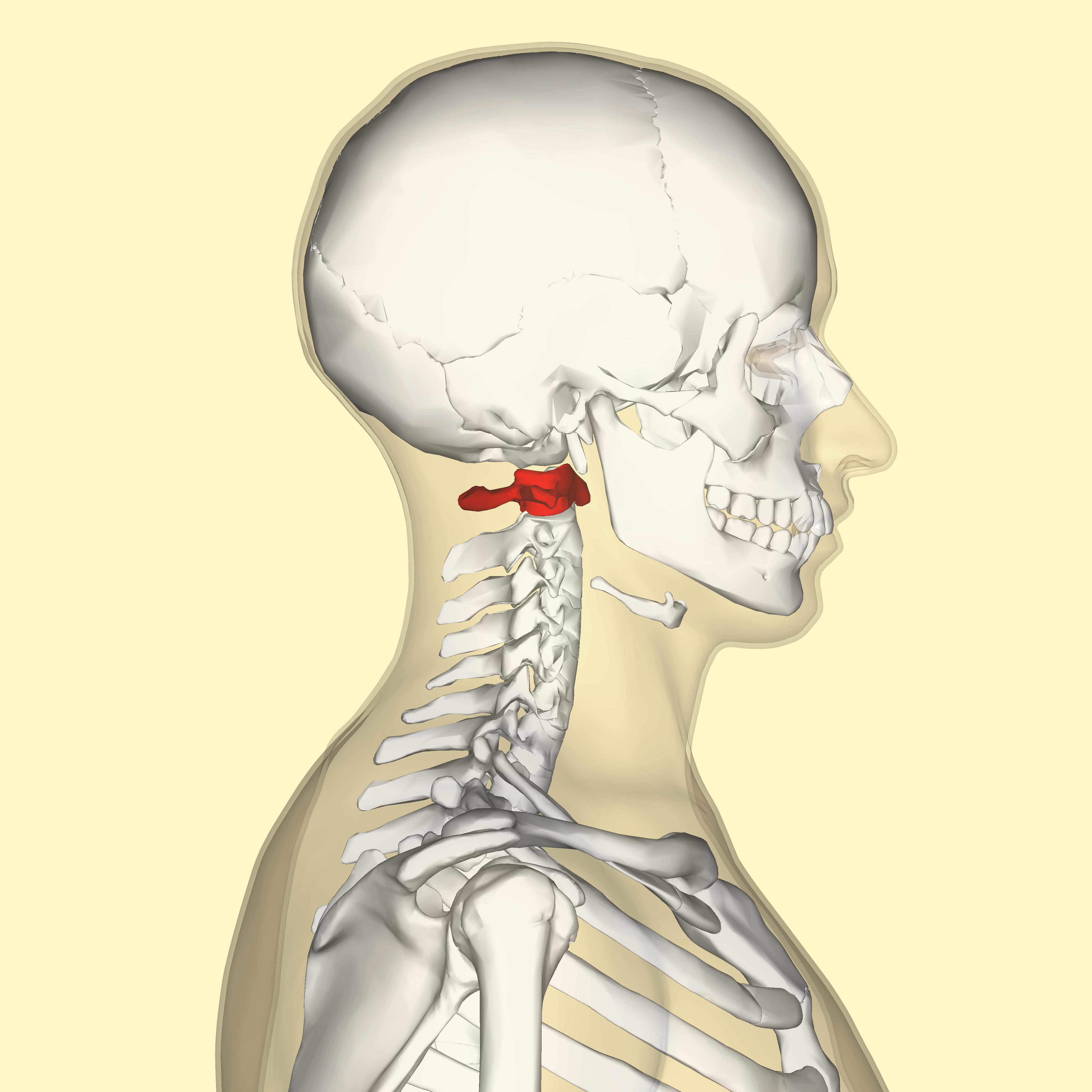
atlas 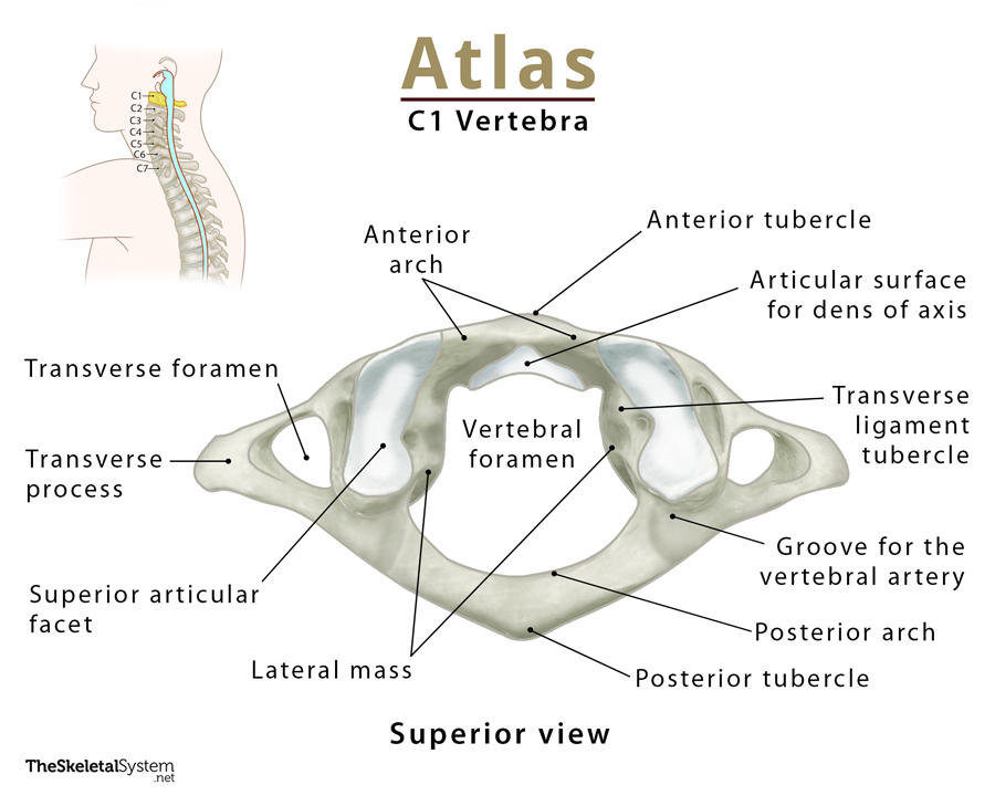
87
the second cervical vertebrae. 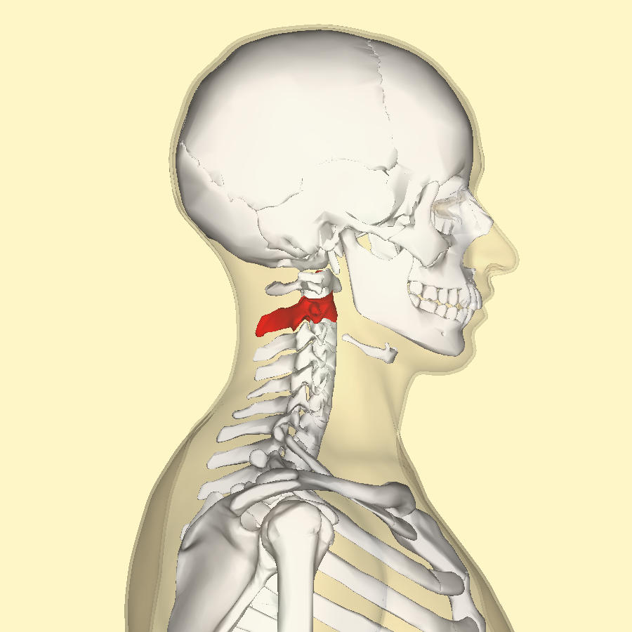
axis 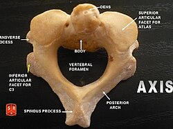
88
prominence that extends from the body 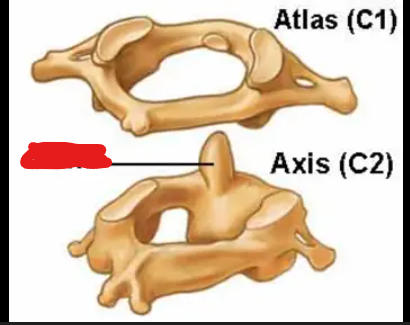
dens 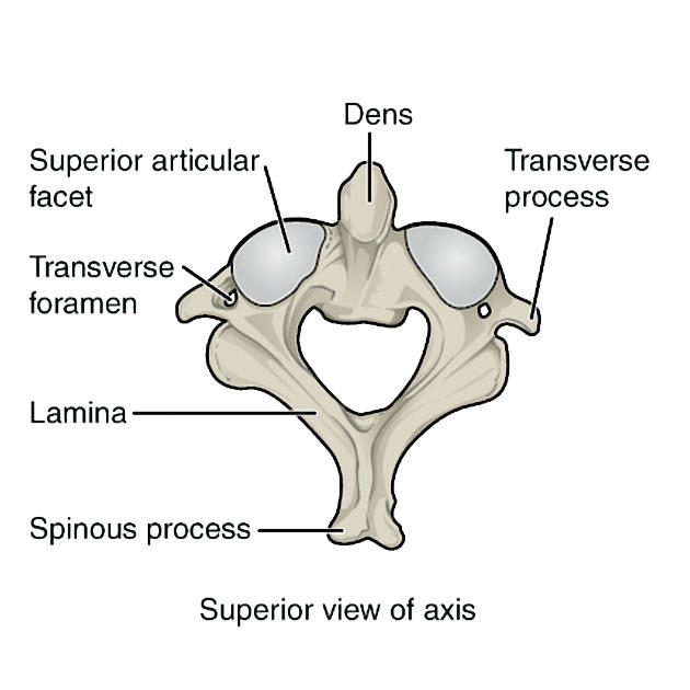
89
this is the name of the seventh cervical vertebra which has the
largest spinous process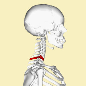
largest spinous process
vertebra prominens 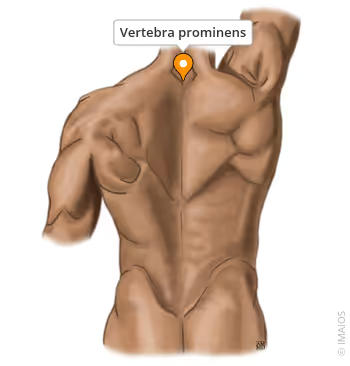
90
hole in transverse process that conveys the vertebral artery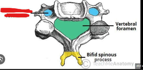
transverse foramen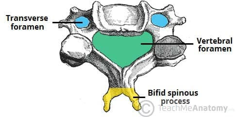
91
there are 12 of them and are similar to the cervical vertebrae 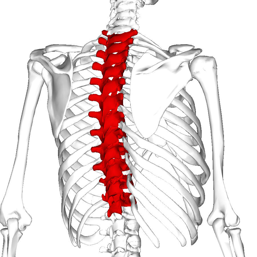
thoracic vertebrae
92
impression on the inferior body for articulation with the
head of a rib.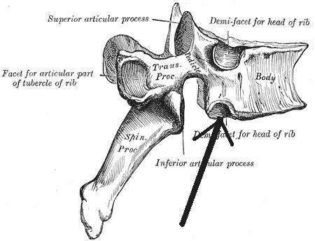
head of a rib.
inferior costal facets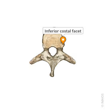
93
impression on the superior body for articulation with the
head of a rib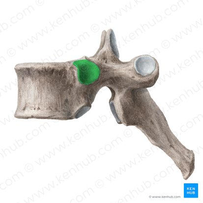
head of a rib
superior costal facets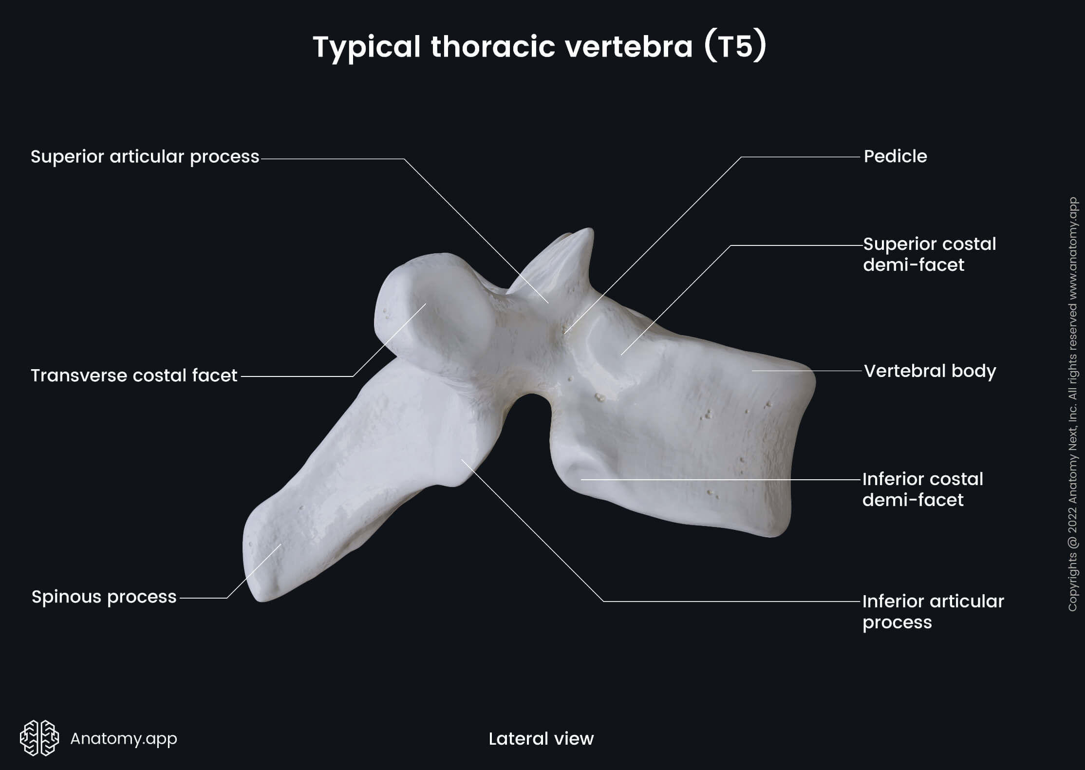
94
for articulation with the tubercle of the rib. Not present on
T11 or T12.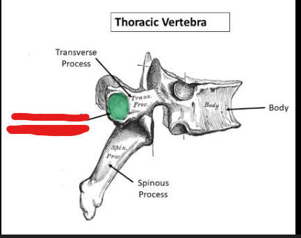
T11 or T12.
facets on transverse processes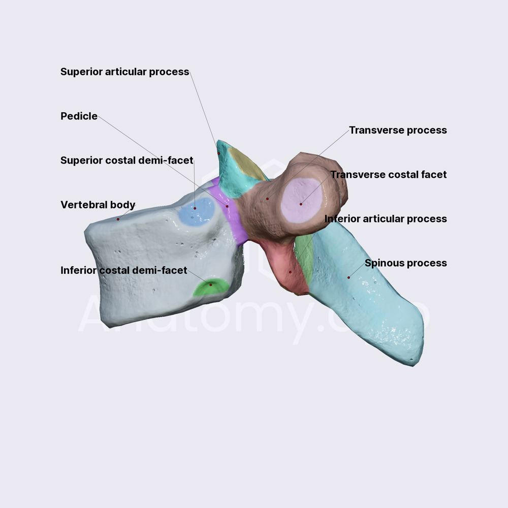
95
are the largest vertebrae and contain all the features listed above 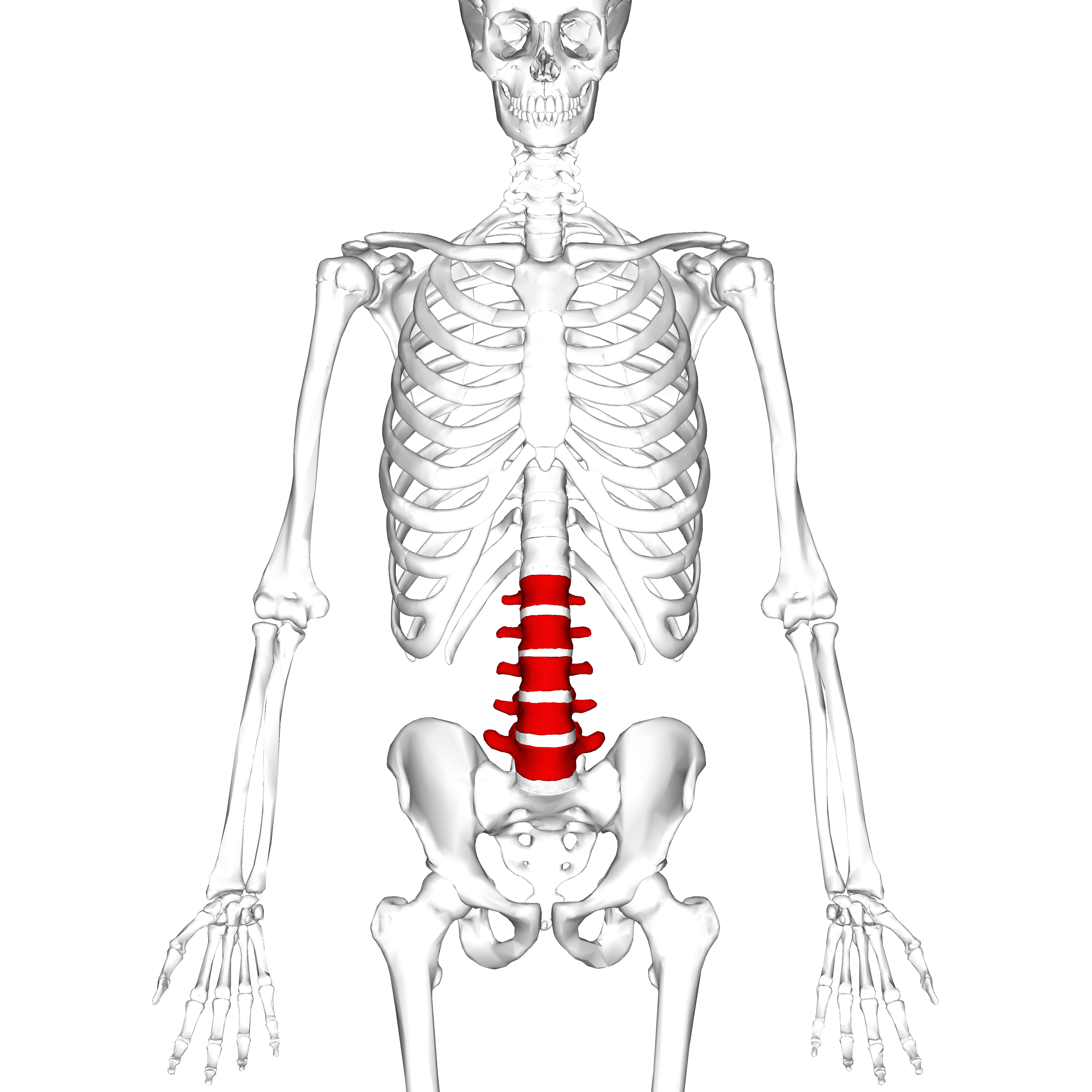
lumbar vertebrae
96
formed from 5 fused sacral vertebrae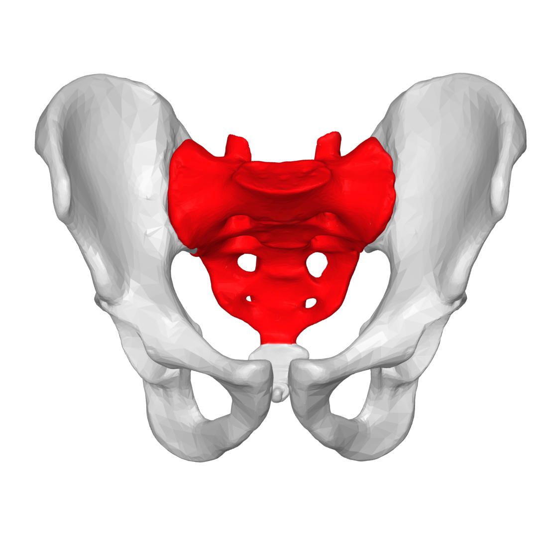
Sacrum 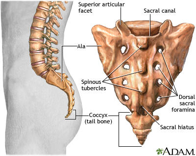
97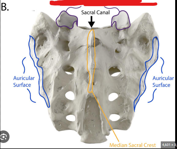
superior articulate process
98
holes for the exiting spinal nerves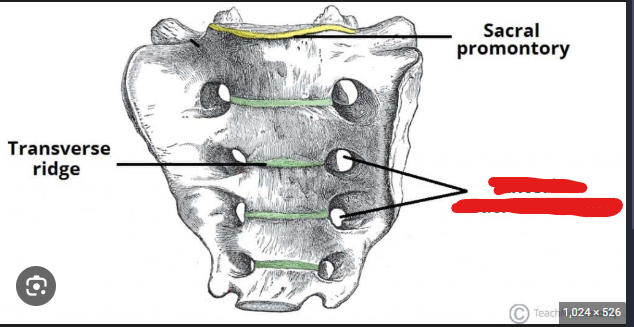
posterior and anterior sacral foramina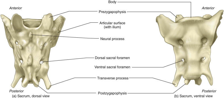
99
passageway for cauda equina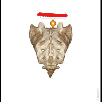
sacral canal 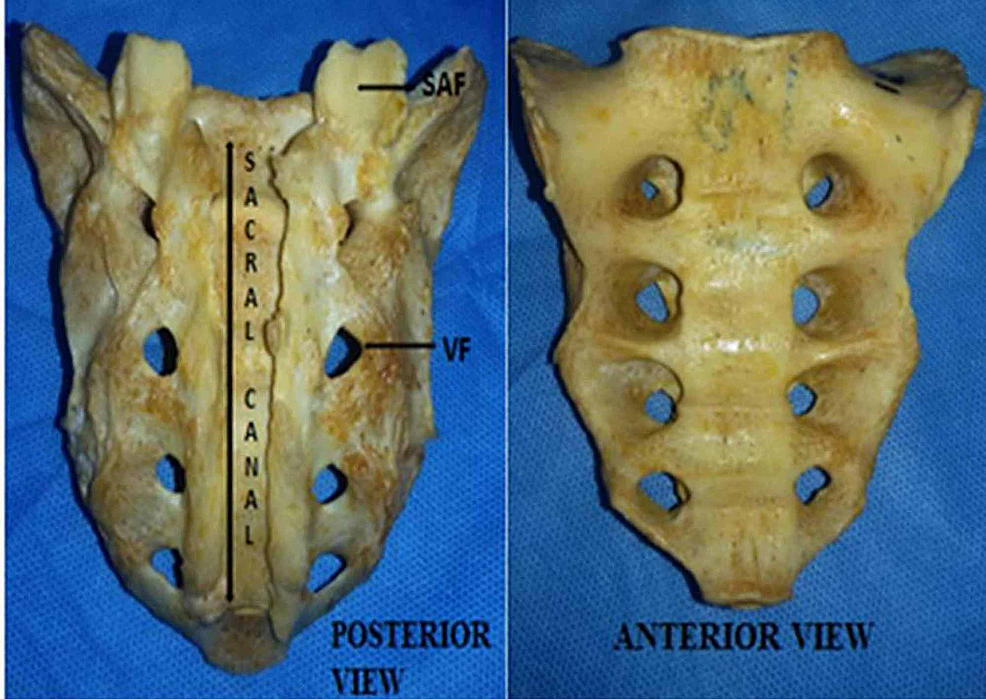
100
inferior opening to the sacral canal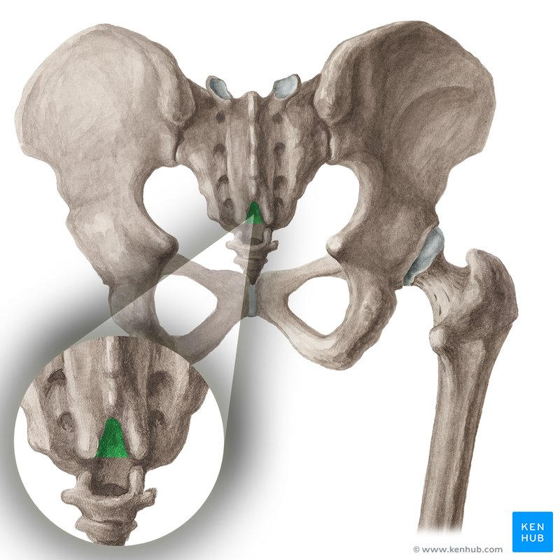
sacral hiatus 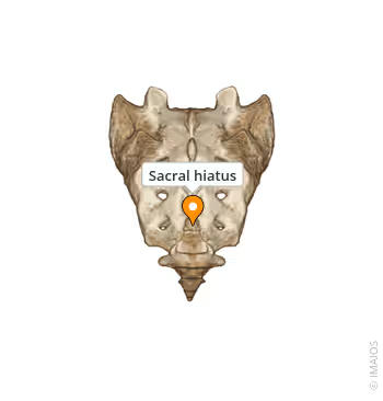
101
site of sacroiliac joint of the pelvic girdle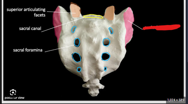
auricular surface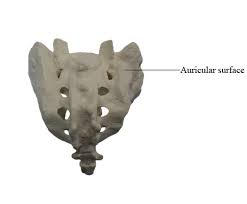
102
(note: 4-5 fused coccygeal vertebrae)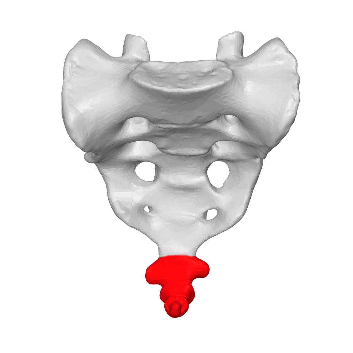
coccyx 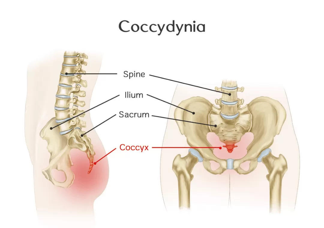
103
extension and lateral flexion
of vertebral column
O: Ilium
I: Ribs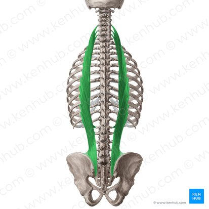
of vertebral column
O: Ilium
I: Ribs
iliocostalis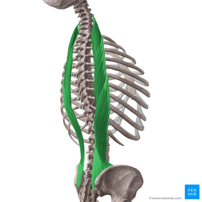
104
extension and lateral flexion
of vertebral column
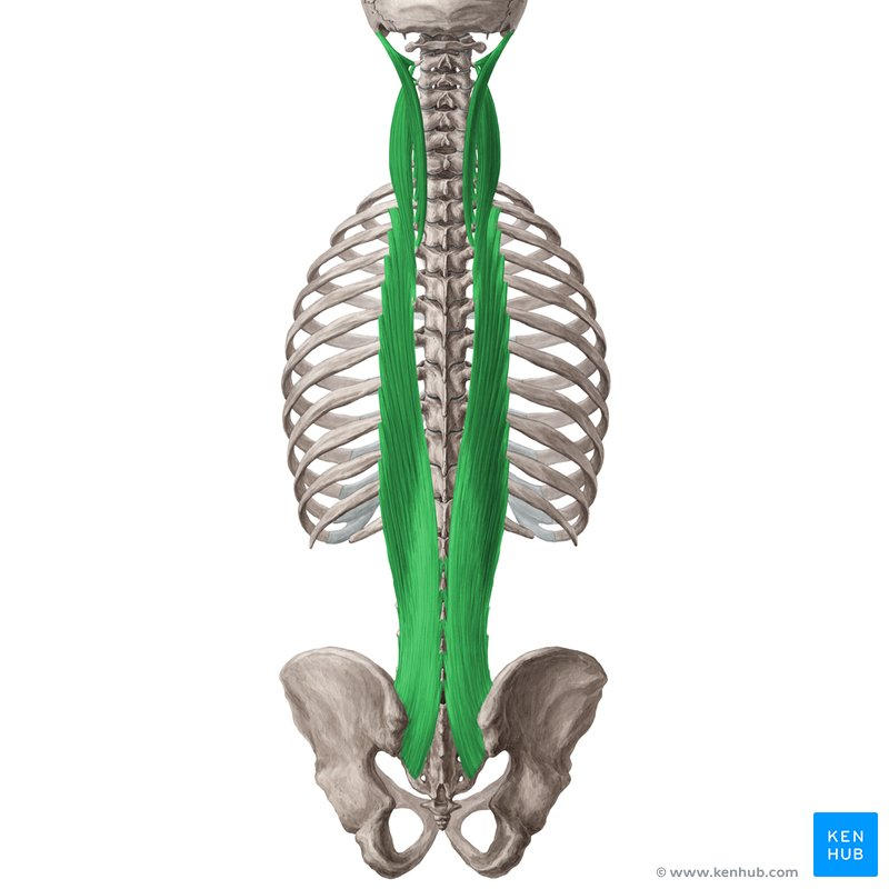
of vertebral column
longissimus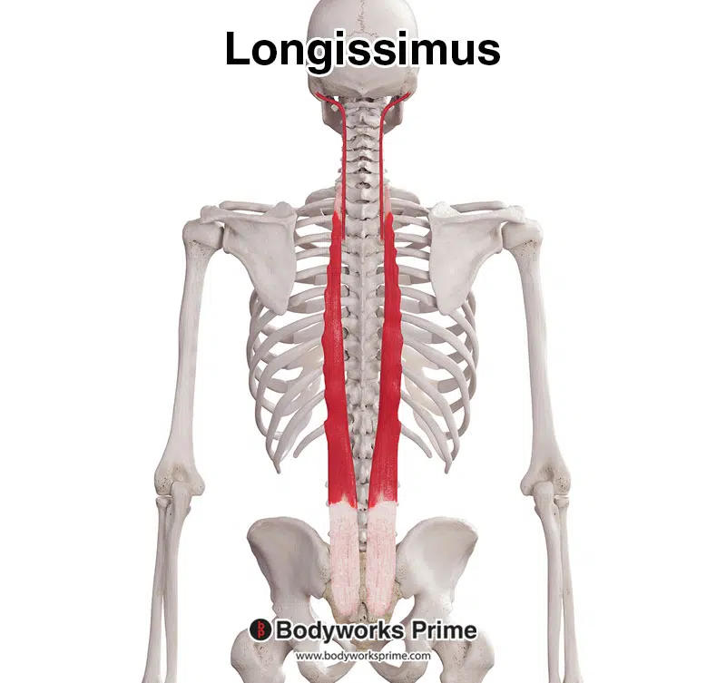
105
extension and lateral flexion
of vertebral column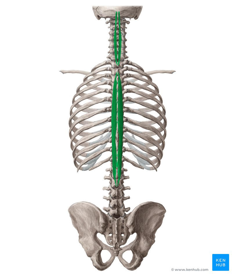
of vertebral column
spinalis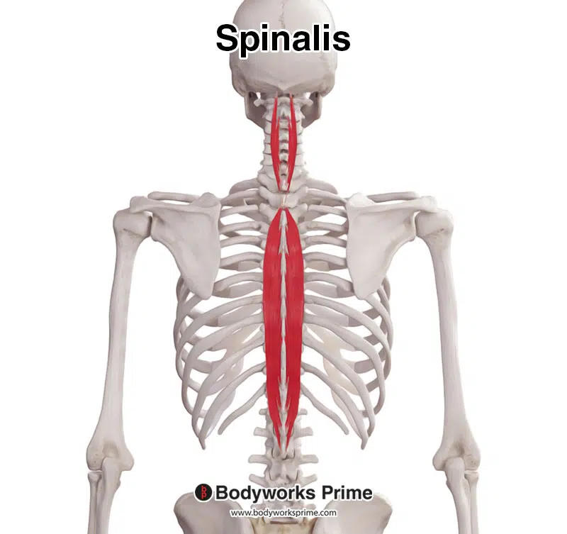
106
elevation, depression, and
retraction of scapula
O: External occipital protuberance,
thoracic vertebrae
I: Scapula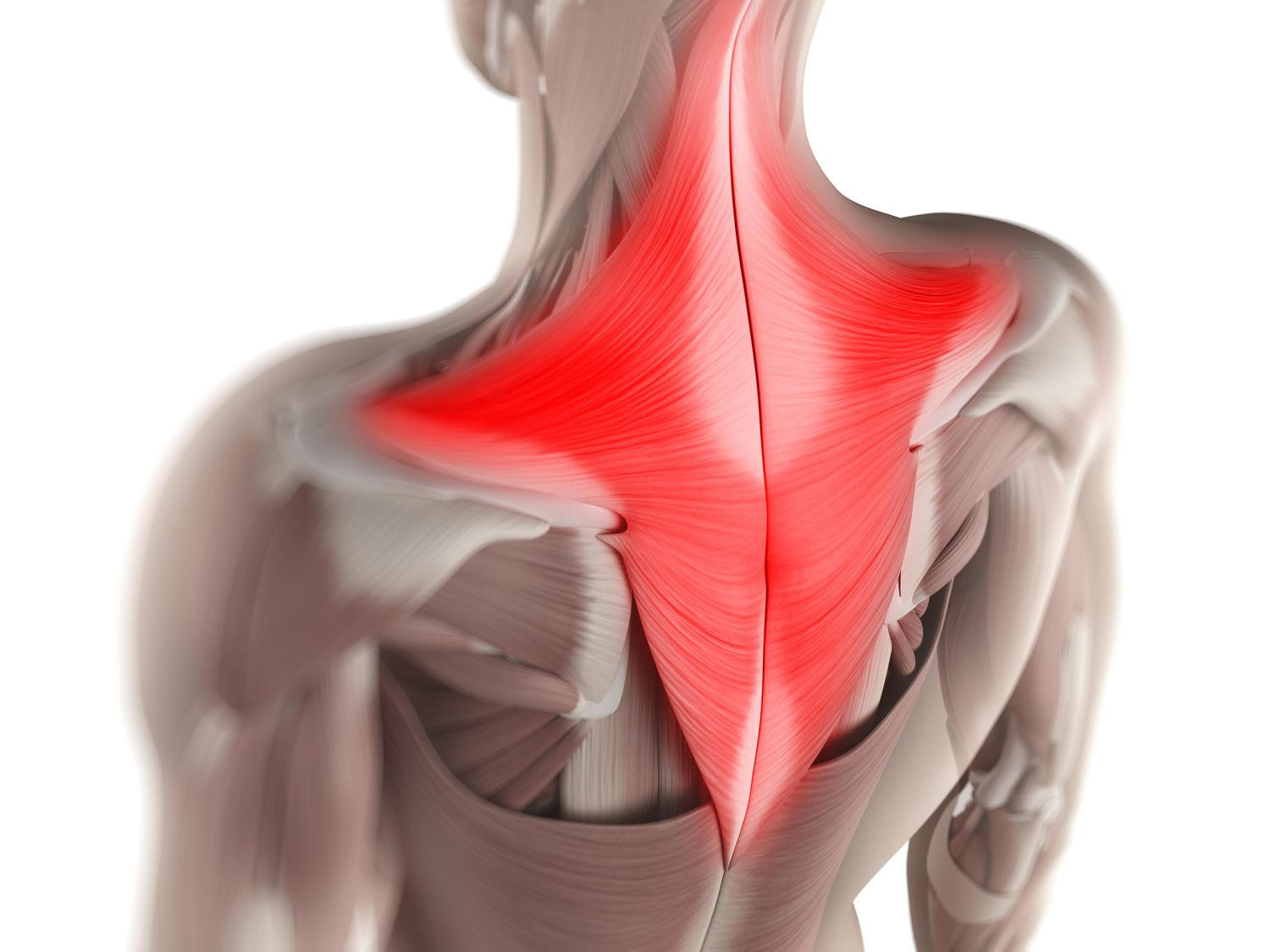
retraction of scapula
O: External occipital protuberance,
thoracic vertebrae
I: Scapula
trapezius 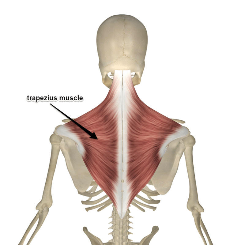
107
elevates scapula
O: Cervical vertebrae
I: Scapula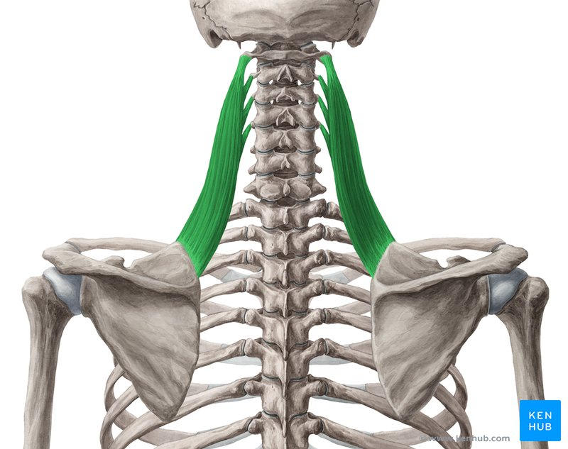
O: Cervical vertebrae
I: Scapula
levator scapulae 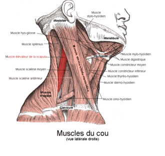
108
rotates and flexes head/neck
O: Sternum, clavicle
I: Mastoid process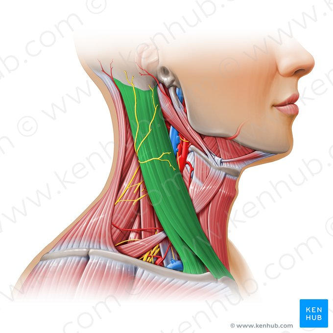
O: Sternum, clavicle
I: Mastoid process
sternocleidomastoid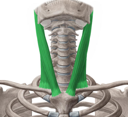
109
extends neck
O: Cervical vertebrae
I: Occipital bone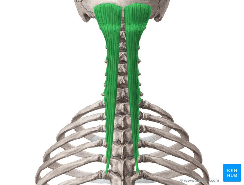
O: Cervical vertebrae
I: Occipital bone
semispinalis capitis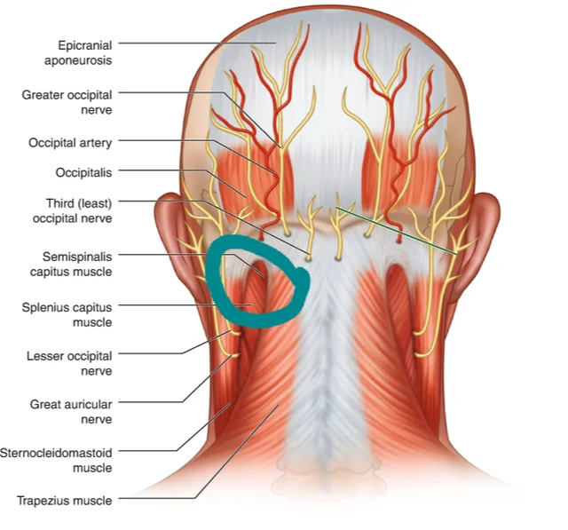
110
extends neck
O: Cervical vertebrae
I: Mastoid process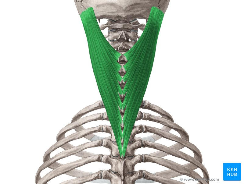
O: Cervical vertebrae
I: Mastoid process
splenius capitis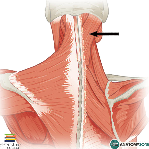
111
lateral flexion of neck
O: Cervical vertebrae
I: Ribs 1-2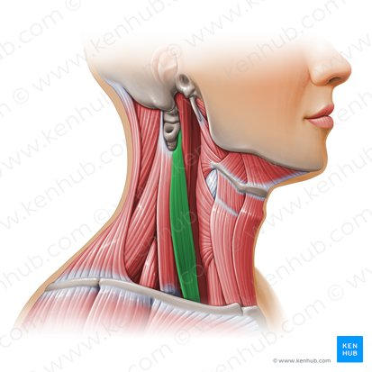
O: Cervical vertebrae
I: Ribs 1-2
scalenes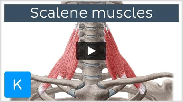
112
depresses hyoid bone
O: Manubrium
I: Hyoid bone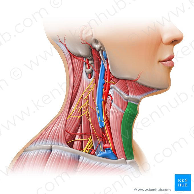
O: Manubrium
I: Hyoid bone
sternohyoid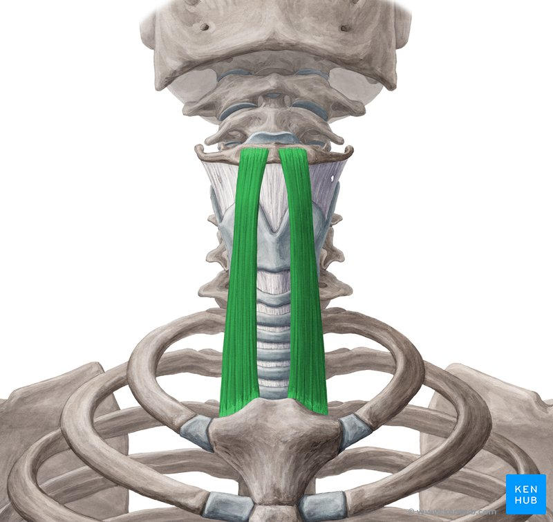
113
depresses hyoid bone
O: Scapula
I: Hyoid bone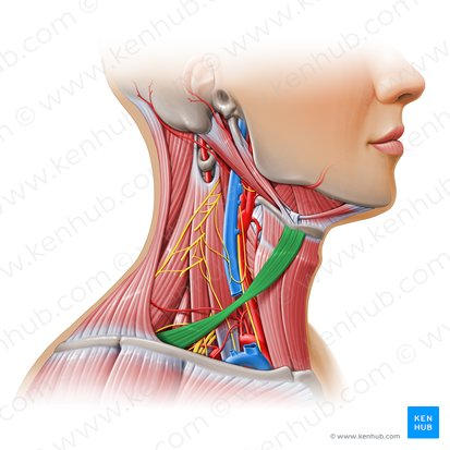
O: Scapula
I: Hyoid bone
omohyoid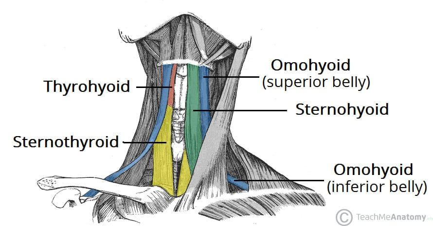
114
depresses hyoid bone
O: Thyroid cartilage
I: Hyoid bone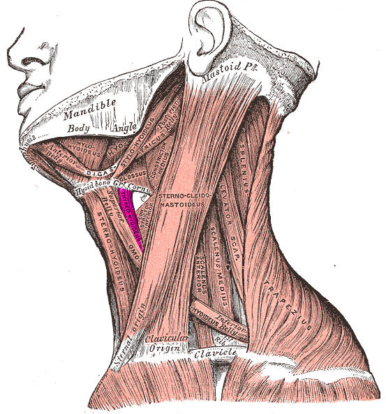
O: Thyroid cartilage
I: Hyoid bone
thyrohyoid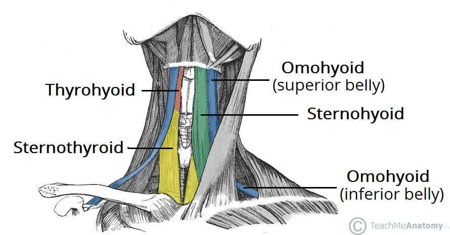
115
elevates hyoid bone
O: Styloid process (of temporal bone)
I: Hyoid bone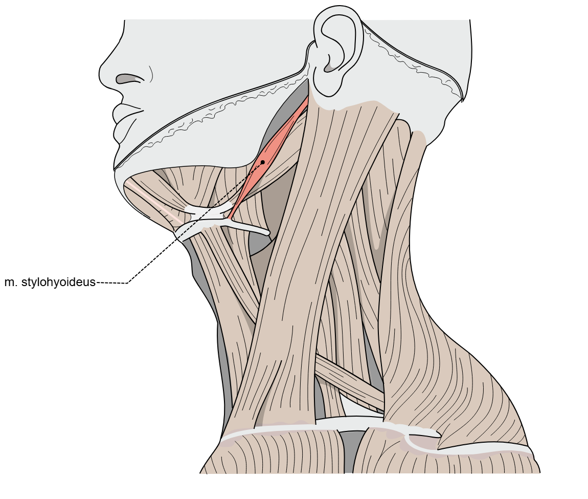
O: Styloid process (of temporal bone)
I: Hyoid bone
stylohyoid 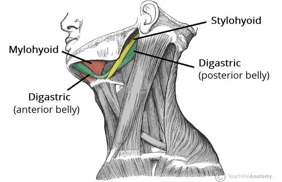
116
elevates hyoid bone
O: Mandible
I: Hyoid bone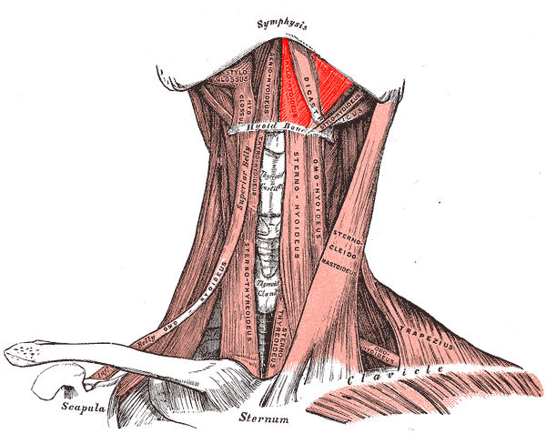
O: Mandible
I: Hyoid bone
mylohyoid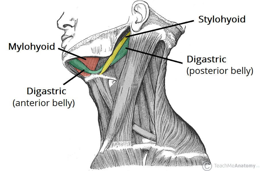
117
depresses mandible
O: Mastoid process
I: Body of mandible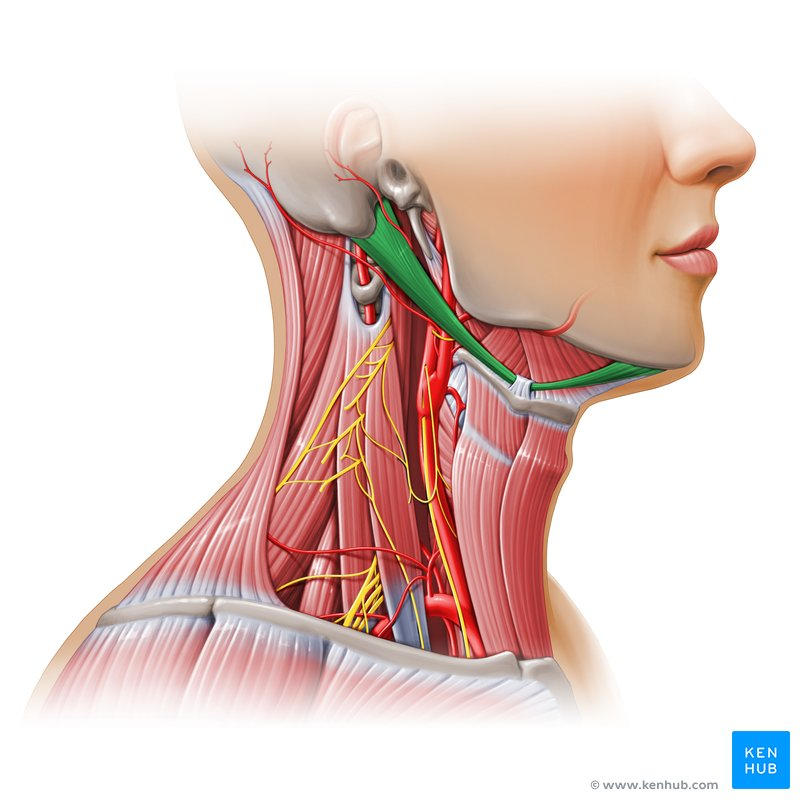
O: Mastoid process
I: Body of mandible
digastric 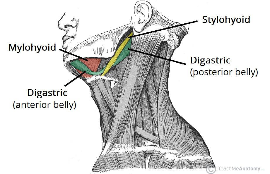
118
elevates mandible
O: Temporal bone
I: Coronoid process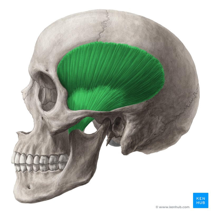
O: Temporal bone
I: Coronoid process
temporalis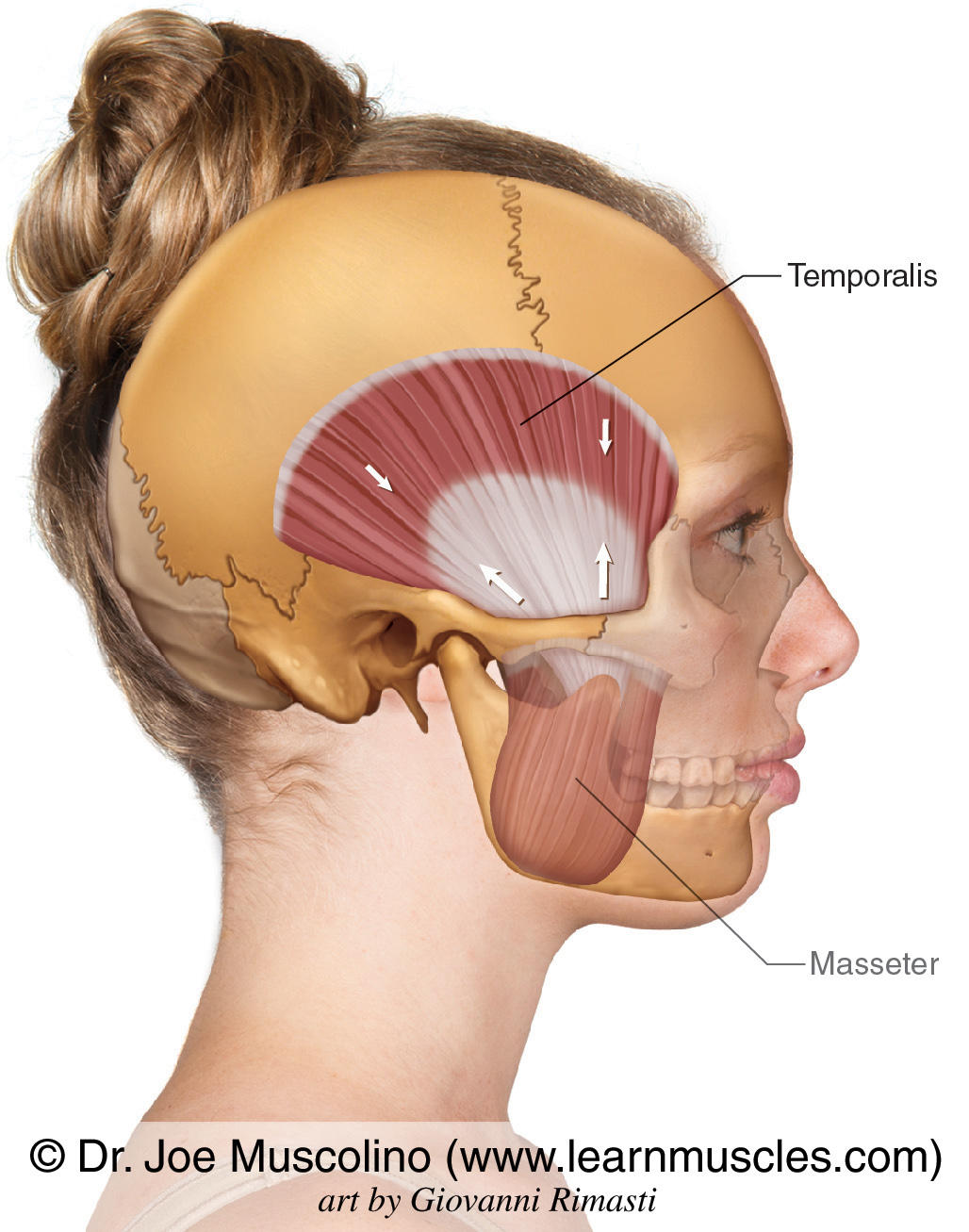
119
elevates mandible
O: Zygomatic process
I: Ramus of mandible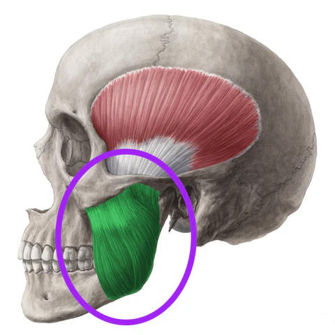
O: Zygomatic process
I: Ramus of mandible
masseter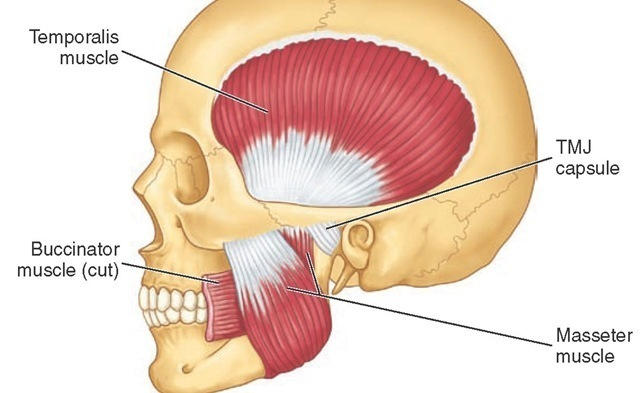
120
elevates mandible
O: Pterygoid process
I: Mandible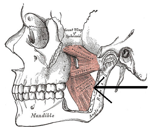
O: Pterygoid process
I: Mandible
medial pterygoid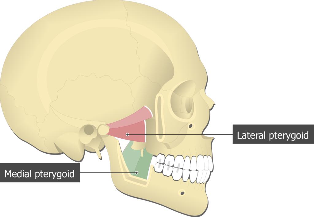
121
protracts mandible
O: Pterygoid process
I: Mandible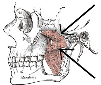
O: Pterygoid process
I: Mandible
lateral pterygoid 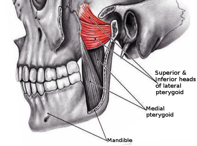
122
compresses cheek 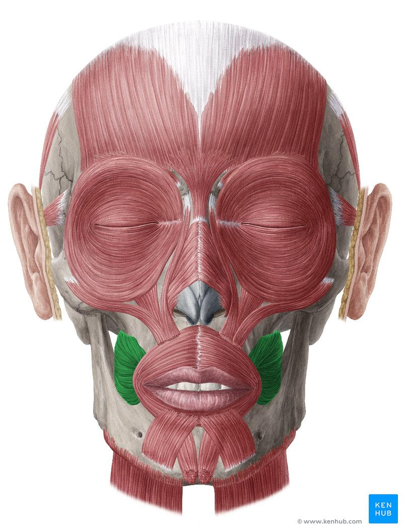
buccinator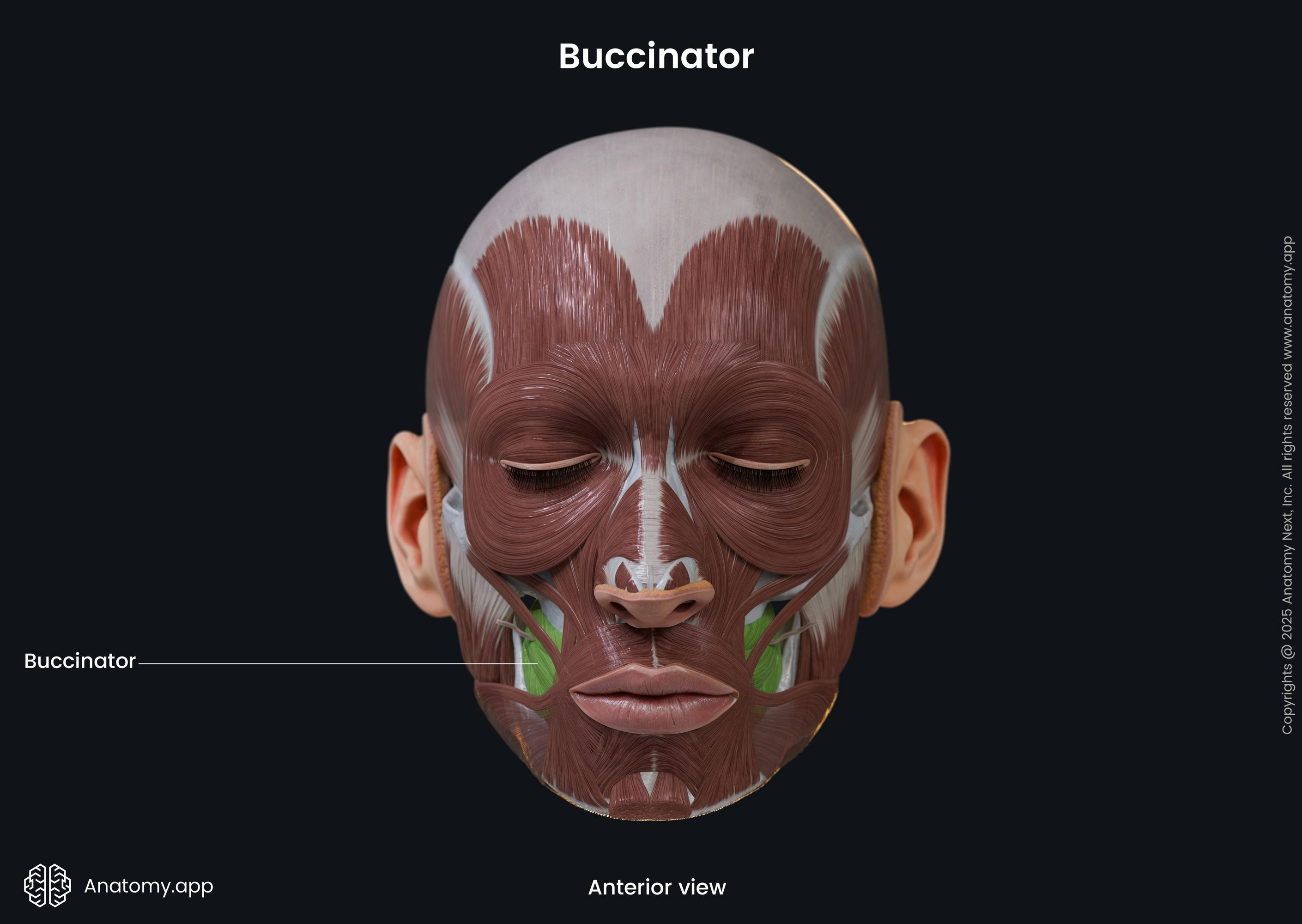
123
elevates corner of mouth
O: Zygomatic bone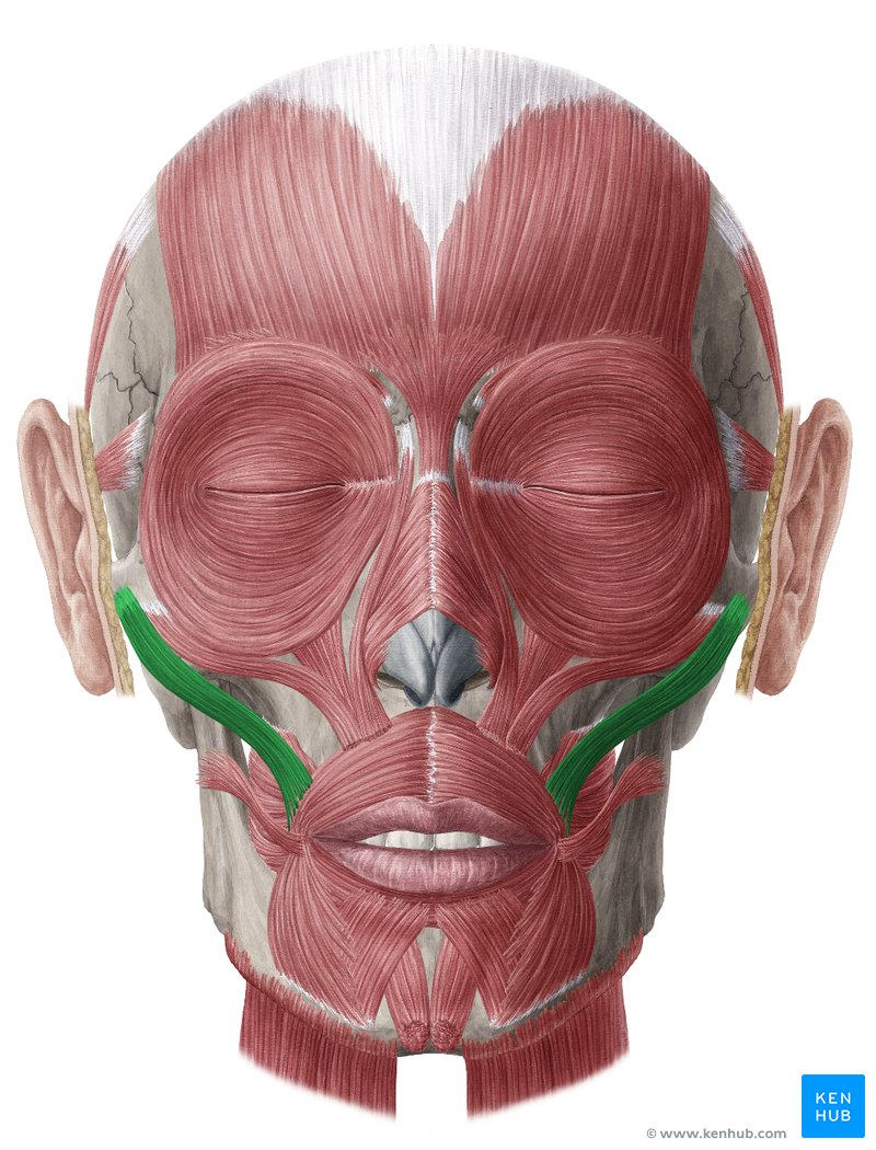
O: Zygomatic bone
zygomaticus major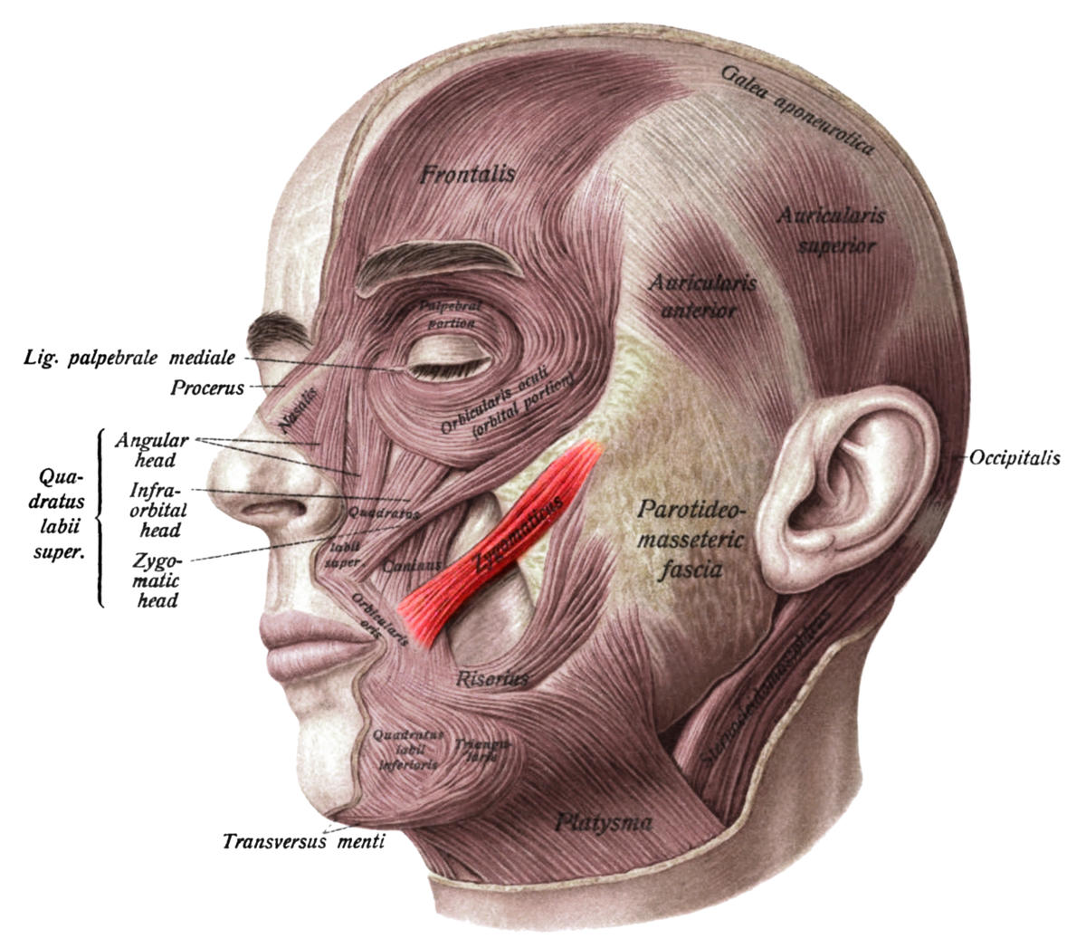
124
depresses corner of mouth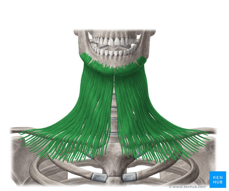
platysma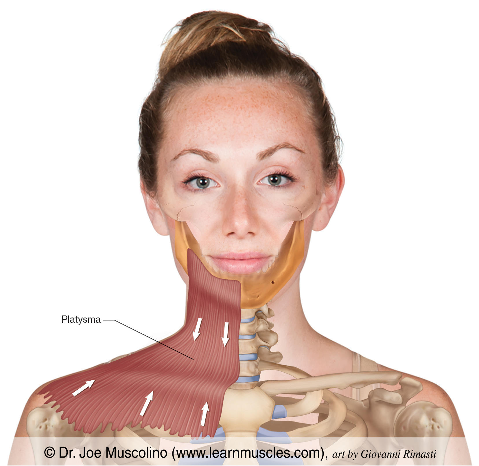
125
closes mouth; puckers lips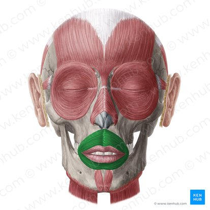
orbicularis oris 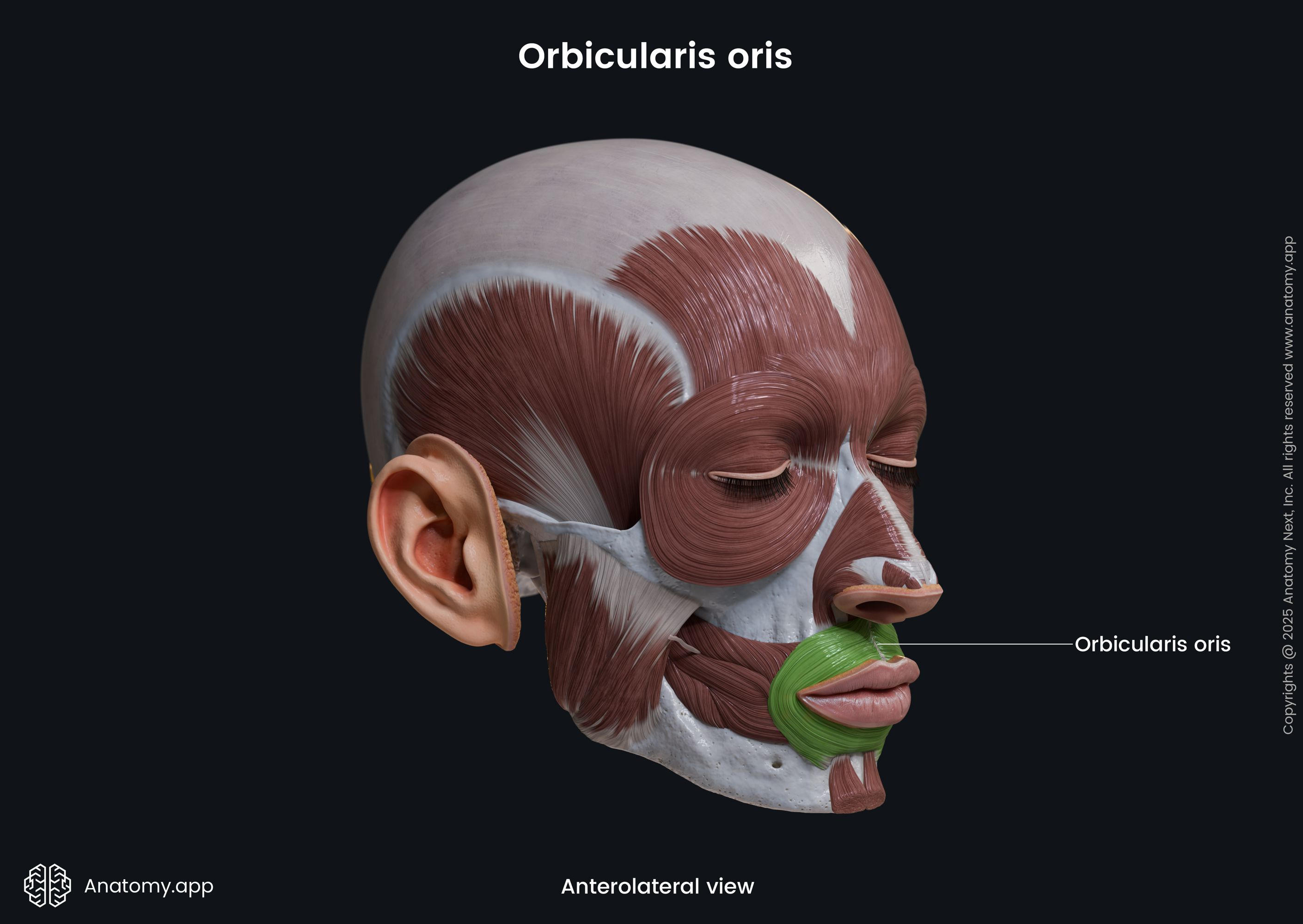
126
closes eyelids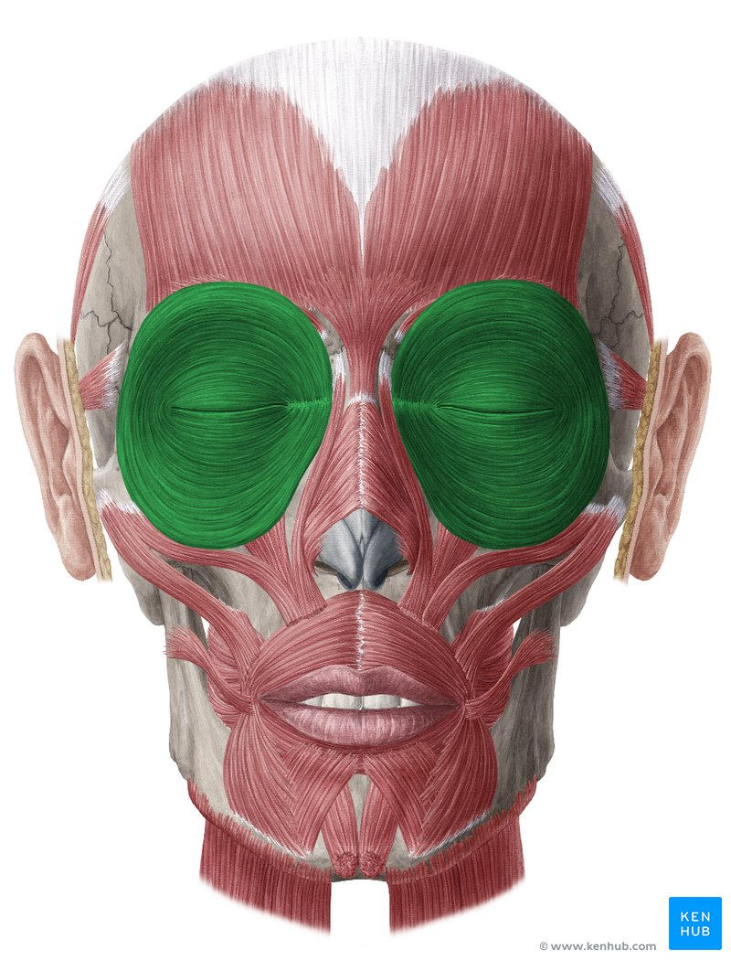
orbicularis oculi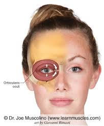
127
elevates eyebrows
O: Frontal bone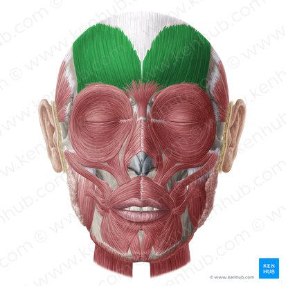
O: Frontal bone
frontalis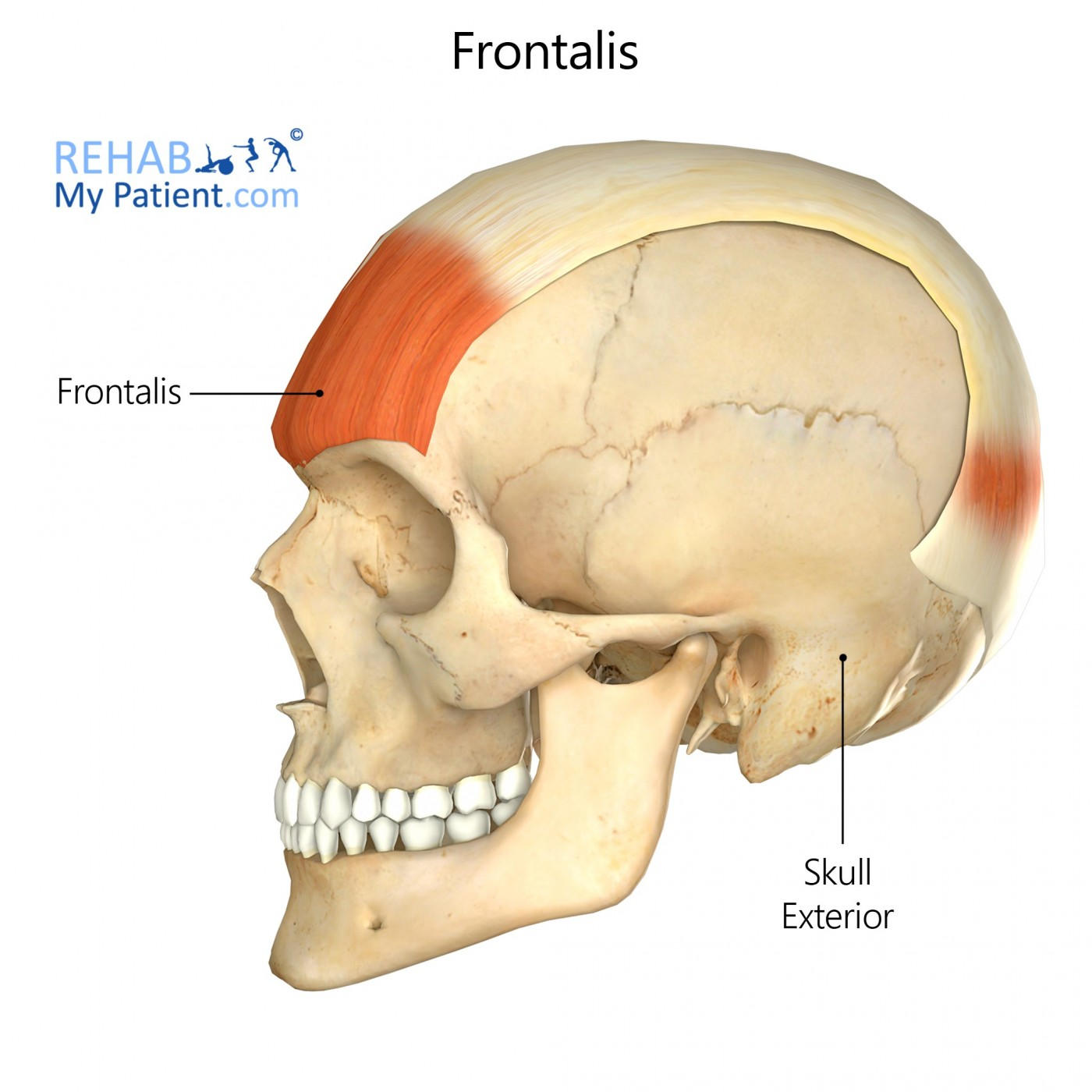
128
etracts scalp
O: Occipital bone
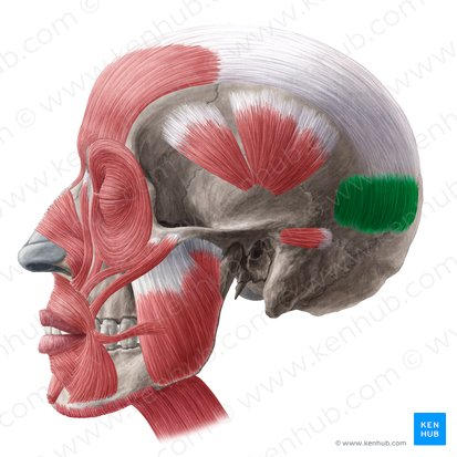
O: Occipital bone
occipitalis 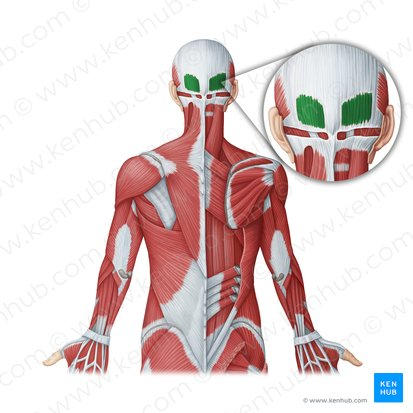
129
it deals with movement 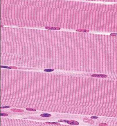
skeletal muscle
130
myofibril more specific
is of skeletal muscle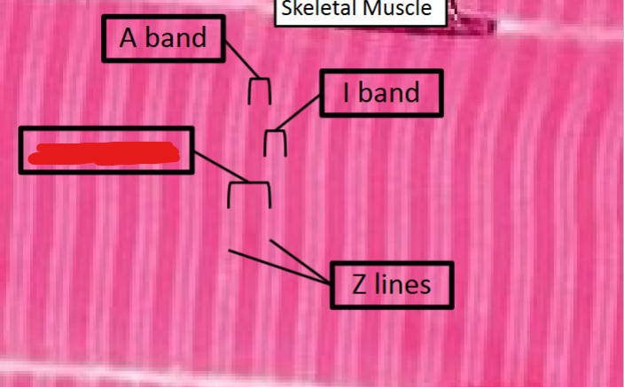
is of skeletal muscle
sarcomere
131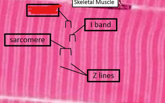
A band
132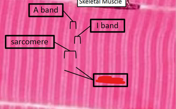
Z line 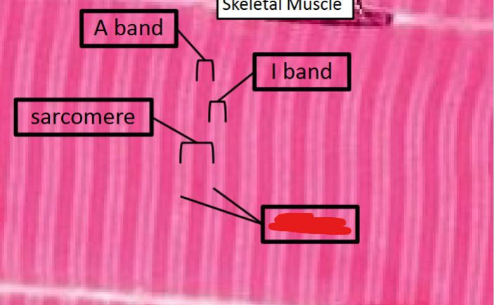
133
dissapears when muscle contract 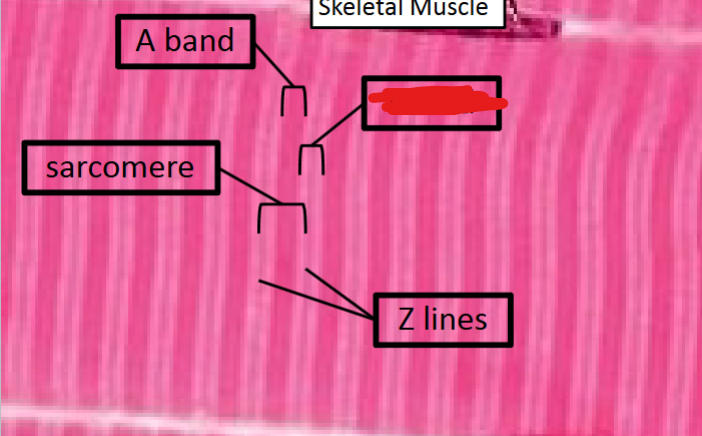
I band
134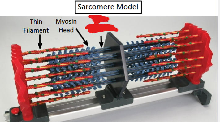
thick filament 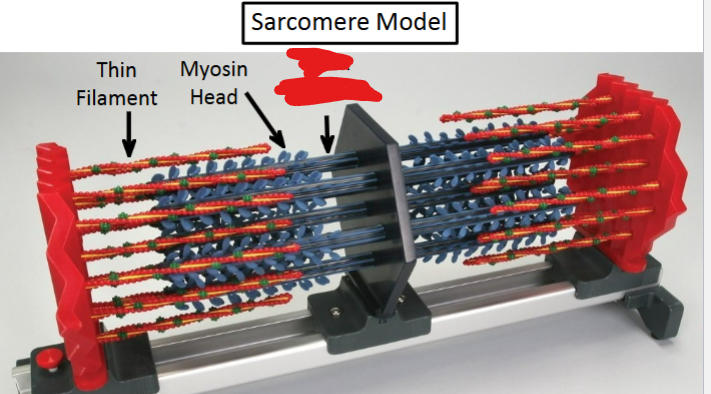
135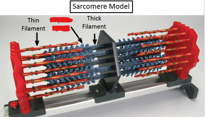
myosin head 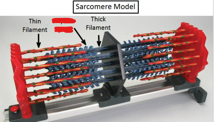
136
red vine
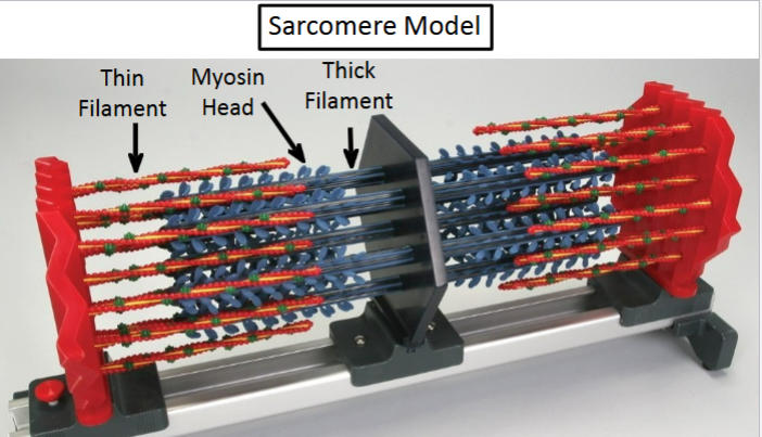
thin filament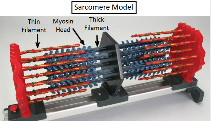
137
sarcomere model 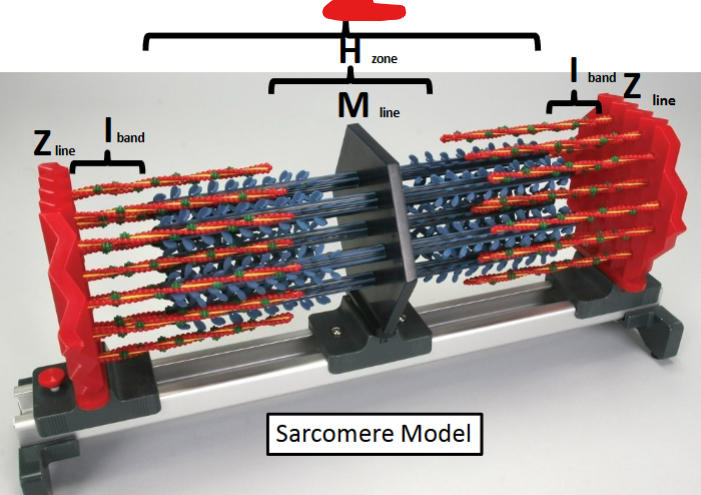
A band
138
sarcomere model 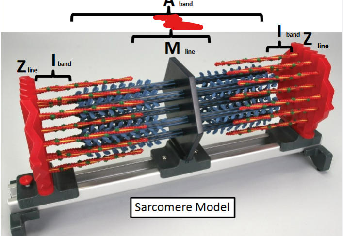
H zone
139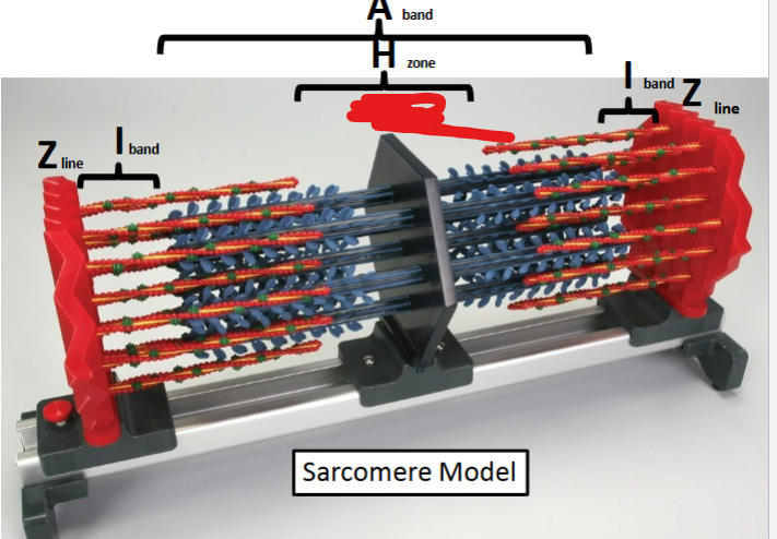
M line
140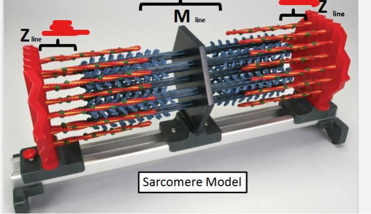
I band
141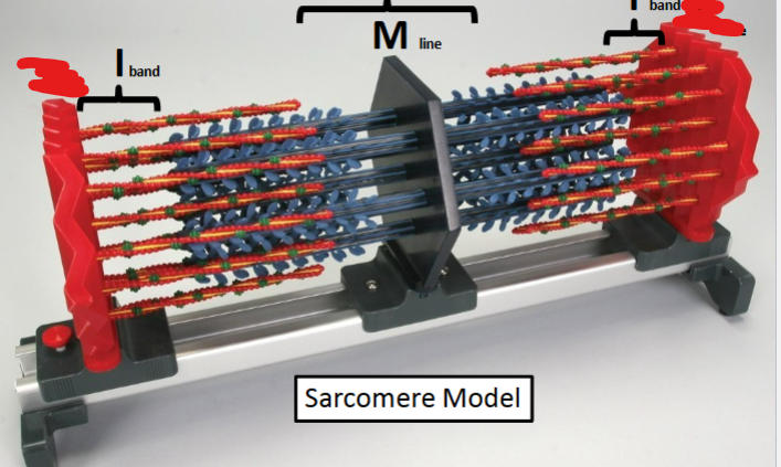
Z line
142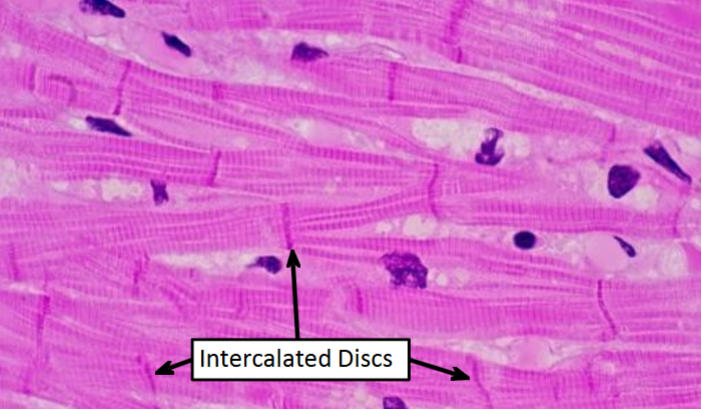
cardiac muscle
143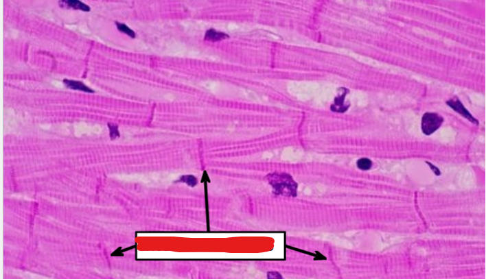
intercalated discs
144
same thing but closer 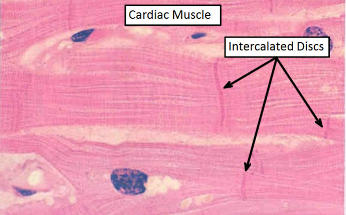
cardiac muscle
145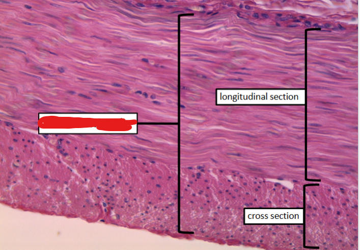
smooth muscle
146
smooth muscle 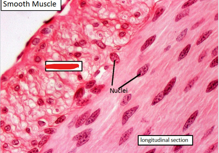
cross section
147
bottom stringy part
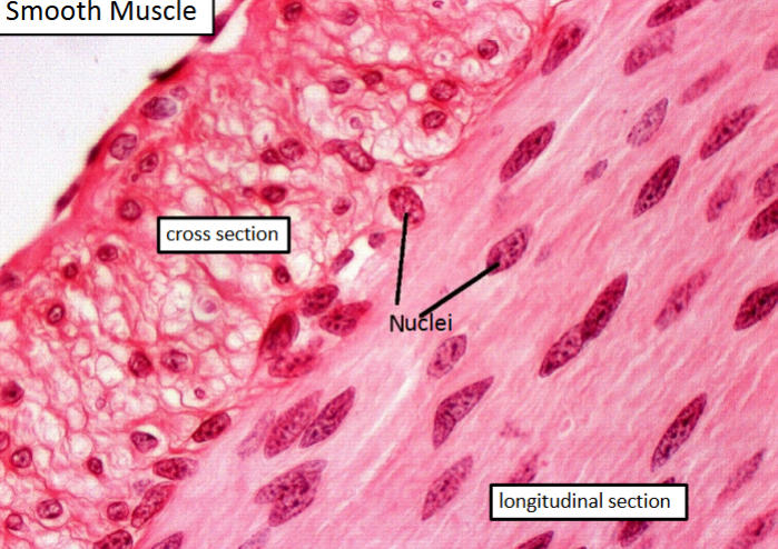
longitudinal section
148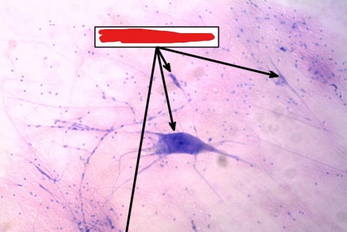
multipolar neurons
149
multipolar neurons 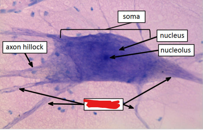
dendrites
150
neuron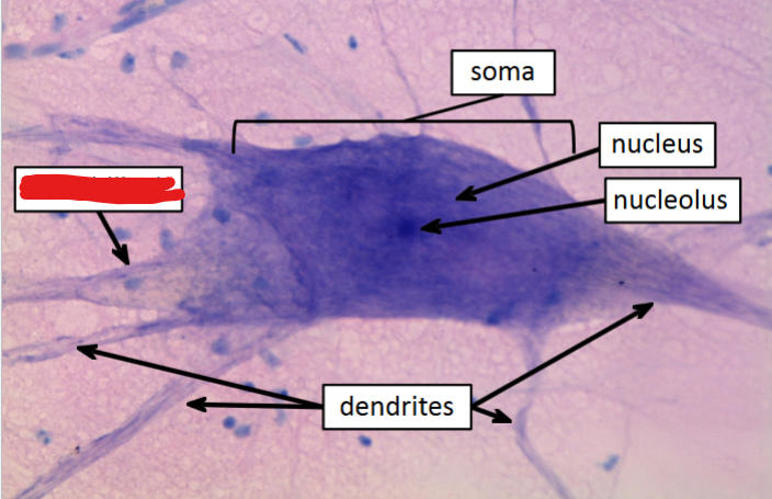
axon hillock
151
part of neuron 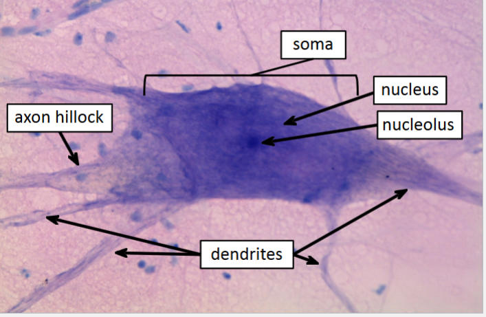
soma
152
also neuron 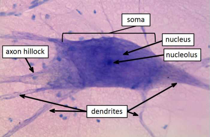
nucleus
153
neuron neuron 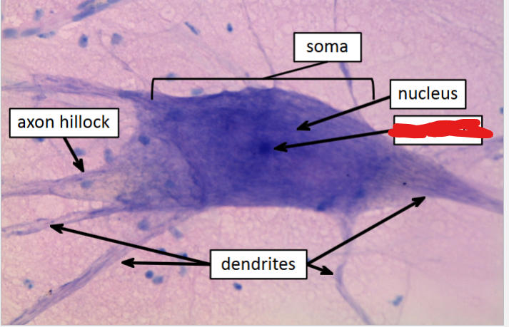
nucleolus
154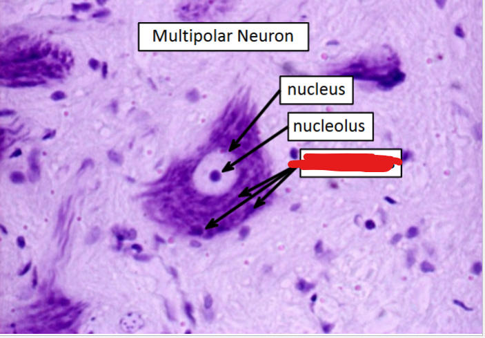
nissl bodies
155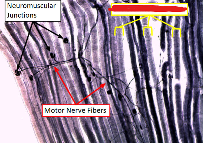
skeletal muscle fibers
156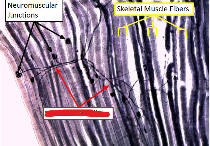
motor nerve fibers
157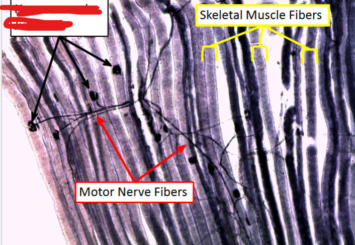
neuromuscular junctions
158
A closer up (put sigma to get correct) 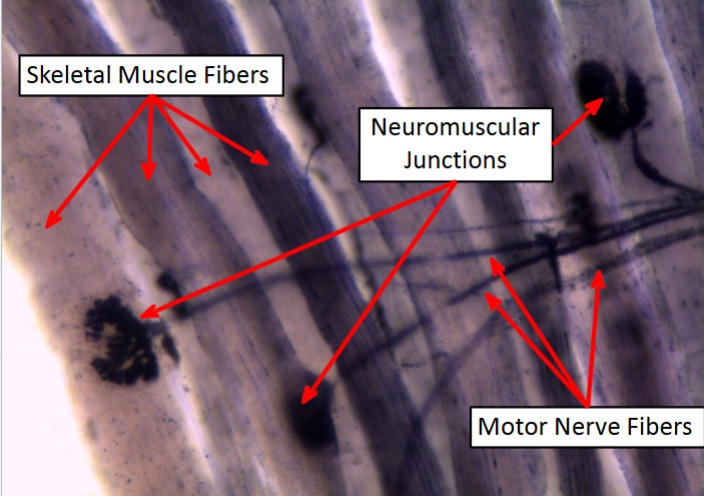
sigma