3.6.4 Homeostasis is the maintenance of a stable internal environment
Study these flashcardsFree · spaced repetition · iPhone, Android, Mac & web
All cards
1
Homeostasis?
Maintainance of an internal environment against external conditions
2
Negative feedback?
Mechanism used to restore any deviations from normal in a system back to its original state
3
What does the pancreas contain:
Has a structure called Islets of Langerhans which has:
- Alpha cells which release glucagon
- Beta cells which release insulin
- Alpha cells which release glucagon
- Beta cells which release insulin
4
What happends if a spike in glucose occurs?
1) Spike is detected by beta cells in islets of langerhan
2) Beta cells release insulin
3)Liver cells become permeable to glucose.
4)Glucose removed from blood and converted to glycogen
2) Beta cells release insulin
3)Liver cells become permeable to glucose.
4)Glucose removed from blood and converted to glycogen
5
Glycogenesis
Formation of glycogen from excess glucose mainly in liver
6
Glycogenolysis
Breaking down glycogen into glucose in liver
7
Gluconeogenesis?
Formation of glucose from non carbohydrate parts like amino acids in liver
8
Where does Osmoregulation occur?
In the nephrons which are long tubules surrounded by capillaries.
9
Pathway of osmoregulation
Renal artery > afferent arterioles > glomerelus > renal capsule/efferent arterioles(if big) > PCT > Loop of henle > DCT > CT
10
How insulin decreases spike in glucose?
-Attaches to receptors on target cell- changing shape of channel proteins (more permeable)
-More protein channels inserted
- Activating glycogenesis enzymes
-More protein channels inserted
- Activating glycogenesis enzymes
11
How glucagon increases glucose? (adrenaline does same except binds to adrenaline complimentary receptors)
- Attaches to receptors on target cells and:
1)Causes a protein to convert into adenylate cyclase which causes ATP to cyclic AMP (cAMP) which cAMP activates protein kinase. This hydrolyses glycogen to glucose
2)Activating gluconeogenesis enzymes
1)Causes a protein to convert into adenylate cyclase which causes ATP to cyclic AMP (cAMP) which cAMP activates protein kinase. This hydrolyses glycogen to glucose
2)Activating gluconeogenesis enzymes
12
Type 1 and 2 diabetes?
Type 1:
Body doesnt produce sufficient amounts of insulin
Type 2:
Receptors in target cells become less responsive to insulin
Body doesnt produce sufficient amounts of insulin
Type 2:
Receptors in target cells become less responsive to insulin
13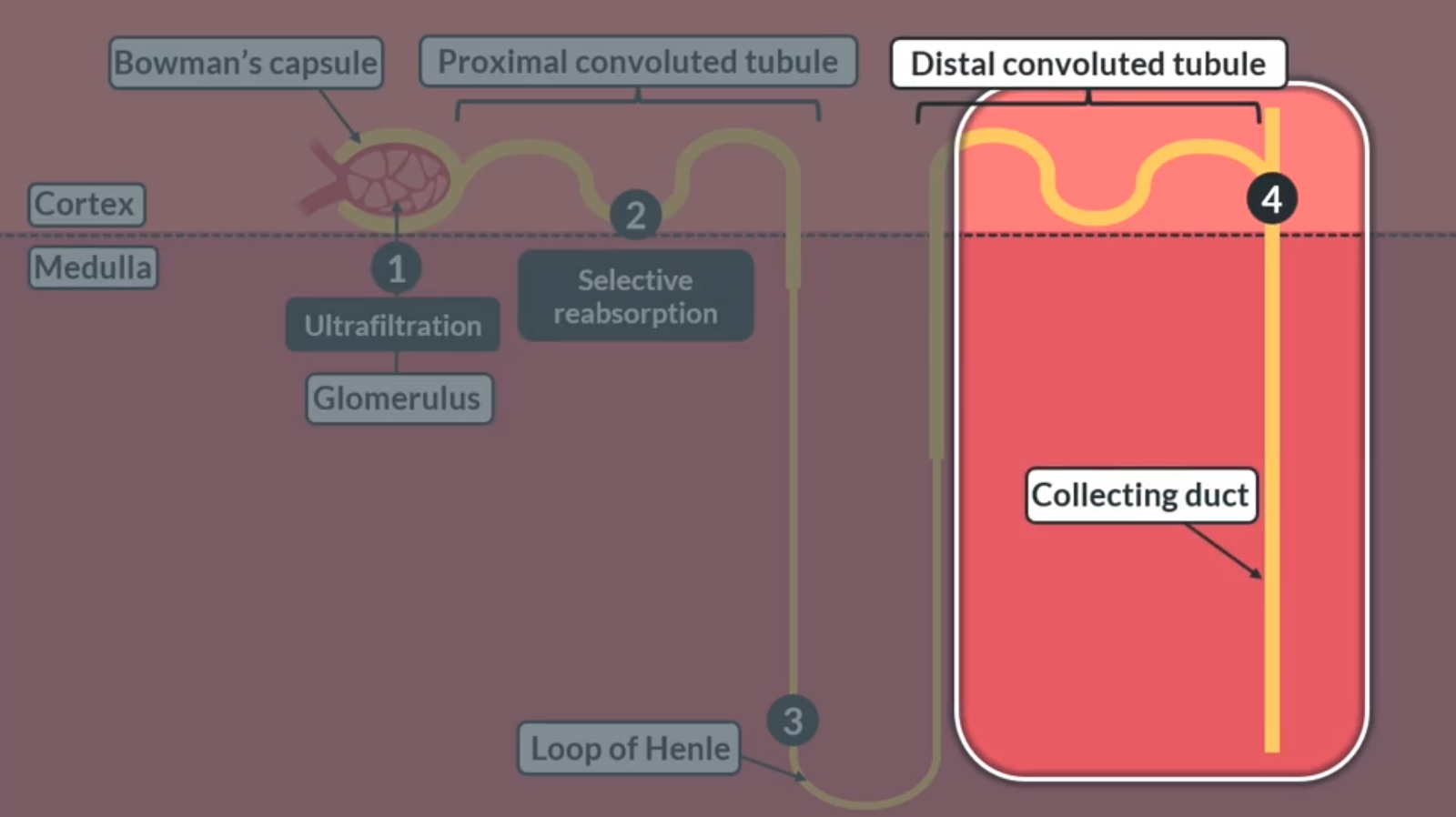
Image occlusion: name the hidden labels
14
Image occlusion: name the hidden labels
15
Image occlusion: name the hidden labels
16
Image occlusion: name the hidden labels
17
Image occlusion: name the hidden labels
18
Image occlusion: name the hidden labels
19
Image occlusion: name the hidden labels
20
How does ultrafiltration work/occur?
1) Blood from afferent arterioles enters glomerulus (lots of capillaries)
2) Since the afferent arterioles diameter is larger than efferent theres a build up of hydrostatic pressure
3) This pressure pushes the small molecules out into the renal capsule. Forming glomerulus filtrate. Large molecules cant fit inside renal capsule
2) Since the afferent arterioles diameter is larger than efferent theres a build up of hydrostatic pressure
3) This pressure pushes the small molecules out into the renal capsule. Forming glomerulus filtrate. Large molecules cant fit inside renal capsule
21
What three things do filtrate need to go through to enter the renal capsule?
1) Gaps between capilary endothelium cells
2) Basement membrane
3) Gaps between podocytes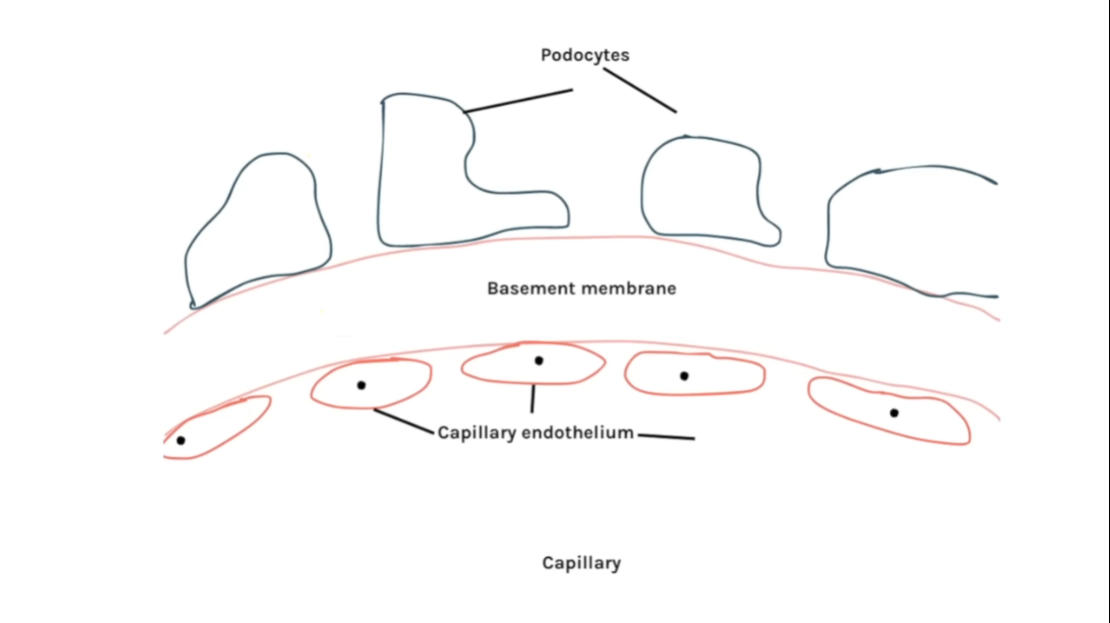
2) Basement membrane
3) Gaps between podocytes
22
Selective reabsorption?
1)Na ions are actively pumped out of PCT into capillaries to decrease conc in PCT
2)Na ions diffuse down into PCT by a co-transporter which also transports glucose against gradient
3)Glucose then facilitates diffuse into capillaries and is reabsorbed
2)Na ions diffuse down into PCT by a co-transporter which also transports glucose against gradient
3)Glucose then facilitates diffuse into capillaries and is reabsorbed
23
Descending loop?
Walls much thinner and is permeable to water
24
Ascending loop?
Walls much thicker and isn't permeable to water
25
Loop of henle stages?
1)Na ions are actively transported out of loop into interstitial area with ATP( from mitochondria)
2)This lowers the WP so water from descending loop diffuse out and reabsorbed by capillaries
3)At the base some Na ions diffuse out as bottom is now diluted
- WP is highest at cortex inside and lowers as you go down medulla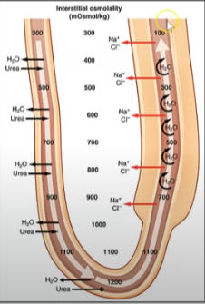
2)This lowers the WP so water from descending loop diffuse out and reabsorbed by capillaries
3)At the base some Na ions diffuse out as bottom is now diluted
- WP is highest at cortex inside and lowers as you go down medulla
26
Distal convuluted tubule and collecting duct?
1)Filtrate is very diluted (due to lots of Na ions being actively transported)
2)More water is diffused out of DCT however the amount is controlled by ADH
3)Urine is formed and passes out of collecting duct
2)More water is diffused out of DCT however the amount is controlled by ADH
3)Urine is formed and passes out of collecting duct
27
What happeneds if water potential of blood is too low?
1) Osmoreceptors detect this as water leaves causing it to shrivel
2)Hypothalamus releases ADH by pituitary gland
3)ADH binds to target cell(DCT/CT)
4) This causes DCT and collecting duct to become more permeable
5)More water is reabsorbed into blood and urine becomes more concentrated
2)Hypothalamus releases ADH by pituitary gland
3)ADH binds to target cell(DCT/CT)
4) This causes DCT and collecting duct to become more permeable
5)More water is reabsorbed into blood and urine becomes more concentrated
28
How does ADH actually make walls more permeable?
1)ADH binds to receptors on DCT/collecting duct
2)This causes phosphorylase to activate
3)Phosphorylase causes vesicles with aquaporins to fuse with membrane
2)This causes phosphorylase to activate
3)Phosphorylase causes vesicles with aquaporins to fuse with membrane
29
What are aquaporins?
Water protein channels