3.3.4 MASS TRANSPORT
Study these flashcardsFree · spaced repetition · iPhone, Android, Mac & web
All cards
1
whats haemoglobin
Made up of 4 polypeptide chains with each having a haem group and these have an iron ion which can bind with one oxygen
2
Affinity?
how likely haemoglobin is to load with oxygen
3
Loading(association)
binding of oxygen to haemoglobin
4
Unloading(dissociating)
releasing oxygen
5
Oxyhaemoglobin dissociation curve?
Expresses that oxygen is loaded in regions with high partial pressure of oxygen and is unloaded in regions with low partial pressure
6
Area with high partial pressure and low partial pressure?
High:Lungs
Low: respiring tissues
Low: respiring tissues
7
co-operative binding?
the binding of the first oxygen changes shape of haemoglobin making it easier for other oxygens to bind
8
The bohr effect?
high carbon dioxide concentration causes curve to shift to the right since affinity decreases as acidic CO2 changes shape of haemoglobin slightly
9
Different animals have different affinities in order to adapt to their surroundings eg:
Foetal and llamas: Have higher affinity
10
myoglobin?.
similiar to haemoglobin but its one haem group has very high affinity and acts as 02 reserve for muscles
11
Double circulatory system meaning?
blood passes through heart twice in one cycle. Once oxygenated and once deoxygenated
12
why double?
To control blood pressure:
In lungs pressure decreases to prevent damage to capillaries and give more time for gas exchange.
Oxygenated blood needs higher pressure to go around the body quicker
In lungs pressure decreases to prevent damage to capillaries and give more time for gas exchange.
Oxygenated blood needs higher pressure to go around the body quicker
13
Cardiac muscle?
Thick muscle in heart that is:
myogenic - can contract/ relax without any stimulation
Doesnt fatigue if it has oxygen
myogenic - can contract/ relax without any stimulation
Doesnt fatigue if it has oxygen
14
Coronary artieries
Supplies cardiac muscles with oxygenated blood
15
4 chambers
Left/ right atrium: Thinner elastic walls
Left/ right ventricle: Thicker walls to create pressure but left is thicker
Left/ right ventricle: Thicker walls to create pressure but left is thicker
16
Veins?(superior vena cava top and inferior bottom)
Vena cava - carries deoxygenated blood from body to right atrium
Pulmonary vein: Carries oxygenated blood from lungs to left atrium
Pulmonary vein: Carries oxygenated blood from lungs to left atrium
17
Arteires
Aorta: carries oxygenated blood from left ventricle to rest body
Pulmonary artery: carries deoxygenated blood from right ventricle to lungs
Pulmonary artery: carries deoxygenated blood from right ventricle to lungs
18
semi lunar valve and AV valves?
pulmonary valve: To pulmonary artery
aortic valve: To aorta
Tricuspid: right side of heart (biology wise)
Bicuspid: left side of heart (biology wise)
aortic valve: To aorta
Tricuspid: right side of heart (biology wise)
Bicuspid: left side of heart (biology wise)
19
septum
Wall that separates the deoxygenated blood from the oxygenated blood
20
Systole and Diastole meaning?
SYSTOLE = Period of contraction
DIASTOLE = Period of relaxation
DIASTOLE = Period of relaxation
21
Diastole?
-Blood enters RIGHT atrium increasing its volume and pressure
-Atrium and ventricles relax
-Atrium and ventricles relax
22
Atrial systole stages?
1)Walls of atria contract
2)This increases its pressure and decreases volume
3)Atriums pressure now higher than ventricles
4)AV valves open and blood moves into ventricles
2)This increases its pressure and decreases volume
3)Atriums pressure now higher than ventricles
4)AV valves open and blood moves into ventricles
23
Ventricular systole?
1)Walls of ventricle contract
2)This increases its pressure extremely and decreases volume
3)AV valves close and semi lunar valves opens
4)Blood moves into arteries and out of heart
2)This increases its pressure extremely and decreases volume
3)AV valves close and semi lunar valves opens
4)Blood moves into arteries and out of heart
24
Whats cardiac output?
Volume of blood that leaves ventricle per minute
Cardiac output = heart rate X stroke volume
Cardiac output = heart rate X stroke volume
25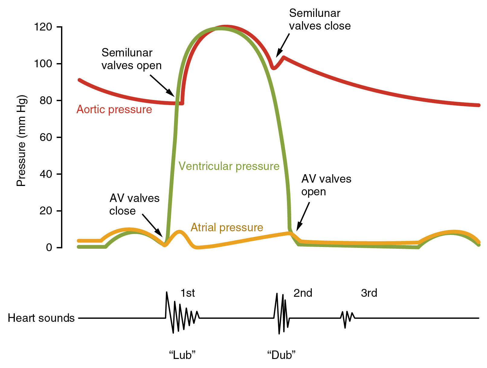
what does this graph show?
26
what is mass flow
Movement of fluid from region of high pressure to region of low pressure. Passive
27
What is tissue fluid
Liquid surrounding cells and is bridge between blood and cells
Contains water,glucose, oxygen, ions etc
Contains water,glucose, oxygen, ions etc
28
Ultrafiltration?
Process of which small molecules pass out of arterial end by high hydrostic pressure from high water potential to low water potential. Large molecules stay.
29
Reabsorption? (tissue fluid)
Pressure is low in venous end and has low water potential due to proteins(etc) so tissue fluid which now has waste products diffuses back in capillaries
30
What happeneds to left over tissue fluid
Excess tissue fluid is absorbed by lymphatic vessels which returns it back to circulatory system to the veins
31
whats oedema
swelling caused by excess tissue fluid
32
How can it occur?
Malnutrition(decrease protein conc)
Increased blood pressure
Damaged capillaries(increase protein conc)
Blockage of lymph vessels
Increased blood pressure
Damaged capillaries(increase protein conc)
Blockage of lymph vessels
33
What is transpiration?
evaporation of water from plant leaves
34
What affects transpiration
Humidity
Air flow
Light intensity
Temperature
Air flow
Light intensity
Temperature
35
COHESION TENSION THEORY
Theory of how water can move from roots to leaves due to COHESION,ADHESION AND ROOT PRESSURE
36
Movement of water up xylem
1)Water evaporates out of stomata decreasing pressure
2)This leads to more water pulling up
3)Since they're cohesive it forms a column of water in xylem
4)Water also adheres to xylem helping to pull upwards and this creates tension
2)This leads to more water pulling up
3)Since they're cohesive it forms a column of water in xylem
4)Water also adheres to xylem helping to pull upwards and this creates tension
37
Phloem tissue components:
Sieve tube elements
Companion cells- provide ATP
Companion cells- provide ATP
38
Translocation stages?
1)Source cell has lots of sucrose so FD into companion cell
2)H+ is actively transported into cell walls by companion cell
3)Forms gradient so H+ diffuse by co transporter which also transports sucrose into sieve tube elements
4)Increase of sucrose in sieve tube decreases WP and so water enters from xylem
5)This increases hydrostatic pressure pushing sucrose down
6)Sucrose actively transported into sink cell, decreasing WP
7)Results in osmosis of some water in sink cell decreasing hydrostatic pressure
2)H+ is actively transported into cell walls by companion cell
3)Forms gradient so H+ diffuse by co transporter which also transports sucrose into sieve tube elements
4)Increase of sucrose in sieve tube decreases WP and so water enters from xylem
5)This increases hydrostatic pressure pushing sucrose down
6)Sucrose actively transported into sink cell, decreasing WP
7)Results in osmosis of some water in sink cell decreasing hydrostatic pressure
39
Arterioles?
Smaller than arteries and connect arteries to capillaries
40
Capillaries?
Connect arterioles to veins
41
Arteries structure?
Thick muscle layer so can constrict/dilate to control volume of blood flow(WITHSTAND HIGH BLOOD PRESSURE)
Thick elastic walls compared to veins to maintain high blood pressure so blood can reach all of the body BY stretching and recoiling(bounce back)
No valves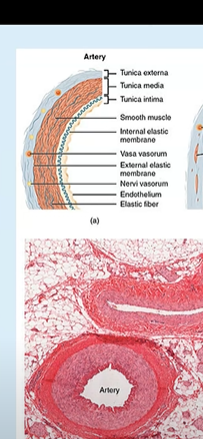
Thick elastic walls compared to veins to maintain high blood pressure so blood can reach all of the body BY stretching and recoiling(bounce back)
No valves
42
Veins structure?
Thin muscle layer so can't control blood flow(as they carry blood away from tissues)
Thin elastic walls as blood pressure is alot lower
Has valves
Thin elastic walls as blood pressure is alot lower
Has valves
43
Capillaries structure?
No muscle and elastic layer so thin membrane so fast diffusion
Lots of them and branches so high SA
Narrow lumen barely fitting RBC lowering blood flow
Lots of them and branches so high SA
Narrow lumen barely fitting RBC lowering blood flow
44
Arterioles? Strucute
Thicker muscle layer(compared to arteries) to restrict blood flow more to the capillaries
Thinner elastic layer as pressure is lower
No valves
Thinner elastic layer as pressure is lower
No valves