Biological Systems
Study these flashcardsFree · spaced repetition · iPhone, Android, Mac & web
All cards
1
THE BODY LIKES HOMEOSTASIS
IF SOMETHING CHANGES THE BODY WILL ATTEMPT TO COMPENSATE AND CORRECT THAT CHANGE
2
nervous system
control and integration of other body systems
adaptive capabilities- homeostasis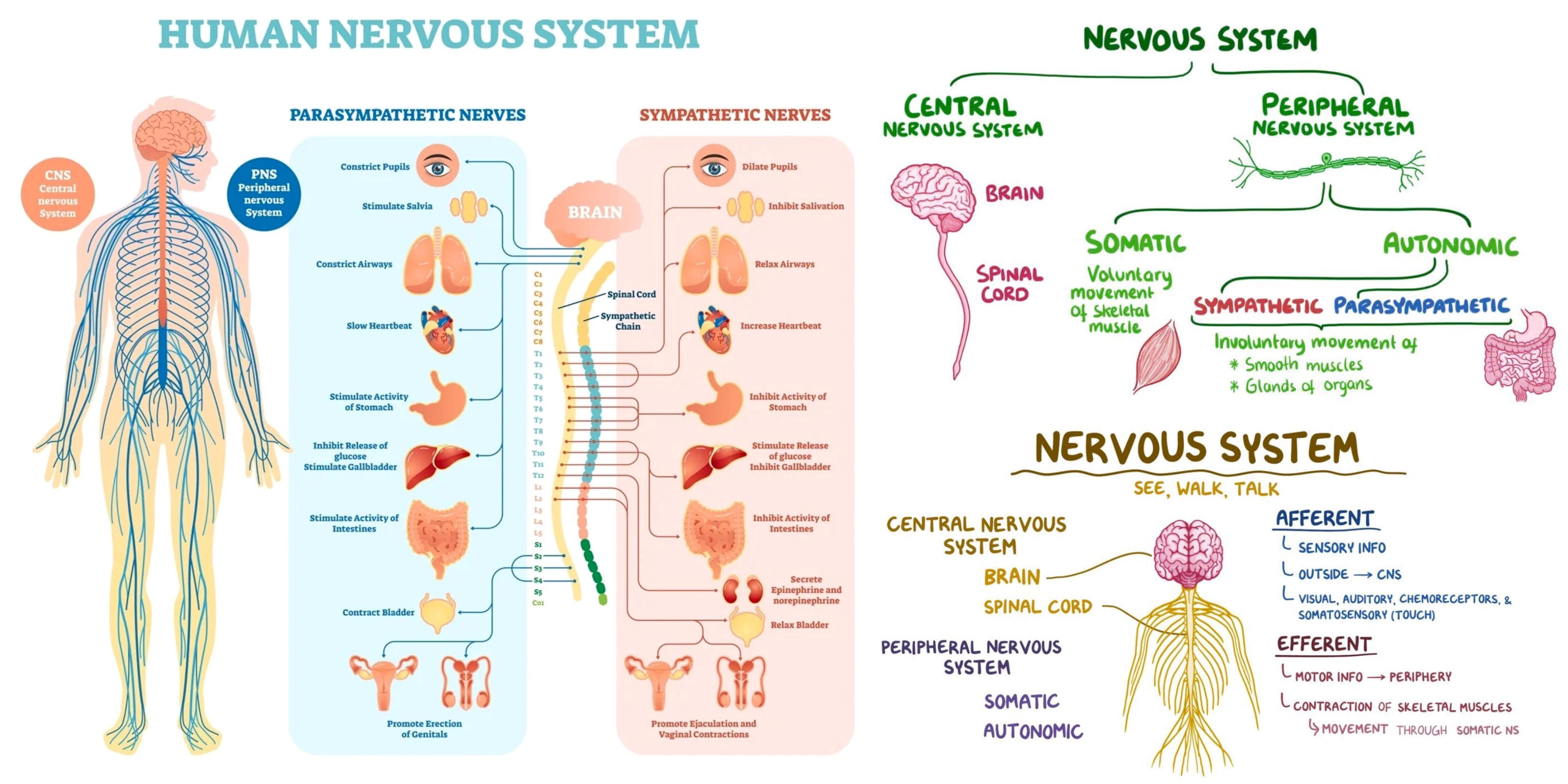
adaptive capabilities- homeostasis
3
cell body
part of neuron containing nucleus, soma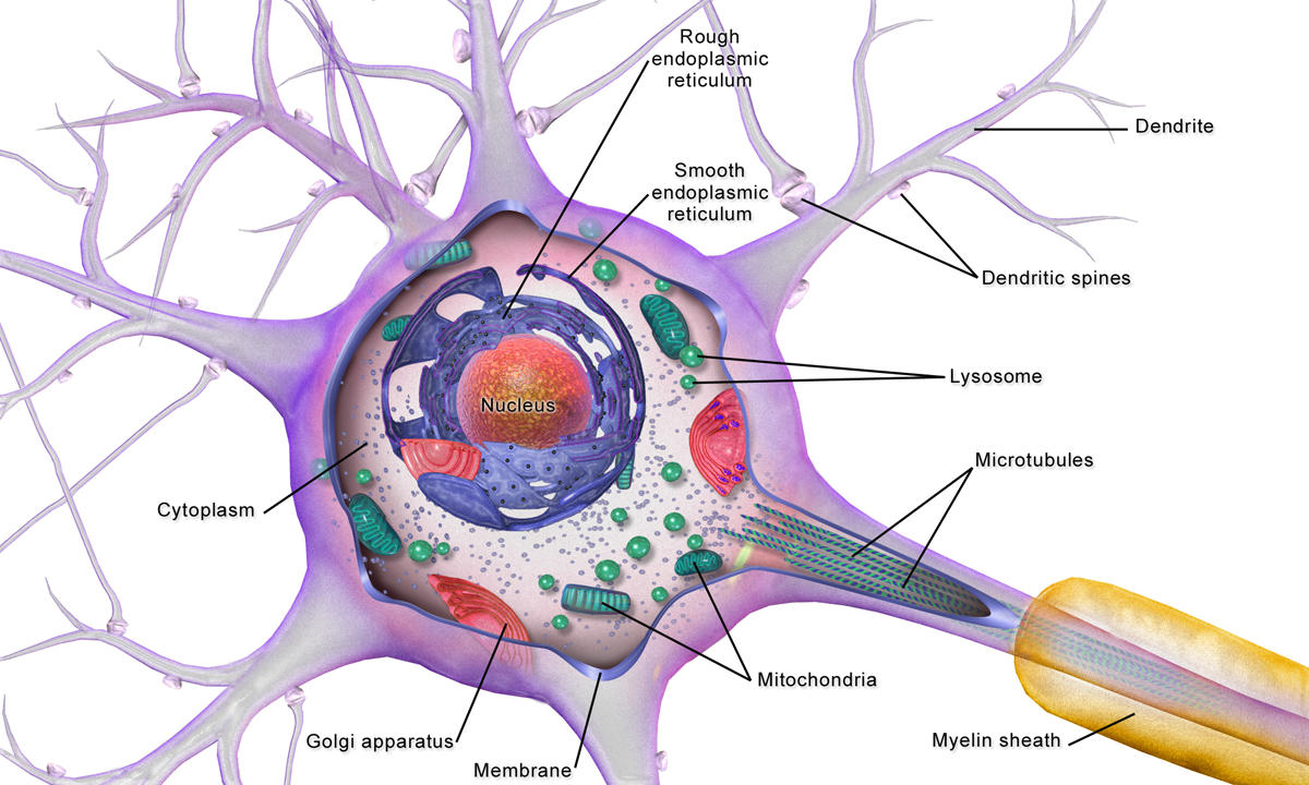
4
axon
part of neuron that sends impulse towards synapse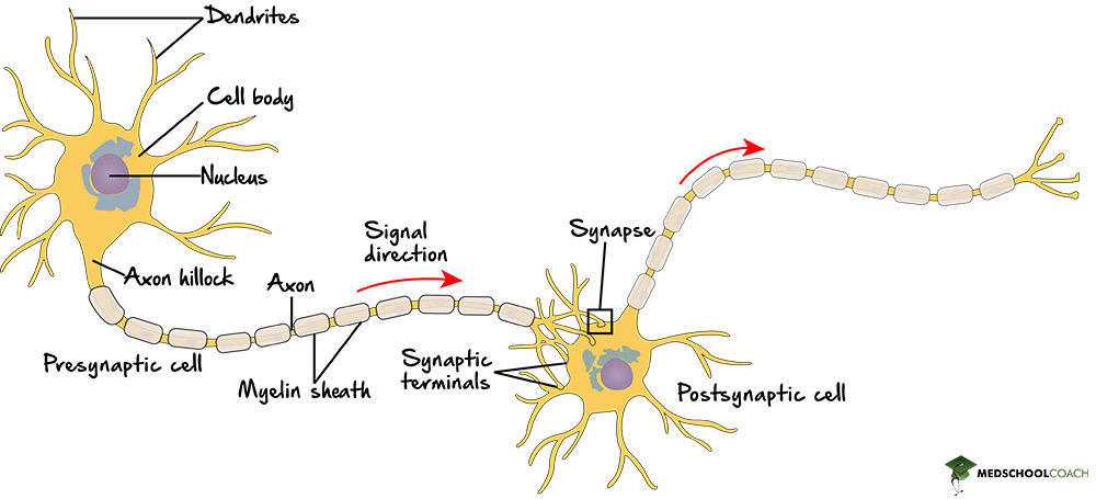
5
dendrites
parts of neuron that receive impulse
bipolar neuron- have only one dendrite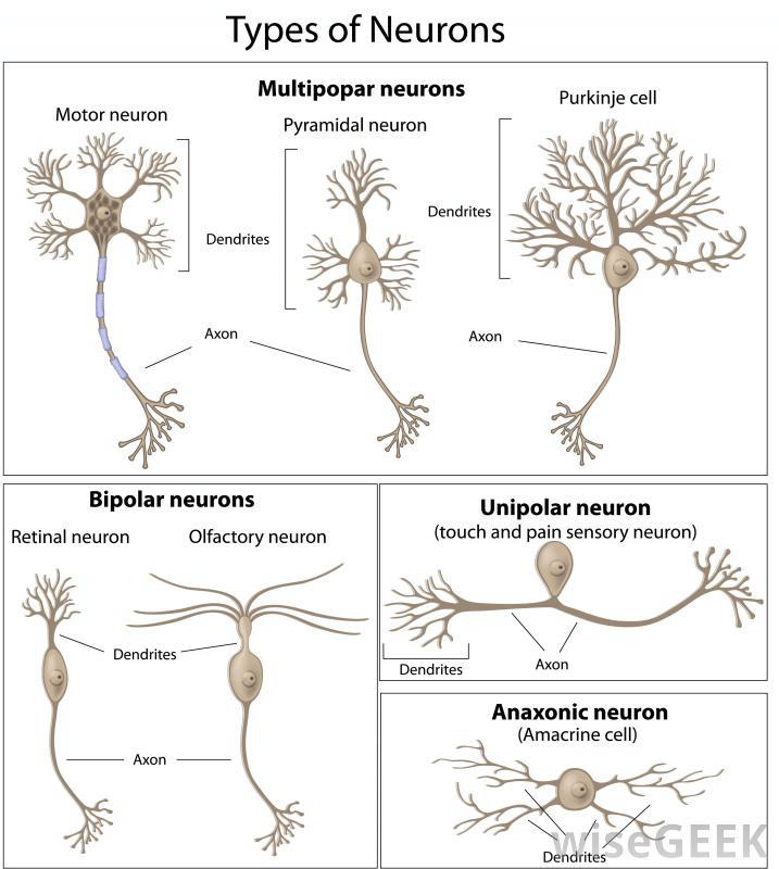
bipolar neuron- have only one dendrite
6
axon hillock
part of neuron that has highest concentration of voltage-gated Na+ channels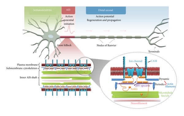
7
myelin sheath
part of neuron that insulates, decreasing surface area to be depolarized and increasing speed of conduction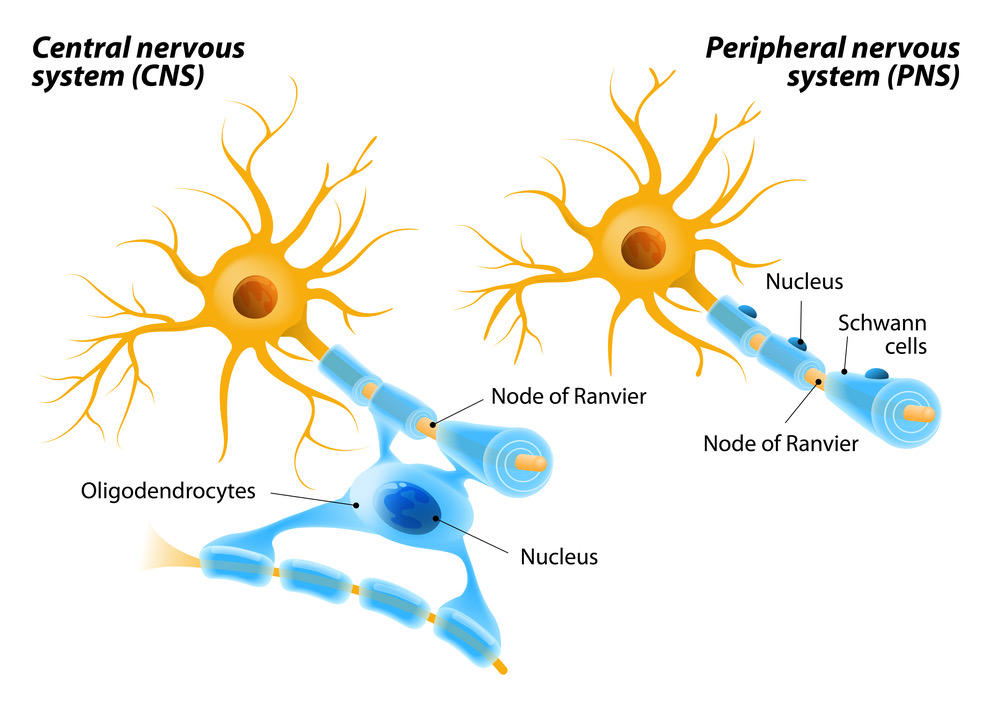
8
synaptic knobs
part of neuron that receives impulse and releases neurotransmitters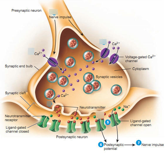
9
Nodes of Ranvier
part of neuron between myelin sheaths that concentrate the ion channels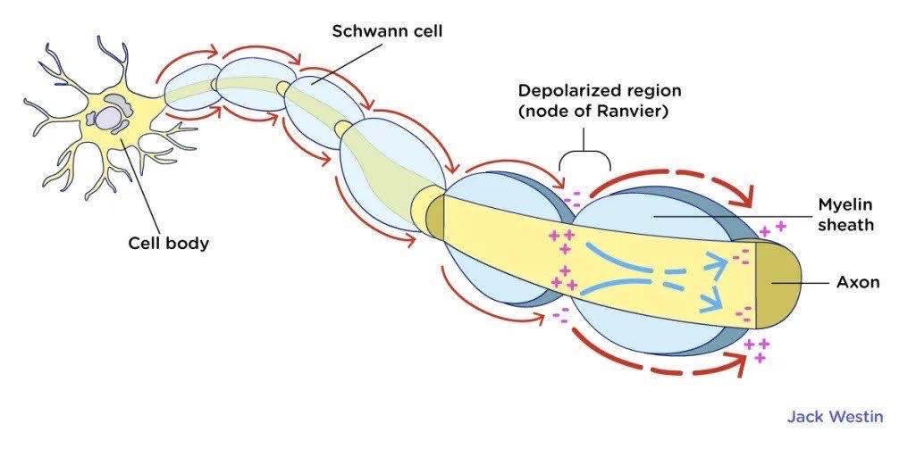
10
resting membrane potential
polarized to -70mV, where the interior of cell is negatively charged
maintained by two things:
1. Na+/K+ pumps- pump 3 Na+ out, 2 K+ in
2. K+ leak channels- allows K+ to leak out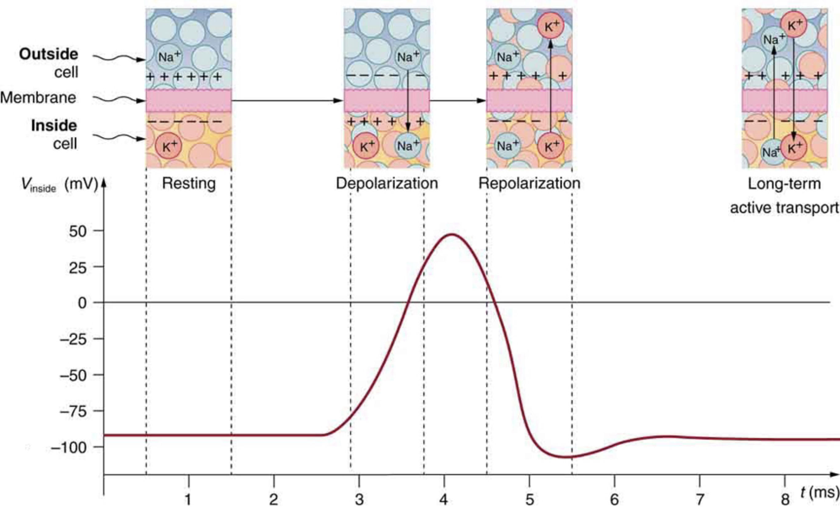
maintained by two things:
1. Na+/K+ pumps- pump 3 Na+ out, 2 K+ in
2. K+ leak channels- allows K+ to leak out
11
action potential
a neural impulse, a brief electrical charge that travels down an axon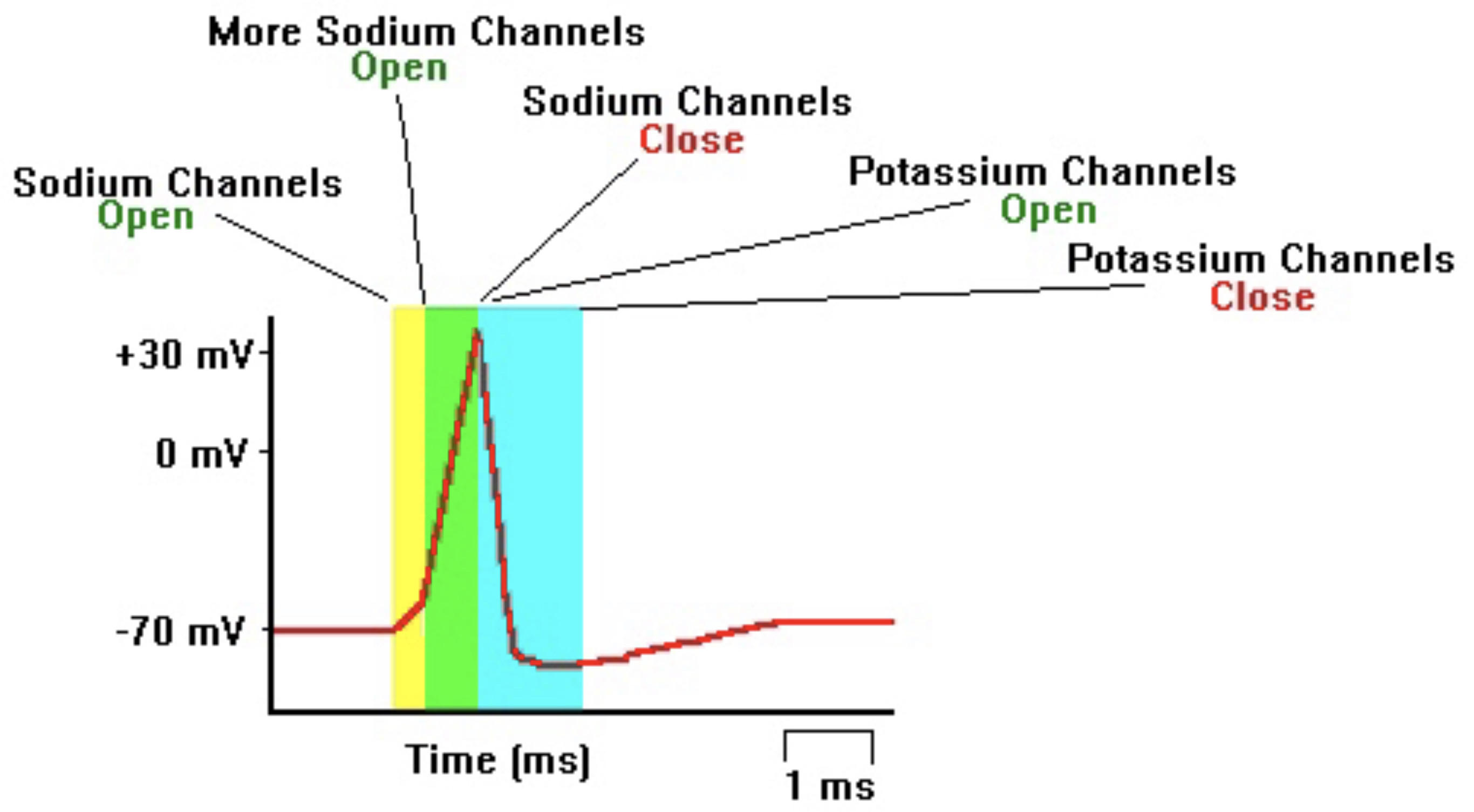
12
depolarization (action potential)
voltage-gated Na+ channels- when membrane potential changes, they open to allow Na+ to flow into cell
they only open at threshold potential of -50 mV
bringing the cell to about +35 mV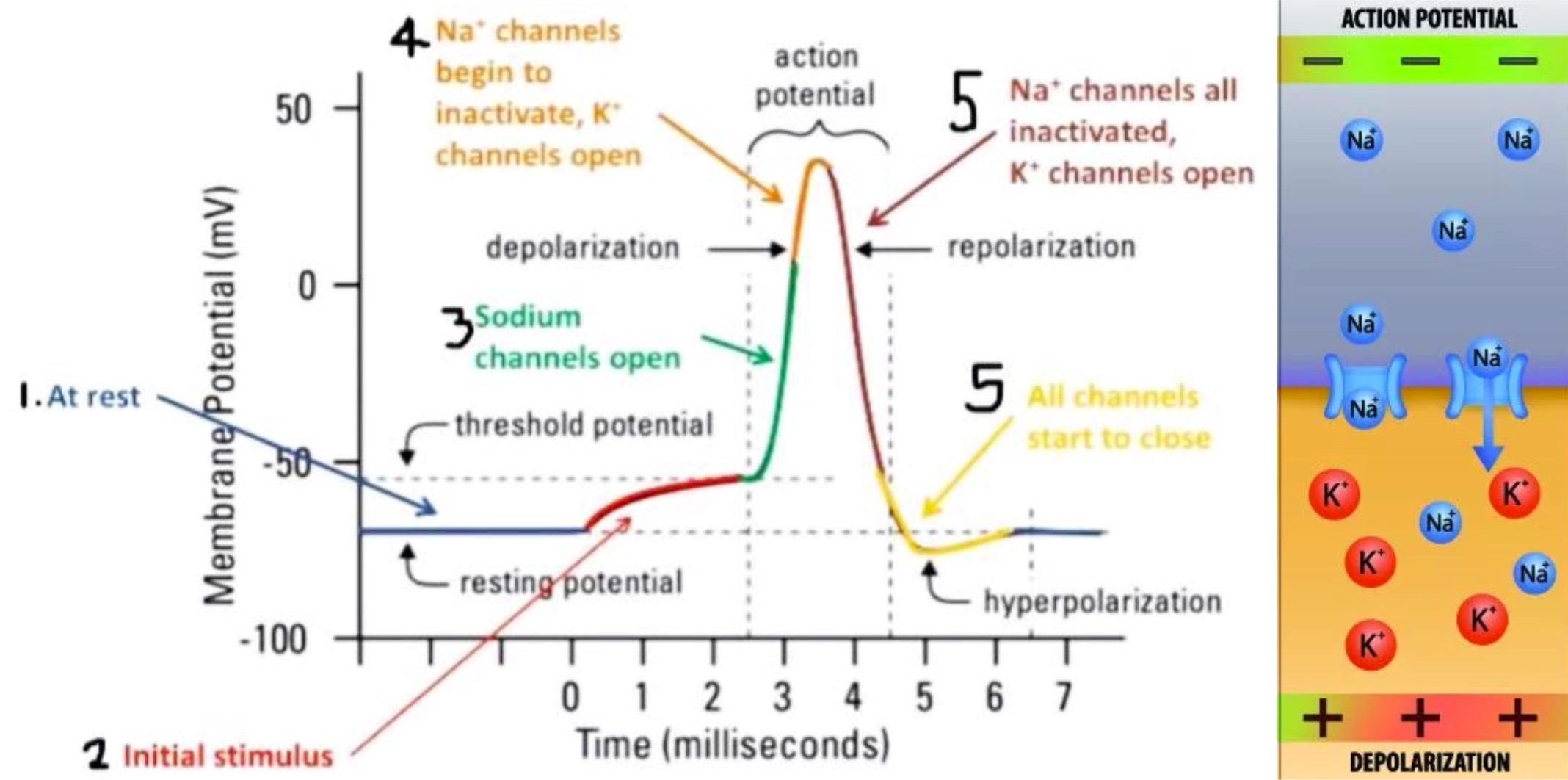
they only open at threshold potential of -50 mV
bringing the cell to about +35 mV
13
repolarization (action potential)
voltage-gated Na+ channels inactivate after opening
voltage-gated K+ channels open slowly in response to depolarization, allowing K+ to flow out of cell
this brings cell to -90 mV, which slowly returns to -70mV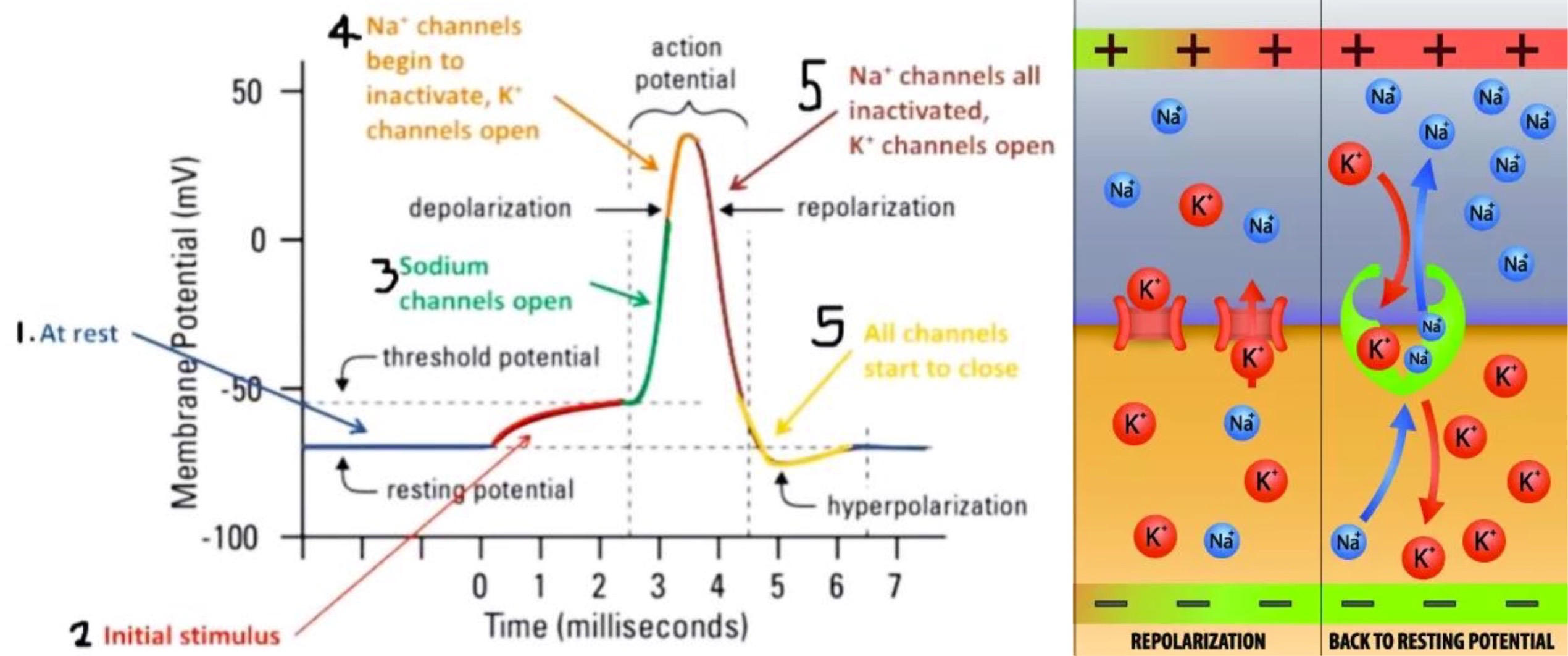
voltage-gated K+ channels open slowly in response to depolarization, allowing K+ to flow out of cell
this brings cell to -90 mV, which slowly returns to -70mV
14
saltatory conduction
Schwann cells and oligodendrocytes wrap axons with myelin, leaving spaces for nodes of Ranvier
forces action potential to jump from node to node, speeding up conduction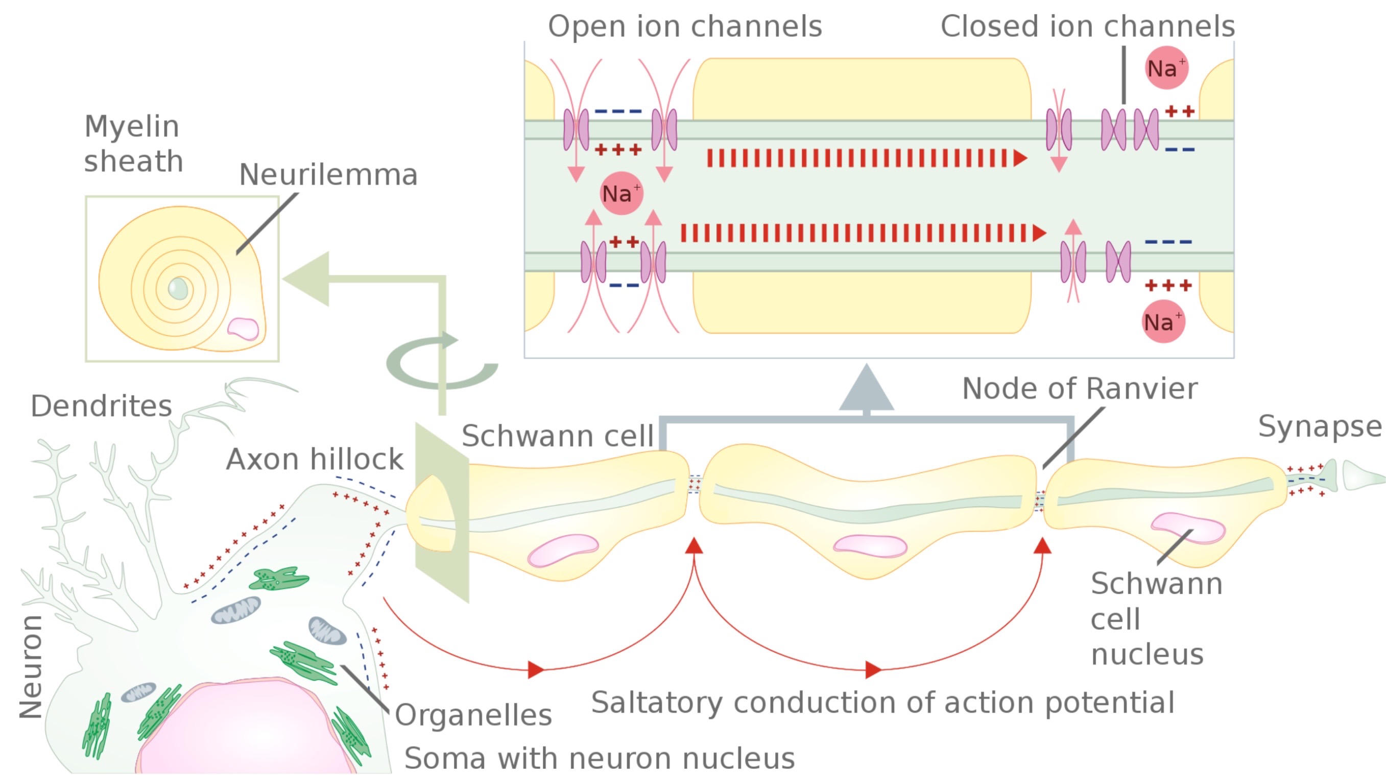
forces action potential to jump from node to node, speeding up conduction
15
refractory period
absolute refractory period- Na+ channels are inactivated when cells are too positive, K+ channels open, near Na+ equilibrium potential
relative refractory period- Na+ channels switch from inactivated to closed (deinactivated) when cells are too negative, K+ channels close as well, near K+ equilibrium potential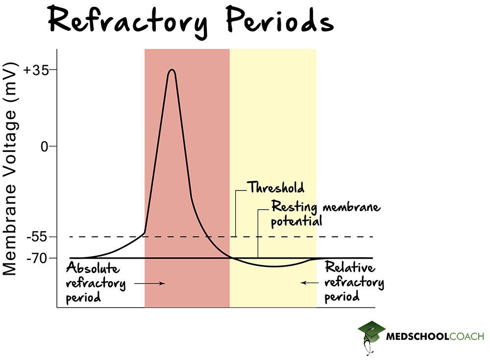
relative refractory period- Na+ channels switch from inactivated to closed (deinactivated) when cells are too negative, K+ channels close as well, near K+ equilibrium potential
16
electrical synapse
gap junction between two cells, bidirectional flow of impulse, rare but important in cardiac muscle cells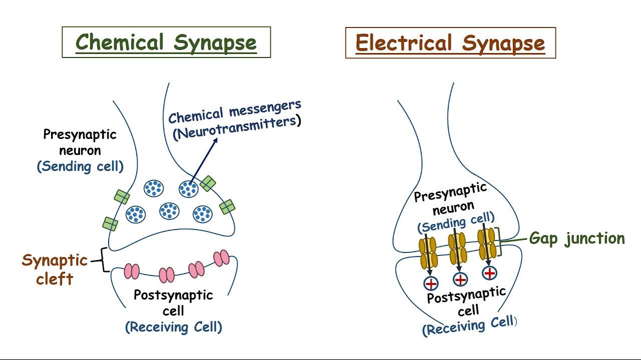
17
chemical synapse
steps:
1. depolarization opens voltage-gated Ca2+ channels
2. Ca2+ influx causes exocytosis of neurotransmitters in secretory vesicles
3. neurotransmitters open ligand-gated Na+ channels on motor endplate
4. Na+ influx triggers action potential
neuromuscular junction- acetylcholine is neurotransmitter
other NTs are GABA, serotonin, dopamine, norepinephrine
1. receptor determines effect, not NT
2. neurons make only one type of NT, can respond to many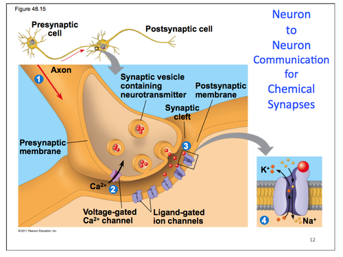
1. depolarization opens voltage-gated Ca2+ channels
2. Ca2+ influx causes exocytosis of neurotransmitters in secretory vesicles
3. neurotransmitters open ligand-gated Na+ channels on motor endplate
4. Na+ influx triggers action potential
neuromuscular junction- acetylcholine is neurotransmitter
other NTs are GABA, serotonin, dopamine, norepinephrine
1. receptor determines effect, not NT
2. neurons make only one type of NT, can respond to many
18
signal summation
action potential is an "all or nothing" response, with the only regulated step being whether the action potential will fire
excitatory postsynaptic potential- depolarize the next neuron, increases chance of action potential firing
inhibitory postsynaptic potential- polarize the next neuron, decreases chance of action potential firing
spatial summation- effect from MULTIPLE presynaptic cells is enough EPSP to fire off postsynaptic cell
temporal summation- SINGLE presynaptic cell, if EPSP are fast enough to fire off postsynaptic cell
tetanus- too many signals building up, blocks inhibitory neurons from releasing neurotransmitters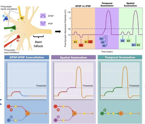
excitatory postsynaptic potential- depolarize the next neuron, increases chance of action potential firing
inhibitory postsynaptic potential- polarize the next neuron, decreases chance of action potential firing
spatial summation- effect from MULTIPLE presynaptic cells is enough EPSP to fire off postsynaptic cell
temporal summation- SINGLE presynaptic cell, if EPSP are fast enough to fire off postsynaptic cell
tetanus- too many signals building up, blocks inhibitory neurons from releasing neurotransmitters
19
astrocytes
guide neuron development, regulate neurotransmitters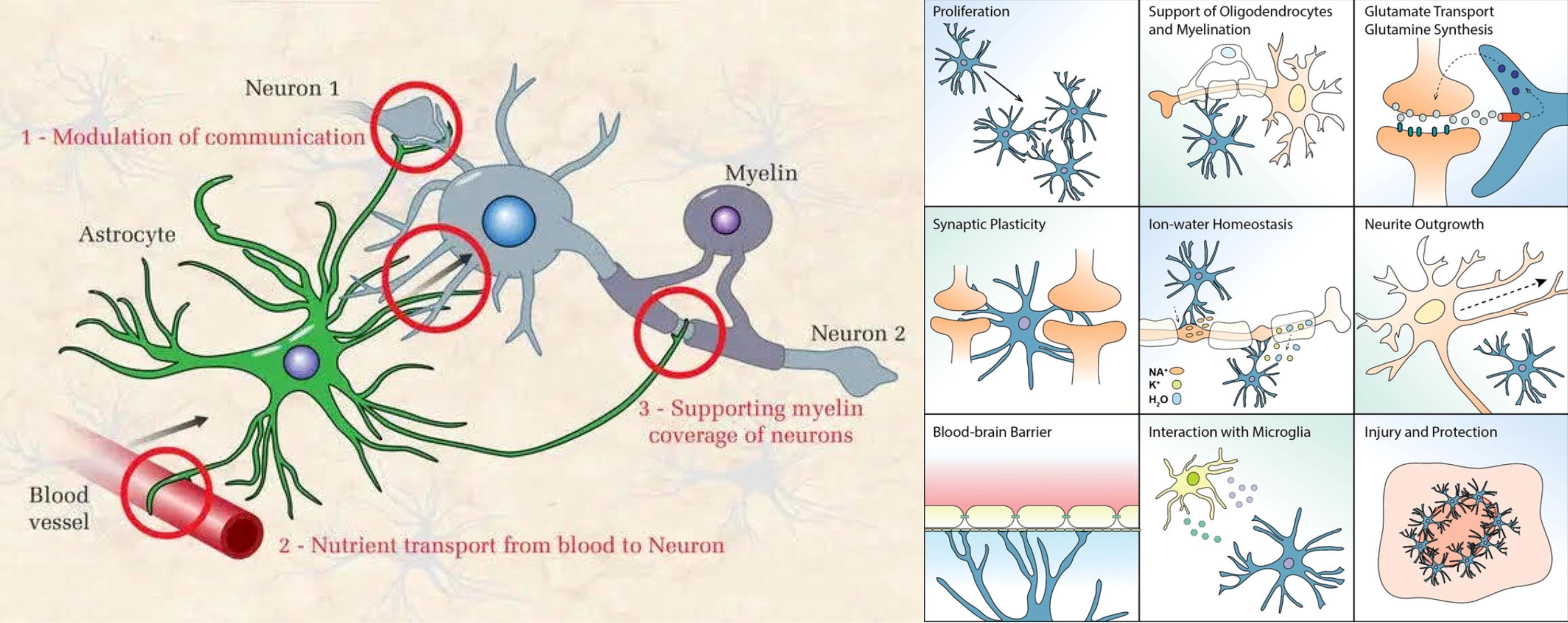
20
microglia
immune system of CNS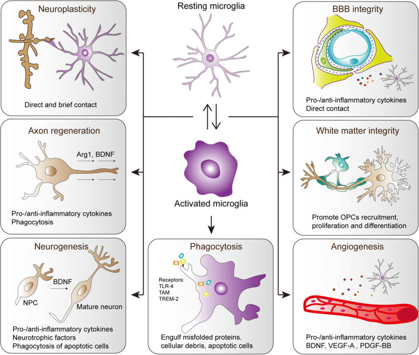
21
ependymal cells
produce and circulate cerebrospinal fluid
one of the 4 glial cells/central neuroglia of the CNS; specialized type of epithelial cells
found lining the inner surface of the ventricles of the brain and the central canal of the spinal cord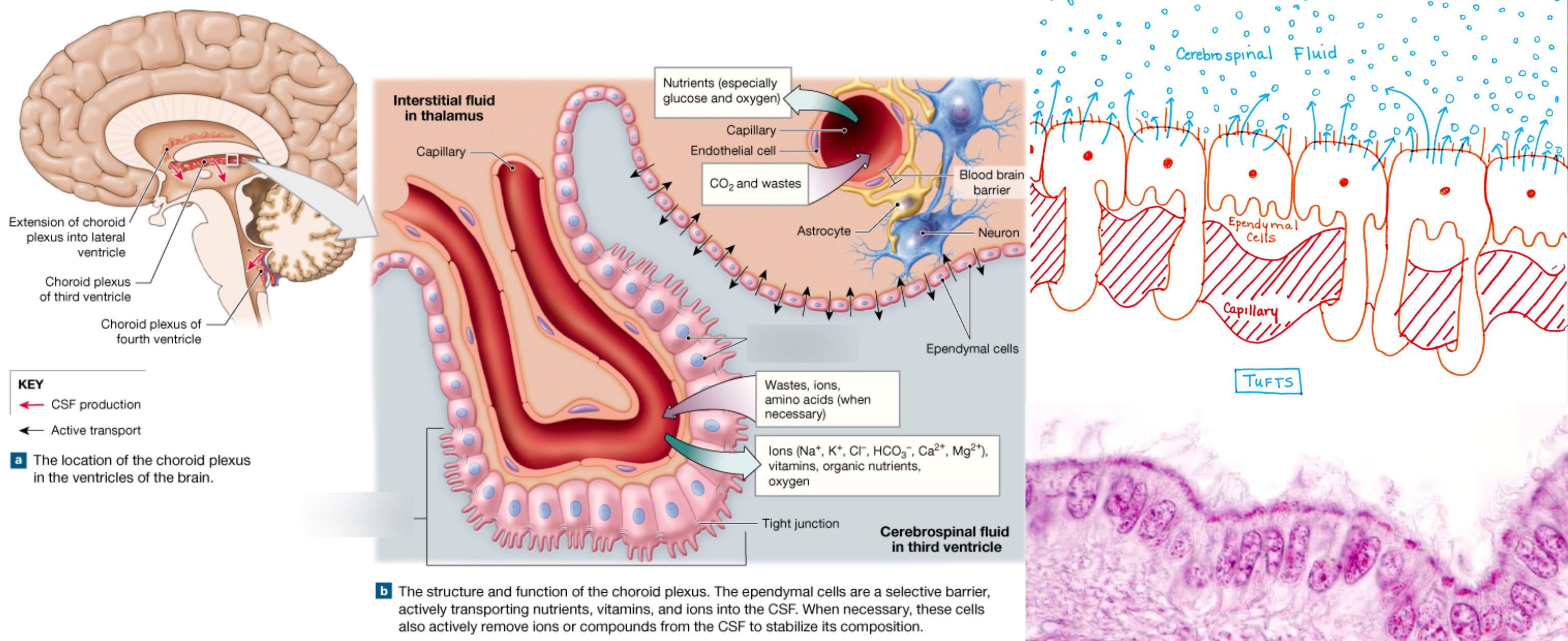
one of the 4 glial cells/central neuroglia of the CNS; specialized type of epithelial cells
found lining the inner surface of the ventricles of the brain and the central canal of the spinal cord
22
afferent/efferent/interneurons
afferent neurons- sensory neurons, to CNS
efferent neurons- motor/effector neurons, from CNS
interneurons- connects afferent and efferent neurons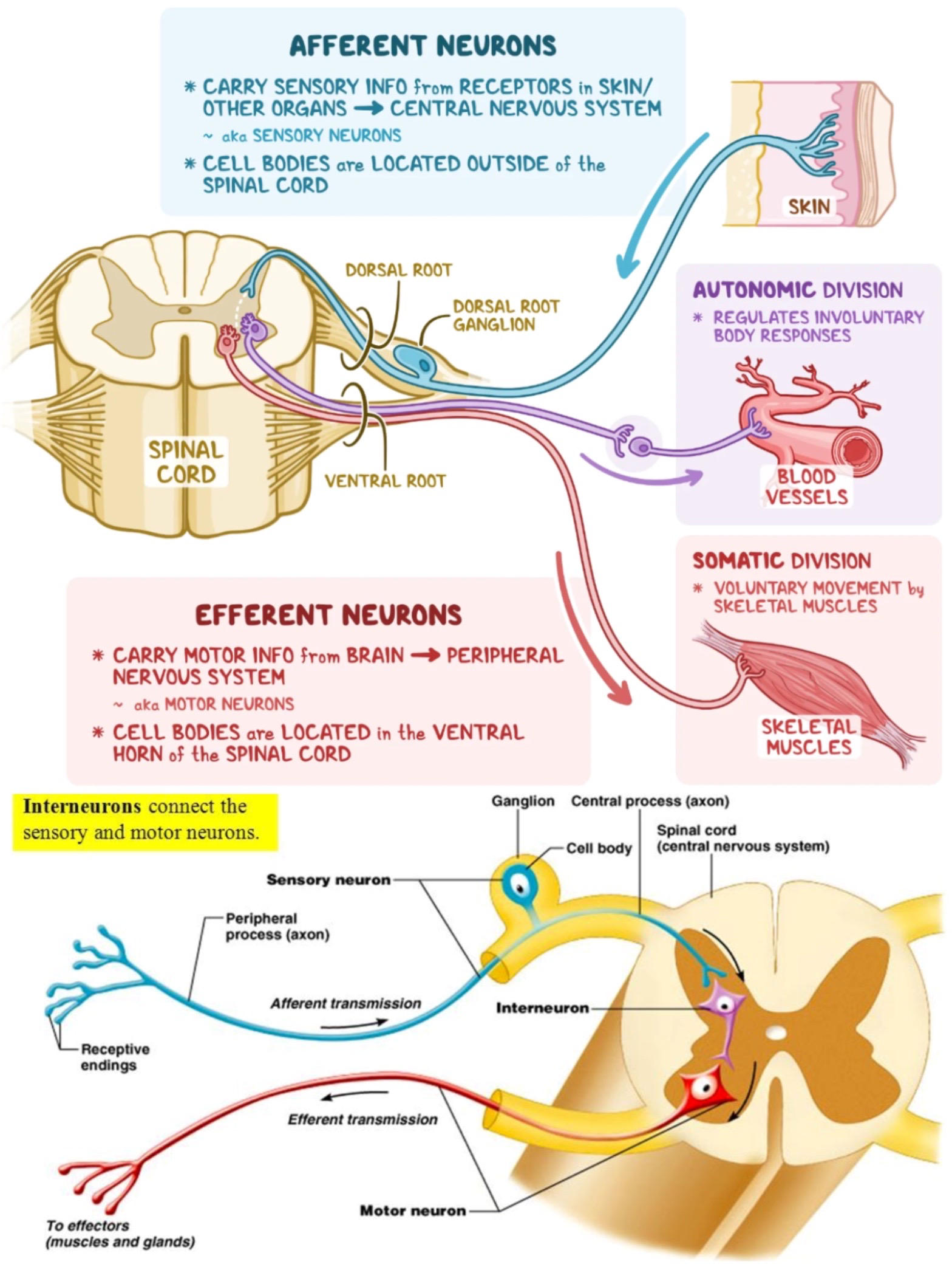
efferent neurons- motor/effector neurons, from CNS
interneurons- connects afferent and efferent neurons
23
reflexes
monosympatic reflex arc- sensory neuron directly connects to motor neuron in the spinal cord
reciprocal inhibition- to contract and reflex muscle pairs
supraspinal circuit- involves input from the brain or brainstem to process a stimuli, unlike most reflex arcs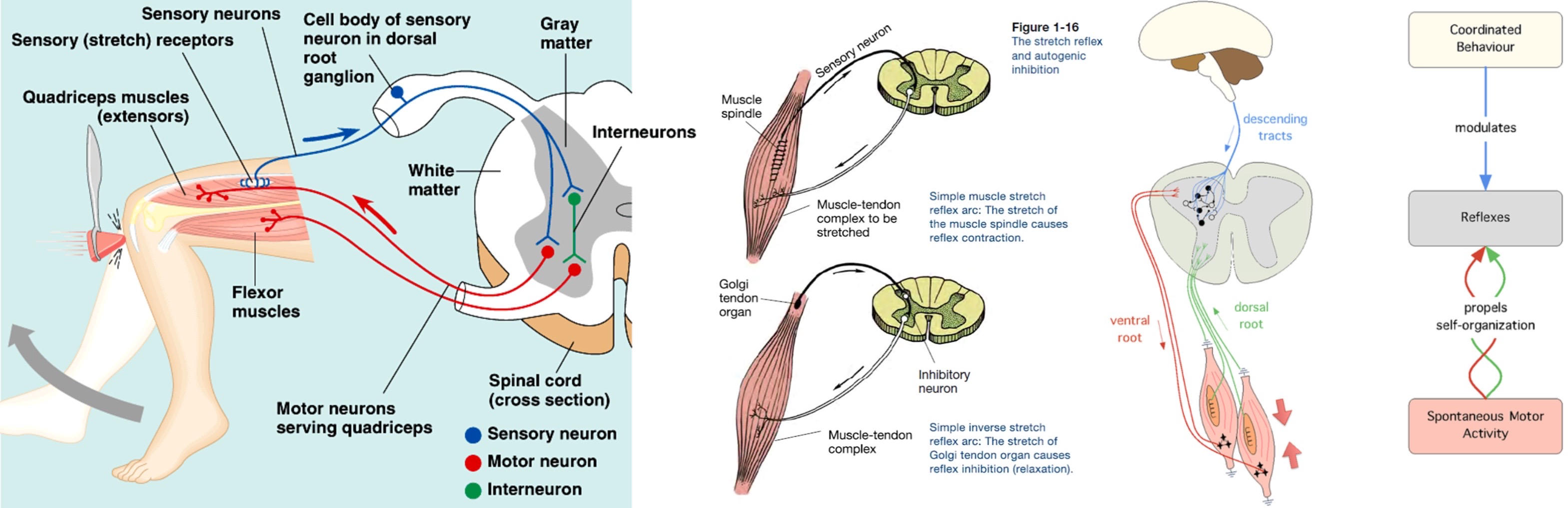
reciprocal inhibition- to contract and reflex muscle pairs
supraspinal circuit- involves input from the brain or brainstem to process a stimuli, unlike most reflex arcs
24
brain subdivisions
hindbrain- medulla, pons, cerebellum
midbrain- Reticular activating system (RAS)
forebrain- diencephalon, telencephalon (cerebral cortex)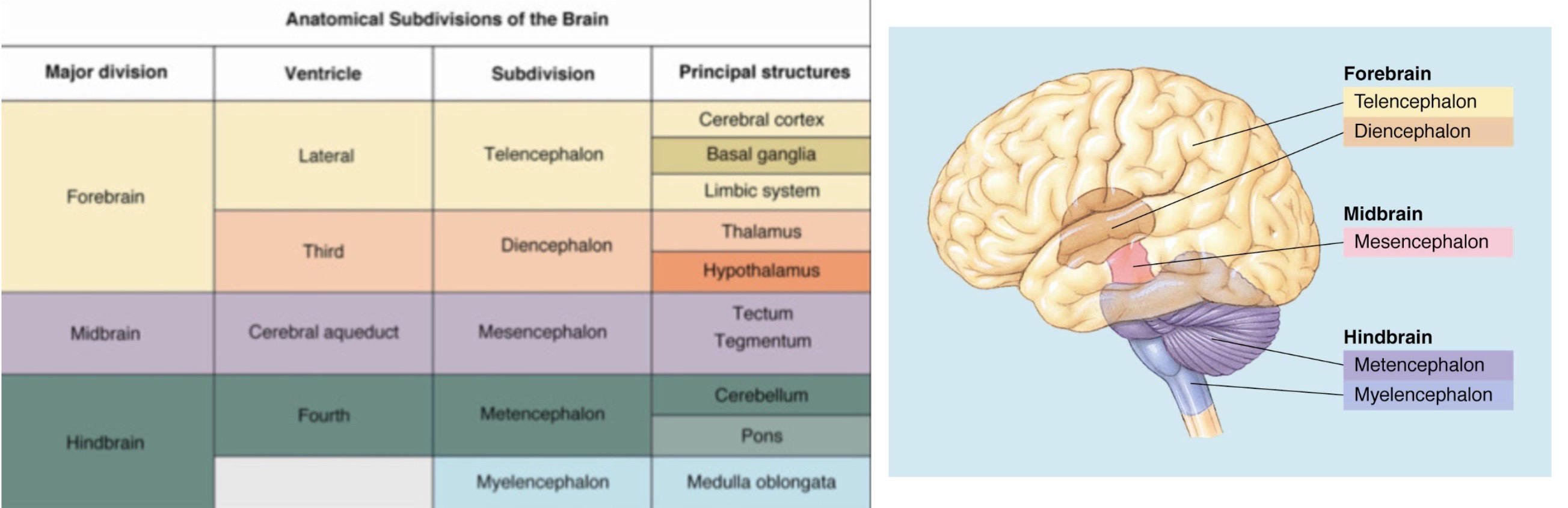
midbrain- Reticular activating system (RAS)
forebrain- diencephalon, telencephalon (cerebral cortex)
25
medulla oblongata
controls heartbeat and breathing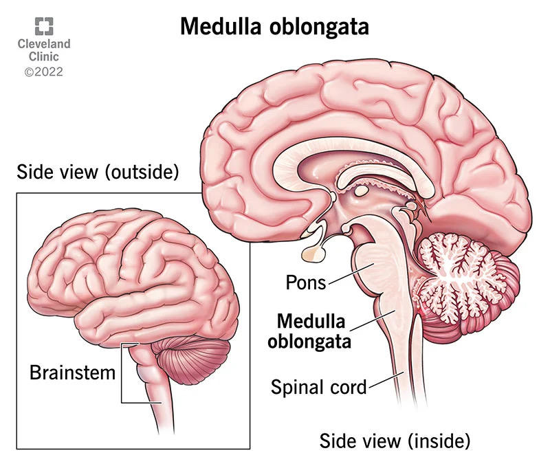
26
pons
coordinating movement and balance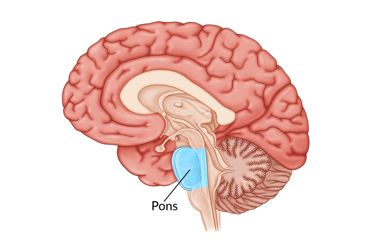
27
cerebellum
coordinating and smoothing out movement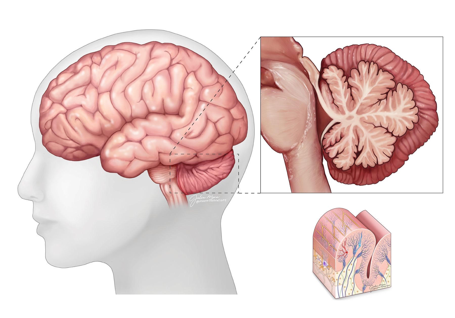
28
reticular activating system
arousal (wakefulness) and awareness 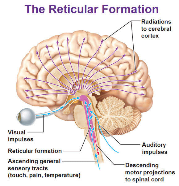
29
thalamus
relay point for all sensory information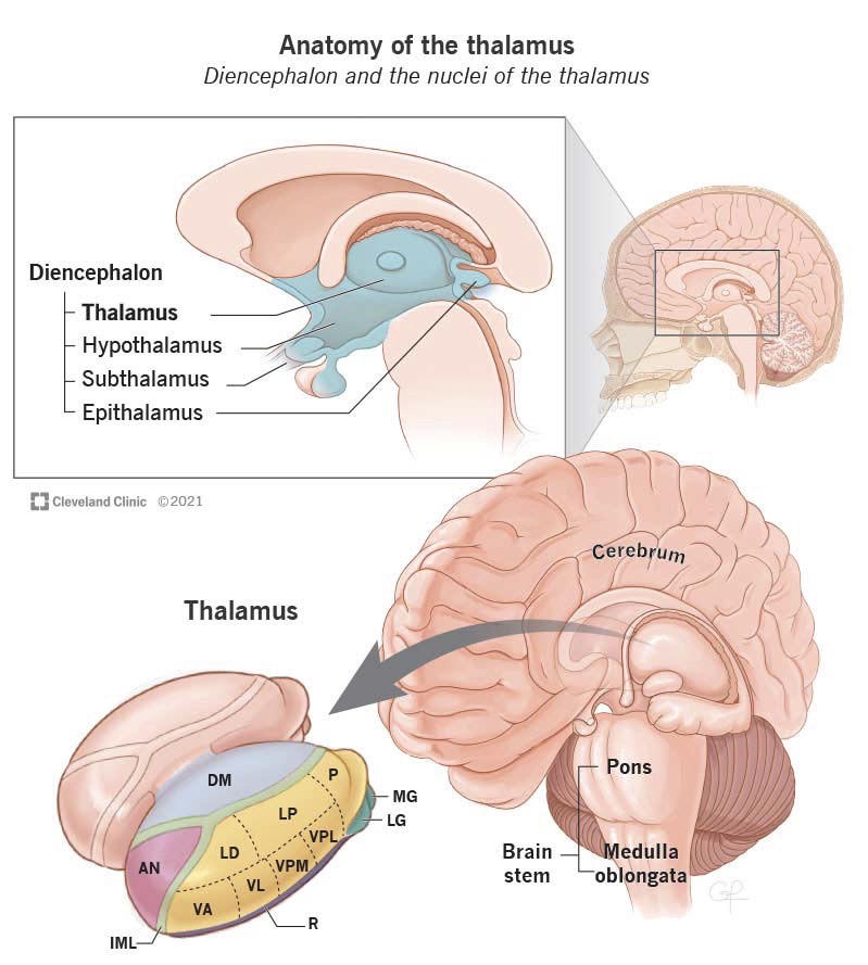
30
hypothalamus
maintain homeostasis, basic needs, controls endocrine system through pituitary gland, hormones affect mood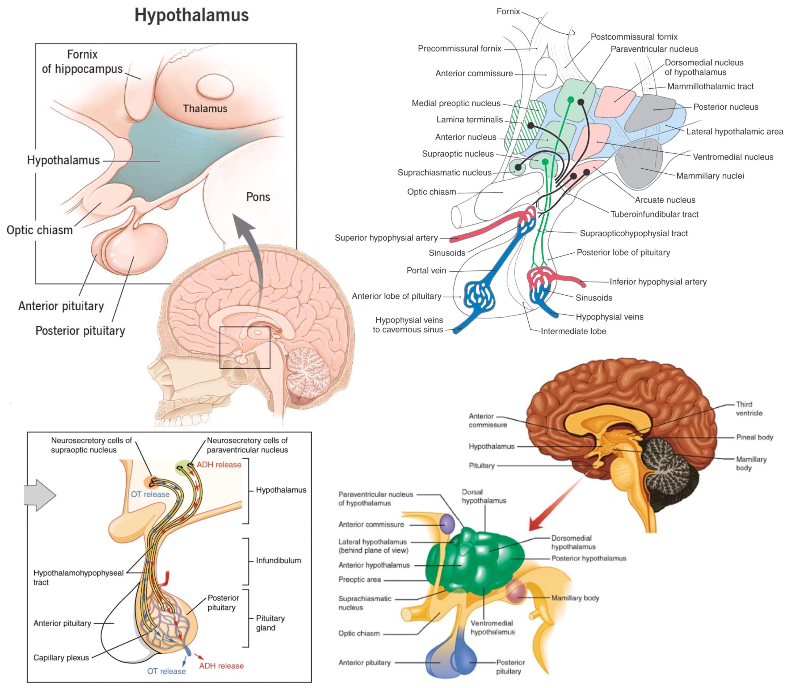
31
spinal cord
primitive reflexes and basic behaviors like walking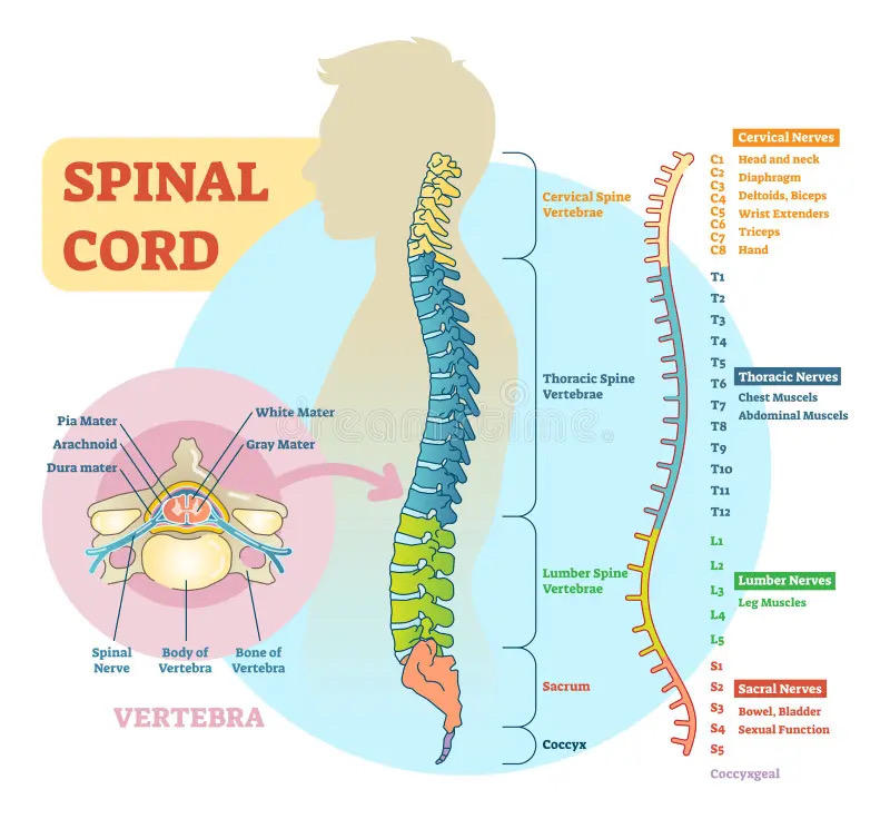
32
white and grey matter
white matter- myelinated axons, highways
1. tract (brain)
2. tract/column (outer spinal cord)
3. nerve (PNS)
grey matter- unmyelinated bodies, cities
1. nucleus (deep brain)
2. cortex (brain surface)
3. horn (inner spinal cord)
4. ganglion (PNS)
think grey is more dense than white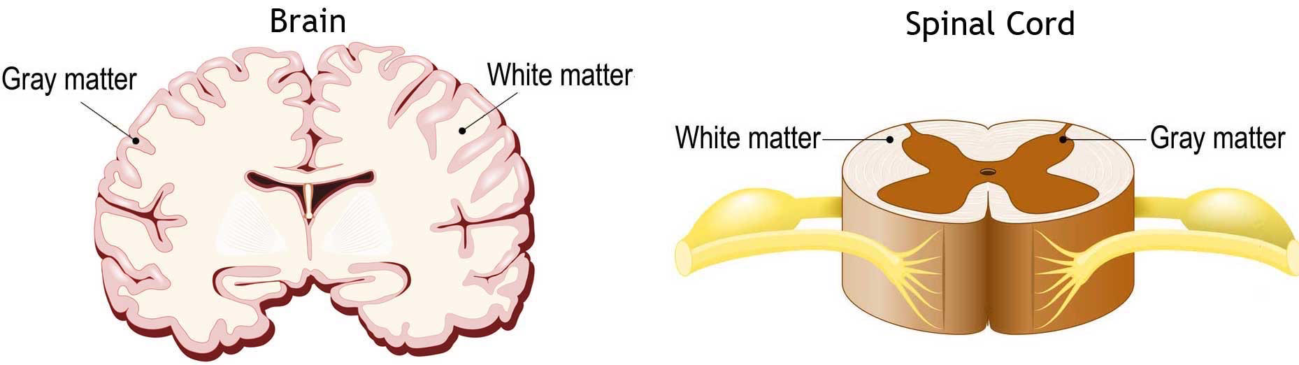
1. tract (brain)
2. tract/column (outer spinal cord)
3. nerve (PNS)
grey matter- unmyelinated bodies, cities
1. nucleus (deep brain)
2. cortex (brain surface)
3. horn (inner spinal cord)
4. ganglion (PNS)
think grey is more dense than white
33
corpus callosum
connects cerebral hemispheres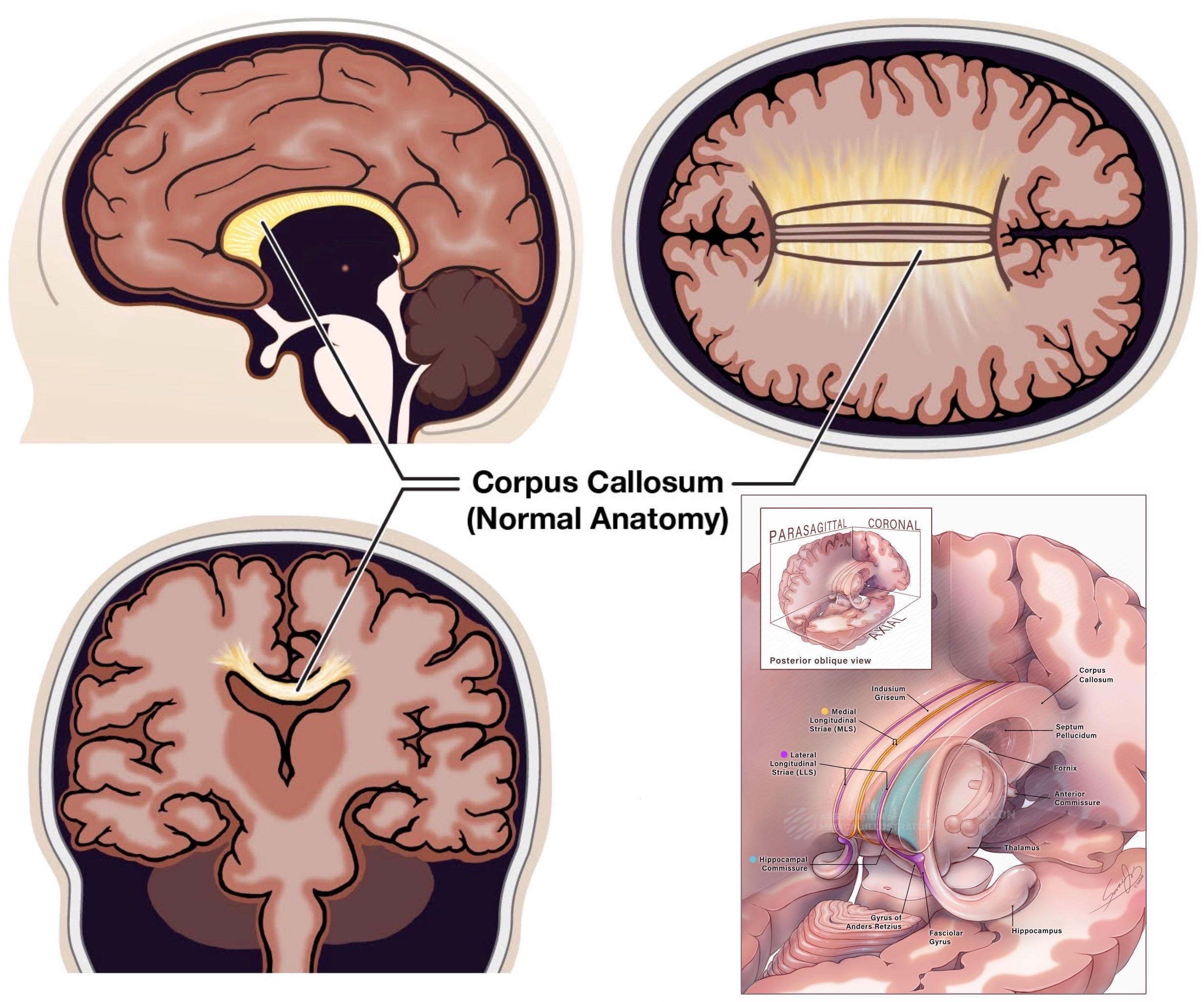
34
basal nuclei and ganglia
voluntary motor control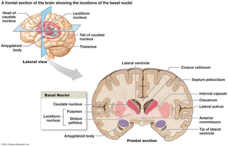
35
amygdala
emotion, fear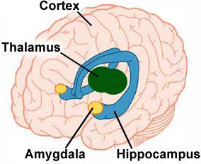
36
hippocampus
encoding to LTM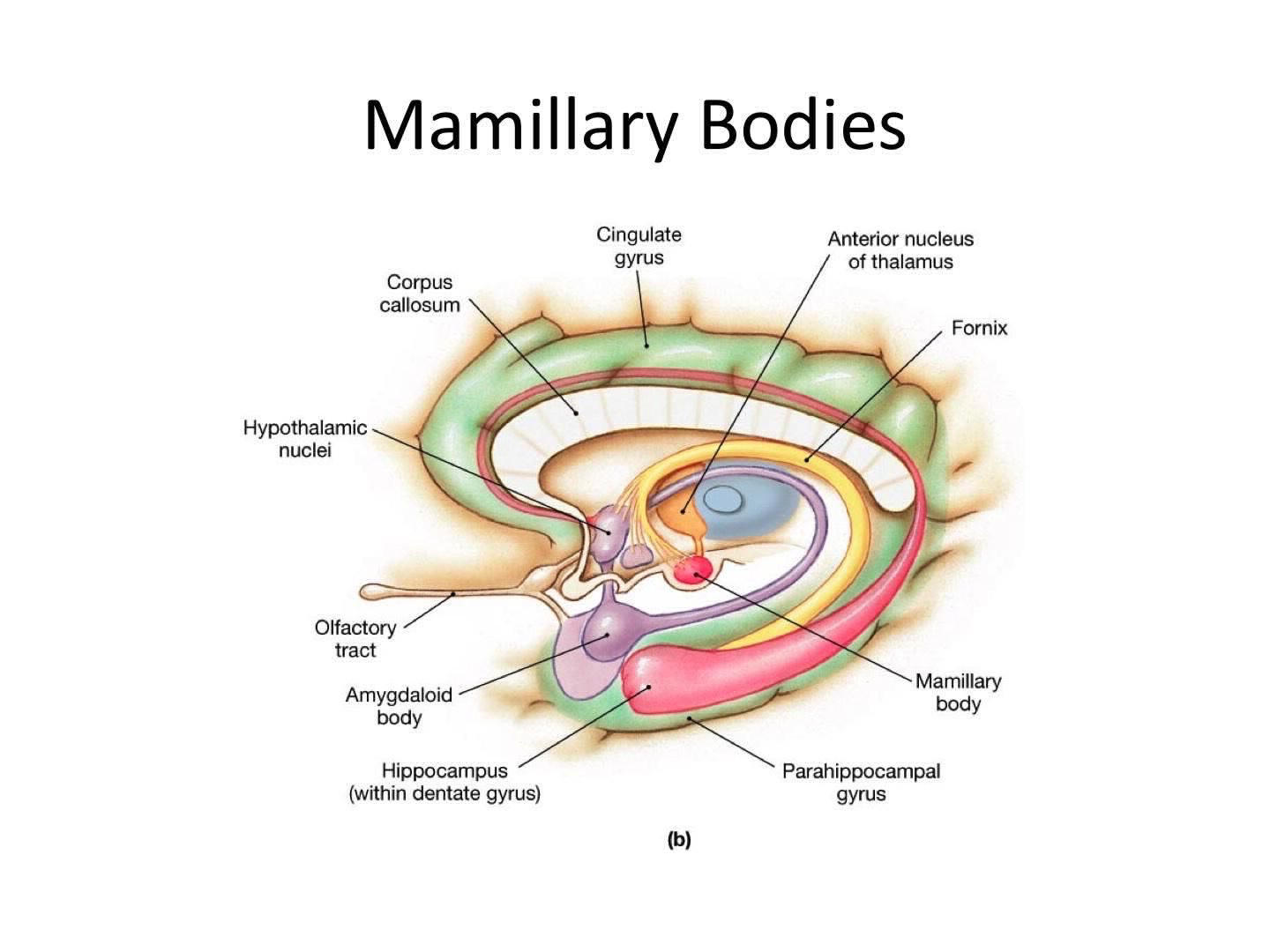
37
hemisphere
left hemisphere:
1. connected to right side of body
2. interprets right side of visual field
3. language processing (on dominant hemisphere)
right hemisphere
1. connected left side of body
2. interprets left side of visual field
3. visuospatial skills
4. emotional processing
5. music perception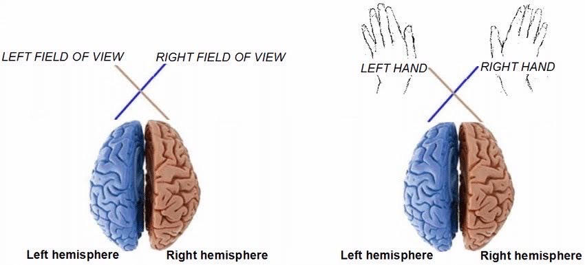
1. connected to right side of body
2. interprets right side of visual field
3. language processing (on dominant hemisphere)
right hemisphere
1. connected left side of body
2. interprets left side of visual field
3. visuospatial skills
4. emotional processing
5. music perception
38
occipital lobe
processes visual information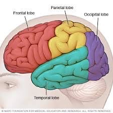
39
parietal lobe
contains somatosensory cortex, integrates sensory information, processes touch and taste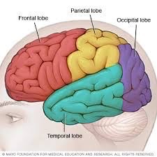
40
temporal lobe
auditory cortex, sound, emotional associations, memories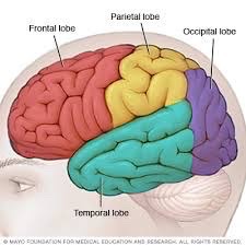
41
frontal lobe
higher-level processing, conscious decision making, executive functions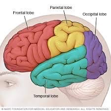
42
lobes
can be called cortices
43
central/peripheral nervous system
CNS- brain and spinal cord
PNS- nerves and sensory structures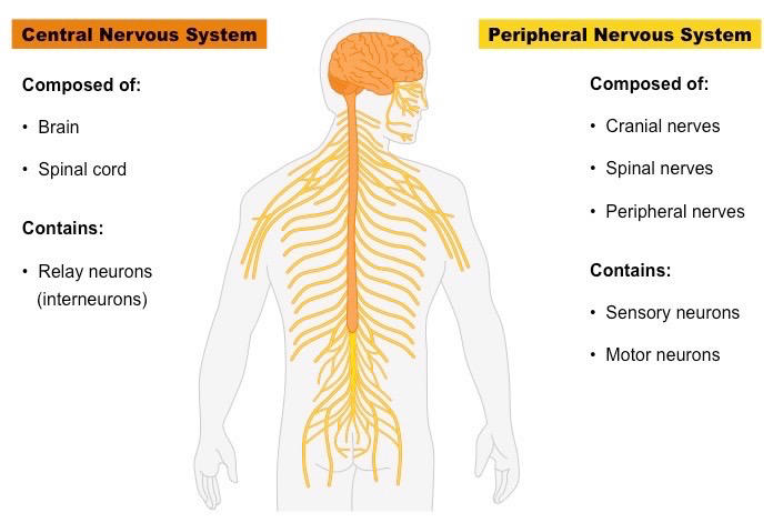
PNS- nerves and sensory structures
44
somatic/autonomic
categories of PNS
somatic- voluntary control of skeletal muscle, signaled by acetylcholine, single neuron coming from CNS
autonomic- involuntary control of glands and smooth muscle (heart, stomach, bladder), signaled by acetylcholine from CNS, can excite or inhibit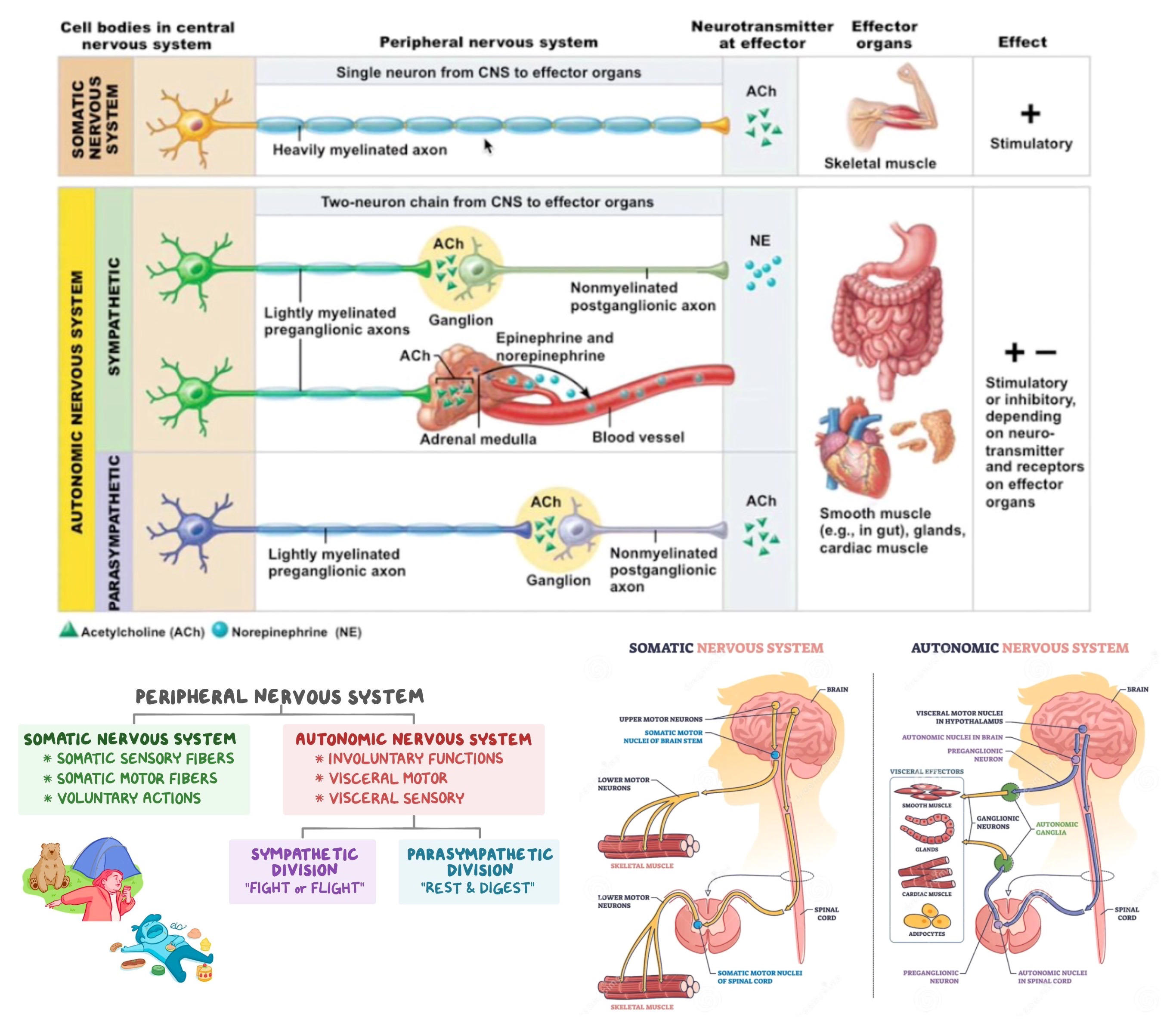
somatic- voluntary control of skeletal muscle, signaled by acetylcholine, single neuron coming from CNS
autonomic- involuntary control of glands and smooth muscle (heart, stomach, bladder), signaled by acetylcholine from CNS, can excite or inhibit
45
parasympathetic
categories of autonomic NS
parasympathetic- "rest and digest," decreased heart rate, respiratory rate, blood pressure, increased digestive and excretory functions
acetylcholine signal from CNS, then acetylcholine to effectors
long presynaptic neuron, short postsynaptic neuron
PARA LONG PRE
vasoconstriction- cut off blood to certain organs
vasodilation- increase blood to certain organs
happens in both systems to shift blood between GI and muscles and brain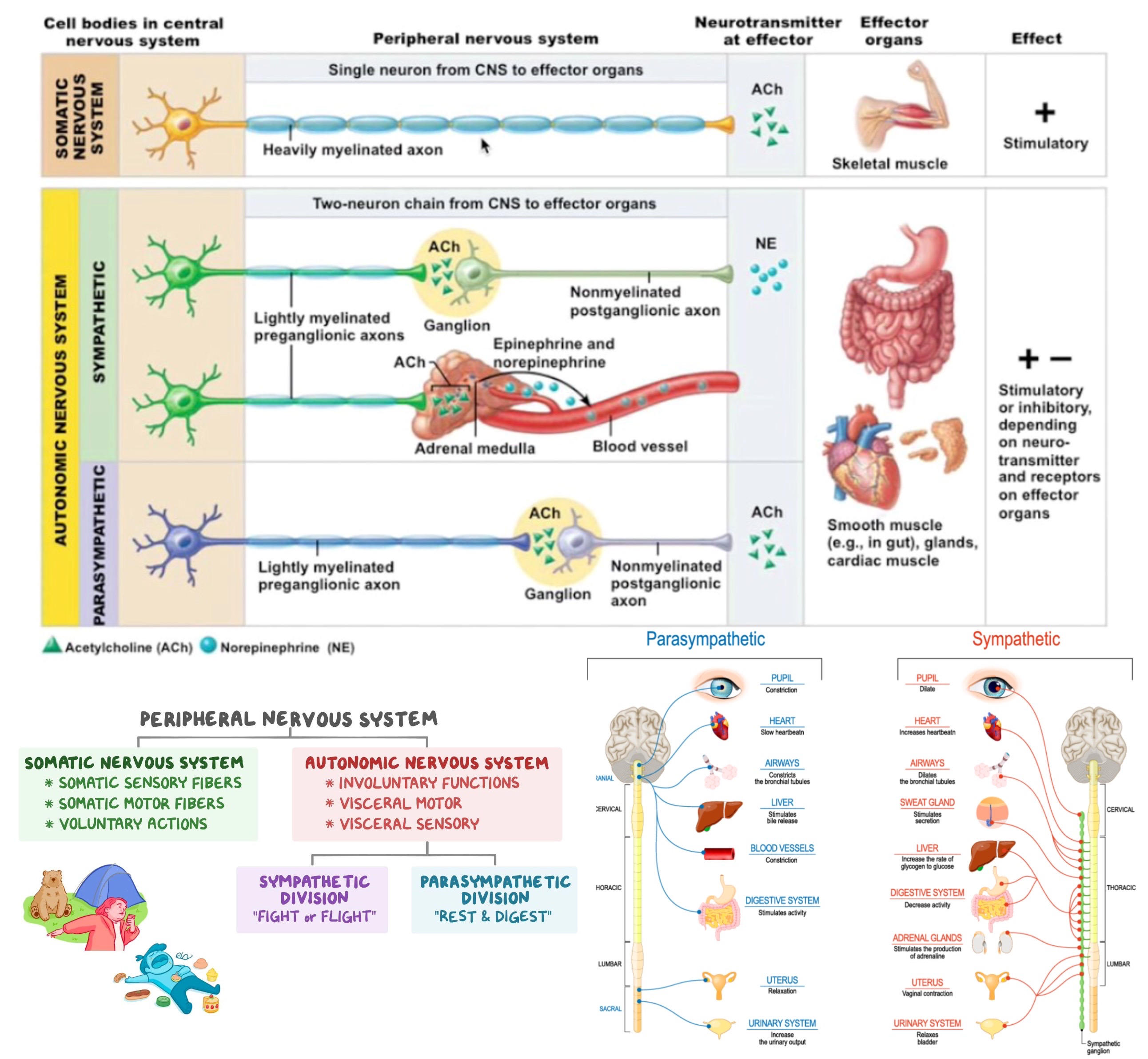
parasympathetic- "rest and digest," decreased heart rate, respiratory rate, blood pressure, increased digestive and excretory functions
acetylcholine signal from CNS, then acetylcholine to effectors
long presynaptic neuron, short postsynaptic neuron
PARA LONG PRE
vasoconstriction- cut off blood to certain organs
vasodilation- increase blood to certain organs
happens in both systems to shift blood between GI and muscles and brain
46
sympathetic
categories of autonomic NS
sympathetic- "fight or flight," increased HR/RR/BP, pupil dilation, decreased digestion, stimulation of adrenal medulla to release epinephrine
increased brain activity, increased glucose metabolism
4Fs = flight, fight, fright, fornicate
acetylcholine signal from CNS, then norepinephrine to effectors
short presynaptic neuron, long postsynaptic neuron
increased BP does not imply vasoconstriction
vasoconstriction- cut off blood to certain organs
vasodilation- increase blood to certain organs
happens in both systems to shift blood between GI and muscles and brain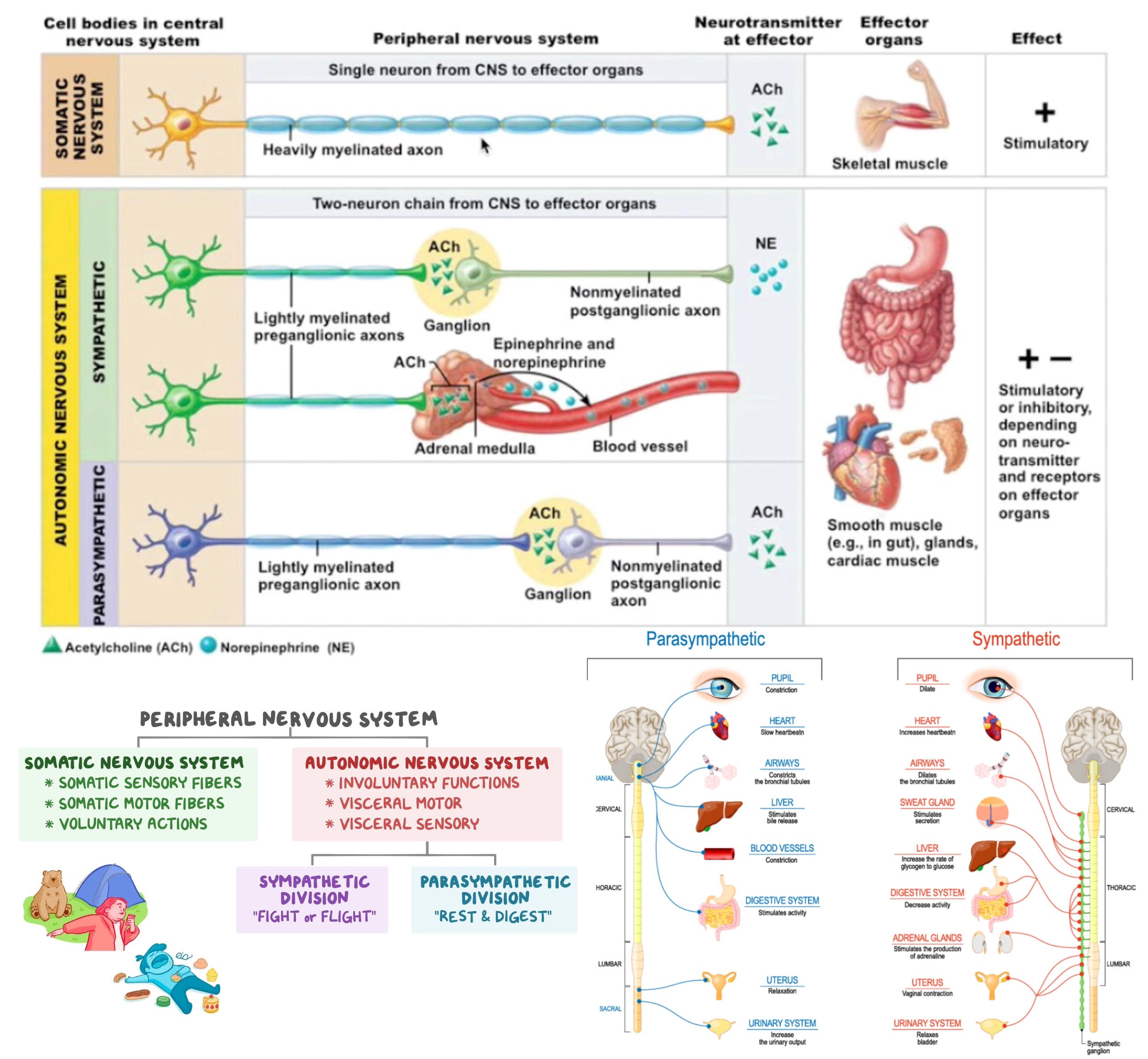
sympathetic- "fight or flight," increased HR/RR/BP, pupil dilation, decreased digestion, stimulation of adrenal medulla to release epinephrine
increased brain activity, increased glucose metabolism
4Fs = flight, fight, fright, fornicate
acetylcholine signal from CNS, then norepinephrine to effectors
short presynaptic neuron, long postsynaptic neuron
increased BP does not imply vasoconstriction
vasoconstriction- cut off blood to certain organs
vasodilation- increase blood to certain organs
happens in both systems to shift blood between GI and muscles and brain
47
PNS nerves
cranial nerves- carry info to/from brain stem
spinal nerves- carry info to/from spinal cord
vagus nerve- cranial nerve, part of parasympathetic NS that decreases HR and increases GI activity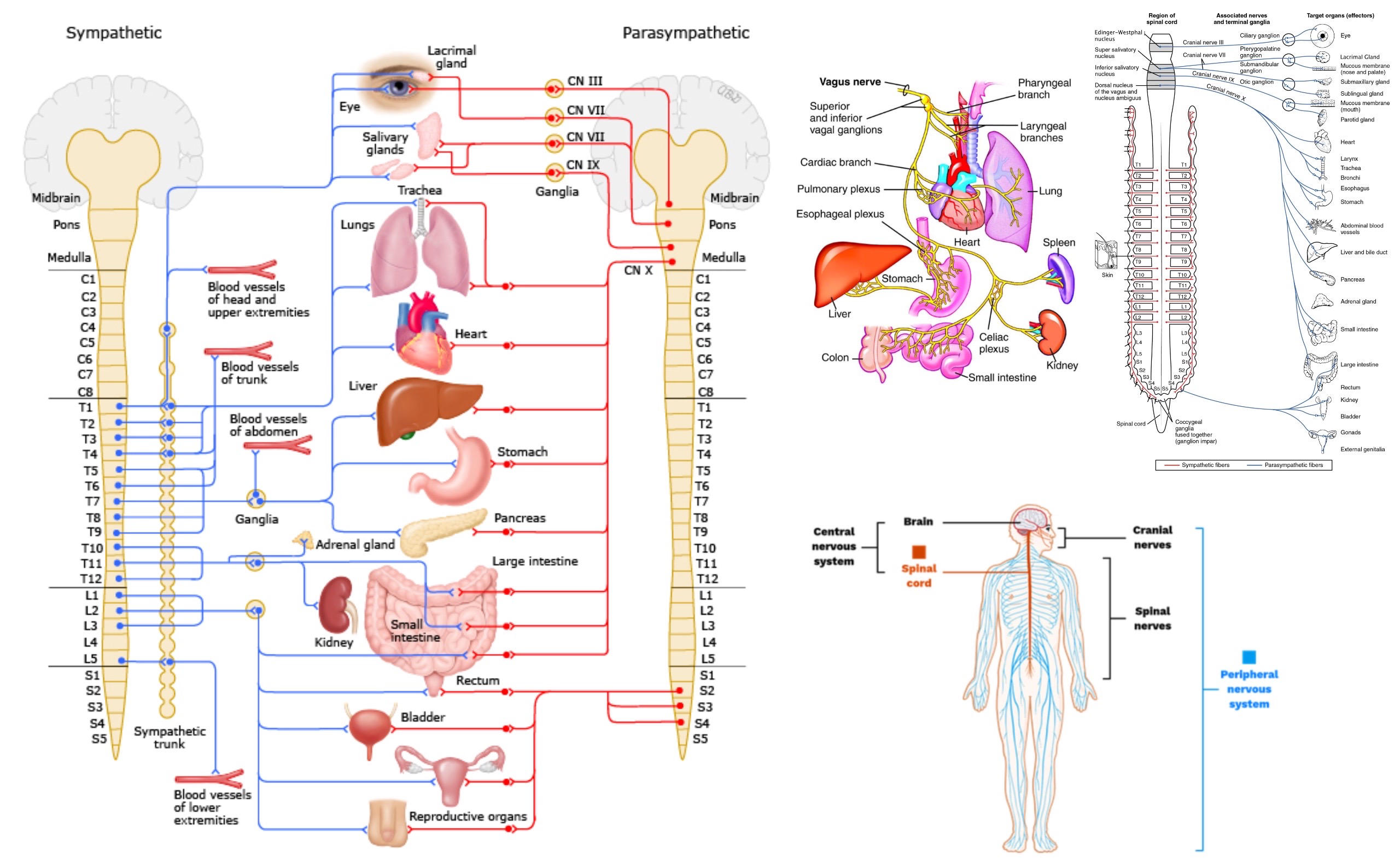
spinal nerves- carry info to/from spinal cord
vagus nerve- cranial nerve, part of parasympathetic NS that decreases HR and increases GI activity
48
dorsal root ganglion
collections of somas of the sensory neurons in the spinal cord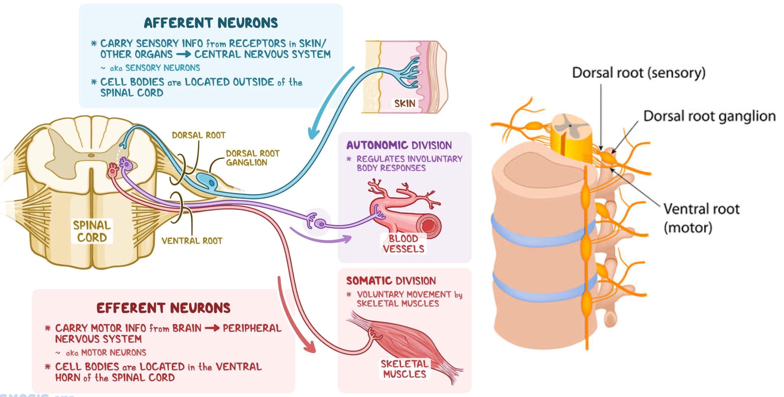
49
meninges
protective sheath of brain and spinal cord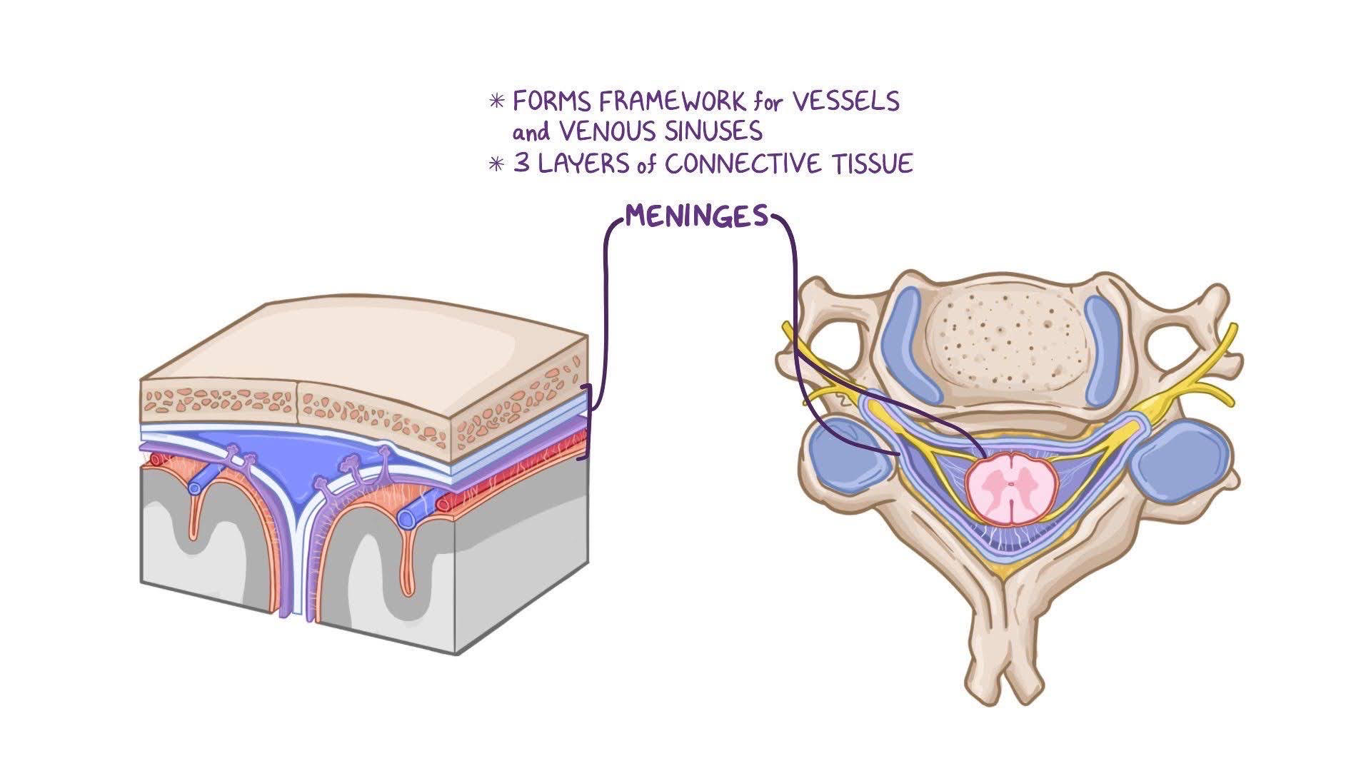
50
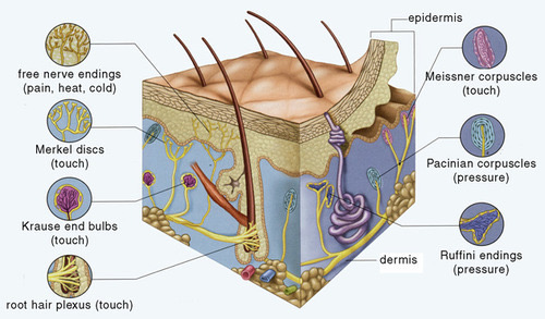
sensory receptors
mechanoreceptors- detect mechanical movement
chemoreceptors- detect chemicals, gustation and olfaction
nociceptors- detect pain
thermoreceptors- detect changes in temperature
photoreceptors- detect EM waves, rods and cones
proprioceptors- detect body position, kinesthetic sense, muscle spindle, (Golgi tendon organs, joint capsule receptors)
chemoreceptors- detect chemicals, gustation and olfaction
nociceptors- detect pain
thermoreceptors- detect changes in temperature
photoreceptors- detect EM waves, rods and cones
proprioceptors- detect body position, kinesthetic sense, muscle spindle, (Golgi tendon organs, joint capsule receptors)
51
gate control theory
spine contains a gate that blocks/allows pain signals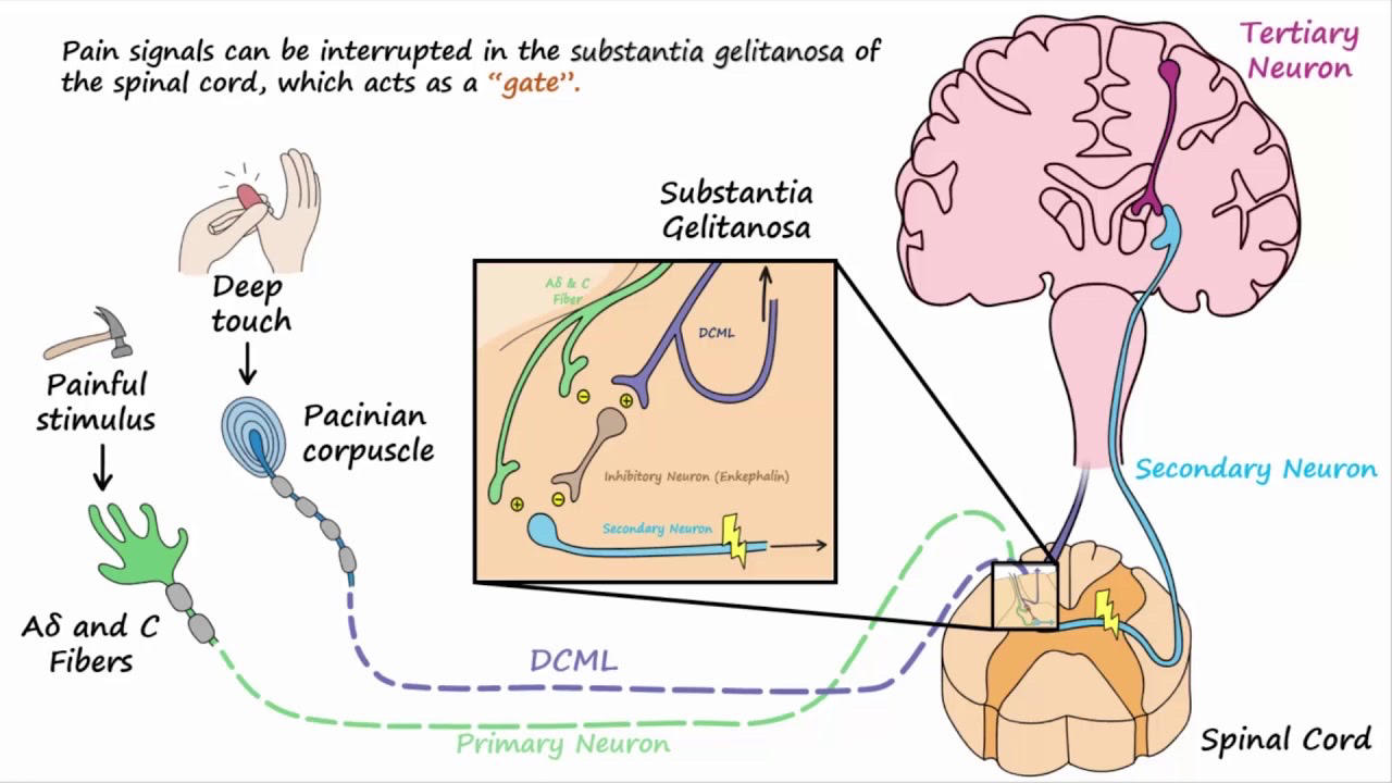
52
olfaction
gustation- taste buds have taste hairs, transmitted to the gustatory cortex
olfaction- smells detected by receptors in nasopharynx, transmitted directly to olfactory bulbs in the olfactory cortex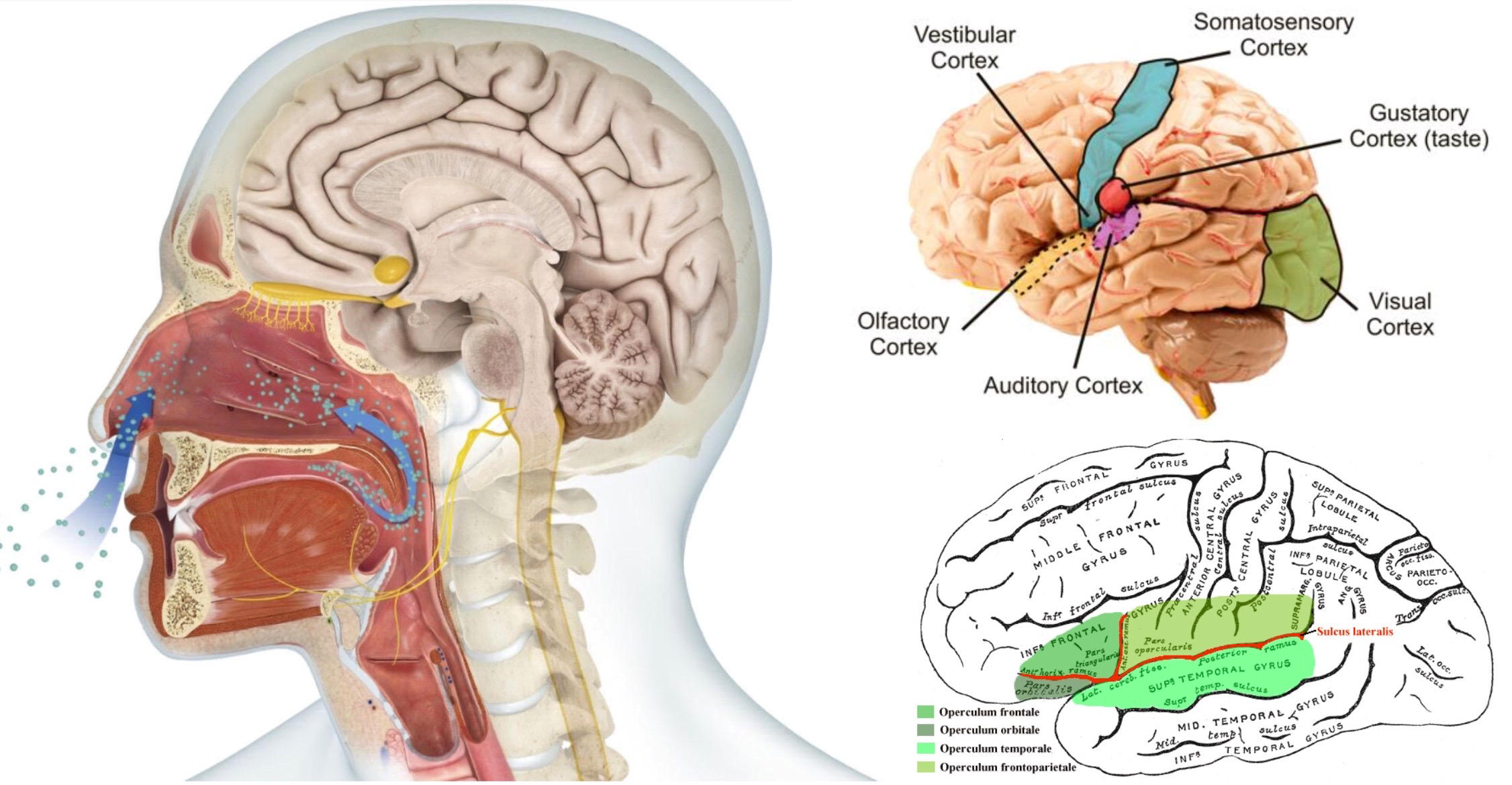
olfaction- smells detected by receptors in nasopharynx, transmitted directly to olfactory bulbs in the olfactory cortex
53
structures of the ear
pinna
auditory canal
eardrum
ossicles (malleus, incus, stapes)
oval window
cochlea (perilymph, endolymph)
organ of Corti (basilar membrane, tectorial membrane, cilia with mechanoreceptors)
semicircular canals, utricle, and saccule
round window
Eustachian tube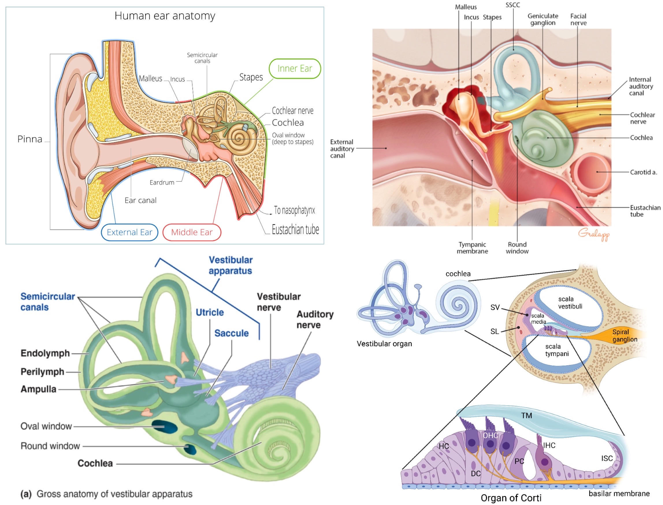
auditory canal
eardrum
ossicles (malleus, incus, stapes)
oval window
cochlea (perilymph, endolymph)
organ of Corti (basilar membrane, tectorial membrane, cilia with mechanoreceptors)
semicircular canals, utricle, and saccule
round window
Eustachian tube
54
sensing sound
pitch- place theory describes how regions of basilar membrane that vibrate determine pitch
loudness- amplitude, generates more action potentials
transmitted to auditory cortex in temporal lobe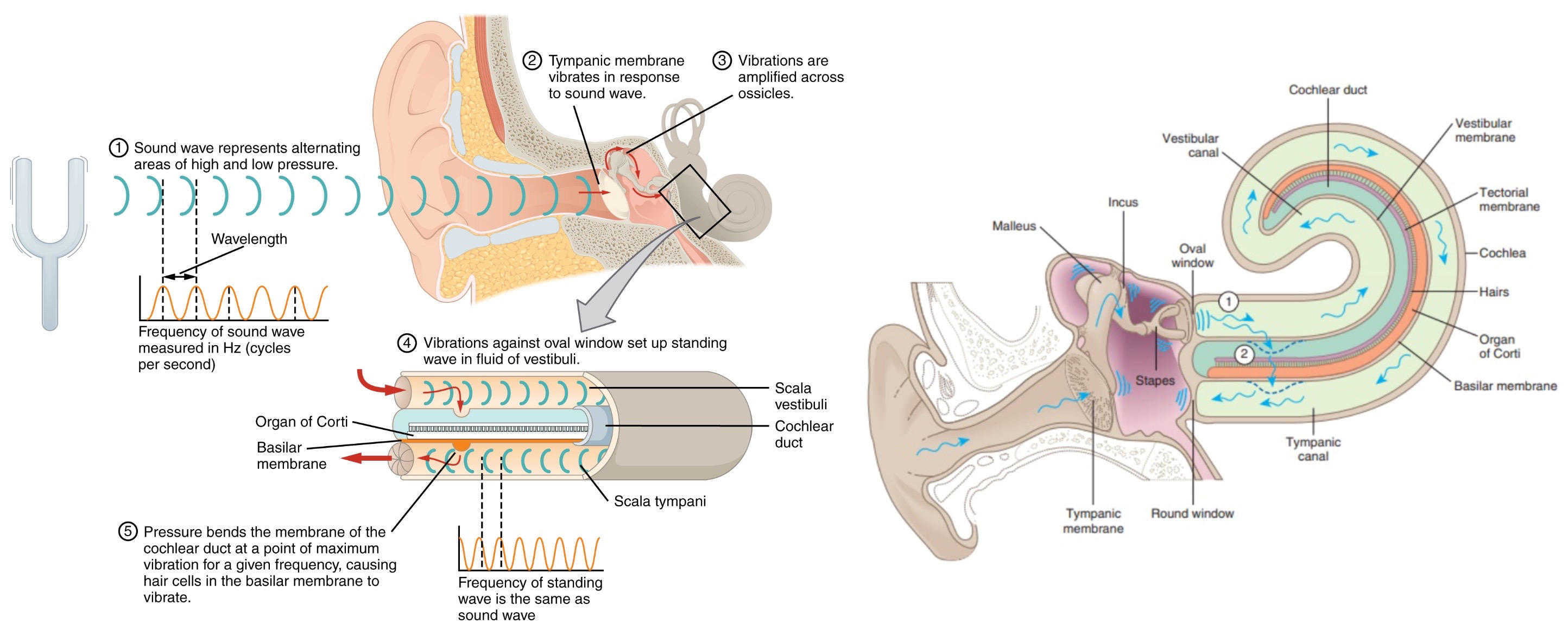
loudness- amplitude, generates more action potentials
transmitted to auditory cortex in temporal lobe
55
vestibular complex
semicircular canals (fluid shifts inside to detect balance), utricle and saccule (cilia detect acceleration), transmit to pons and cerebellum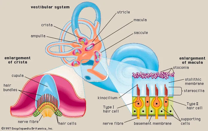
56
structures of the eye
conjunctiva- mucous membrane
sclera- thick outer coat, whites
cornea
pupil
iris
lens
retina
fovea centralis
optic disk
optic nerve
anterior chamber
posterior chamber
vitreous chamber
rods and cones
bipolar cells
ganglion cells (optic nerve)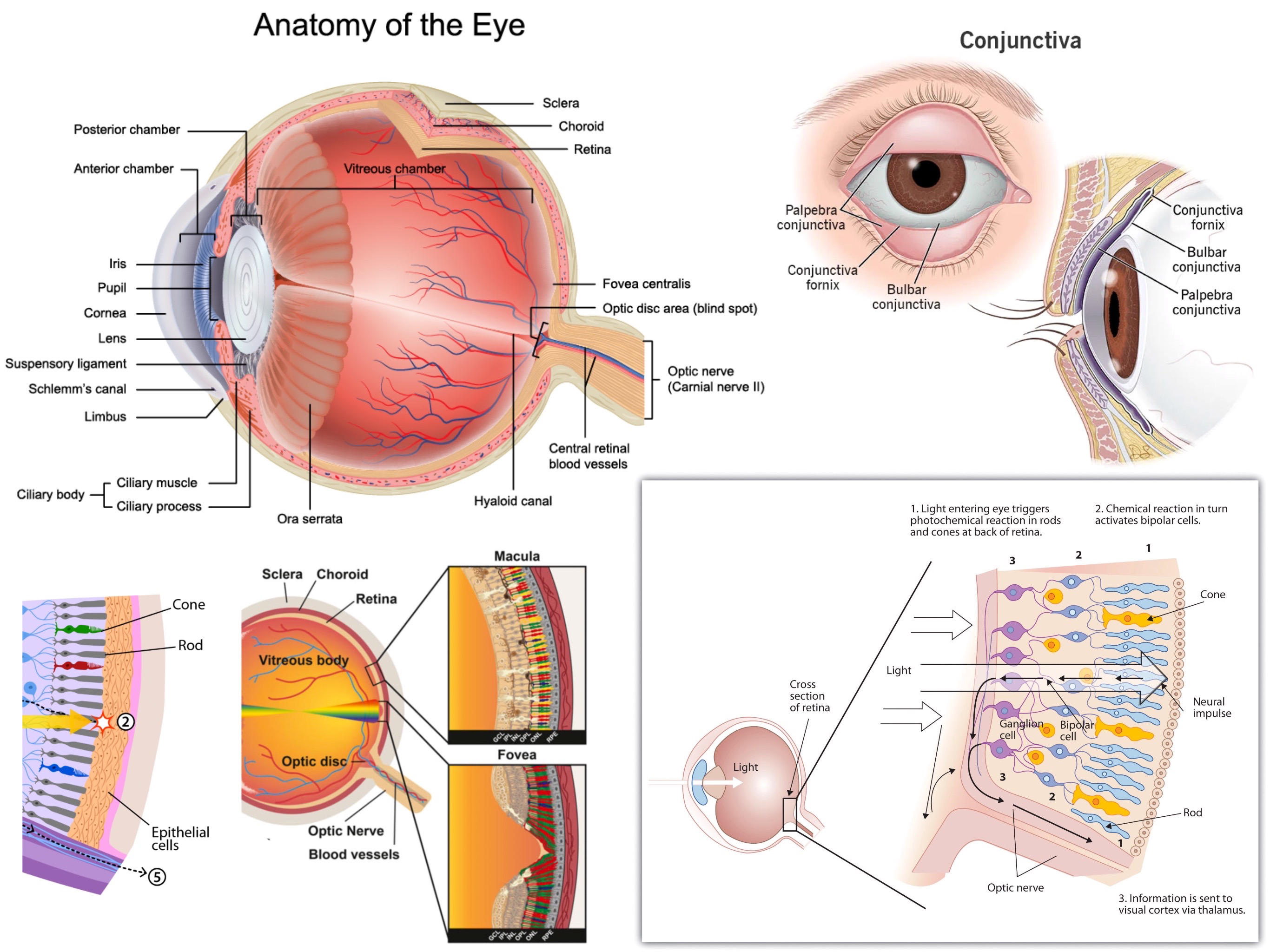
sclera- thick outer coat, whites
cornea
pupil
iris
lens
retina
fovea centralis
optic disk
optic nerve
anterior chamber
posterior chamber
vitreous chamber
rods and cones
bipolar cells
ganglion cells (optic nerve)
57
sensing light
photoreceptors are rods (best at detecting low light) and cones (best at detecting color)
opsins are proteins on photoreceptors
cones are concentrated in the fovea, cones have pigments that absorb green, red, blue, intensity of each color determines the final color perceived
photoreceptors are default on (bipolar and ganglion cells turn on/off)
retinal bound to opsins convert to all-trans on absorbing a photon, triggers polarization of cell by blocking Na+ channels
that stops release of glutamate to bipolar cells
ganglion cells transmit to through optic nerve
*optical chasm* divides the signal so left visual field goes to right brain, right visual field goes to left brain,
first hits lateral geniculate nucleus in thalamus, then goes to respective occipital lobes
signal intensity is determined by how many action potentials are sent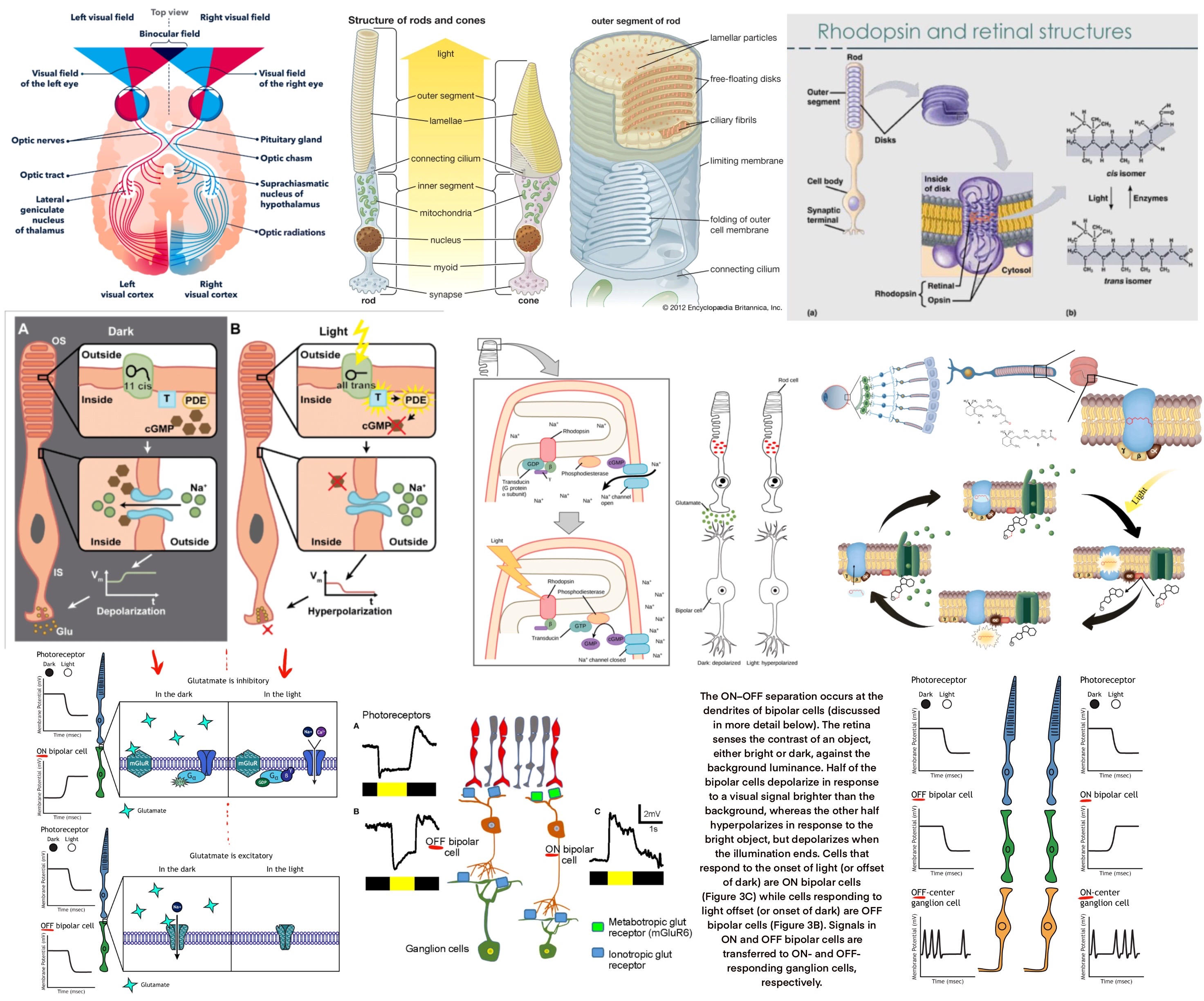
opsins are proteins on photoreceptors
cones are concentrated in the fovea, cones have pigments that absorb green, red, blue, intensity of each color determines the final color perceived
photoreceptors are default on (bipolar and ganglion cells turn on/off)
retinal bound to opsins convert to all-trans on absorbing a photon, triggers polarization of cell by blocking Na+ channels
that stops release of glutamate to bipolar cells
ganglion cells transmit to through optic nerve
*optical chasm* divides the signal so left visual field goes to right brain, right visual field goes to left brain,
first hits lateral geniculate nucleus in thalamus, then goes to respective occipital lobes
signal intensity is determined by how many action potentials are sent
58
visual field
left visual field hits right side of retina, goes to right hemisphere
right visual field hits left side of retina, goes to left hemisphere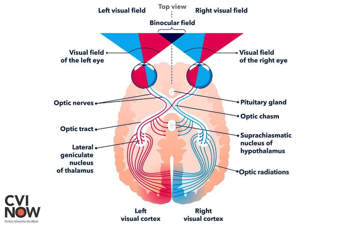
right visual field hits left side of retina, goes to left hemisphere
59
feature detection theory
color- trichromatic theory
form- parvocellular cells are neurons that detect shapes and boundaries
motion- magnocellular cells are neurons that detect motion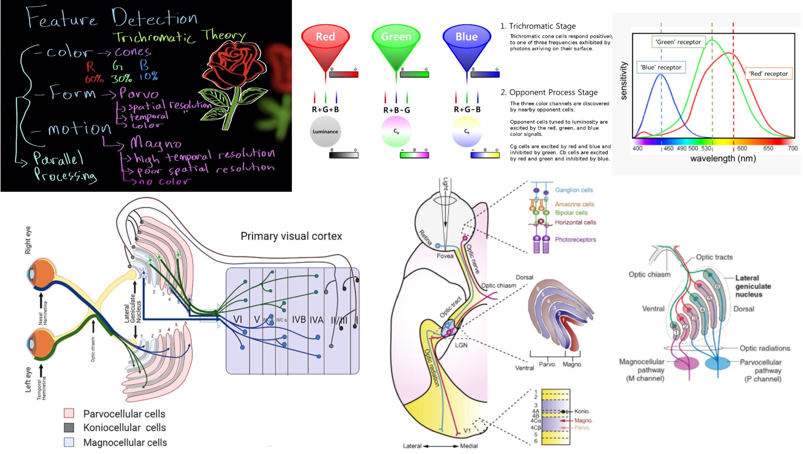
form- parvocellular cells are neurons that detect shapes and boundaries
motion- magnocellular cells are neurons that detect motion
60
vision defects
emmetropia- normal vision
myopia- nearsightedness, needs concave lens
hyperopia- farsightedness, needs convex lens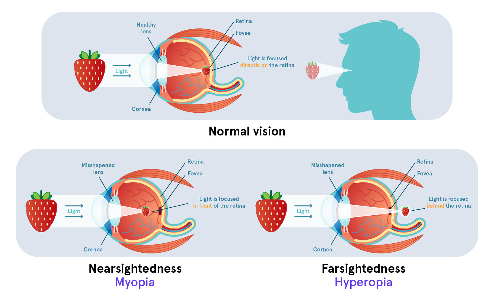
myopia- nearsightedness, needs concave lens
hyperopia- farsightedness, needs convex lens
61
adrenal glands
sits on top of the kidneys
adrenal medulla- releases *epinephrine*, sympathetic NS
adrenal cortex- releases *aldosterone and cortisol* (both steroid hormones), stress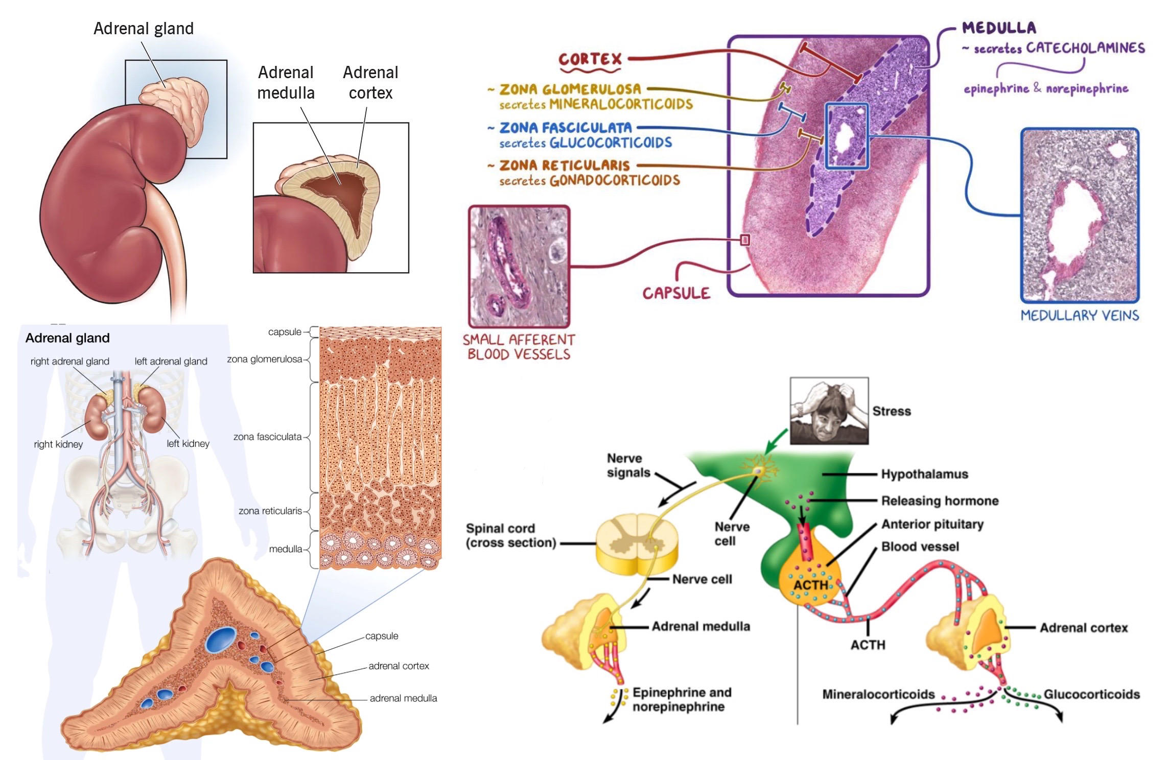
adrenal medulla- releases *epinephrine*, sympathetic NS
adrenal cortex- releases *aldosterone and cortisol* (both steroid hormones), stress
62
endocrine glands
produce hormones
sent directly to blood
next to capillaries, no ducts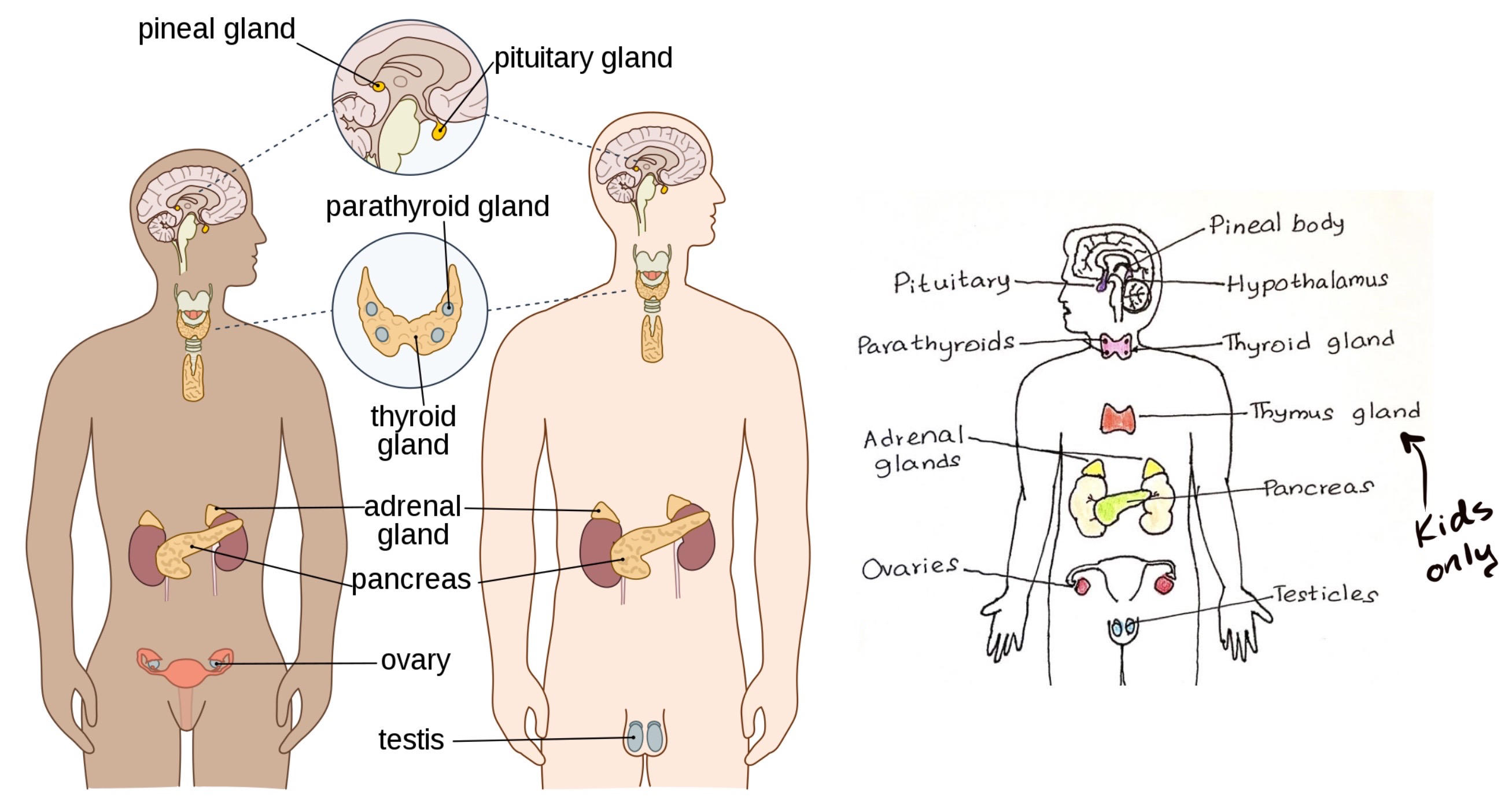
sent directly to blood
next to capillaries, no ducts
63
exocrine glands
produce sweat, tears, mucus, earwax, saliva, stomach acid, bile, semen, breast milk, enzymes
sent onto a body surface or cavity
all have ducts except for mucus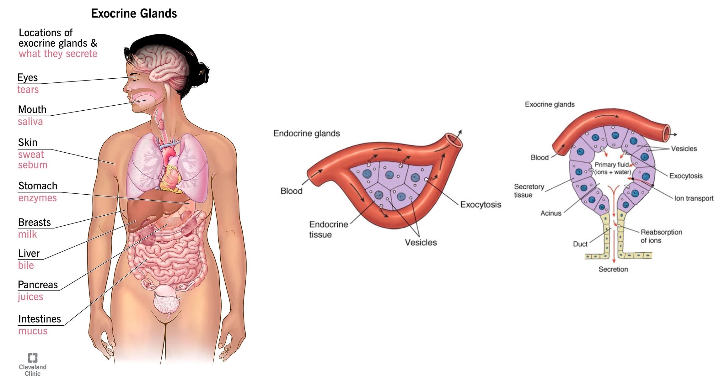
sent onto a body surface or cavity
all have ducts except for mucus
64
peptide/steroid hormones
peptide- made from amino acids, bind with cell surface receptor, which uses 2nd messenger system like *cAMP*, soluble in blood
fast and temporary!
ex: *insulin, epinephrine*
steroid- made from cholesterol, bind with intracellular receptor, which binds DNA to alter transcription, *require transport proteins to move through blood*
slow and more permanent!
ex: *aldosterone, cortisol*, ends with one/en/ol
exception: thyroid hormone is a peptide that acts like steroid
memorize hormones on pg. 317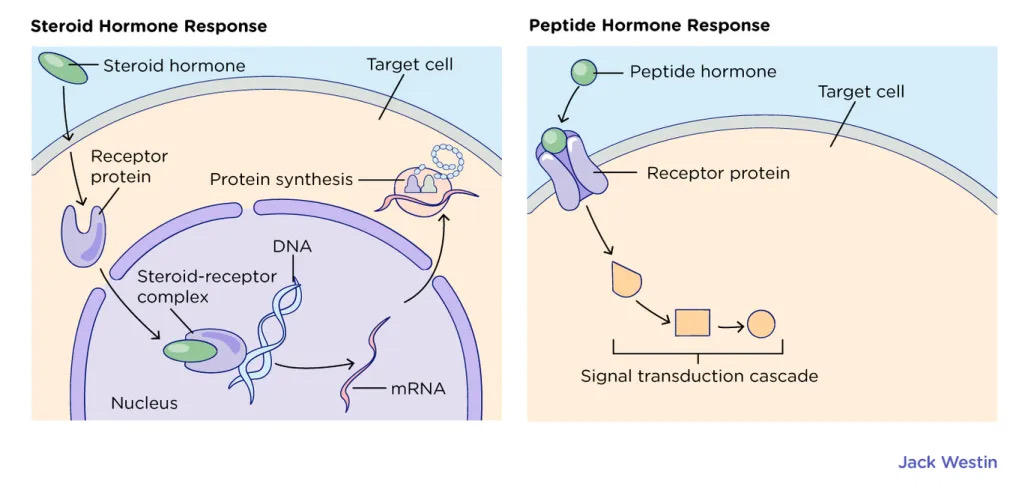
fast and temporary!
ex: *insulin, epinephrine*
steroid- made from cholesterol, bind with intracellular receptor, which binds DNA to alter transcription, *require transport proteins to move through blood*
slow and more permanent!
ex: *aldosterone, cortisol*, ends with one/en/ol
exception: thyroid hormone is a peptide that acts like steroid
memorize hormones on pg. 317
65
hormone regulation
neural- hypothalamus sends action potential to releases hormone, epinephrine released from adrenal medulla
hormonal- tropic hormones release other hormones, ACTH stimulates release of cortisol from adrenal cortex
humoral- blood component release hormone, glucose triggers release of insulin, lack of glucose triggers release of glucagon
will attempt to compensate for insufficiencies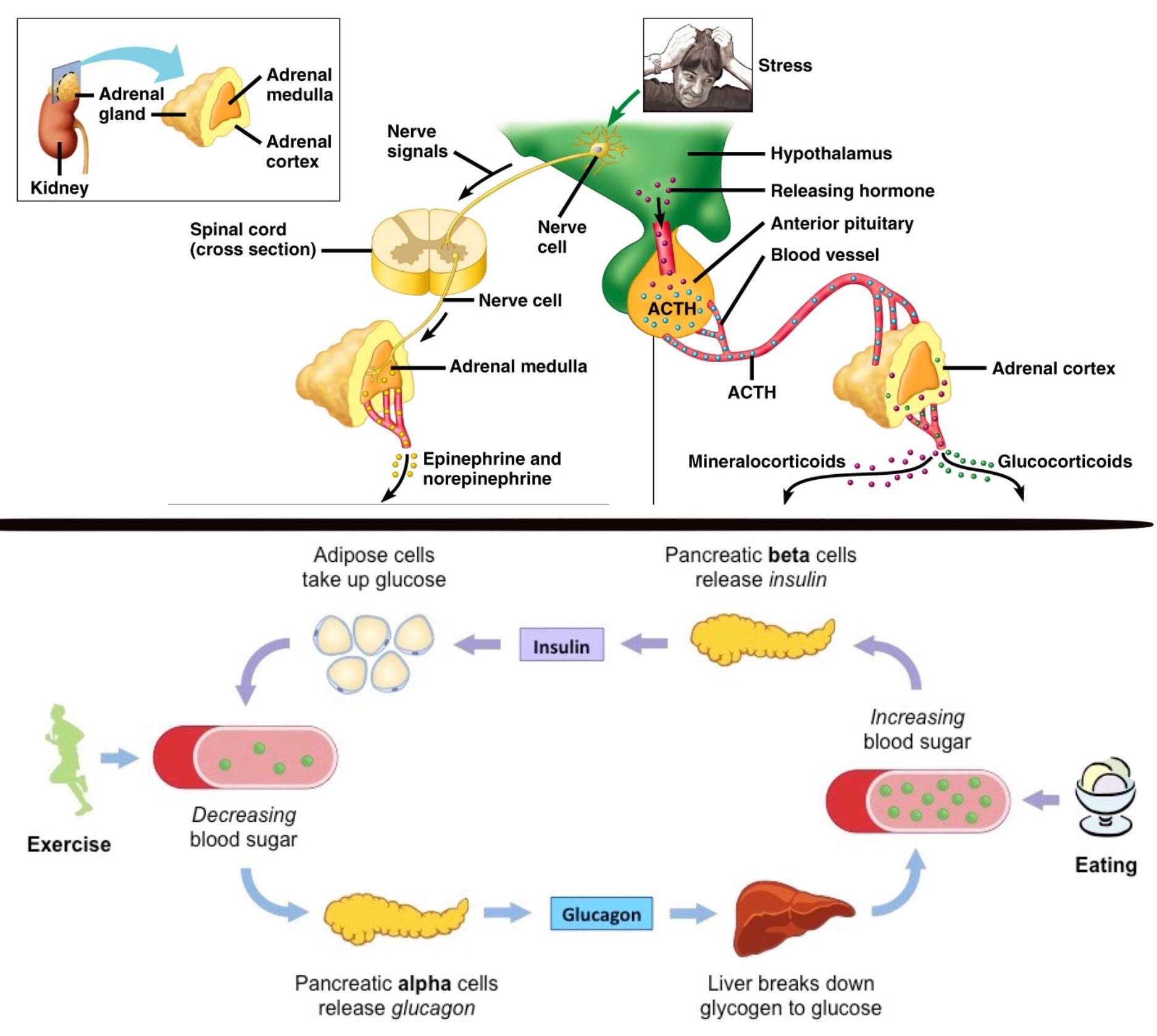
hormonal- tropic hormones release other hormones, ACTH stimulates release of cortisol from adrenal cortex
humoral- blood component release hormone, glucose triggers release of insulin, lack of glucose triggers release of glucagon
will attempt to compensate for insufficiencies
66
hypothalamic-pituitary control axis
anterior pituitary- adrenohypophysis, controlled indirectly by hypothalamus with tropic hormones, makes and secretes FLAT PEG:
1. FSH, LH for testosterone/estrogen
2. ACTH for adrenal cortex (cortisol, aldosterone)
3. TSH for thyroid gland, stimulate metabolism
4. Prolactin for milk
5. Endorphins
6. Growth hormone, stimulate growth
capillary cells, hormone making cells, uses portal veins
posterior pituitary- neurohypophysis, composed of neurons from hypothalamus that directly release hormones, stores and releases oxytocin and ADH (vasopressin)
neuroendocrine cells- neurons that release hormones, action potential travels down cell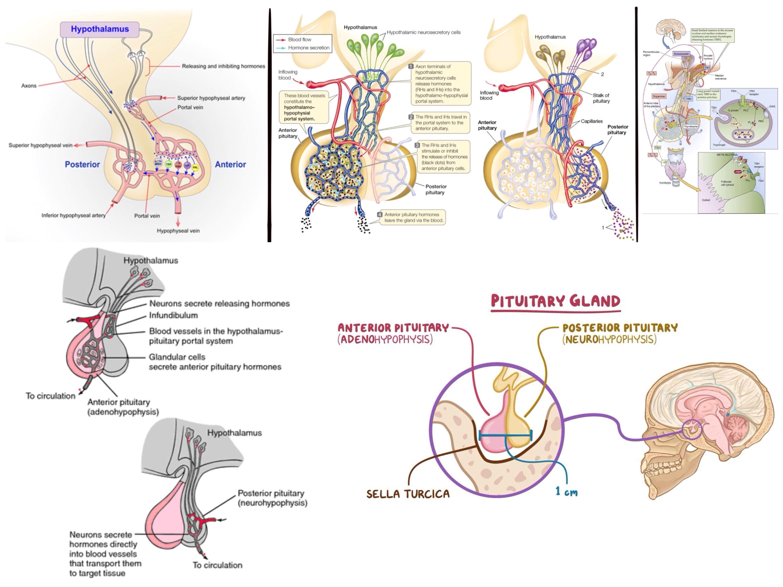
1. FSH, LH for testosterone/estrogen
2. ACTH for adrenal cortex (cortisol, aldosterone)
3. TSH for thyroid gland, stimulate metabolism
4. Prolactin for milk
5. Endorphins
6. Growth hormone, stimulate growth
capillary cells, hormone making cells, uses portal veins
posterior pituitary- neurohypophysis, composed of neurons from hypothalamus that directly release hormones, stores and releases oxytocin and ADH (vasopressin)
neuroendocrine cells- neurons that release hormones, action potential travels down cell
67
perfusion
flow of blood through tissue
ischemia- inadequate blood flow
hypoxia- inadequate O2 supply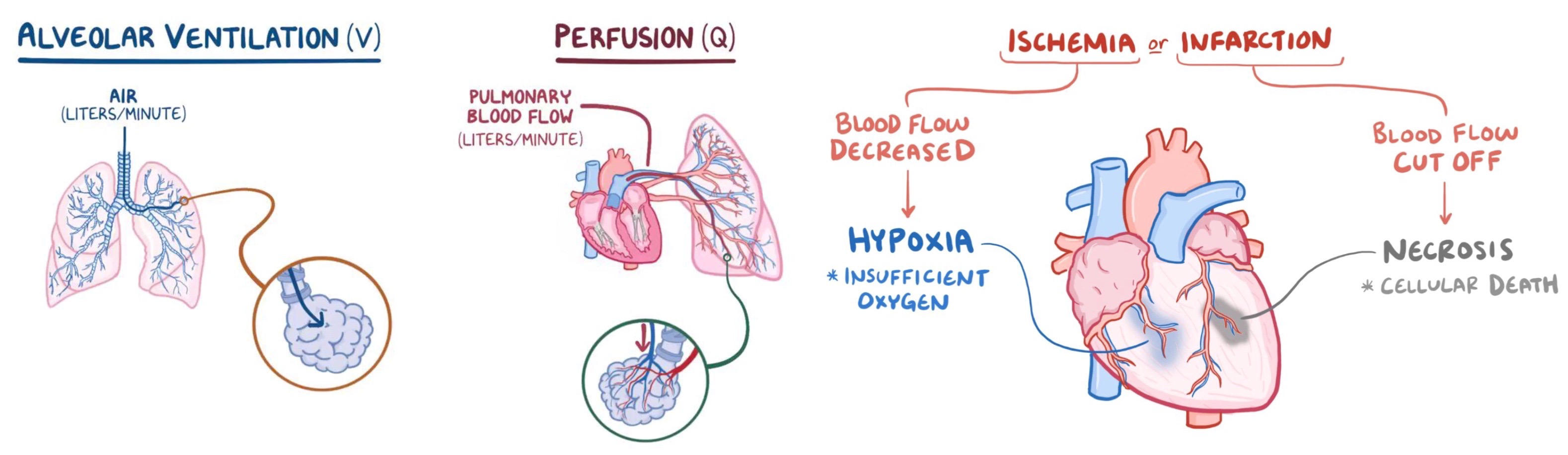
ischemia- inadequate blood flow
hypoxia- inadequate O2 supply
68
arteries
away from heart, high pressure
muscular, elastic walls that regulate flow
arterioles- small arteries just before capillaries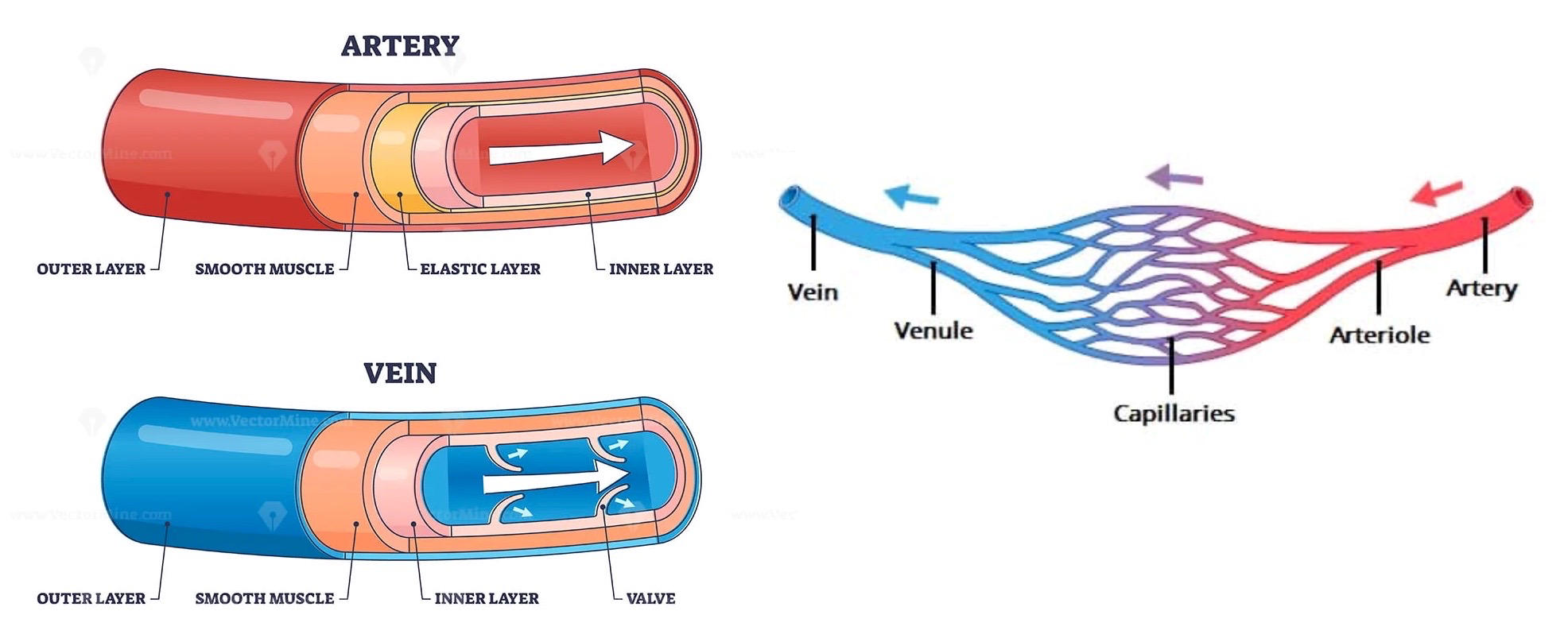
muscular, elastic walls that regulate flow
arterioles- small arteries just before capillaries
69
veins
towards the heart, low pressure
muscles and organs that squish against the vessels, valves ensure unidirectional since veins are not muscular or elastic
venules- small veins just after capillaries
varicose veins- when valves fail, venous pressure builds up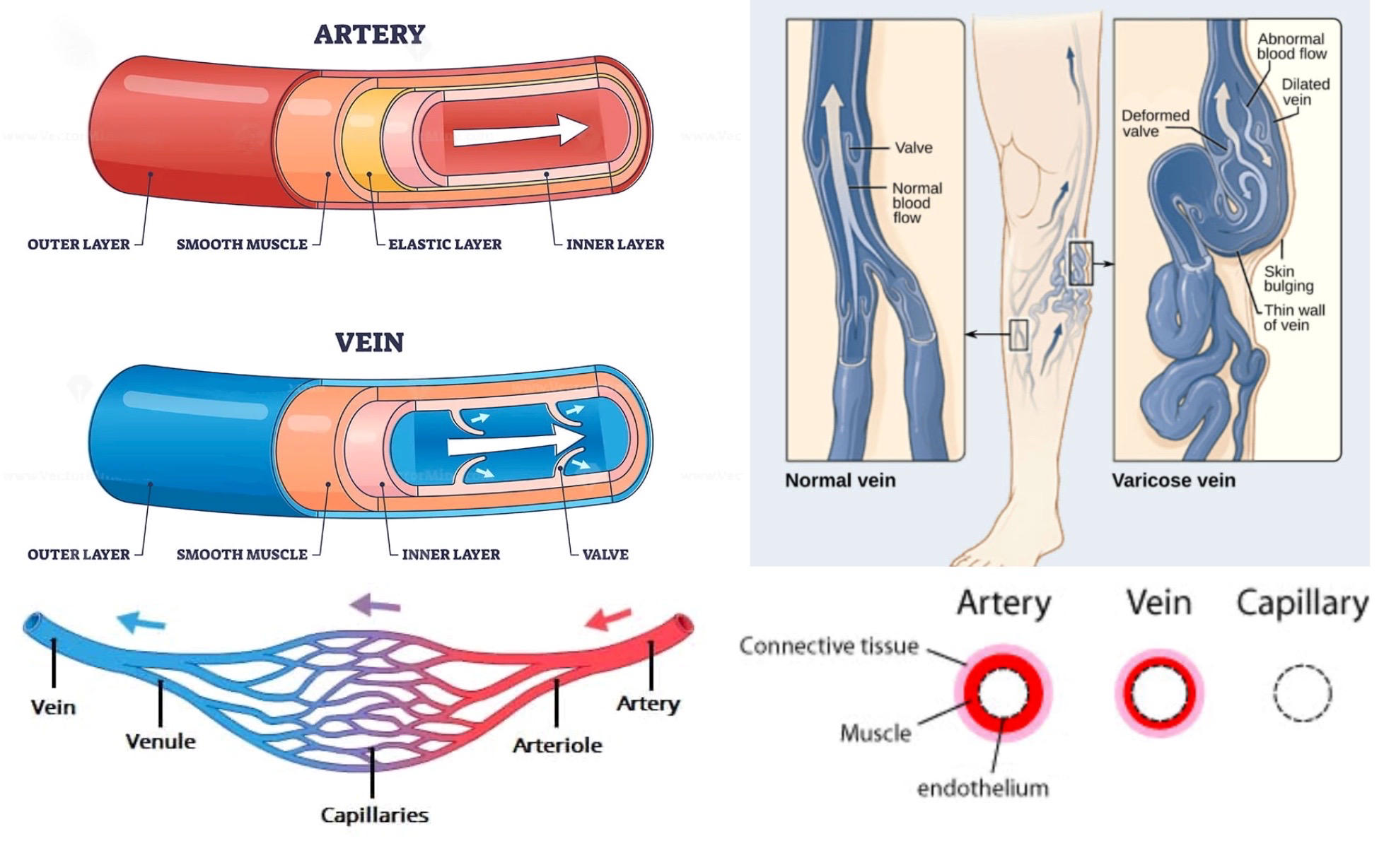
muscles and organs that squish against the vessels, valves ensure unidirectional since veins are not muscular or elastic
venules- small veins just after capillaries
varicose veins- when valves fail, venous pressure builds up
70
capillaries
nutrient and waste exchange, walls are single cell thick
fluid pushed out due to pressure, fluid pushed back in afterwards due to osmosis
capillaries have lower pressure and lower velocity of blood flow because total cross sectional area is greater than arteries
BP drops to 0 mm Hg when it reaches vena cava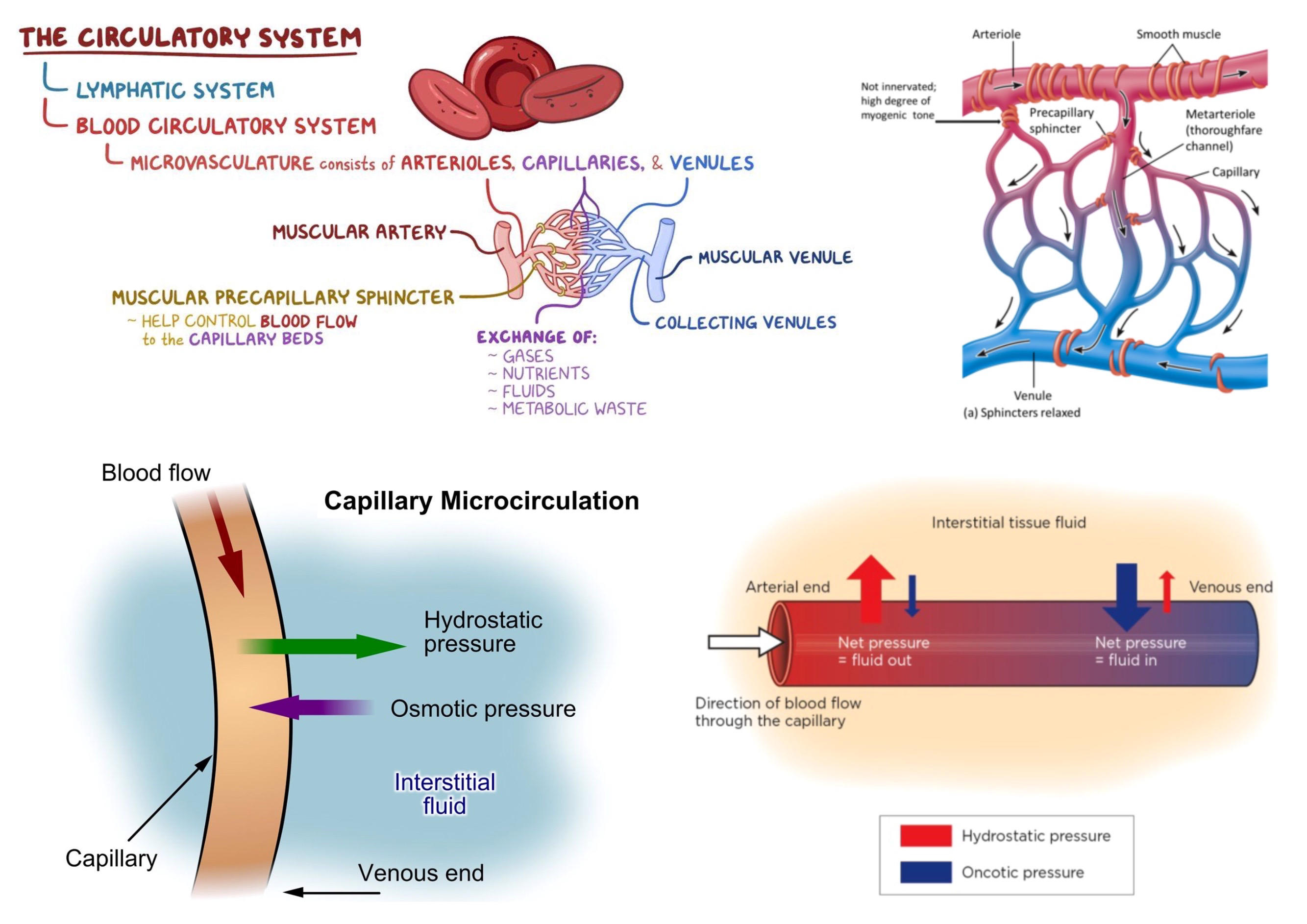
fluid pushed out due to pressure, fluid pushed back in afterwards due to osmosis
capillaries have lower pressure and lower velocity of blood flow because total cross sectional area is greater than arteries
BP drops to 0 mm Hg when it reaches vena cava
71
endothelial cells
inner lining of all blood vessels, type of epithelial cells
4 functions:
1. vasoconstriction- maintain BP
2. inflammation- recruit WBCs
3. angiogenesis- form new blood vessels
4. thrombosis- control blood clotting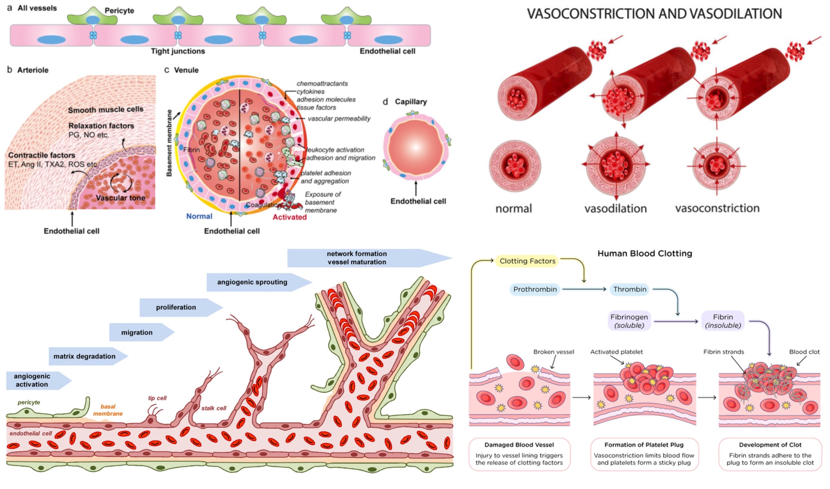
4 functions:
1. vasoconstriction- maintain BP
2. inflammation- recruit WBCs
3. angiogenesis- form new blood vessels
4. thrombosis- control blood clotting
72
circulation
pulmonary circulation- flow of blood from heart to the lungs and back, right side of heart pumps it
systemic circulation- flow of blood from heart to body and back, left side of heart pumps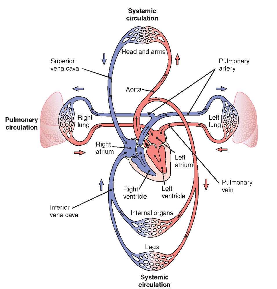
systemic circulation- flow of blood from heart to body and back, left side of heart pumps
73
portal systems
most blood passes through one set of capillaries before returning to the heart
hepatic portal system- heart to intestine to liver to heart, transports nutrients
hypothalamic-hypophysial portal system- heart to hypothalamus to pituitary gland to heart, transports hormones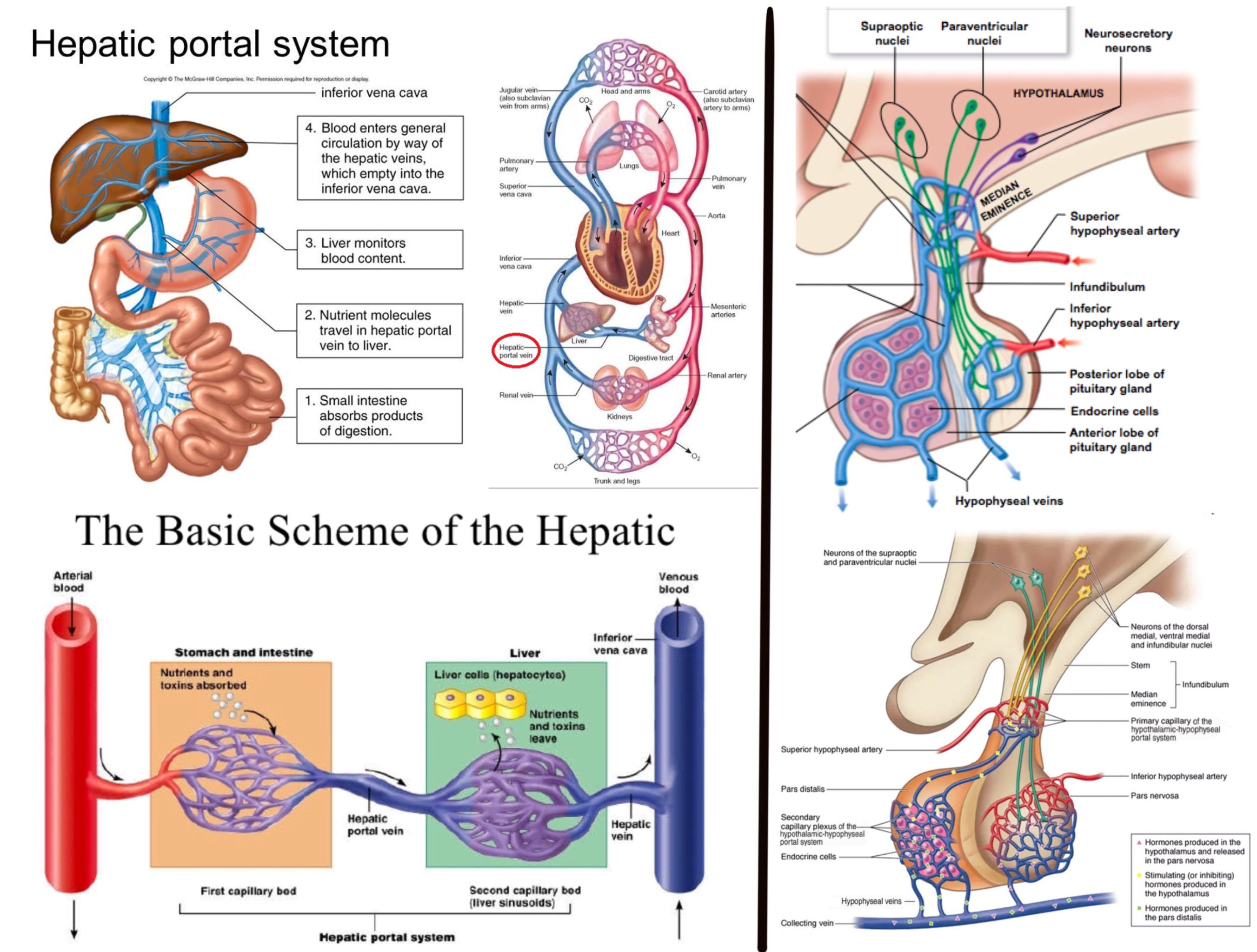
hepatic portal system- heart to intestine to liver to heart, transports nutrients
hypothalamic-hypophysial portal system- heart to hypothalamus to pituitary gland to heart, transports hormones
74
lymphatic system
retrieve water, WBCs, proteins from tissues and return to circulation
connected to circulatory system
*lymph nodes*- location of immune system cells, filter fluid from tissues
*spleen*- filters blood, removes old RBCs
thoracic duct- near the neck, where lymphatic system dumps fluid back into circulatory system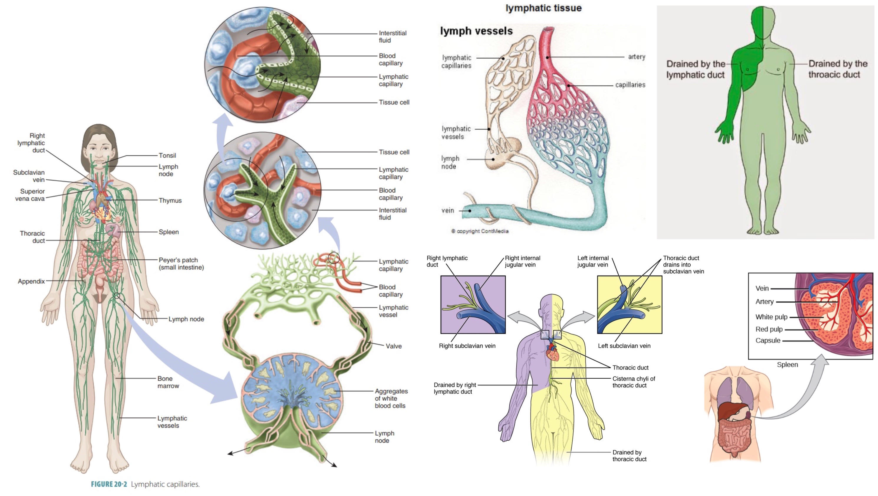
connected to circulatory system
*lymph nodes*- location of immune system cells, filter fluid from tissues
*spleen*- filters blood, removes old RBCs
thoracic duct- near the neck, where lymphatic system dumps fluid back into circulatory system
75
heart
superior/inferior vena cava- receives deoxygenated blood from body
right atrium- waiting room
tricuspid AV valve
right ventricle- pumps deoxygenated blood to lungs
pulmonary semilunar valve
pulmonary artery- carries deoxygenated blood to lungs
pulmonary vein- carries oxygenated blood from lungs
left atrium- waiting room
bicuspid/mitral AV valve
left ventricle- pumps oxygenated blood to body
aortic semilunar valve
aortic arch- carries oxygenated blood to body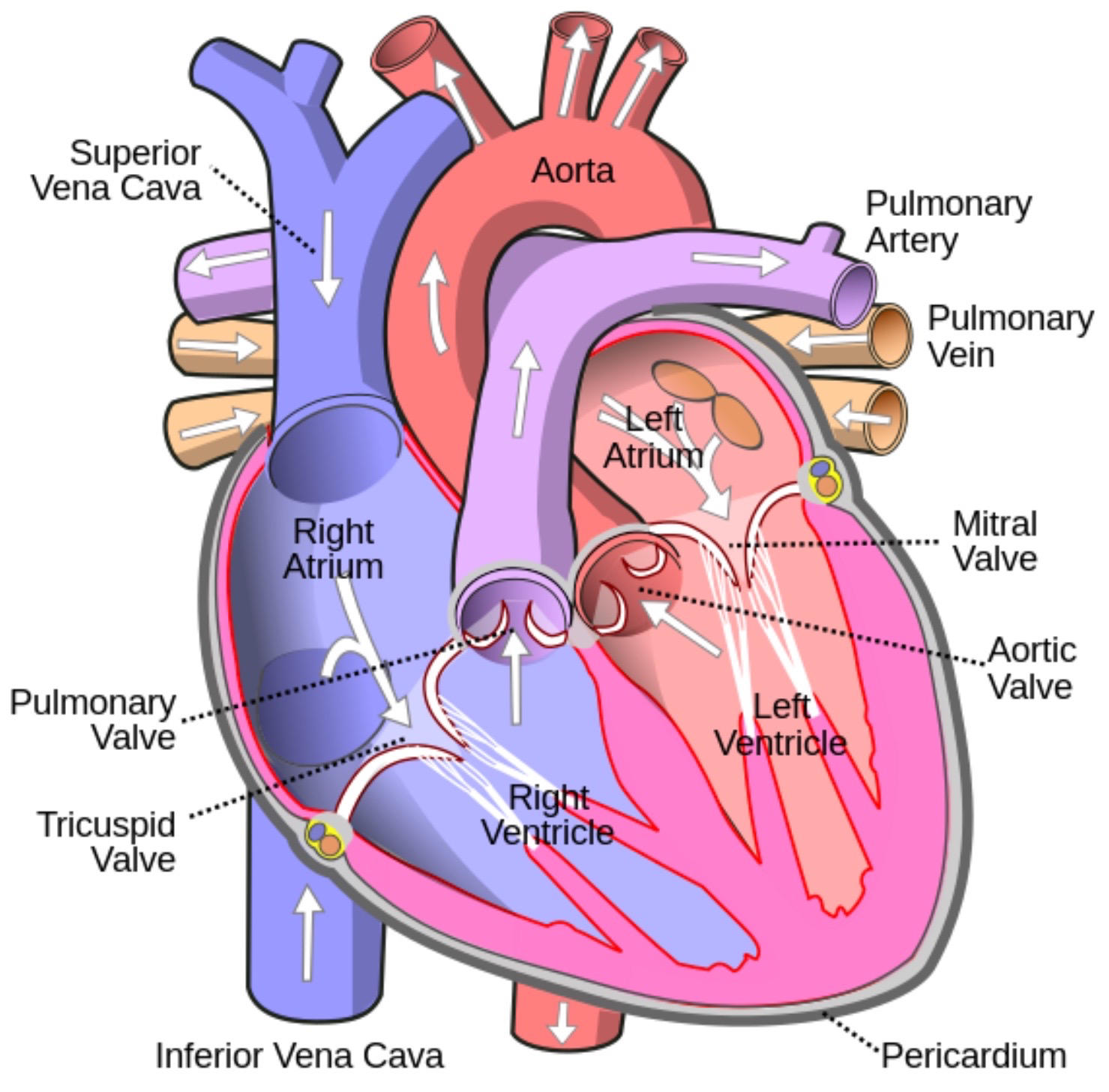
right atrium- waiting room
tricuspid AV valve
right ventricle- pumps deoxygenated blood to lungs
pulmonary semilunar valve
pulmonary artery- carries deoxygenated blood to lungs
pulmonary vein- carries oxygenated blood from lungs
left atrium- waiting room
bicuspid/mitral AV valve
left ventricle- pumps oxygenated blood to body
aortic semilunar valve
aortic arch- carries oxygenated blood to body
76
coronary arteries
aorta branches off to supply blood to walls of heart
coronary veins carry deoxygenated blood to coronary sinus, which drains directly into right atrium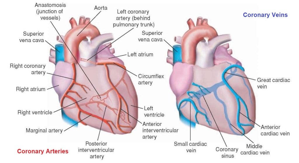
coronary veins carry deoxygenated blood to coronary sinus, which drains directly into right atrium
77
heart sounds
lub- AV valves close, systole begins
dup- semilunar valves close, diastole begins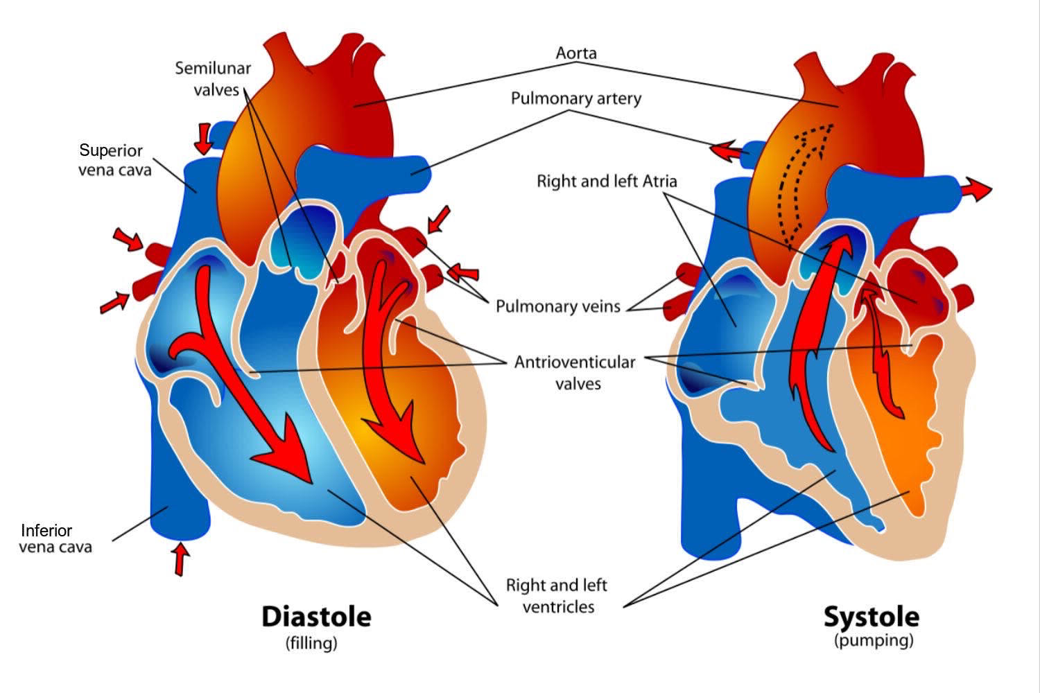
dup- semilunar valves close, diastole begins
78
blood pressure and cardiac cycle
systole- heart contracts, ventricles pump blood to body
systolic pressure- BP in arteries when heart contracts
diastole- heart relaxes, atria pump blood to ventricles
diastolic pressure- BP in arteries when heart relaxes
pulse pressure- difference between SP and DP
BP is directly proportional to cardiac output and peripheral resistance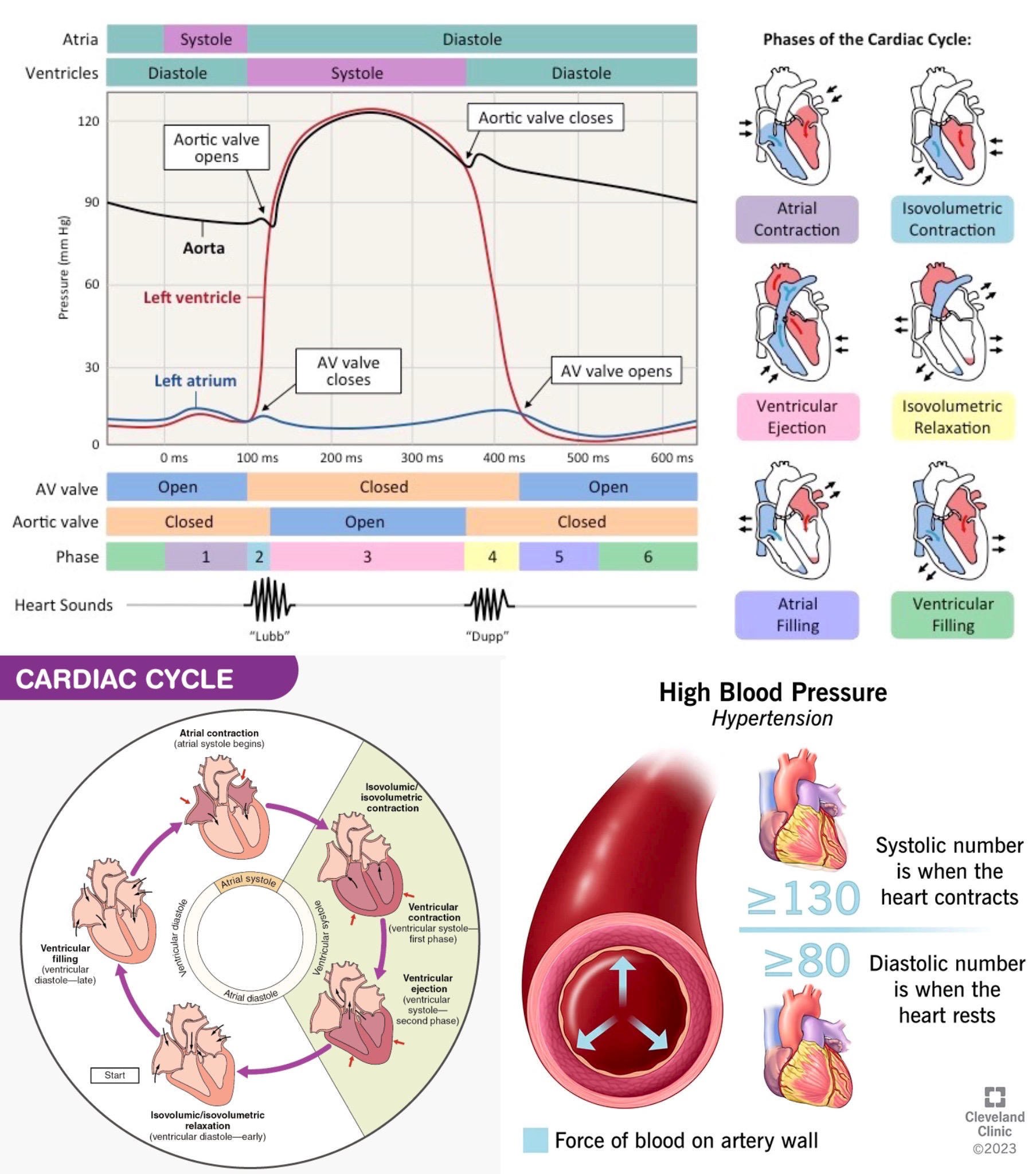
systolic pressure- BP in arteries when heart contracts
diastole- heart relaxes, atria pump blood to ventricles
diastolic pressure- BP in arteries when heart relaxes
pulse pressure- difference between SP and DP
BP is directly proportional to cardiac output and peripheral resistance
79
cardiac output
cardiac output (L/min) = stroke volume (L/ beats) x heart rate (beats/min)
stroke volume is blood pumped with each systole, affected:
1. Frank-Starling mechanism- greater venous return stretches heart, so that it contracts harder (increase venous return by not peeing)
2. change activity level for stronger heart
3. change in posture (orthostatic hypotension)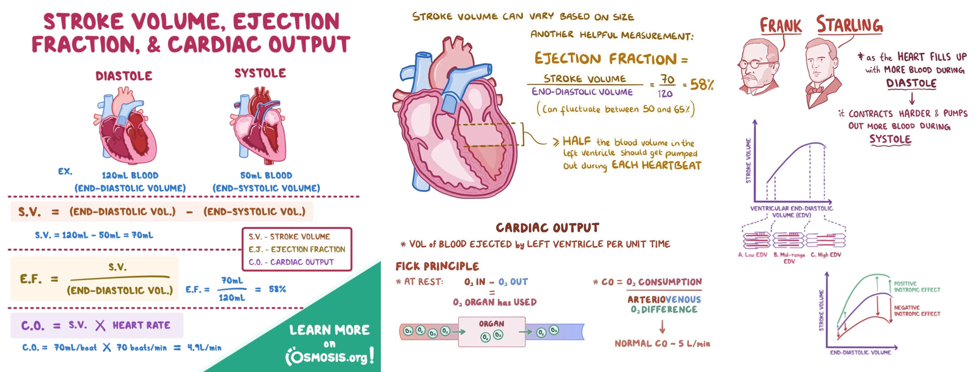
stroke volume is blood pumped with each systole, affected:
1. Frank-Starling mechanism- greater venous return stretches heart, so that it contracts harder (increase venous return by not peeing)
2. change activity level for stronger heart
3. change in posture (orthostatic hypotension)
80
cardiac muscle cells
functional syncytium- the individual cells work with adjacent cells for coordinated action. Rapid transmission of electrical impulses transfers between cells to trigger simultaneous contraction of the heart muscle. Electrical synapses, no chemical synapses
intercalated disks- connection between cardiac muscle cells, contain gap junctions
cardiac conduction system- atria signal is transmitted to ventricles after a delay, since they are separate systems not connected by gap junctions
voltage-gated Ca2+ channels- in addition to voltage-gated Na+ channels, these stay open longer for longer depolarization, greater contraction of cardiac muscle, and less tetany (twitches)
T tubules- plasma membrane dips into cytoplasm, more surface area for Ca2+ channels, also sarcoplasmic reticulum
actin-myosin fibers- contract in response to influx of Ca2+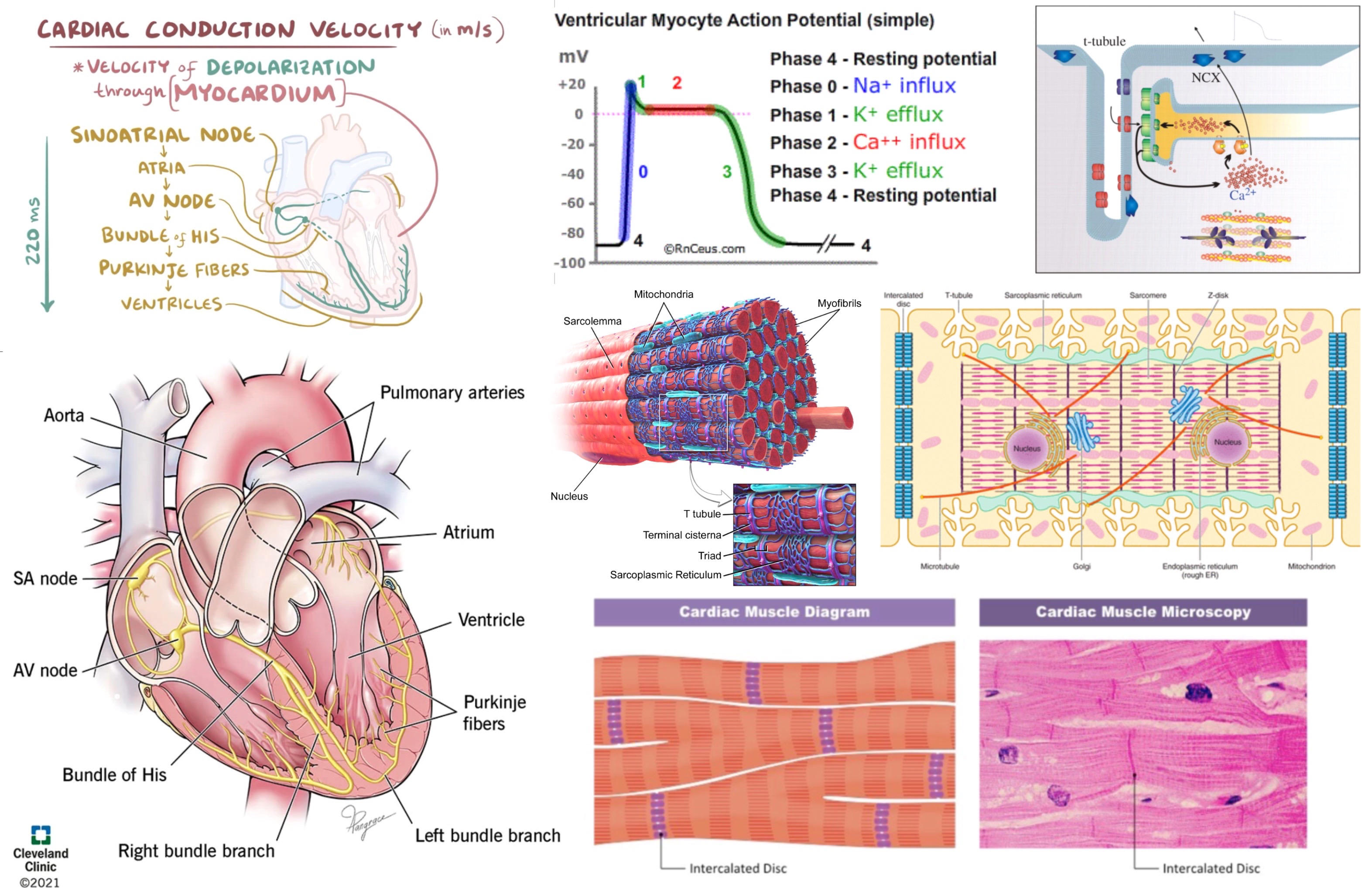
intercalated disks- connection between cardiac muscle cells, contain gap junctions
cardiac conduction system- atria signal is transmitted to ventricles after a delay, since they are separate systems not connected by gap junctions
voltage-gated Ca2+ channels- in addition to voltage-gated Na+ channels, these stay open longer for longer depolarization, greater contraction of cardiac muscle, and less tetany (twitches)
T tubules- plasma membrane dips into cytoplasm, more surface area for Ca2+ channels, also sarcoplasmic reticulum
actin-myosin fibers- contract in response to influx of Ca2+
81
excitation of SA node
sinoatrial (SA) node- pacemaker of the heart
Phase 4- unstable resting potential, Na+ leak channels slowly depolarize cell until threshold potential for voltage-gated Ca+ channels
Phase 0- depolarization, influx of Ca2+ from voltage-gated Ca2+ channels
Phase 3- repolarization, closure of Ca2+ channels, K+ leak channels open and K+ flows out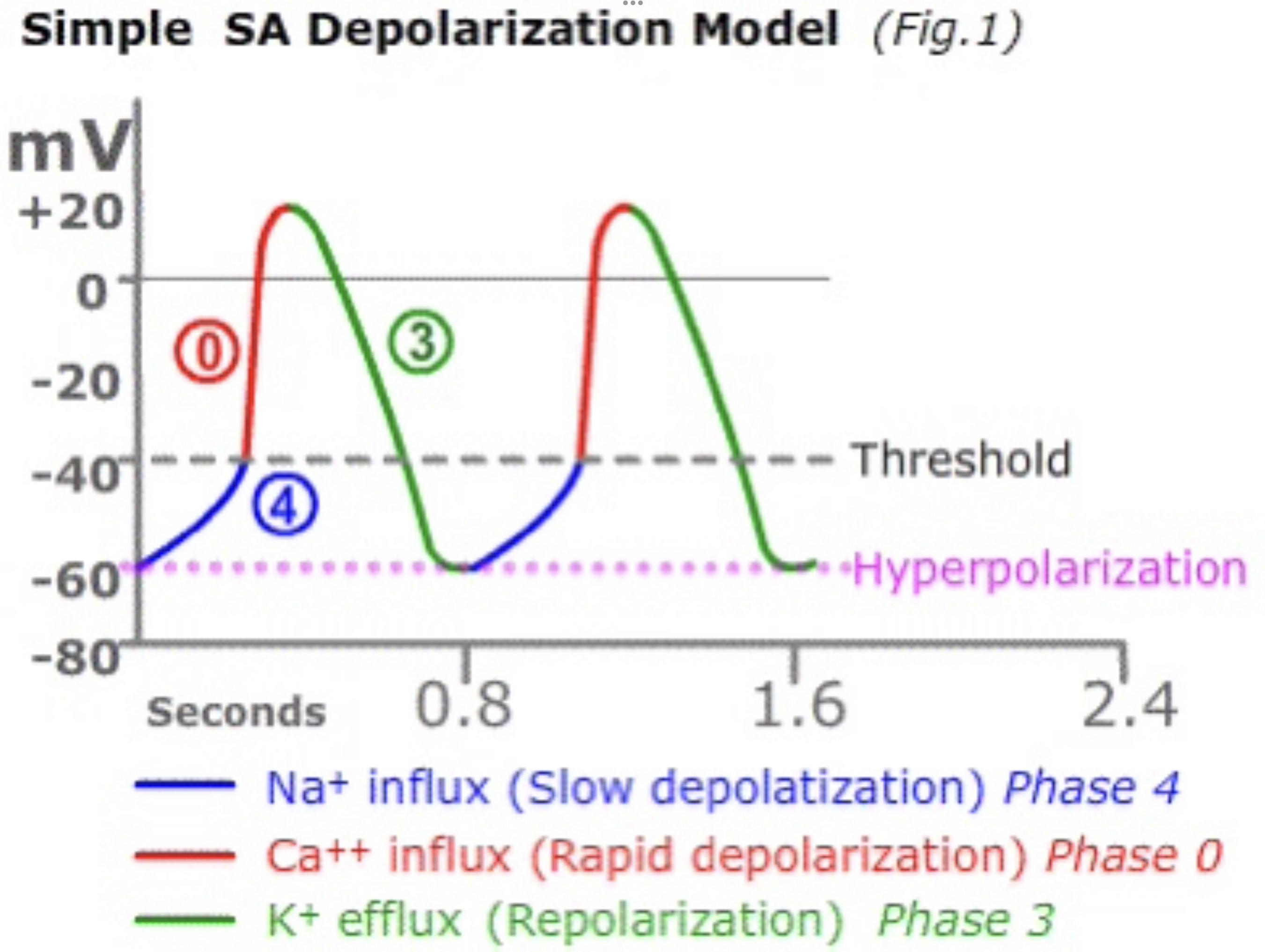
Phase 4- unstable resting potential, Na+ leak channels slowly depolarize cell until threshold potential for voltage-gated Ca+ channels
Phase 0- depolarization, influx of Ca2+ from voltage-gated Ca2+ channels
Phase 3- repolarization, closure of Ca2+ channels, K+ leak channels open and K+ flows out
82
excitation of cardiac muscle cells
Phase 0- depolarization, influx of Na+
Phase 1- initial repolarization, closure of Na+ channels, K+ leak channels open and K+ flows out
Phase 2- plateau phase, influx of Ca2+ from voltage-gated Ca2+ channels balances K+ leak
Phase 3- repolarization, closure of Ca2+ channels, K+ leak channels keep leaking
Phase 4- resting potential, closure of K+ leak channels, maintained by Na+/K+ ATPase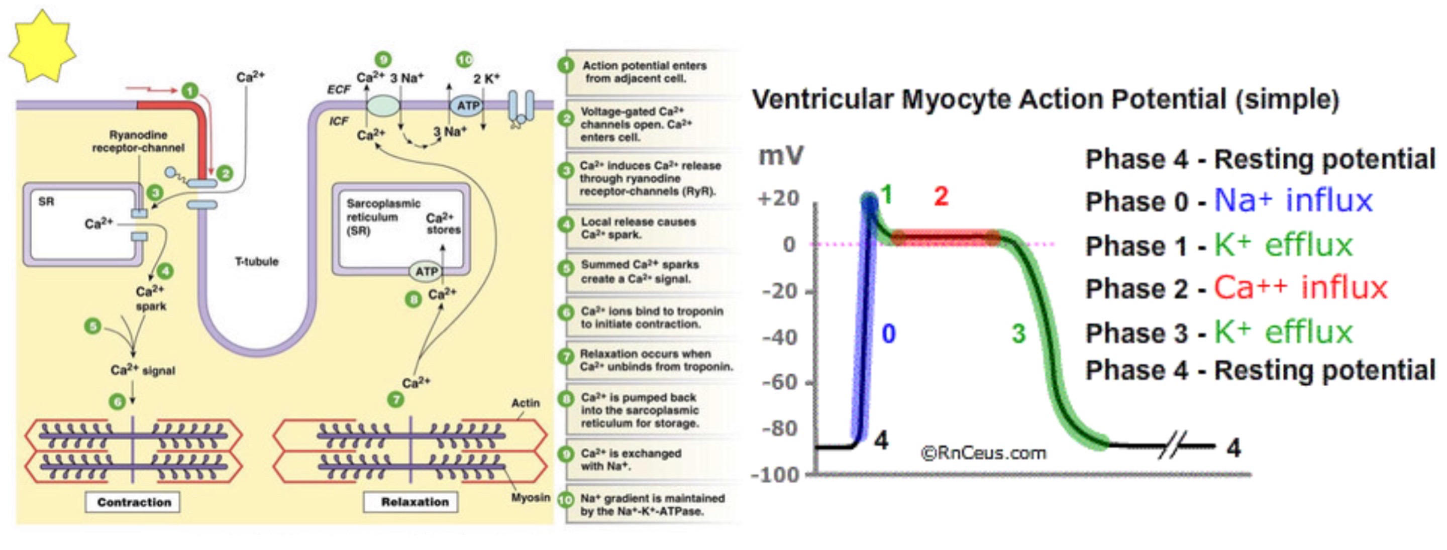
Phase 1- initial repolarization, closure of Na+ channels, K+ leak channels open and K+ flows out
Phase 2- plateau phase, influx of Ca2+ from voltage-gated Ca2+ channels balances K+ leak
Phase 3- repolarization, closure of Ca2+ channels, K+ leak channels keep leaking
Phase 4- resting potential, closure of K+ leak channels, maintained by Na+/K+ ATPase
83
cardiac conduction system
sinoatrial (SA) node- sends impulse to atria
internodal tract
atrioventricular (AV) node- delays impulse to ventricles
bundle of His (AV bundle)
Purkinje fibers- distributes electrical impulse starting at the apex of the heart and moving upwards to pump upwards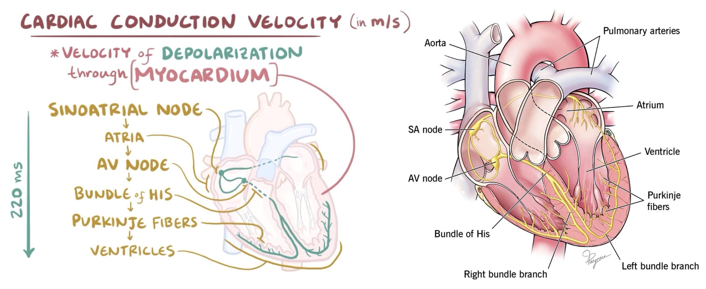
internodal tract
atrioventricular (AV) node- delays impulse to ventricles
bundle of His (AV bundle)
Purkinje fibers- distributes electrical impulse starting at the apex of the heart and moving upwards to pump upwards
84
autonomic NS regulates heart
parasympathetic NS inhibits rapid automaticity- vagus nerve release acetylcholine to signal SA node to slow down
*vagus tone*- degree of inhibition
sympathetic NS stimulates HR- norepinephrine and epinephrine stimulate cardiac muscle cells
baroreceptors- monitor BP, high BP sends signal for vagal tone increase and sympathetic decrease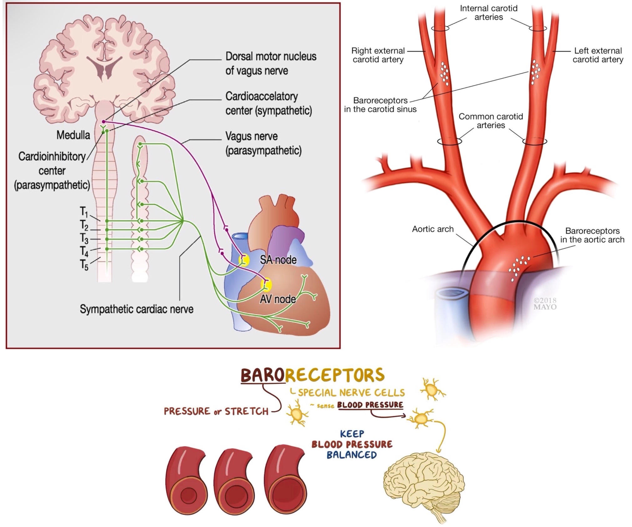
*vagus tone*- degree of inhibition
sympathetic NS stimulates HR- norepinephrine and epinephrine stimulate cardiac muscle cells
baroreceptors- monitor BP, high BP sends signal for vagal tone increase and sympathetic decrease
85
peripheral resistance
pressure gradient = cardiac output (L/min) * peripheral resistance
*adrenergic tone*- sympathetic NS controls peripheral resistance, increase BP
sympathetic NS can also preferentially perfuse certain tissues
local autoregulation- happens with coronary blood flow, certain metabolic wastes build up, automatically trigger high BP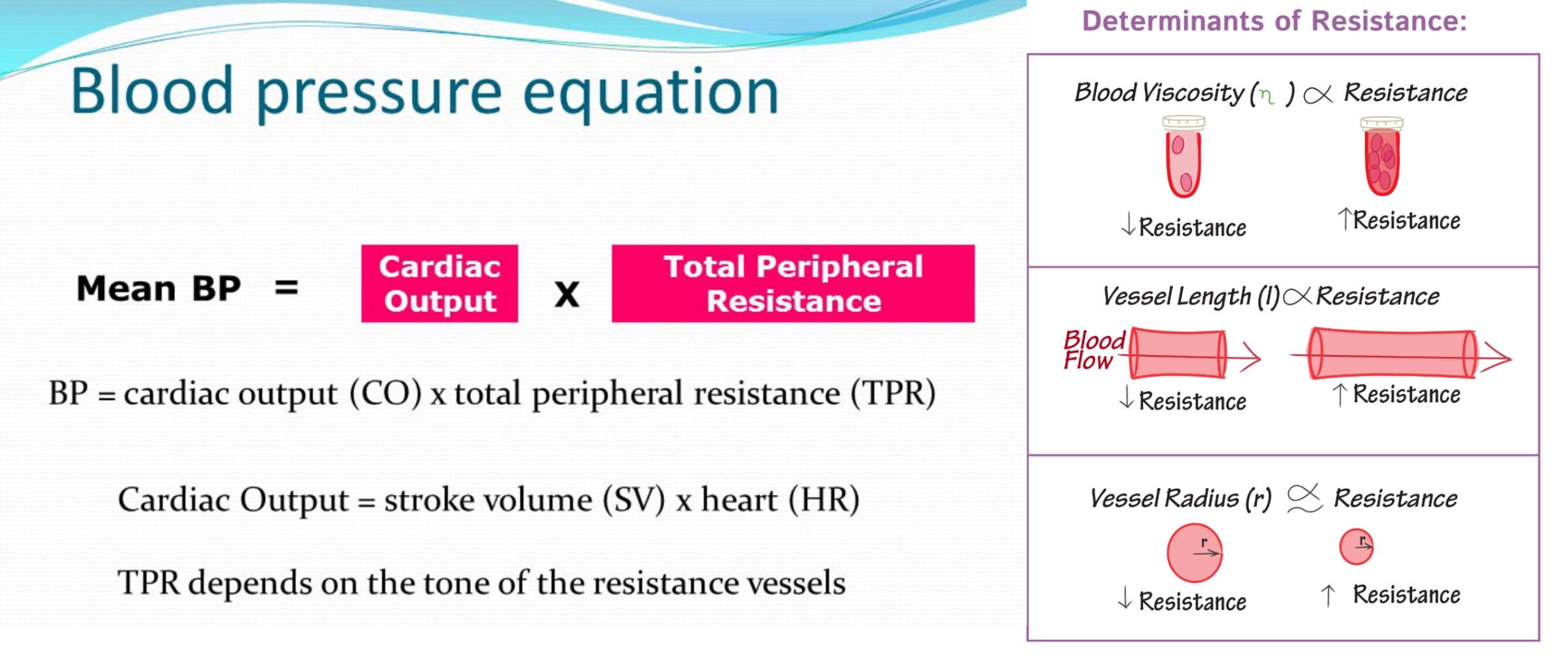
*adrenergic tone*- sympathetic NS controls peripheral resistance, increase BP
sympathetic NS can also preferentially perfuse certain tissues
local autoregulation- happens with coronary blood flow, certain metabolic wastes build up, automatically trigger high BP
86
components of blood
plasma (55%): (mostly water)
1. electrolytes- Na+, K+, Cl-, Ca2+, Mg2+
2. buffers- bicarbonate, maintain pH 7.4
3. glucose
4. albumin- maintain osmotic pressure in capillaries, mobilizes proteins/lipids in serum
5. immunoglobulins- immune system
6. fibrinogen- blood clotting
7. lipoproteins- transport lipids
8. waste- urea (breakdown of amino acids), bilirubin (breakdown of heme)
hematocrit (45%)-include red blood cells
leukocytes (1%)- white blood cells
blood forms 3 layers when centrifuged
most things in blood came from bone marrow stem cells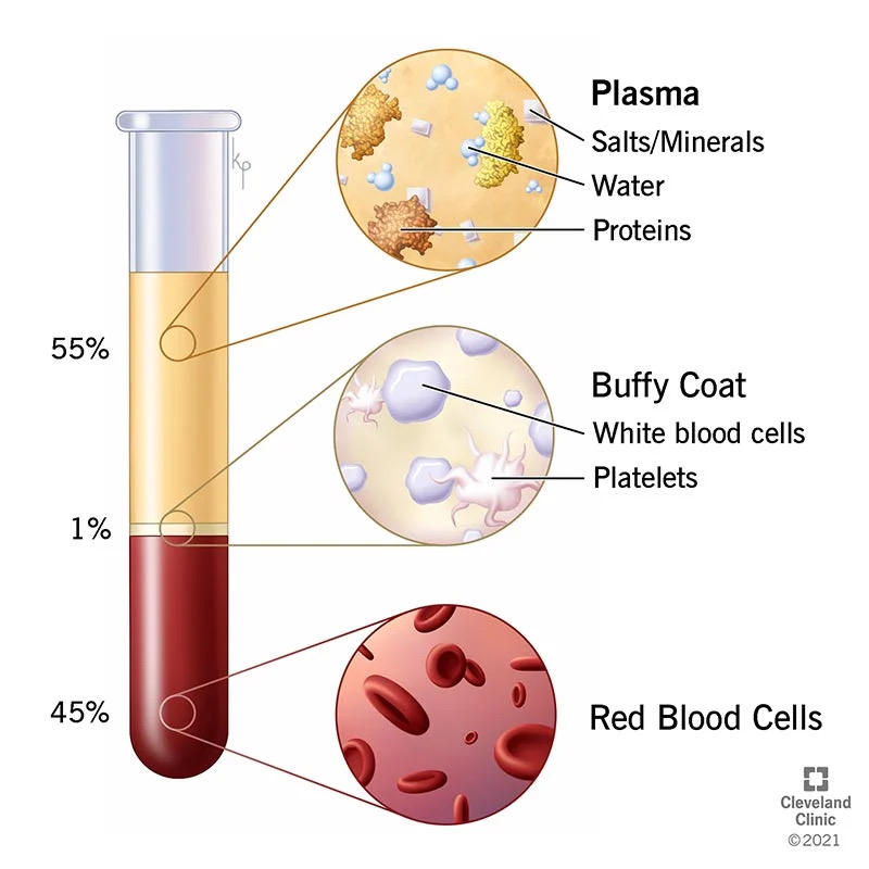
1. electrolytes- Na+, K+, Cl-, Ca2+, Mg2+
2. buffers- bicarbonate, maintain pH 7.4
3. glucose
4. albumin- maintain osmotic pressure in capillaries, mobilizes proteins/lipids in serum
5. immunoglobulins- immune system
6. fibrinogen- blood clotting
7. lipoproteins- transport lipids
8. waste- urea (breakdown of amino acids), bilirubin (breakdown of heme)
hematocrit (45%)-include red blood cells
leukocytes (1%)- white blood cells
blood forms 3 layers when centrifuged
most things in blood came from bone marrow stem cells
87
erythrocytes
erythropoietin- hormone made in kidney, stimulates red blood cell production in red bone marrow
RBCs are made in red bone marrow, stored and destroyed in spleen
fun facts:
1. has no organelles, no DNA
2. rely on glycolysis for ATP synthesis
3. contains hemoglobin to transport O2, also CO2
4. biconcave shape has large surface area for gas exchange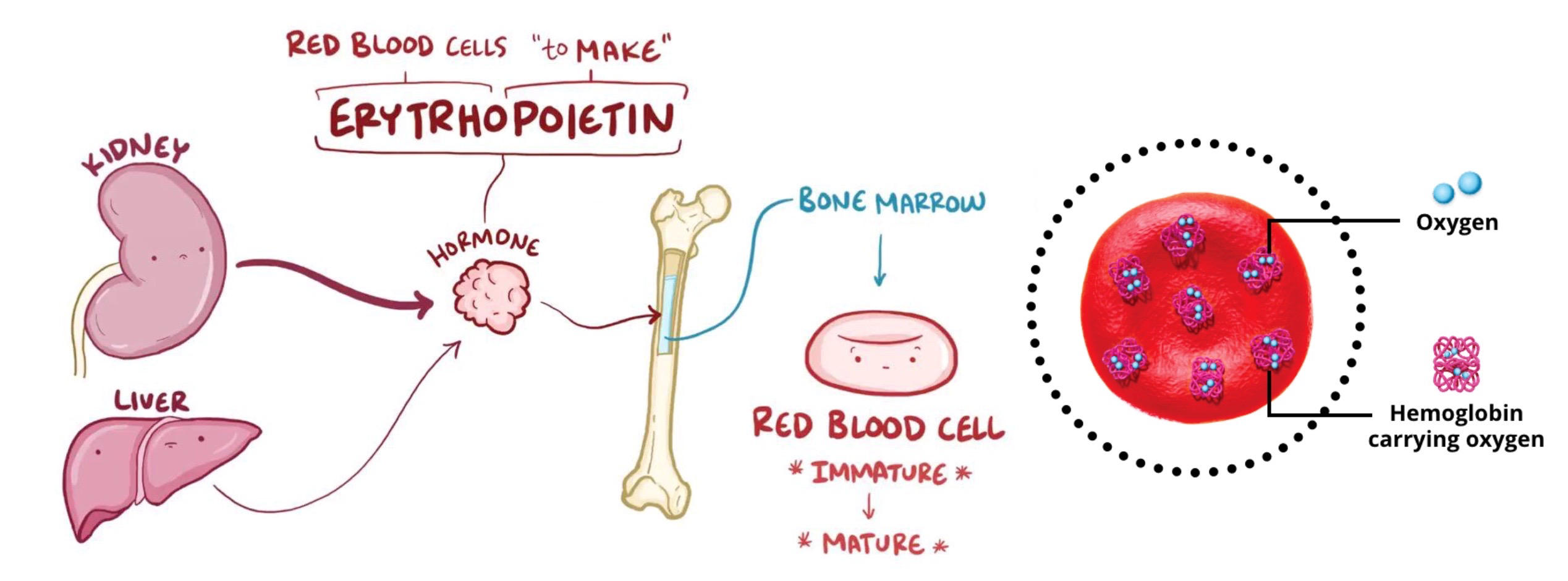
RBCs are made in red bone marrow, stored and destroyed in spleen
fun facts:
1. has no organelles, no DNA
2. rely on glycolysis for ATP synthesis
3. contains hemoglobin to transport O2, also CO2
4. biconcave shape has large surface area for gas exchange
88
transfusion reaction
incorrect A/B/Rh antigen in blood causes clumping and destruction of RBCs
sensitization- Rh- patient must be exposed to Rh+ blood first to develop antigen, then future exposure to Rh+ blood will cause transfusion reaction
hemolytic disease of the newborn:
1. Rh- mother delivers Rh+ child, becoming sensitized to Rh+
2. future Rh+ babies at risk since Rh antibodies can cross placental barrier to destroy baby's RBCs
3. injecting mother with anti-Rh during first child can prevent sensitization
AB+ blood has all 3 antigens, universal recipient
O- has no antigens, universal donor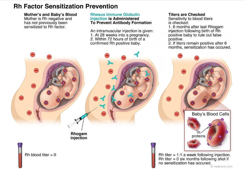
sensitization- Rh- patient must be exposed to Rh+ blood first to develop antigen, then future exposure to Rh+ blood will cause transfusion reaction
hemolytic disease of the newborn:
1. Rh- mother delivers Rh+ child, becoming sensitized to Rh+
2. future Rh+ babies at risk since Rh antibodies can cross placental barrier to destroy baby's RBCs
3. injecting mother with anti-Rh during first child can prevent sensitization
AB+ blood has all 3 antigens, universal recipient
O- has no antigens, universal donor
89
hemostasis
body's ability to prevent bleeding, uses platelets
platelets- thrombocytes, produced in the bone marrow, cell fragments that do not have nuclei
platelet plug- platelets aggregate at site of damage
fibrin- protein that holds the platelet plug together
thrombin- converts fibrinogen to fibrin
thrombus- blood clot
hemophilia- defect in a clotting protein, excessive bleeding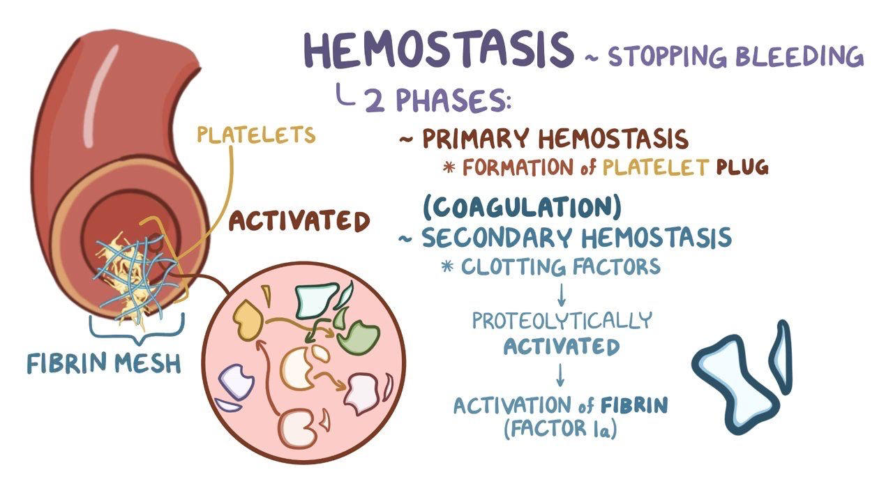
platelets- thrombocytes, produced in the bone marrow, cell fragments that do not have nuclei
platelet plug- platelets aggregate at site of damage
fibrin- protein that holds the platelet plug together
thrombin- converts fibrinogen to fibrin
thrombus- blood clot
hemophilia- defect in a clotting protein, excessive bleeding
90
O2 transport
hemoglobin- 4 identical subunits with a heme (Fe in middle to bind O2) in each, carried on RBCs and are not in plasma!
cooperativity- when one subunit goes from tense to relaxed and binds O2, the other subunits become relaxed as well and thus have higher affinity for O2
Hill coefficient- 1 is no cooperativity, 4 is perfect cooperativity
4 factors decrease hemoglobin affinity for O2:
1. low O2 environment- cooperativity
2. low pH- Bohr effect
3. high CO2 environment
4. high temperature
% saturation = hemes with O2/total hemes
O2-hemoglobin dissociation curve show cooperativity
fetal Hb has higher affinity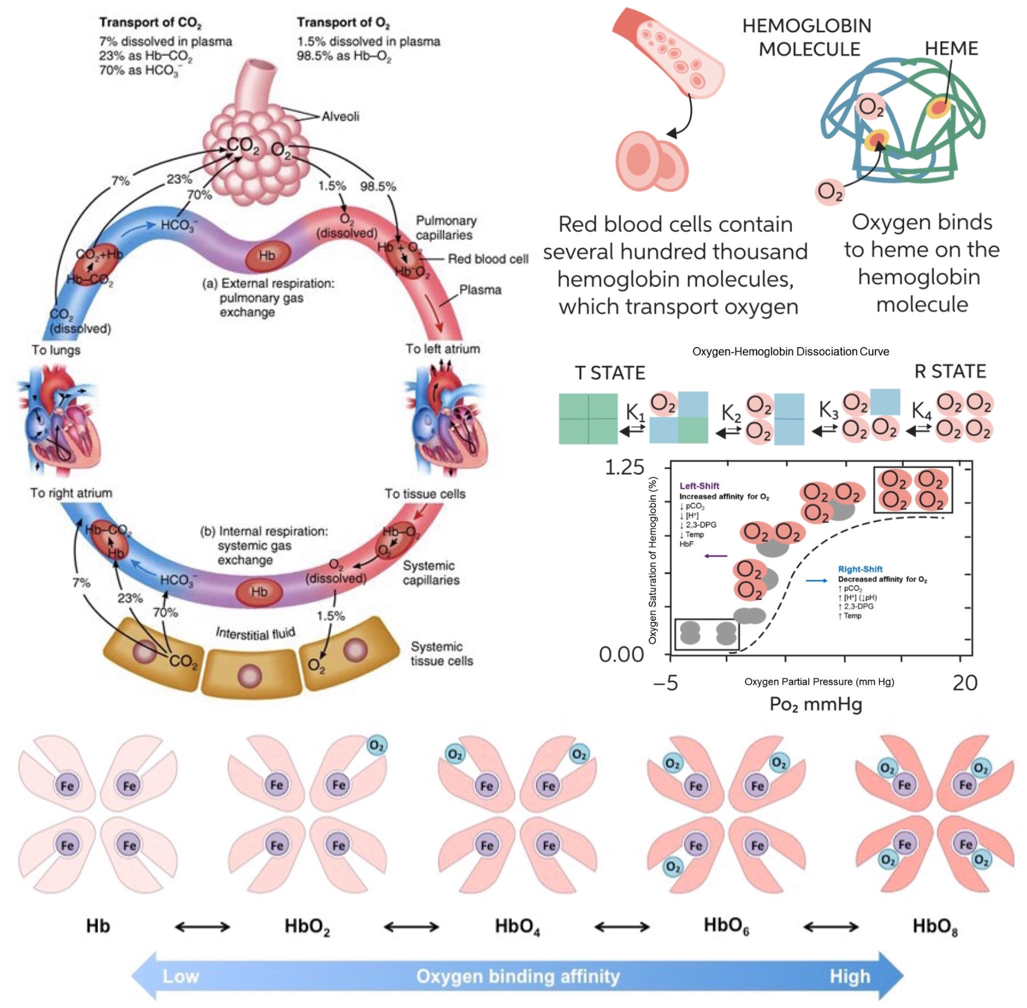
cooperativity- when one subunit goes from tense to relaxed and binds O2, the other subunits become relaxed as well and thus have higher affinity for O2
Hill coefficient- 1 is no cooperativity, 4 is perfect cooperativity
4 factors decrease hemoglobin affinity for O2:
1. low O2 environment- cooperativity
2. low pH- Bohr effect
3. high CO2 environment
4. high temperature
% saturation = hemes with O2/total hemes
O2-hemoglobin dissociation curve show cooperativity
fetal Hb has higher affinity
91
oxygen debt
during physical activity, muscles do not get enough oxygen
1. anaerobic glycolysis- produce more ATP without oxygen, lactic acid is a byproduct
2. fast twitch fibers (white)- less mitochondria, use anaerobic glycolysis unlike slow twitch fibers (red)
3. creatine phosphate- supplemental energy reserve that produces more ATP, creatine is a byproduct
4. myoglobin- O2 is taken from hemoglobin and stored in myoglobin (higher O2 affinity) in muscles, can be accessed if more oxygen is needed
5. Bohr effect- oxygen saturation curve shifts down because of increased CO2/H+, allows more O2 to be dumped from hemoglobin at muscles
6. increased RR/HR- increase oxygenation
7. vasodilation- increase oxygenation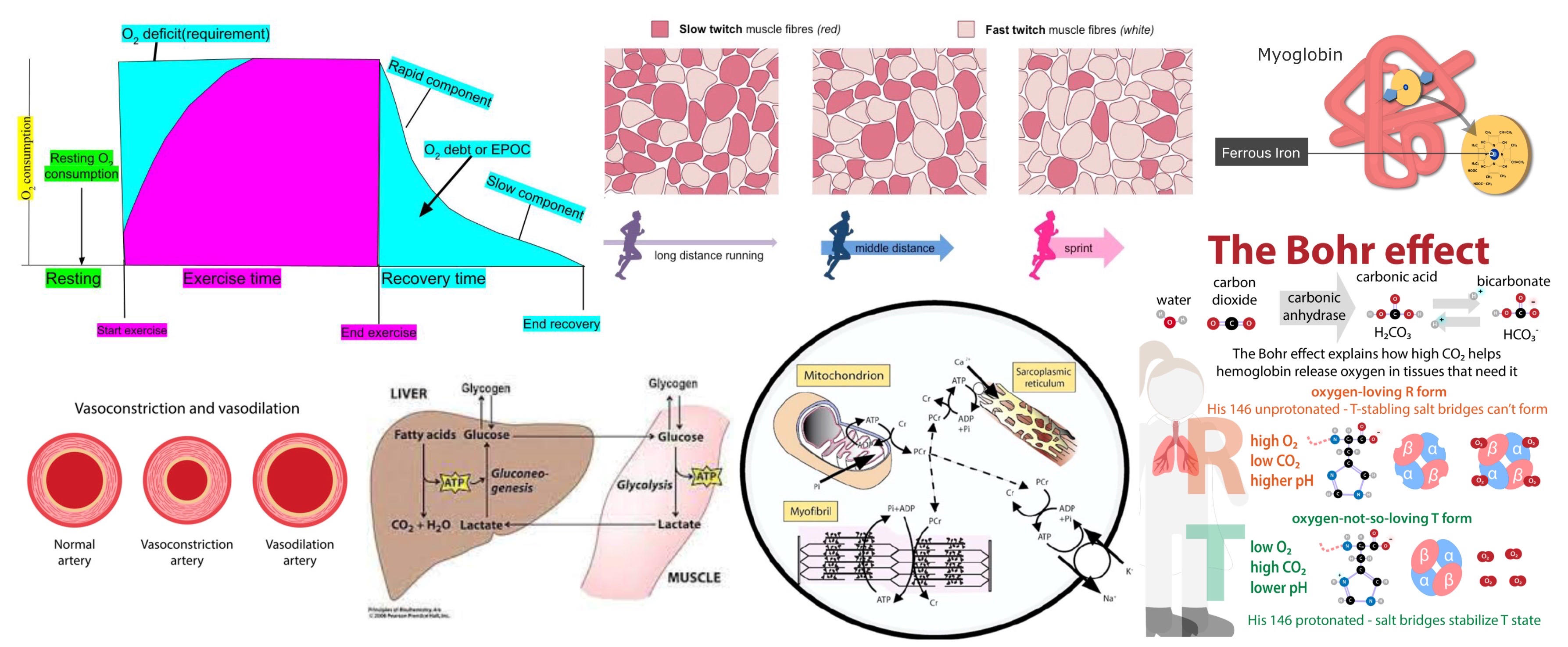
1. anaerobic glycolysis- produce more ATP without oxygen, lactic acid is a byproduct
2. fast twitch fibers (white)- less mitochondria, use anaerobic glycolysis unlike slow twitch fibers (red)
3. creatine phosphate- supplemental energy reserve that produces more ATP, creatine is a byproduct
4. myoglobin- O2 is taken from hemoglobin and stored in myoglobin (higher O2 affinity) in muscles, can be accessed if more oxygen is needed
5. Bohr effect- oxygen saturation curve shifts down because of increased CO2/H+, allows more O2 to be dumped from hemoglobin at muscles
6. increased RR/HR- increase oxygenation
7. vasodilation- increase oxygenation
92
CO2 transport
CO2 + H2O <-> H2CO3 (carbonic acid) <-> HCO3- (bicarbonate) + H+
this reaction occurs within RBCs, HCO3- exits RBC by exchanging with Cl-
3 ways that CO2 is transported:
1. carbonic anhydrase- enzyme in RBCs that catalyzes rxn
2. attached to hemoglobin (not in O2 position)
3. more water soluble than O2, so in plasma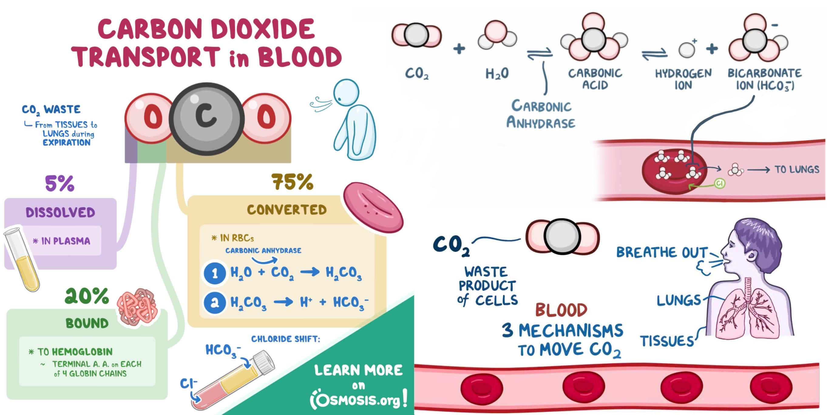
this reaction occurs within RBCs, HCO3- exits RBC by exchanging with Cl-
3 ways that CO2 is transported:
1. carbonic anhydrase- enzyme in RBCs that catalyzes rxn
2. attached to hemoglobin (not in O2 position)
3. more water soluble than O2, so in plasma
93
capillary exchange
things that must be exchanged in capillaries:
1. nutrients- amino acids and glucose
2. waste- liver removes wastes and secretes bile
3. lymphocytes- WBCs to attack invaders in tissue
4. water- albumin maintains oncotic pressure to keep water in blood vessels
5. inflammation- WBCs and water flow out of blood vessels to cause edema
nutrient exchange:
1. hepatic portal vein- carries amino acids and glucose from digestive tract to liver
2.chylomicrons- lipoproteins that store fat, enter lacteals in lymphatic system, taken up by liver, converted, carries fat to adipocytes for storage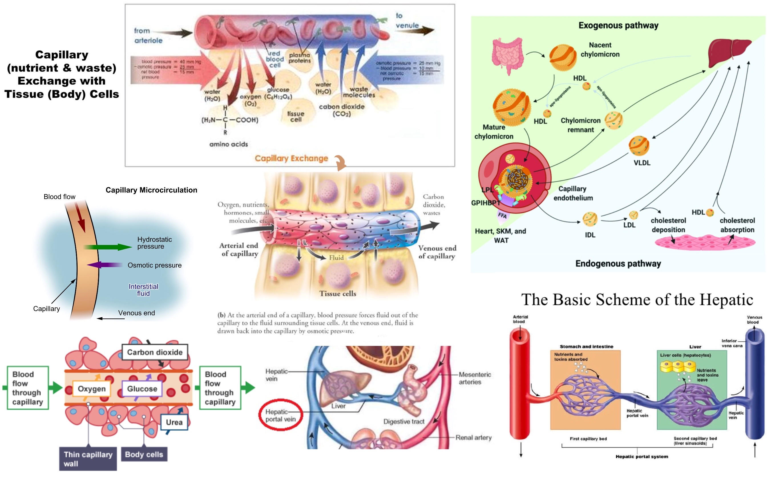
1. nutrients- amino acids and glucose
2. waste- liver removes wastes and secretes bile
3. lymphocytes- WBCs to attack invaders in tissue
4. water- albumin maintains oncotic pressure to keep water in blood vessels
5. inflammation- WBCs and water flow out of blood vessels to cause edema
nutrient exchange:
1. hepatic portal vein- carries amino acids and glucose from digestive tract to liver
2.chylomicrons- lipoproteins that store fat, enter lacteals in lymphatic system, taken up by liver, converted, carries fat to adipocytes for storage
94
innate/natural immunity
general protection against invaders:
1. skin
2. lysozyme- enzyme in tears/saliva/ blood that kills bacteria
3. stomach acidity
4. macrophages and neutrophils
5. complement system- blood proteins that nonspecifically bind to foreign cells, signal for destruction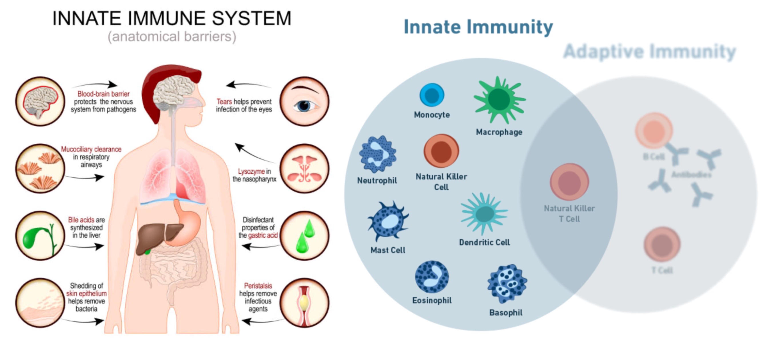
1. skin
2. lysozyme- enzyme in tears/saliva/ blood that kills bacteria
3. stomach acidity
4. macrophages and neutrophils
5. complement system- blood proteins that nonspecifically bind to foreign cells, signal for destruction
95
humoral immunity
specific protection by antibodies (immunoglobulins), produced by B cells
antibody structure:
1. light and heavy chains joined by disulfide bonds
2. constant region- classes include IgG, IgA, IgM, IgD, IgE
3. variable region- specific antigen binding
antibody function:
1. epitope- small binding site on antigen
2. hapten- small molecule that is only recognized after bound to large carrier protein
3. can directly inactivate antigen, induce phagocytosis, or activate complement system
antibodies generated in one species will get recognized as a foreign substance in a different species
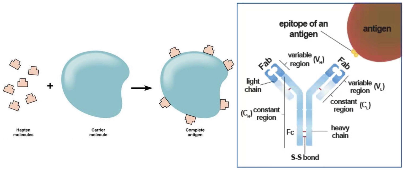
antibody structure:
1. light and heavy chains joined by disulfide bonds
2. constant region- classes include IgG, IgA, IgM, IgD, IgE
3. variable region- specific antigen binding
antibody function:
1. epitope- small binding site on antigen
2. hapten- small molecule that is only recognized after bound to large carrier protein
3. can directly inactivate antigen, induce phagocytosis, or activate complement system
antibodies generated in one species will get recognized as a foreign substance in a different species
96
B cells
B cell development:
1. lymphocytes in humoral immunity derived from bone marrow stem cells
2. antibody proteins assembled by recombination to create different B cells, allows for clonal selection
3. antigen binding triggers differentiation to plasma cell or memory cell
4. plasma cells- actively secrete antibody protein into plasma
5. memory cells- become dormant, waiting for same antigen to reappear then becomes activated
primary immune response- just plasma cells on first exposure to antigen
secondary immune response- memory cells respond quickly, vaccination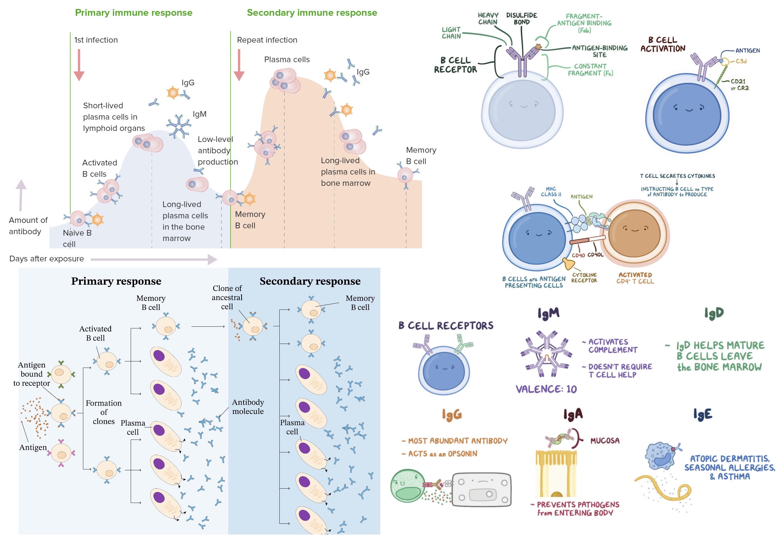
1. lymphocytes in humoral immunity derived from bone marrow stem cells
2. antibody proteins assembled by recombination to create different B cells, allows for clonal selection
3. antigen binding triggers differentiation to plasma cell or memory cell
4. plasma cells- actively secrete antibody protein into plasma
5. memory cells- become dormant, waiting for same antigen to reappear then becomes activated
primary immune response- just plasma cells on first exposure to antigen
secondary immune response- memory cells respond quickly, vaccination
97
T cells
T-cell development:
1. lymphocytes in cell-mediated immunity, T helpers and T killers
2. T cells are produced in bone marrow, but mature in *thymus gland*
3. T helpers (CD4)- activates B cells and T killer cells, communicates with hormones (lymphokines, interleukins), host of HIV
4. T killers (CD8)- destroy abnormal hosts cells (virus infected, cancer, foreign grafts)
MHC I- every cell has them, displays foreign protein on surface of cell to be *detected by T killers*
MHC II- only on antigen-presenting cells (macrophages and B cells), displays foreign protein after chopping it up to be *detected by T helpers*
T cell activation only occurs when T cell binds to both antigen and MHC molecule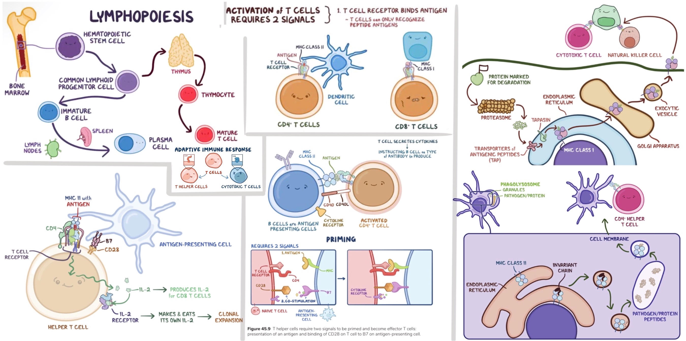
1. lymphocytes in cell-mediated immunity, T helpers and T killers
2. T cells are produced in bone marrow, but mature in *thymus gland*
3. T helpers (CD4)- activates B cells and T killer cells, communicates with hormones (lymphokines, interleukins), host of HIV
4. T killers (CD8)- destroy abnormal hosts cells (virus infected, cancer, foreign grafts)
MHC I- every cell has them, displays foreign protein on surface of cell to be *detected by T killers*
MHC II- only on antigen-presenting cells (macrophages and B cells), displays foreign protein after chopping it up to be *detected by T helpers*
T cell activation only occurs when T cell binds to both antigen and MHC molecule
98
leukocytes
white blood cells
3 types:
1. lymphocytes- B cells, T cells, NK cells
2. neutrophils- phagocytosis, most common
3. monocytes- phagocytosis, move to tissue through blood to become macrophages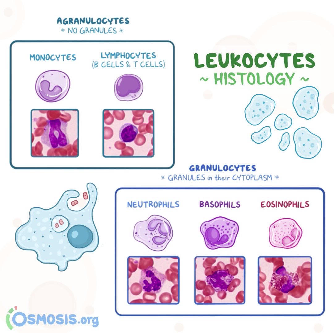
3 types:
1. lymphocytes- B cells, T cells, NK cells
2. neutrophils- phagocytosis, most common
3. monocytes- phagocytosis, move to tissue through blood to become macrophages
99
innate vs adaptive immunity
innate/natural- general immune response, monocytes, neutrophils, NK cells
adaptive immunity- antigen specific response, B cells (humoral), T cells (cell-mediated)
humoral/cell-mediated are actually parts of both systems (innate and adaptive)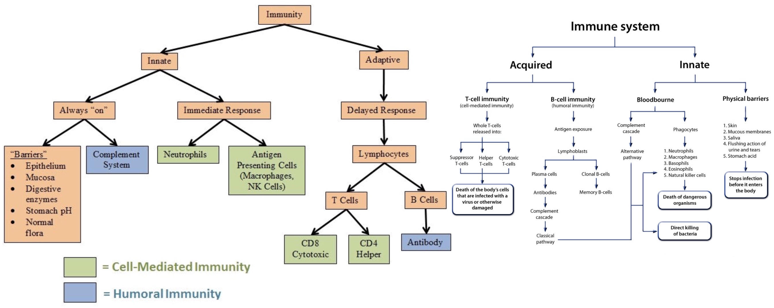
adaptive immunity- antigen specific response, B cells (humoral), T cells (cell-mediated)
humoral/cell-mediated are actually parts of both systems (innate and adaptive)
100
passive immunity
an individual does not produce his or her own antibodies, but rather receives them directly from another source, such as mother to infant through breast milk
101
other tissues in immune response
bone marrow- synthesis of all cells in blood
lymph nodes
spleen- filters blood, destroys old RBCs
thymus gland- T cell maturation
tonsils and appendix- catch pathogens, not necessary
lymph nodes
spleen- filters blood, destroys old RBCs
thymus gland- T cell maturation
tonsils and appendix- catch pathogens, not necessary
102
autoimmunity
tolerance- immune system ignores normal cells
B cells are selected in bone marrow, T cells in thymus gland
autoimmunity- those that attack normal cells become anergic (unresponsive) or undergo apoptosis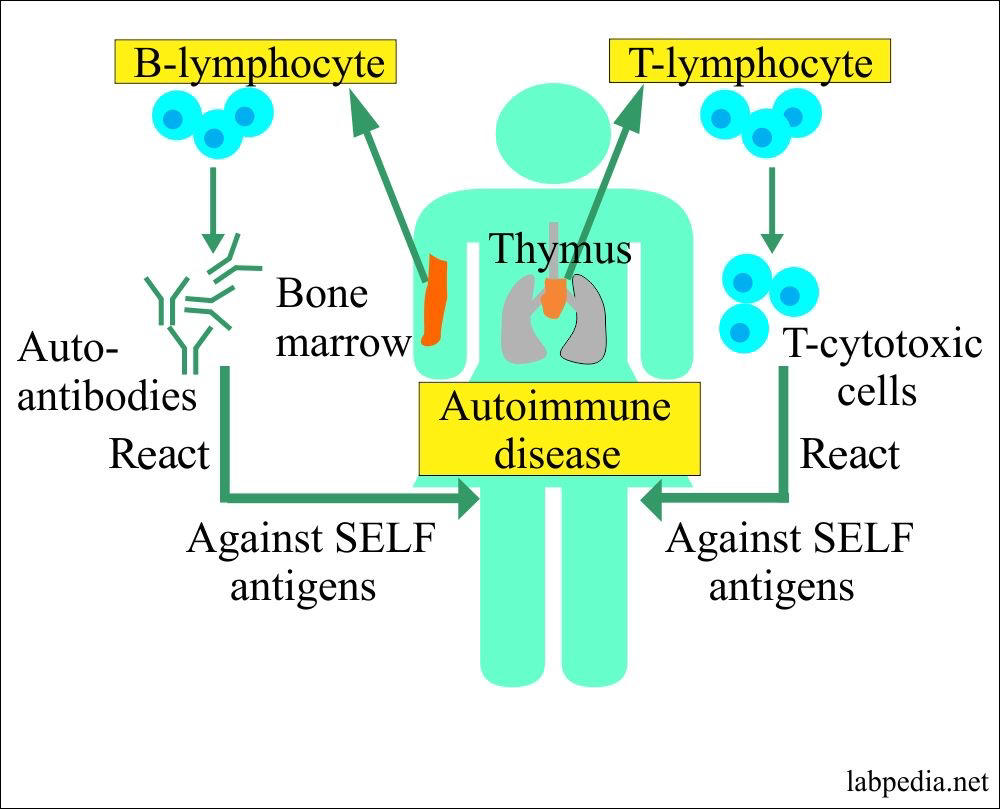
B cells are selected in bone marrow, T cells in thymus gland
autoimmunity- those that attack normal cells become anergic (unresponsive) or undergo apoptosis
103
excretory system
liver- breaks down hydrophobic large waste products, secretes bile, synthesizes urea from free ammonia (toxic)
colon- large intestine reabsorbs water and ions from feces
skin- produces sweat, contains water, ions, and urea
kidneys- keeps everything at optimum levels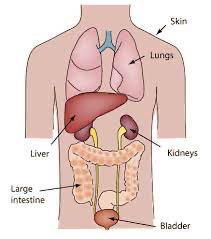
colon- large intestine reabsorbs water and ions from feces
skin- produces sweat, contains water, ions, and urea
kidneys- keeps everything at optimum levels
104
urine formation
filtration- pressurized blood passed over filter, water and small molecules pass into renal tubule to form filtrate
selective reabsorption- glucose, water, proteins are put back in blood
secretion- more waste is added to the filtrate
concentration- more reabsorption of water, makes concentrated urine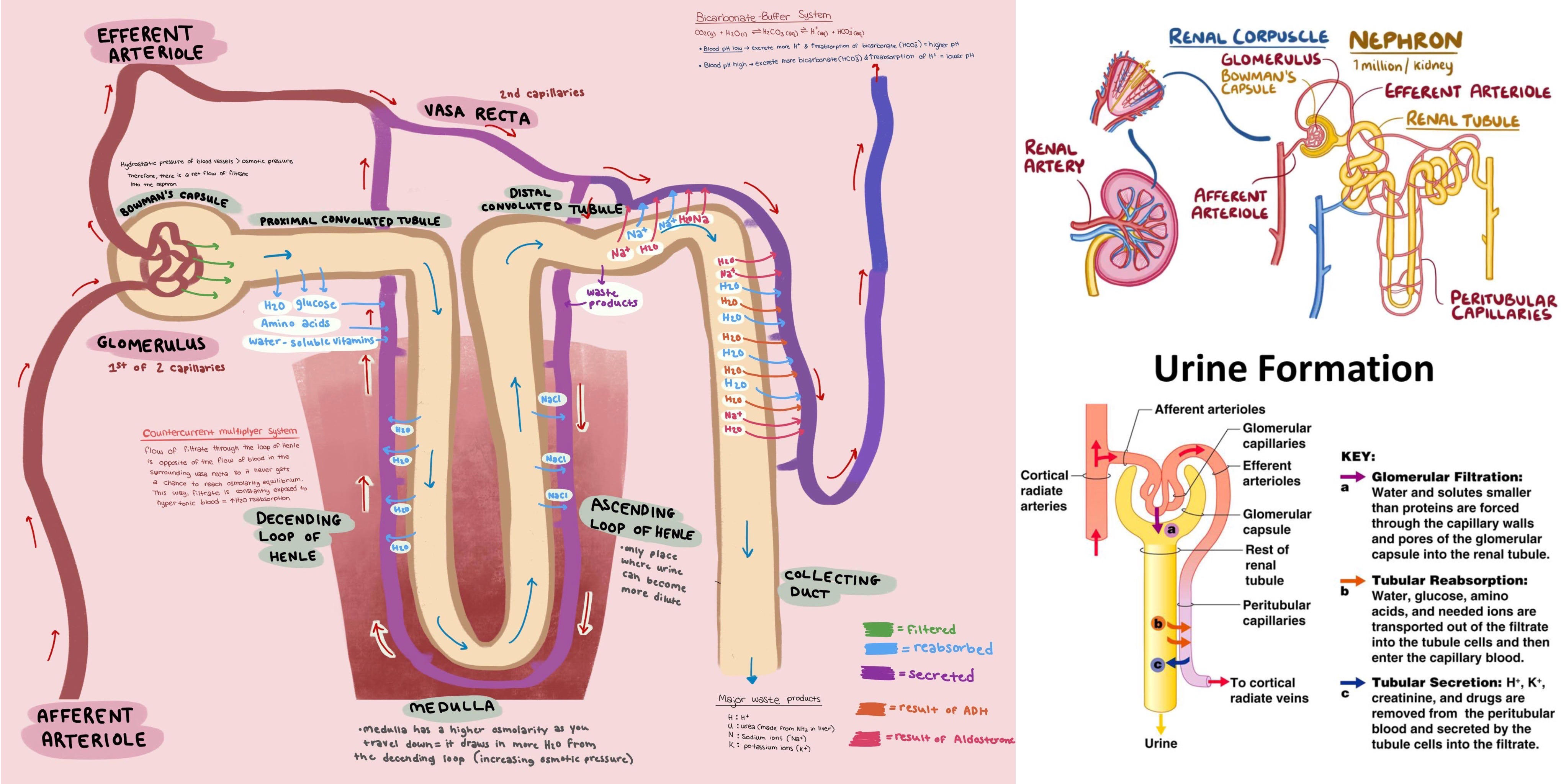
selective reabsorption- glucose, water, proteins are put back in blood
secretion- more waste is added to the filtrate
concentration- more reabsorption of water, makes concentrated urine
105
urinary system
renal artery
kidney
ureter
urinary bladder
internal/external sphincter
urethra
cortex
medulla
collecting ducts
papilla
calyx
renal pelvis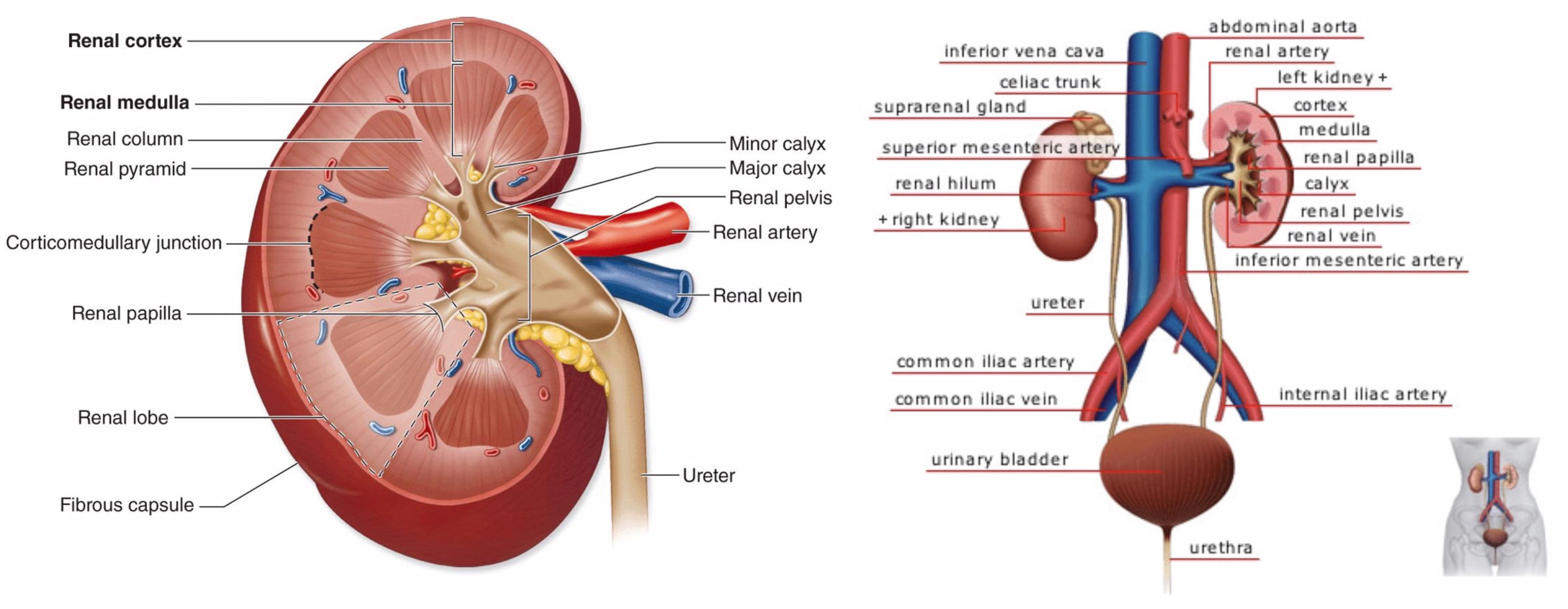
kidney
ureter
urinary bladder
internal/external sphincter
urethra
cortex
medulla
collecting ducts
papilla
calyx
renal pelvis
106
parts of the nephron
1. afferent arteriole
2. glomerulus
3. efferent arteriole- includes cells and proteins
4. Bowman's capsule
5. proximal convoluted tubule- water and solute reabsorption
6. loop of Henle- countercurrent multiplier, descending limb reabsorbs water, ascending limb reabsorbs ions to medulla, allows for better water reabsorption in collecting duct
7. distal convoluted tubule- more regulated reabsorption, secretion of toxins into urine
8. collecting duct- secretion of toxins into urine
9. calyces
peritubular capillaries- reabsorption via active transport, drain back to renal vein
vasa recta- countercurrent to loop of Henle, brings reabsorbed water back to blood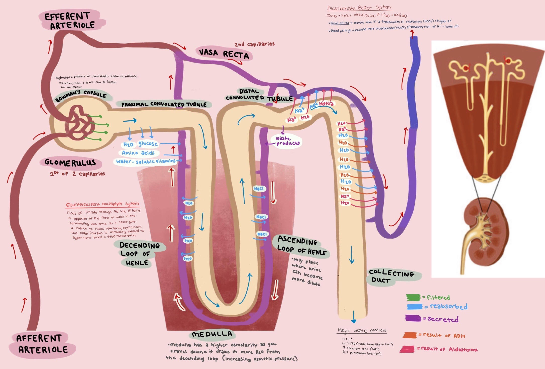
2. glomerulus
3. efferent arteriole- includes cells and proteins
4. Bowman's capsule
5. proximal convoluted tubule- water and solute reabsorption
6. loop of Henle- countercurrent multiplier, descending limb reabsorbs water, ascending limb reabsorbs ions to medulla, allows for better water reabsorption in collecting duct
7. distal convoluted tubule- more regulated reabsorption, secretion of toxins into urine
8. collecting duct- secretion of toxins into urine
9. calyces
peritubular capillaries- reabsorption via active transport, drain back to renal vein
vasa recta- countercurrent to loop of Henle, brings reabsorbed water back to blood
107
glomerulus
uses passive flow due to pressure difference
filters blood into Bowman's capsule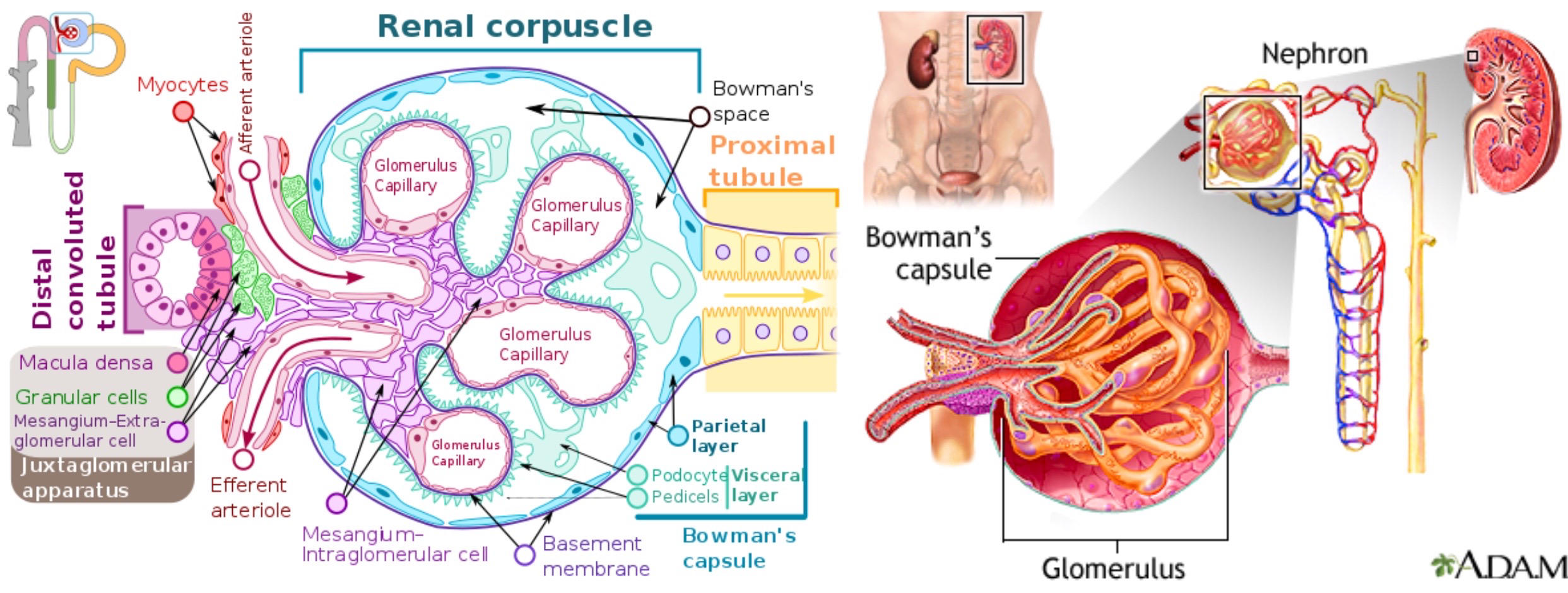
filters blood into Bowman's capsule
108
collecting duct
cortical portion- first
medullary portion- second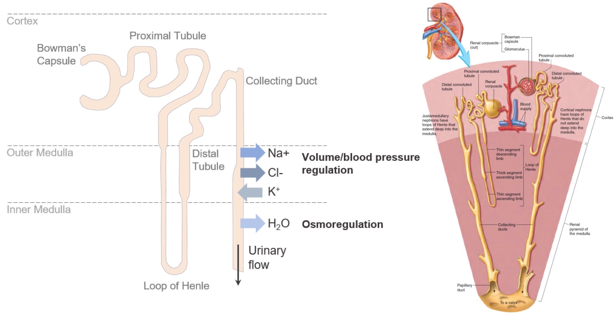
medullary portion- second
109
kidneys control blood pressure
antidiuretic hormone (*ADH*)- increase water reabsorption in distal tubule and collecting duct, increases BP
*aldosterone*- increase Na+ reabsorption in distal tubule and collecting duct, increases plasma osmolarity, increases water retention, increases BP
*renin-angiotensin* system:
macula densa/juxtaglomerular cells- have baroreceptors to sense BP
renin- when BP drops, JG cells secrete renin to blood, converts angiotensinogen (made in liver) into angiotensin I
angiotensin-converting enzyme (ACE)- converts angiotensin I to angiotensin II
*angiotensin II*-vasoconstrictor (fast), also stimulates release of *aldosterone* (slow) to increase BP
ANP- from heart, vasodilator, inhibits renin release, inhibits aldosterone release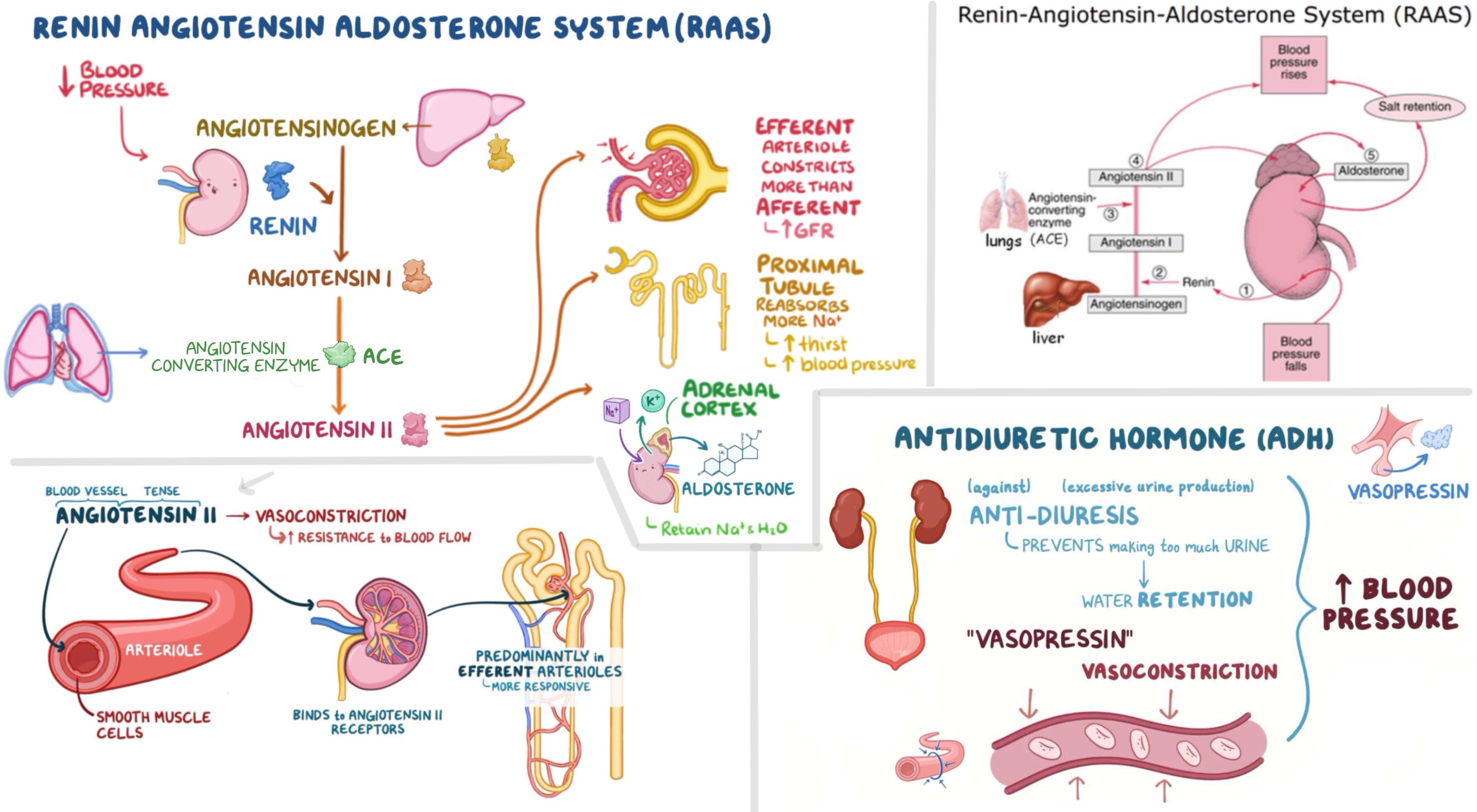
*aldosterone*- increase Na+ reabsorption in distal tubule and collecting duct, increases plasma osmolarity, increases water retention, increases BP
*renin-angiotensin* system:
macula densa/juxtaglomerular cells- have baroreceptors to sense BP
renin- when BP drops, JG cells secrete renin to blood, converts angiotensinogen (made in liver) into angiotensin I
angiotensin-converting enzyme (ACE)- converts angiotensin I to angiotensin II
*angiotensin II*-vasoconstrictor (fast), also stimulates release of *aldosterone* (slow) to increase BP
ANP- from heart, vasodilator, inhibits renin release, inhibits aldosterone release
110
kidneys control pH
when pH is too high, HCO3- is excreted
when pH is too low, H+ is excreted
carbonic anhydrase converts CO2 to H2CO3
usually reabsorb bicarbonate and excrete H+
hyperventilation also raises pH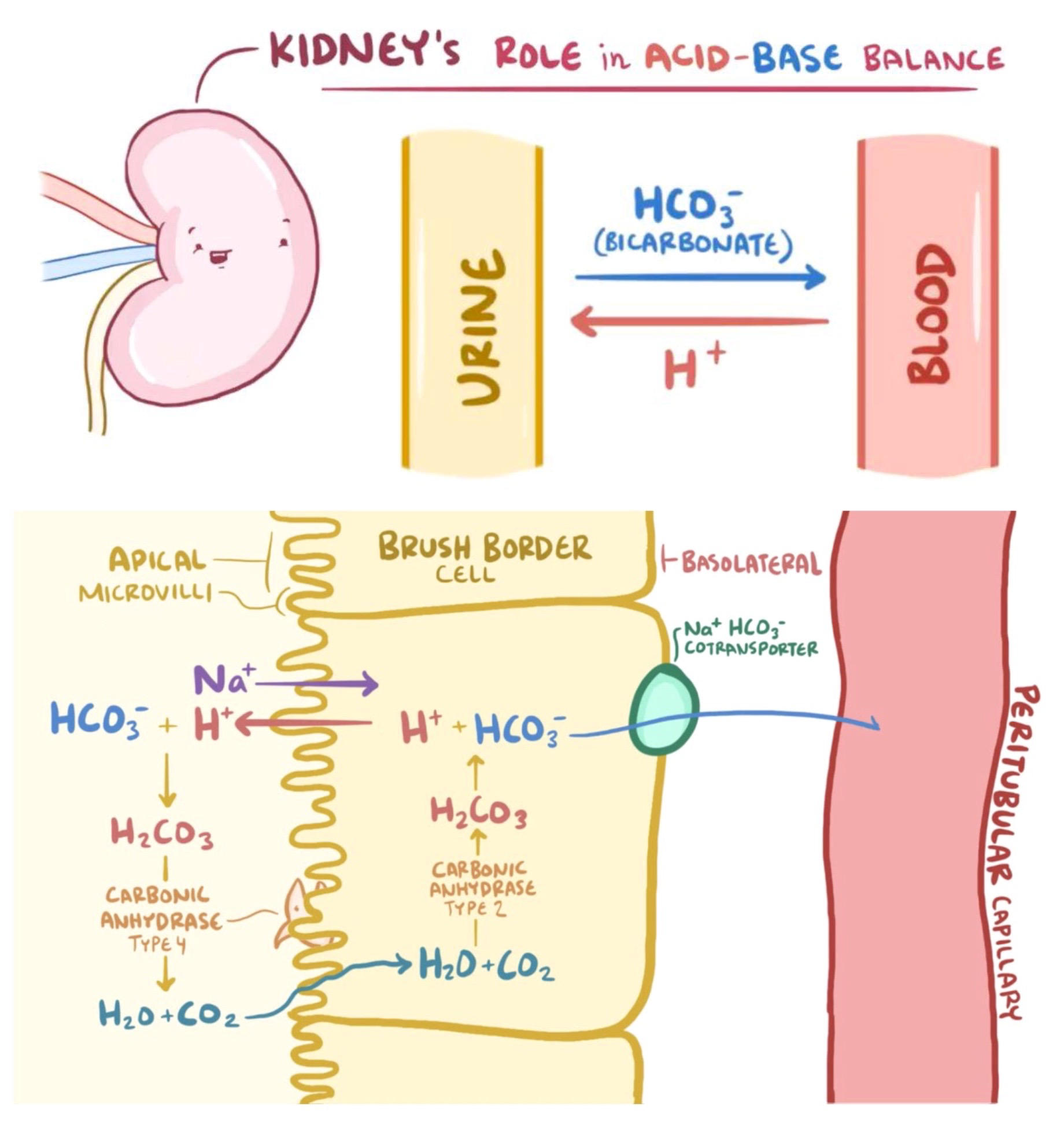
when pH is too low, H+ is excreted
carbonic anhydrase converts CO2 to H2CO3
usually reabsorb bicarbonate and excrete H+
hyperventilation also raises pH
111
alimentary canal
gastrointestinal (GI) tract extending from mouth to anus
made of smooth muscle
peristalsis- moving bolus of food down GI tract:
1. longitudinal layer- propel bolus forward
2. circular layer- prevents backflow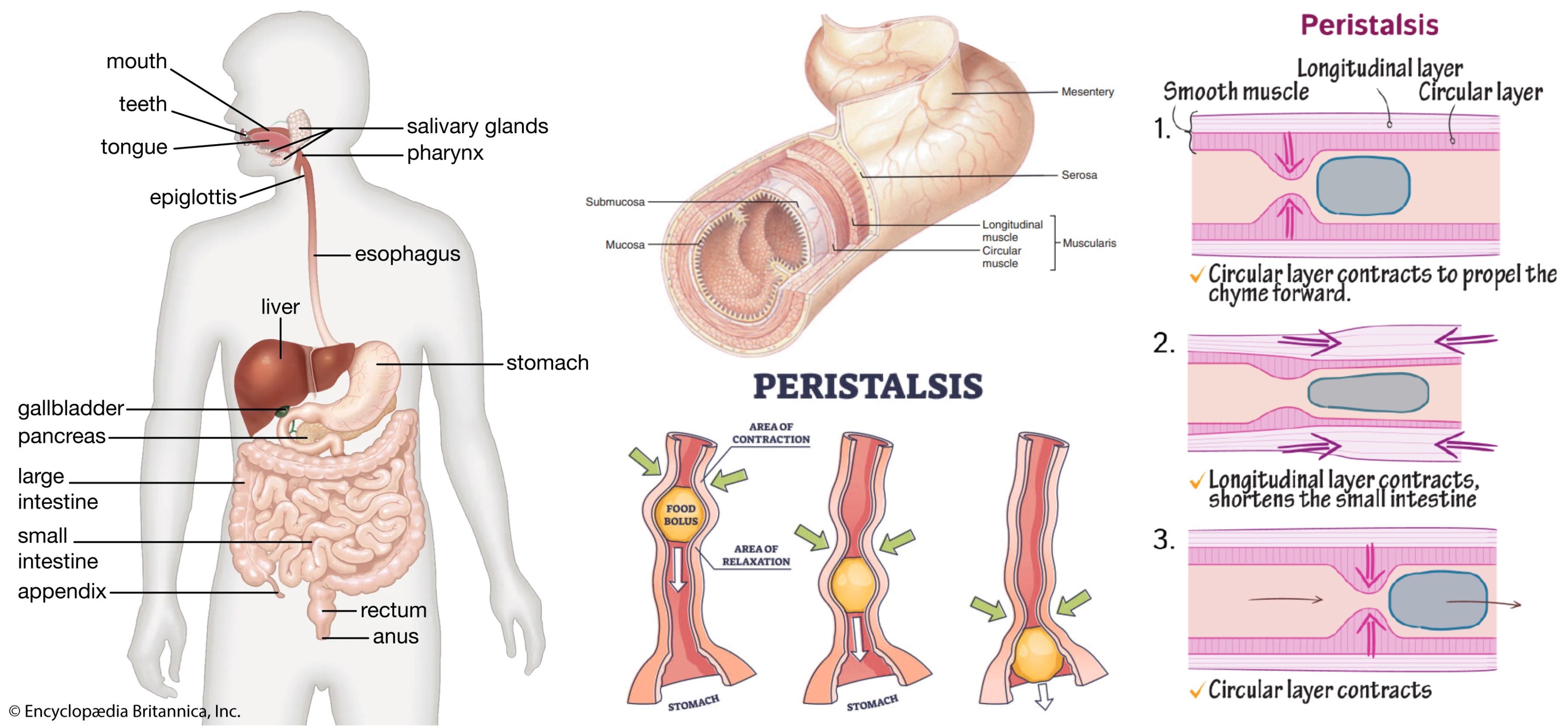
made of smooth muscle
peristalsis- moving bolus of food down GI tract:
1. longitudinal layer- propel bolus forward
2. circular layer- prevents backflow
112
parts of the GI tract: mouth
1. mastication- chewing with incisors, cuspids, molars
2. saliva- salivary amylase which hydrolyzes starch to disaccharides, lingual lipase which digests fat, lysozyme which attack bacterial cell walls
3. bolus- ball of digested food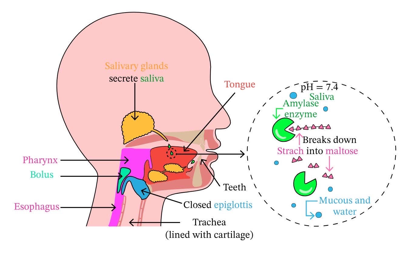
2. saliva- salivary amylase which hydrolyzes starch to disaccharides, lingual lipase which digests fat, lysozyme which attack bacterial cell walls
3. bolus- ball of digested food
113
parts of the GI tract: esophagus
1. pharynx- throat, has openings to trachea and esophagus
2. epiglottis- cartilage flap to cover up trachea
3. upper esophageal sphincter- muscular ring at top
4. lower esophageal sphincter- muscular ring near stomach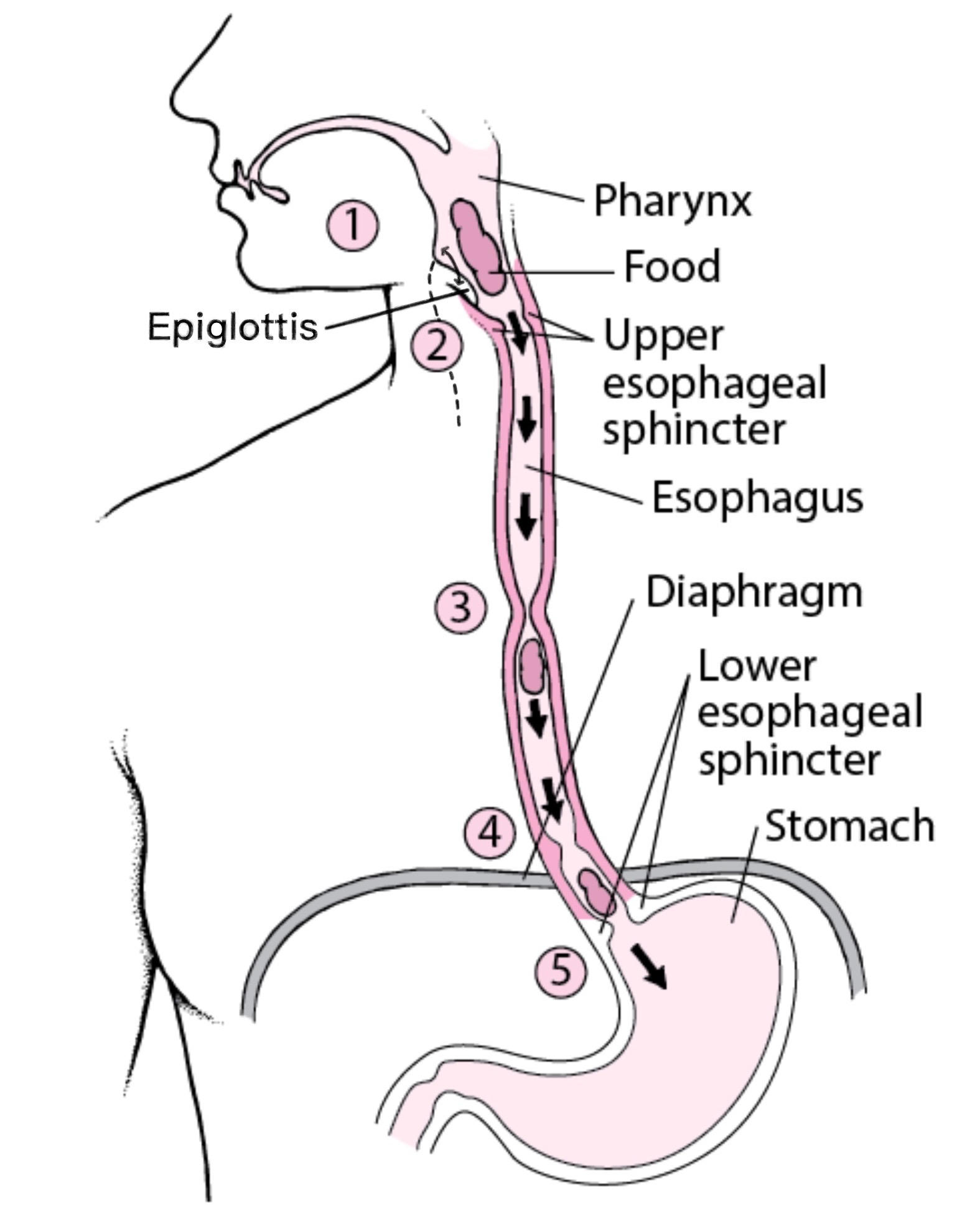
2. epiglottis- cartilage flap to cover up trachea
3. upper esophageal sphincter- muscular ring at top
4. lower esophageal sphincter- muscular ring near stomach
114
parts of the GI tract: stomach
1. acidity- *gastric pH is 2* due to HCl secretion by parietal cells, functions: destroys microorganisms, acid-catalyzed hydrolysis of proteins, converts pepsinogen to pepsin, can also denature important proteins like antibodies!
2. *pepsin*- pepsinogen (zymogen) is secreted by chief cells and converted to pepsin, catalyzes proteolysis
3. motility- churns food, creates chyme (food + secretions)
4. pyloric sphincter- regulates food entering duodenum, cholecystokinin is hormone that signals to close it
5. *gastrin*- G cells secrete this hormone that stimulate acid and pepsin secretion, histamine binds to parietal cells to stimulate acid secretion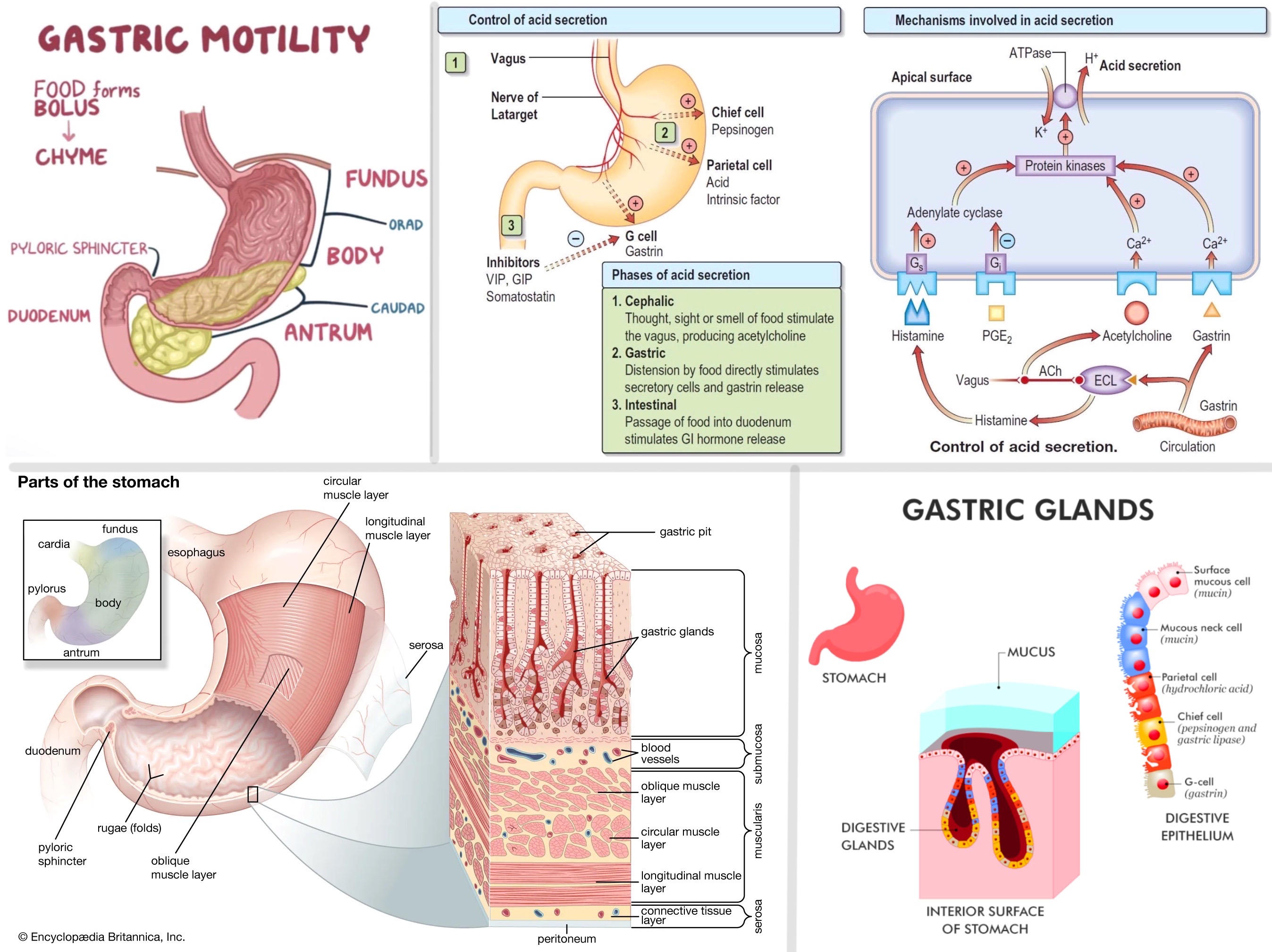
2. *pepsin*- pepsinogen (zymogen) is secreted by chief cells and converted to pepsin, catalyzes proteolysis
3. motility- churns food, creates chyme (food + secretions)
4. pyloric sphincter- regulates food entering duodenum, cholecystokinin is hormone that signals to close it
5. *gastrin*- G cells secrete this hormone that stimulate acid and pepsin secretion, histamine binds to parietal cells to stimulate acid secretion
115
parts of the GI tract: small intestine (wall)
1. villi- multicellular projections that make up *brush border*
2. hepatic portal vein- villi have capillaries that connect
3. lacteals- lymphatic vessels to absorb fat
4. Peyer's patches- lymphocytes to monitor for pathogens
5. 3 segments: *duodenum, jejunum, ileum* (Don’t Jiggle It)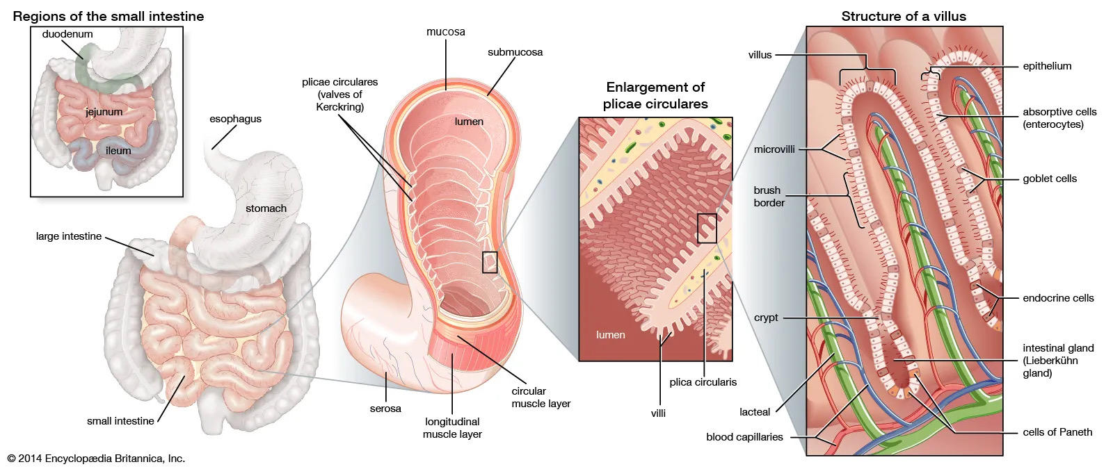
2. hepatic portal vein- villi have capillaries that connect
3. lacteals- lymphatic vessels to absorb fat
4. Peyer's patches- lymphocytes to monitor for pathogens
5. 3 segments: *duodenum, jejunum, ileum* (Don’t Jiggle It)
116
parts of the GI tract: small intestine (duodenum)
1. *pancreatic duct*- brings exocrine secretions (digestive enzymes) from pancreas
2. *common bile duct*- brings bile (waste vehicle, digest fat) from gallbladder
3. sphincter of Oddi- bile and pancreatic ducts empty here
4. duodenal enzymes- enterokinase (creates trypsin), brush border enzymes (hydrolyze disaccs/dipeps to monos)
5. duodenal hormones- cholecystokinin (to digest fat), secretin (to increase pH from stomach since duodenal pH must be neutral), enterogastrone (to stop stomach emptying)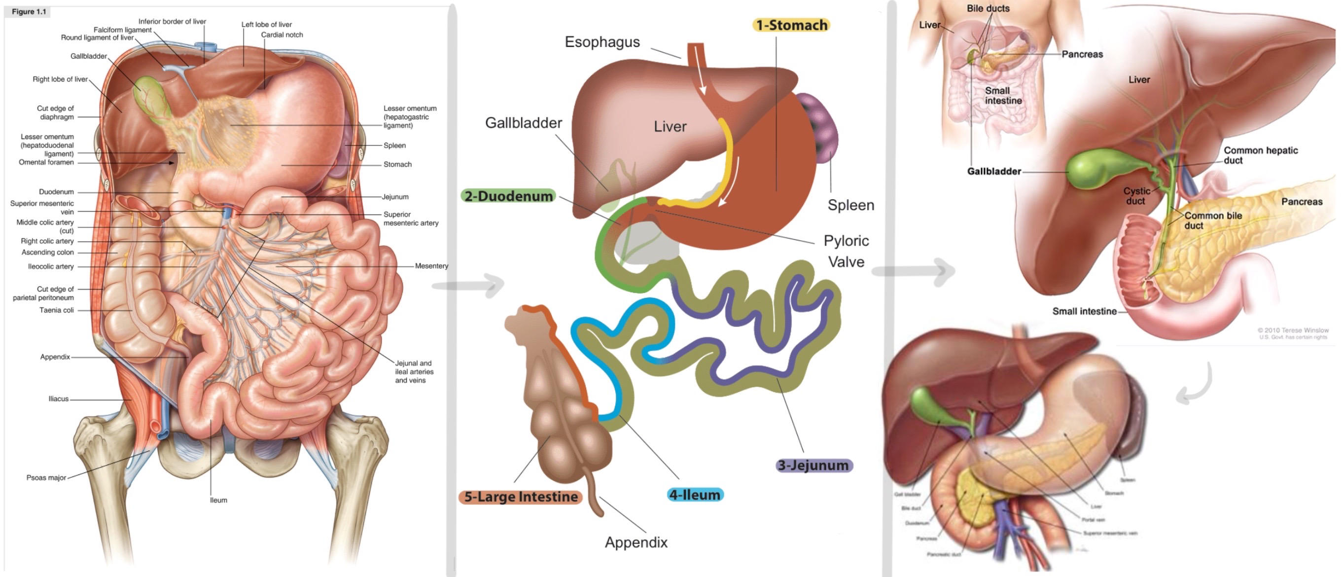
2. *common bile duct*- brings bile (waste vehicle, digest fat) from gallbladder
3. sphincter of Oddi- bile and pancreatic ducts empty here
4. duodenal enzymes- enterokinase (creates trypsin), brush border enzymes (hydrolyze disaccs/dipeps to monos)
5. duodenal hormones- cholecystokinin (to digest fat), secretin (to increase pH from stomach since duodenal pH must be neutral), enterogastrone (to stop stomach emptying)
117
parts of the GI tract: large intestine
1. ileocecal valve- separate small intestine from large
2. function: absorb water and minerals, form feces
3. segments: *cecum, appendix, rectum, anal sphincter*
4. colonic bacteria- supply vitamin K, outcompete dangerous bacteria for space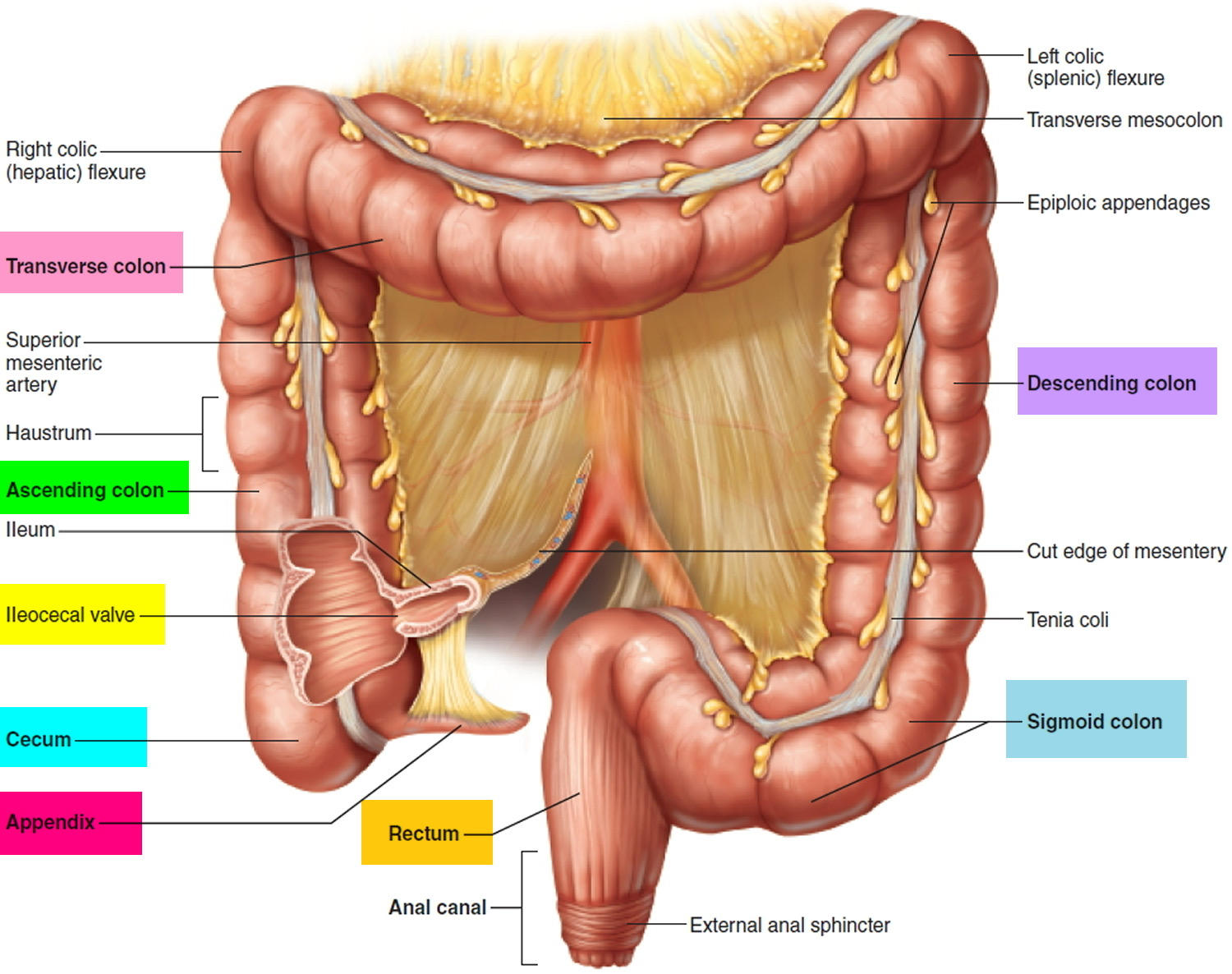
2. function: absorb water and minerals, form feces
3. segments: *cecum, appendix, rectum, anal sphincter*
4. colonic bacteria- supply vitamin K, outcompete dangerous bacteria for space
118
enteric nervous system
branch of autonomic NS, independent of para/sympathetic
controls everything in GI tract, pancreas, and gall bladder
2 networks:
1. myenteric plexus- controls gut motility
2. submucosal plexus- controls blood flow, enzyme secretion, ion exchange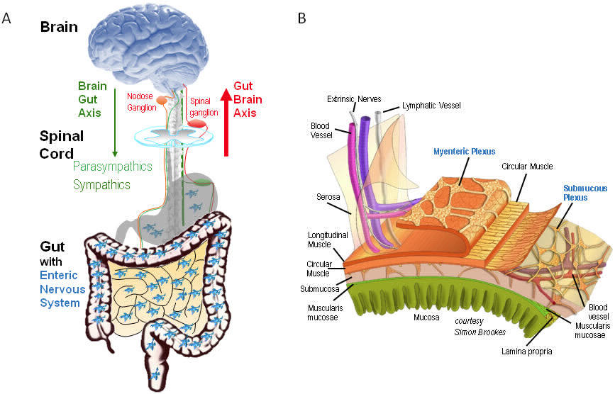
controls everything in GI tract, pancreas, and gall bladder
2 networks:
1. myenteric plexus- controls gut motility
2. submucosal plexus- controls blood flow, enzyme secretion, ion exchange
119
GI secretions
exocrine secretions:
1. acini- sacs of epithelial cells that secrete products into GI
2. goblet cells- secrete mucus
3. gastric glands- secrete acid in stomach
4. pancreatic acinar cells- pancreas
endocrine secretions:
1. go to capillaries, do not empty into ducts
2. islets of Langerhans- pancreas, produce insulin, glucagon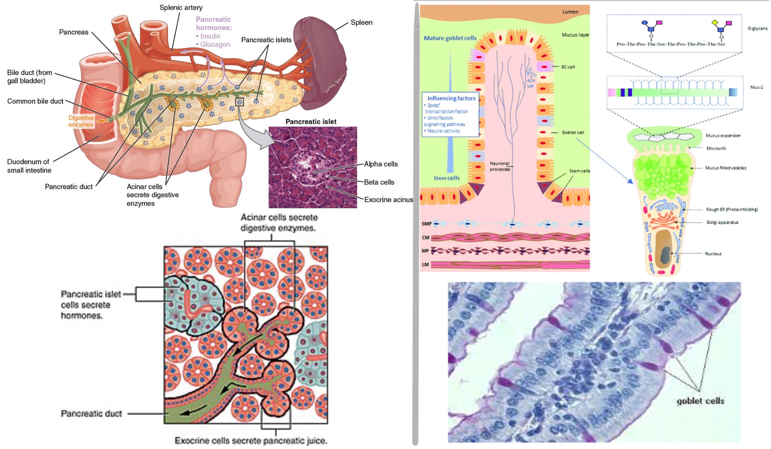
1. acini- sacs of epithelial cells that secrete products into GI
2. goblet cells- secrete mucus
3. gastric glands- secrete acid in stomach
4. pancreatic acinar cells- pancreas
endocrine secretions:
1. go to capillaries, do not empty into ducts
2. islets of Langerhans- pancreas, produce insulin, glucagon
120
GI accessory organs
pancreas:
endocrine (produce insulin, glucagon) and exocrine role (releases digestive enzymes, *zymogen* form needs to be activated)
liver:
1. produce bile (digest fat in small intestine)
2. hepatic portal vein- liver monitors blood from stomach, ex: if glucose is low, liver is only organ than can break down glycogen
3. urea- ammonia is created during amino acid breakdown, liver converts to urea
4. lipid metabolism- chylomicrons broken down are brought by lymphatic system to liver, liver makes lipoproteins with remnants, makes *cholesterol*
5. produces plasma proteins- albumin, fibrinogens, clotting factors
6. detoxification- smooth ER has enzymes to break down drugs
gallbladder:
bile is stored and concentrated here; gallstones: incorrect ingredients in bile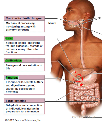
endocrine (produce insulin, glucagon) and exocrine role (releases digestive enzymes, *zymogen* form needs to be activated)
liver:
1. produce bile (digest fat in small intestine)
2. hepatic portal vein- liver monitors blood from stomach, ex: if glucose is low, liver is only organ than can break down glycogen
3. urea- ammonia is created during amino acid breakdown, liver converts to urea
4. lipid metabolism- chylomicrons broken down are brought by lymphatic system to liver, liver makes lipoproteins with remnants, makes *cholesterol*
5. produces plasma proteins- albumin, fibrinogens, clotting factors
6. detoxification- smooth ER has enzymes to break down drugs
gallbladder:
bile is stored and concentrated here; gallstones: incorrect ingredients in bile
121
regulating blood glucose
lowering glucose (hyperglycemia won't kill you):
1. insulin
raising glucose (hypoglycemia will kill you):
1. glucagon- peptide from pancreas
2. epinephrine- aa-derivative from adrenal medulla
3. cortisol- steroid from adrenal cortex
1. insulin
raising glucose (hypoglycemia will kill you):
1. glucagon- peptide from pancreas
2. epinephrine- aa-derivative from adrenal medulla
3. cortisol- steroid from adrenal cortex
122
appetite
ghrelin- gastric cells secrete to stimulate appetite
peptide YY- jejunum secretes to reduce appetite
leptin- adipose tissues secretes to reduce appetite
peptide YY- jejunum secretes to reduce appetite
leptin- adipose tissues secretes to reduce appetite
123
vitamins
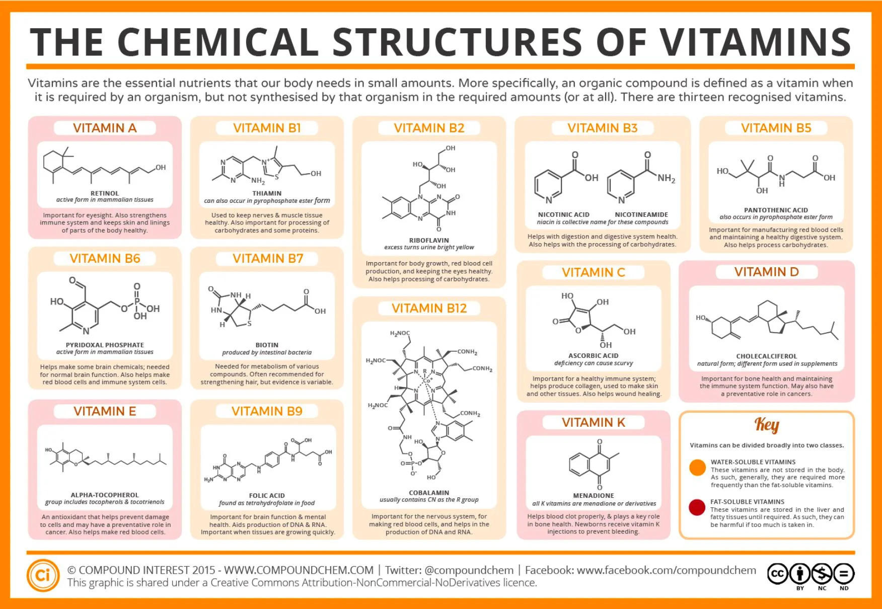
excess fat-soluble vitamins are stored in adipose tissue:
1. A- visual pigment
2. D- stimulates Ca2+ absorption in gut
3. E- prevent oxidation of unsaturated fat
4. K- formation of blood clotting factors
ADEK fat soluble
too much fat would sequester vitamins, causing deficiency
excess water-soluble vitamins are excreted in urine:
1. C- collagen formation
2. biotin- CO2 transport
3. B’s- *FAD/NAD*, also thiamine for decarboxylation enzyme!
BC water soluble
1. A- visual pigment
2. D- stimulates Ca2+ absorption in gut
3. E- prevent oxidation of unsaturated fat
4. K- formation of blood clotting factors
ADEK fat soluble
too much fat would sequester vitamins, causing deficiency
excess water-soluble vitamins are excreted in urine:
1. C- collagen formation
2. biotin- CO2 transport
3. B’s- *FAD/NAD*, also thiamine for decarboxylation enzyme!
BC water soluble
124
muscle tissue
3 types:
1. skeletal (striated) muscle- voluntary muscle, striated
2. cardiac muscle- heart, striated
3. smooth muscle- walls of all hollow organs, smooth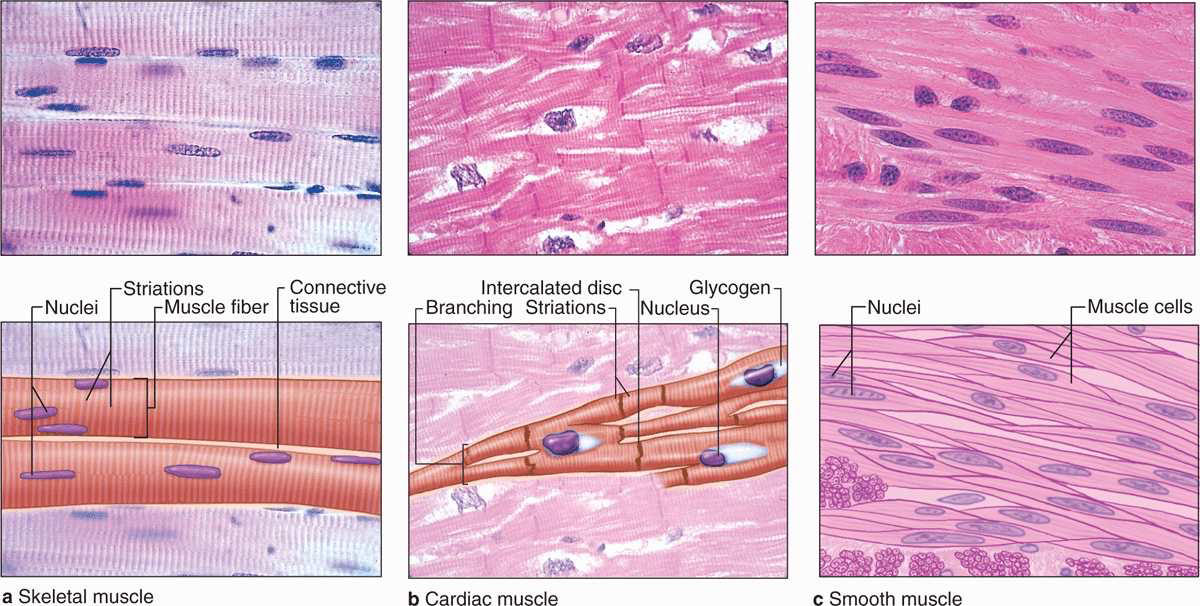
1. skeletal (striated) muscle- voluntary muscle, striated
2. cardiac muscle- heart, striated
3. smooth muscle- walls of all hollow organs, smooth
125
joint movement (skeletal muscle)
tendons- collagen, connective tissue
origin- point that muscle attaches to bone that stays still
insertion- point that muscle attaches to bone that moves
antagonistic- muscles moving in opposite directions
synergistic- muscles moving in same directions
movement:
1. flexing- reduce angle
2. extending- increase angle
3. abducting- move away from body midline
4. adducting- move towards body midline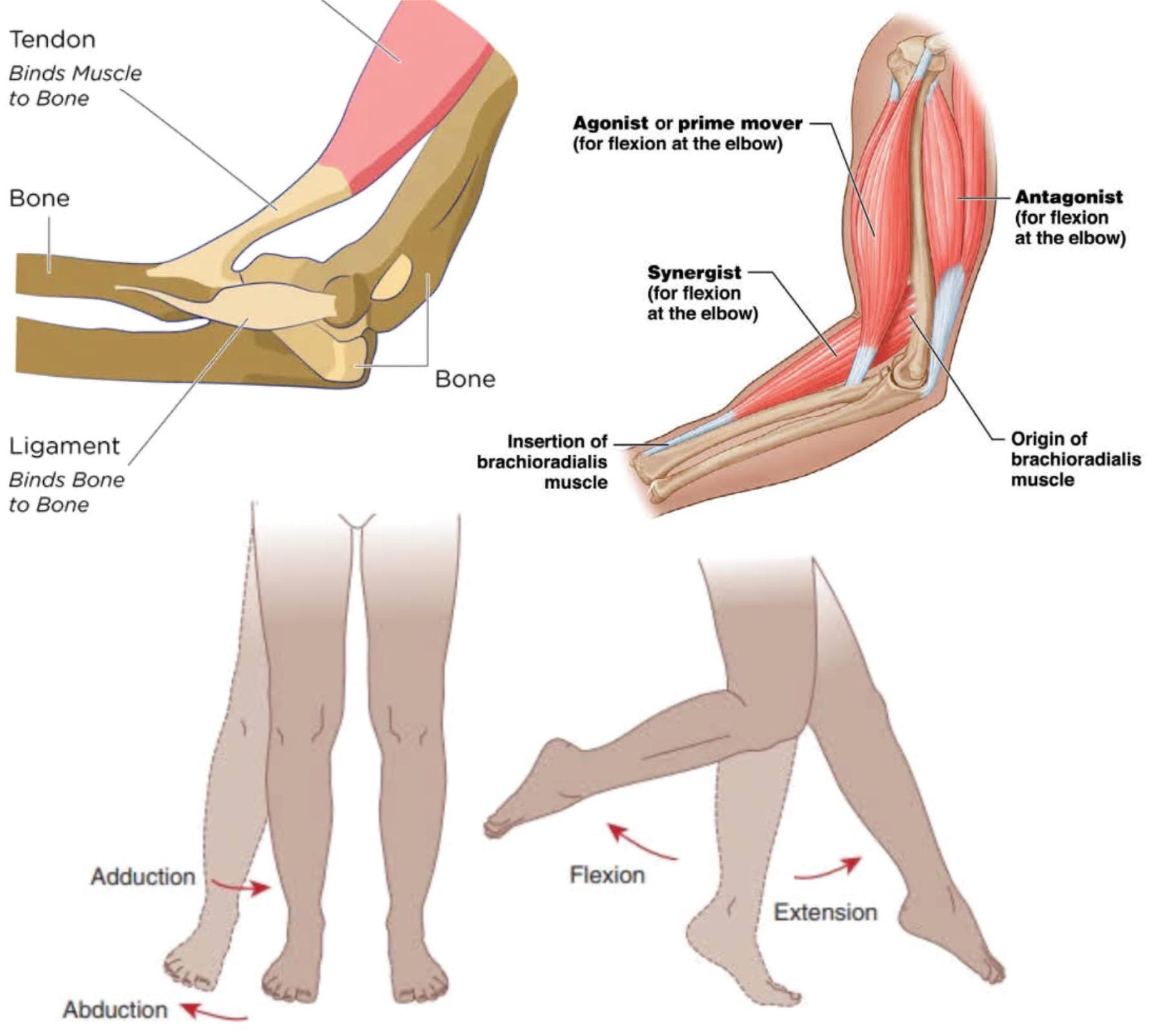
origin- point that muscle attaches to bone that stays still
insertion- point that muscle attaches to bone that moves
antagonistic- muscles moving in opposite directions
synergistic- muscles moving in same directions
movement:
1. flexing- reduce angle
2. extending- increase angle
3. abducting- move away from body midline
4. adducting- move towards body midline
126
muscle structure (skeletal muscle)
fascicles- bundles of muscle tissue
myofibers- single muscle cell formed from fusion of cells
multinucleate syncytia- fusion of cells, controlled by 1 nerve
sarcolemma- cell membrane with a layer of collagen and polysaccharides to help connect with tendons
myofibrils- organelles inside muscle cell that contracts, made of actin and myosin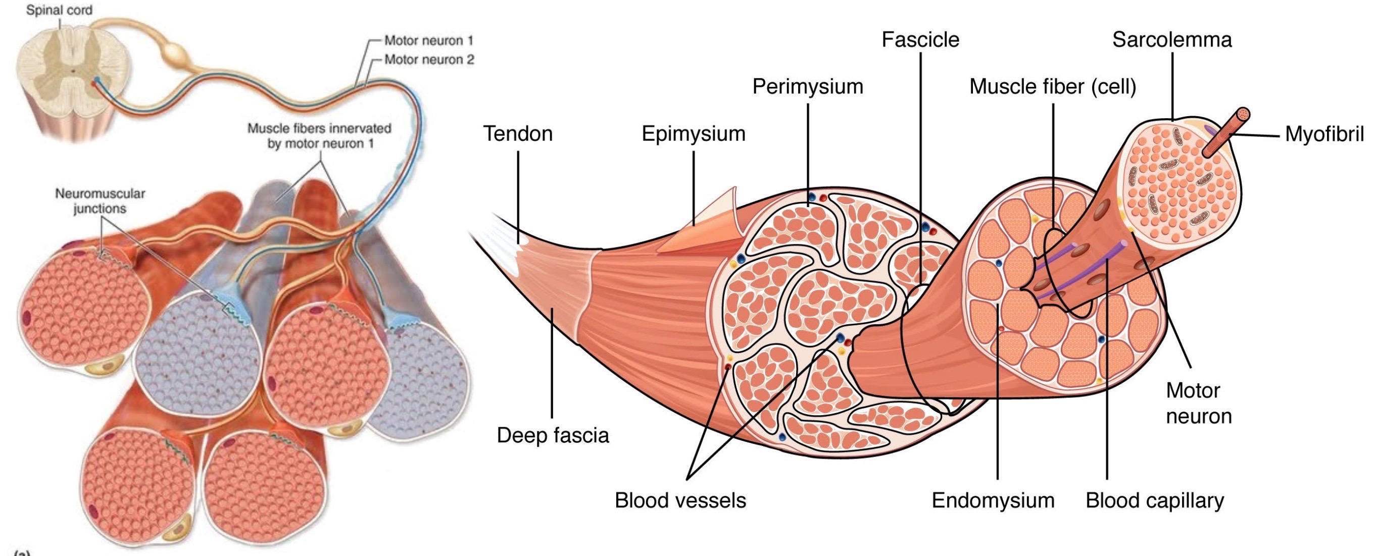
myofibers- single muscle cell formed from fusion of cells
multinucleate syncytia- fusion of cells, controlled by 1 nerve
sarcolemma- cell membrane with a layer of collagen and polysaccharides to help connect with tendons
myofibrils- organelles inside muscle cell that contracts, made of actin and myosin
127
myofiber structure (skeletal muscle)
sarcomere- segments of myofibrils connected end-to-end
thin filaments (I-band) - actin is on both ends of sarcomere
thick filaments (A-band) - myosin is in middle of sarcomere
Z line- thin filaments from each sarcomere connect
H-band - thick filament segment not overlapping with thin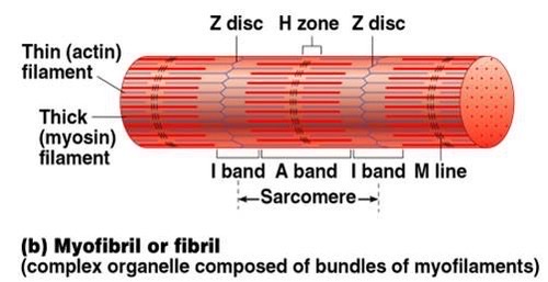
thin filaments (I-band) - actin is on both ends of sarcomere
thick filaments (A-band) - myosin is in middle of sarcomere
Z line- thin filaments from each sarcomere connect
H-band - thick filament segment not overlapping with thin
128
cross bridge contractile cycle (skeletal muscle)
myosin- ATP powered motor protein, head attached to actin via a cross bridge, tail attached to thick filament
steps:
1. myosin has ADP and P bound
2. power stroke- head moves to low energy conformation, pulls actin toward center of sarcomere, releases ADP
3. ATP binds, actin is released by myosin head
4. ATP hydrolysis occurs, reset the myosin head back
fun facts:
1. ATP is used to reset myosin, not during power stroke
2. rigor mortis- no ATP means myosin cannot release actin
3. myosin head groups operate asynchronously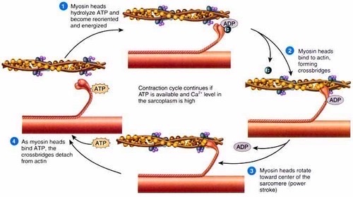
steps:
1. myosin has ADP and P bound
2. power stroke- head moves to low energy conformation, pulls actin toward center of sarcomere, releases ADP
3. ATP binds, actin is released by myosin head
4. ATP hydrolysis occurs, reset the myosin head back
fun facts:
1. ATP is used to reset myosin, not during power stroke
2. rigor mortis- no ATP means myosin cannot release actin
3. myosin head groups operate asynchronously
129
troponin-tropomyosin complex (skeletal muscle)
contractile cycle will occur spontaneously, but in myofiber contraction only occurs after Ca2+ influx
tropomyosin- fibrous protein that winds around actin, blocking myosin binding sites
troponin- globular protein on tropomyosin that binds Ca2+ and moves tropomyosin away from myosin binding sites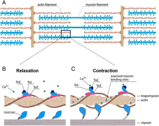
tropomyosin- fibrous protein that winds around actin, blocking myosin binding sites
troponin- globular protein on tropomyosin that binds Ca2+ and moves tropomyosin away from myosin binding sites
130
neuromuscular junction (skeletal muscle)
synapse between axon terminus and a myofiber, acetylcholine is the neurotransmitter
steps:
1. presynaptic membrane has voltage-gated Ca2+ channels, Ca2+ influx triggers release of vesicles of acetylcholine
2. postsynaptic membrane is called motor end plate, has ligand-gated Na+ channels (acetylcholine receptors)
3. end plate potential (EPP)- depolarization of myofiber
4. miniature EPP (MEPP)- EPP by a single ACh vesicle
5. *acetylcholinesterase*- hydrolyzes acetylcholine to stop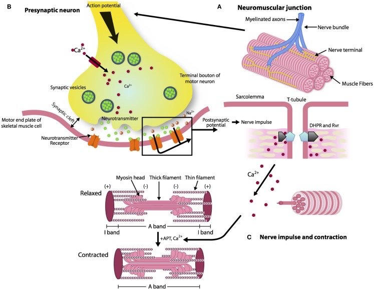
steps:
1. presynaptic membrane has voltage-gated Ca2+ channels, Ca2+ influx triggers release of vesicles of acetylcholine
2. postsynaptic membrane is called motor end plate, has ligand-gated Na+ channels (acetylcholine receptors)
3. end plate potential (EPP)- depolarization of myofiber
4. miniature EPP (MEPP)- EPP by a single ACh vesicle
5. *acetylcholinesterase*- hydrolyzes acetylcholine to stop
131
action potential (skeletal muscle)
steps:
1. when EPP reaches threshold, voltage-gated Na+ channels open, causing Na+ influx and depolarization
2. transverse tubules (T-tubules)- invaginations of cell membrane into cytoplasm to depolarize interior as well
3. sarcoplasmic reticulum (SR)- net organelle of smooth ER that wraps around each myofibril, active transporters brings Ca2+ from sarcoplasm (myofiber cytoplasm) into SR
4. SR has mechanical/voltage/???-gated Ca2+ channels that further allow Ca2+ influx into sarcoplasm
5. contractile cycle occurs
6. repolarization- Ca2+ sequestered back into SR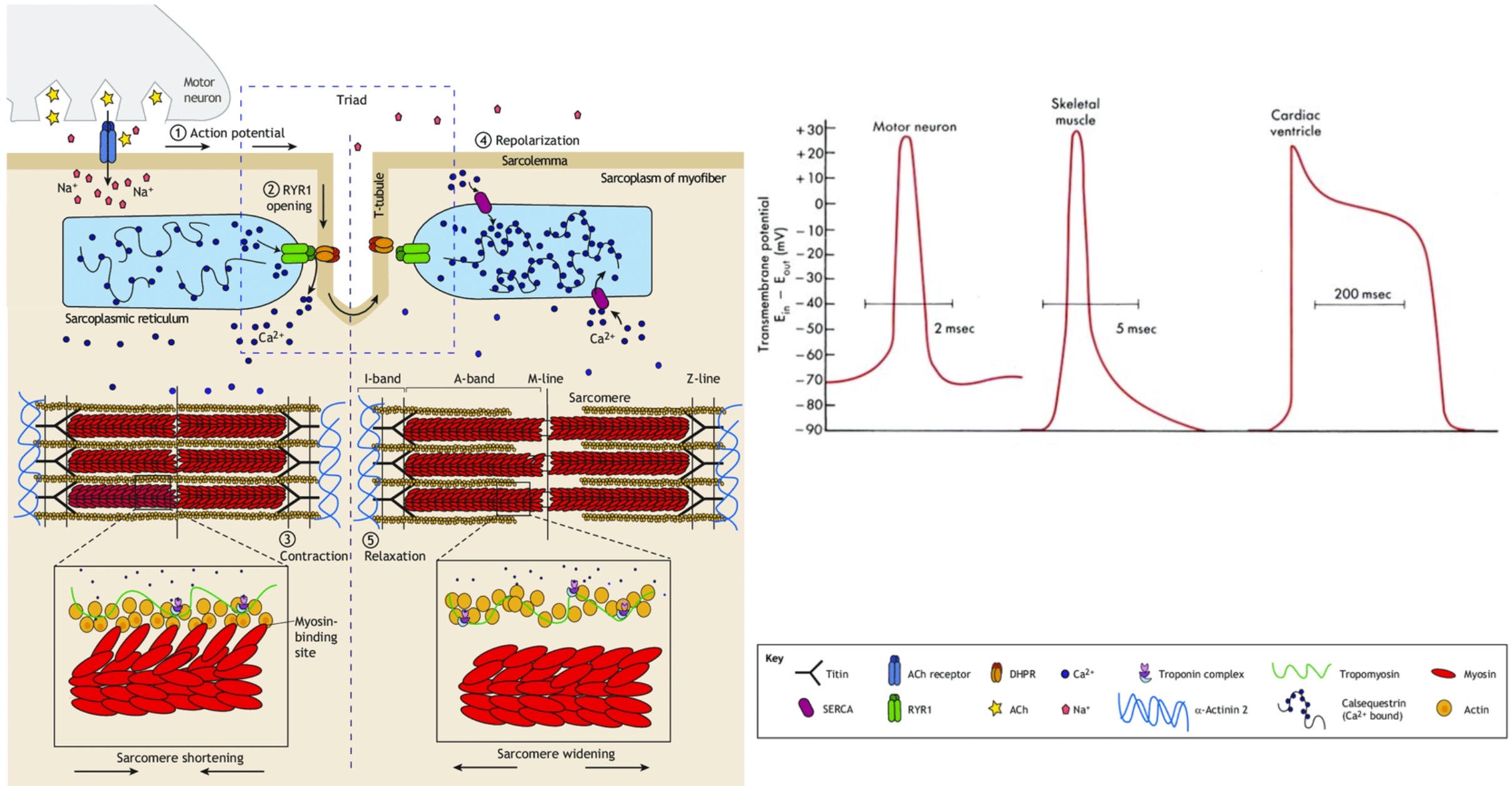
1. when EPP reaches threshold, voltage-gated Na+ channels open, causing Na+ influx and depolarization
2. transverse tubules (T-tubules)- invaginations of cell membrane into cytoplasm to depolarize interior as well
3. sarcoplasmic reticulum (SR)- net organelle of smooth ER that wraps around each myofibril, active transporters brings Ca2+ from sarcoplasm (myofiber cytoplasm) into SR
4. SR has mechanical/voltage/???-gated Ca2+ channels that further allow Ca2+ influx into sarcoplasm
5. contractile cycle occurs
6. repolarization- Ca2+ sequestered back into SR
132
increasing muscle contraction (skeletal muscle)
twitch- smallest measurable muscle contraction
tetanus- strongest possible contraction (frequency summation, not a good thing)
3 methods:
1. motor unit recruitment- activate more myofibers with each motor neuron axon
2. frequency summation- rapid contractions don't allow Ca2+ to be sequestered, can build off each other
3. length-tension relationship- muscle contracts most forcibly at an optimum length, maximum degree of overlap between thick and thin filaments without obstruction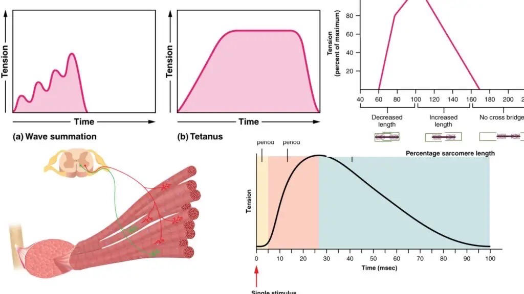
tetanus- strongest possible contraction (frequency summation, not a good thing)
3 methods:
1. motor unit recruitment- activate more myofibers with each motor neuron axon
2. frequency summation- rapid contractions don't allow Ca2+ to be sequestered, can build off each other
3. length-tension relationship- muscle contracts most forcibly at an optimum length, maximum degree of overlap between thick and thin filaments without obstruction
133
myofiber energy storage (skeletal muscle)
creatine phosphate- hydrolyzed to creatine to generate ATP to provide myofiber a fast energy source, since it's faster than TCA/ glycolysis
myoglobin- takes oxygen from hemoglobin, provides a quick oxygen reserve for muscles
check the oxygen debt card for more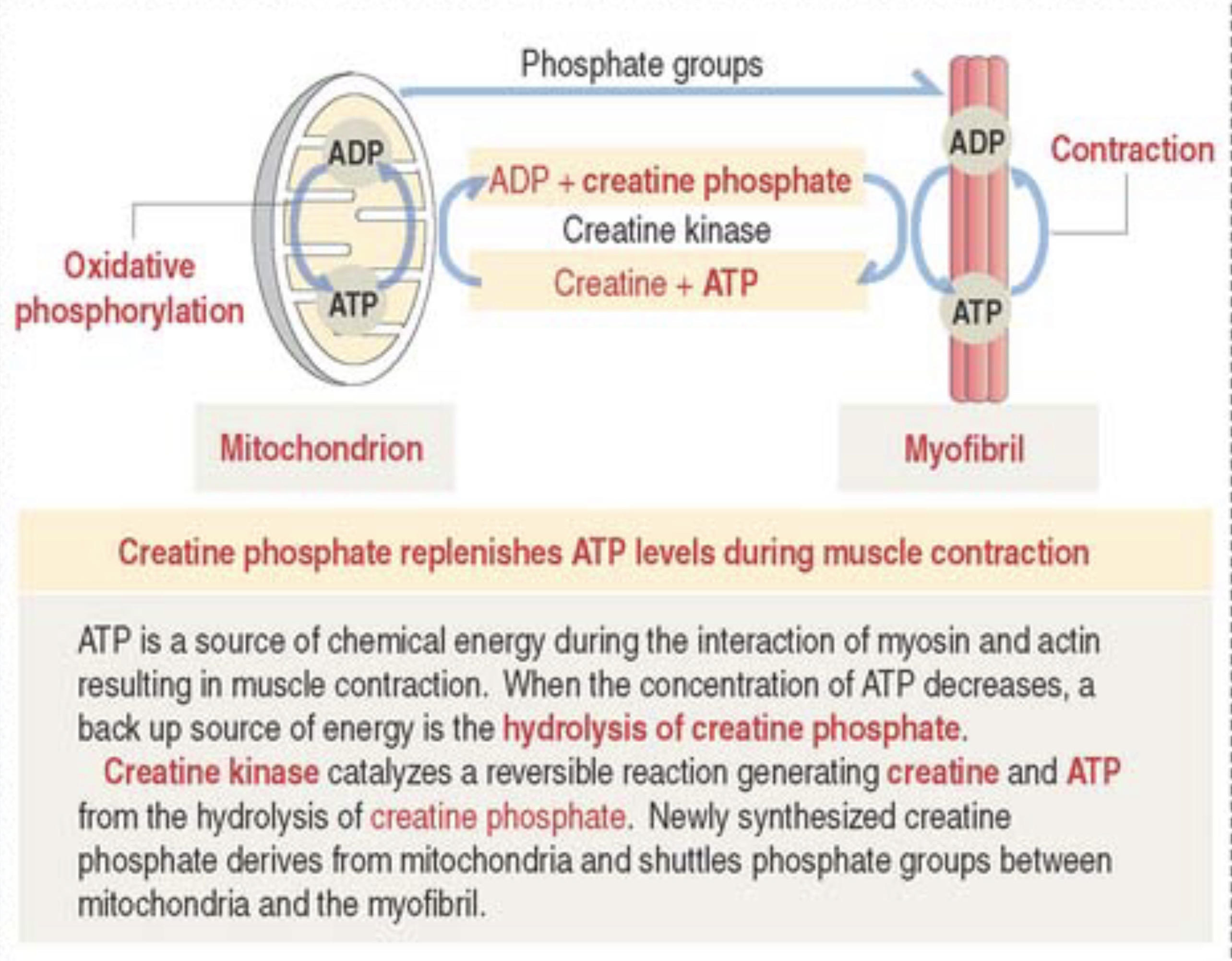
myoglobin- takes oxygen from hemoglobin, provides a quick oxygen reserve for muscles
check the oxygen debt card for more
134
muscle fiber types (skeletal muscle)
type I slow twitch fiber (red oxidative)- slow, low force, good capillary network with lots of myoglobin
type IIA fast twitch fiber - medium everything
type IIB fast twitch fiber (white)- explosive force, fatigue quickly, very few mitochondria, anaerobic glycolysis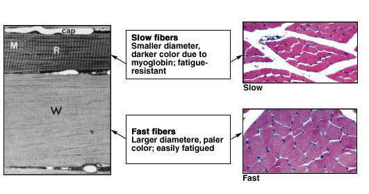
type IIA fast twitch fiber - medium everything
type IIB fast twitch fiber (white)- explosive force, fatigue quickly, very few mitochondria, anaerobic glycolysis
135
cardiac muscle
differences from skeletal muscle (1-4):
1. functional syncytium- not multinucleate syncytium, since only has 1 nucleus per cell, but has intercalated disks (gap junctions) that connect all cells to send action potentials
2. voltage-gated Ca2+ channels- on plasma membrane in addition to being on SR, they trigger contraction and propogate action potential during depolarization
3. plateau phase- due to voltage-gated Ca2+ channels, longer contraction for better heart pumping and to prevent disorganized impulses
4. SA node- not motor neurons, stimulate contraction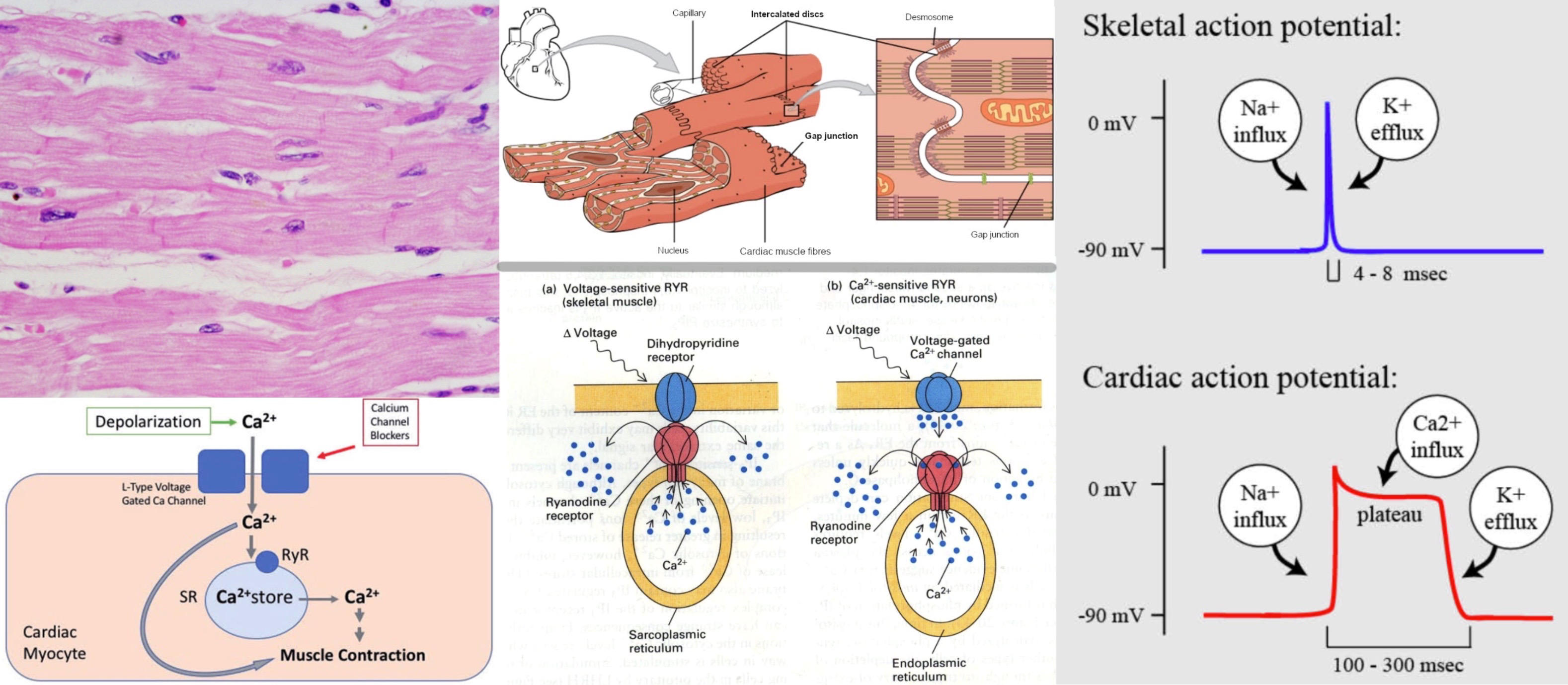
1. functional syncytium- not multinucleate syncytium, since only has 1 nucleus per cell, but has intercalated disks (gap junctions) that connect all cells to send action potentials
2. voltage-gated Ca2+ channels- on plasma membrane in addition to being on SR, they trigger contraction and propogate action potential during depolarization
3. plateau phase- due to voltage-gated Ca2+ channels, longer contraction for better heart pumping and to prevent disorganized impulses
4. SA node- not motor neurons, stimulate contraction
136
smooth muscle
differences from skeletal muscle:
1. functional syncytium- not multinucleate syncytium, since only has 1 nucleus per cell, but has gap junctions that connect all cells to send action potentials
2. smaller- so they do not have T-tubules
3. no sarcomeres- thick/thin filaments are in cytoplasm
4. calmodulin regulation- binds Ca2+, activates MLCK which phosphorylates myosin to activate it, no troponin!
5. voltage-gated Ca2+ channels- much more reliant on these than the SR ones
6. no voltage-gated Na+ channels- so slower than skeletal but faster than cardiac
7. slow waves- constantly changing resting potential, parasympathetic NS primes smooth muscle cells to push waves past threshold and release *spike potential*, acetylcholine from parasympathetic NS increases waves, norepinephrine from sympathetic NS decreases waves
8. autonomic motor neurons- not somatic like skeletal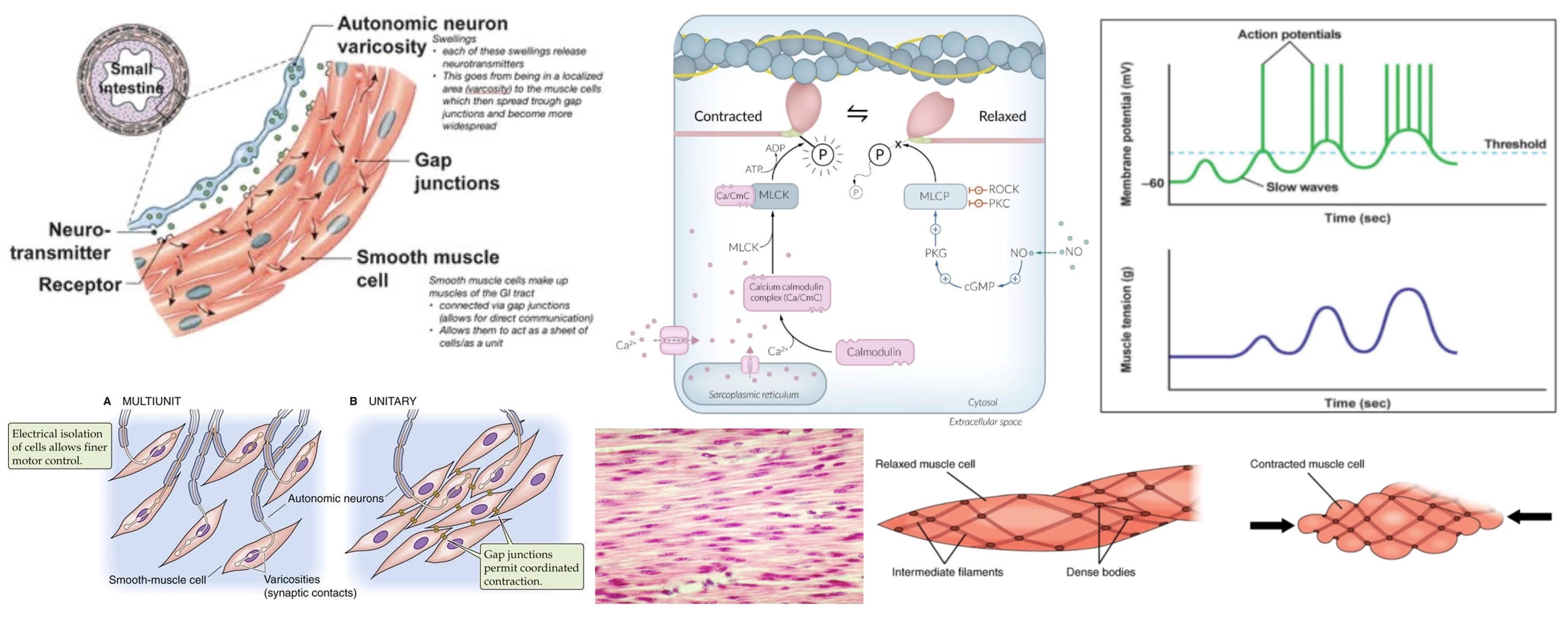
1. functional syncytium- not multinucleate syncytium, since only has 1 nucleus per cell, but has gap junctions that connect all cells to send action potentials
2. smaller- so they do not have T-tubules
3. no sarcomeres- thick/thin filaments are in cytoplasm
4. calmodulin regulation- binds Ca2+, activates MLCK which phosphorylates myosin to activate it, no troponin!
5. voltage-gated Ca2+ channels- much more reliant on these than the SR ones
6. no voltage-gated Na+ channels- so slower than skeletal but faster than cardiac
7. slow waves- constantly changing resting potential, parasympathetic NS primes smooth muscle cells to push waves past threshold and release *spike potential*, acetylcholine from parasympathetic NS increases waves, norepinephrine from sympathetic NS decreases waves
8. autonomic motor neurons- not somatic like skeletal
137
connective tissue
*fibroblasts*- adipocytes (fat cells), chondrocytes (cartilage cells), osteocytes (bone cells) are all derived from fibroblasts, which secrete collagen, a fibrous protein
elastin- another fibrous protein, stretchy
extracellular matrix- most of connective tissue is just ECM, which has collagen, elastin, fibronectin, integrin, a lot of water, *proteoglycans* (ground substance, just carbohydrates attached to proteins)
loose connective tissue- adipose and areolar tissue
dense connective tissue- tendons, cartilage, bone (lots of collagen)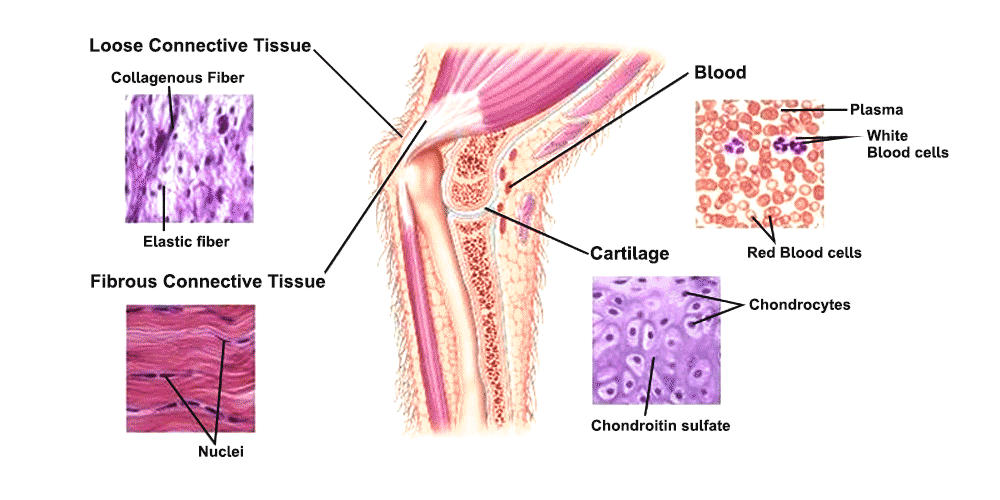
elastin- another fibrous protein, stretchy
extracellular matrix- most of connective tissue is just ECM, which has collagen, elastin, fibronectin, integrin, a lot of water, *proteoglycans* (ground substance, just carbohydrates attached to proteins)
loose connective tissue- adipose and areolar tissue
dense connective tissue- tendons, cartilage, bone (lots of collagen)
138
bone structure (micro)
collagen- fibrous protein in ICM
hydroxyapatite- calcium phosphate crystals that form around collagen framework to give bones strength
spongy bone:
spicules (trabeculae)- spikes of bone
bone marrow
compact bone:
osteon (Haversian system)- basic unit, has a central canal (Haversian canal) with blood, lymph vessels, and nerves
lamellae- concentric rings of bone around central canal
canaliculi- tiny channels branching from central canal
lacunae- lakes that canaliculi connect to with one osteocyte
osteocytes- bone cell, contact each other with long processes that have gap junctions, exchange nutrients/waste
perforating canals (Volkmann's canals)- connect different osteons, have blood, lymph, nerves as well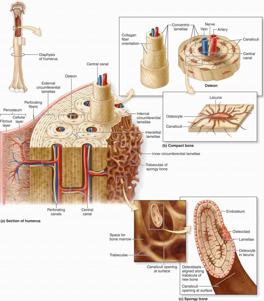
hydroxyapatite- calcium phosphate crystals that form around collagen framework to give bones strength
spongy bone:
spicules (trabeculae)- spikes of bone
bone marrow
compact bone:
osteon (Haversian system)- basic unit, has a central canal (Haversian canal) with blood, lymph vessels, and nerves
lamellae- concentric rings of bone around central canal
canaliculi- tiny channels branching from central canal
lacunae- lakes that canaliculi connect to with one osteocyte
osteocytes- bone cell, contact each other with long processes that have gap junctions, exchange nutrients/waste
perforating canals (Volkmann's canals)- connect different osteons, have blood, lymph, nerves as well
139
axial/appendicular skeleton
axial- skull, spinal cord, rib cage
appendicular- limbs and pelvis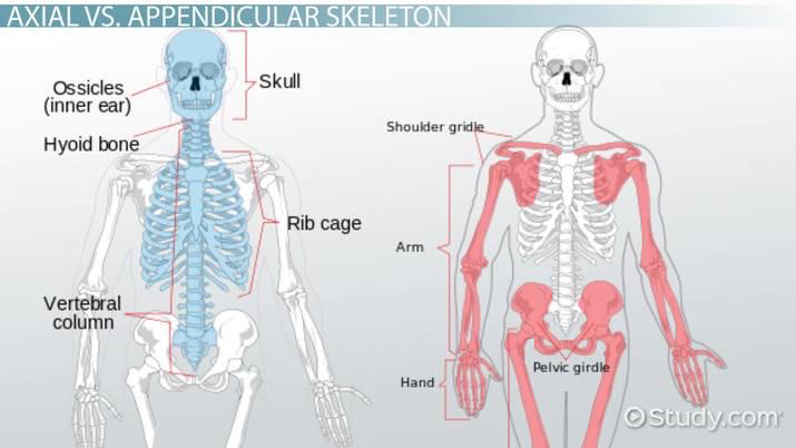
appendicular- limbs and pelvis
140
bone structure (macro)
flat bones- organ protection, hematopoiesis, compact shell with spongy core with red marrow (hematopoiesis), activity responds to erythropoietin
long bones- support and movement, compact:
diaphysis (main shaft)- compact with yellow marrow (fat), inactive
epiphysis (flared end)- compact shell with spongy core with normal marrow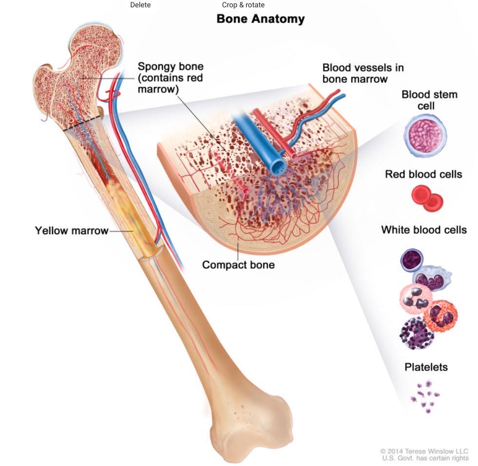
long bones- support and movement, compact:
diaphysis (main shaft)- compact with yellow marrow (fat), inactive
epiphysis (flared end)- compact shell with spongy core with normal marrow
141
cartilage
avascular- no blood vessels, receives nutrition and immune proection from surrounding fluid
chondrocytes- secrete cartilage
hyaline cartilage- strong but flexible, larynx and trachea are reinforced by it, joints that have it call it articular cartilage
elastic cartilage- more flexible since it contains elastin, ear and epiglottis have it
fibrous cartilage- very rigid, intervertebral disks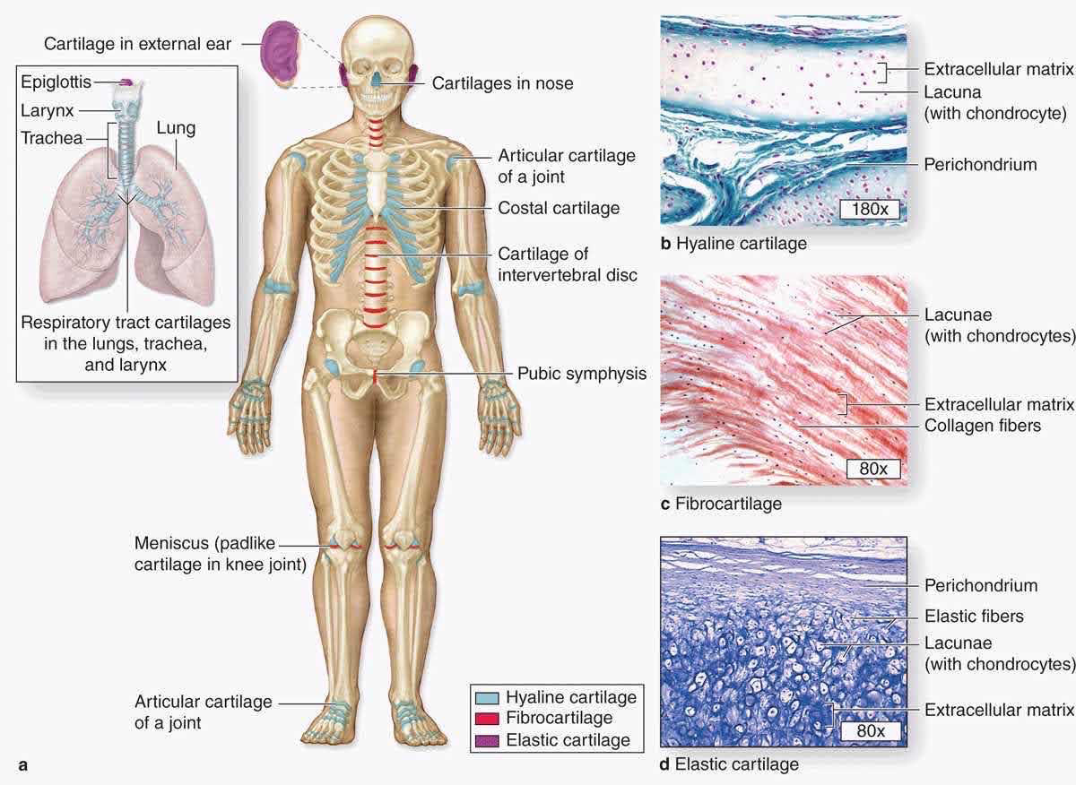
chondrocytes- secrete cartilage
hyaline cartilage- strong but flexible, larynx and trachea are reinforced by it, joints that have it call it articular cartilage
elastic cartilage- more flexible since it contains elastin, ear and epiglottis have it
fibrous cartilage- very rigid, intervertebral disks
142
joints
3 types:
1. synarthroses- immovable joints, two bones fused together, skull is example
2. amphiarthroses- slightly movable joints, vertebral joints are an example
3. diarthroses- freely movable joints, ball and sockets are an example
other terms:
1. ligaments- connect bone to other bones, dense connective tissue
2. tendons- connect bone to muscles, dense connective tissue
3. synovial fluid- lubricate joints, kept within synovial capsule
4. articular cartilage- smoothes bone contact with each other
5. arthritis- joint inflammation, damages articular cartilage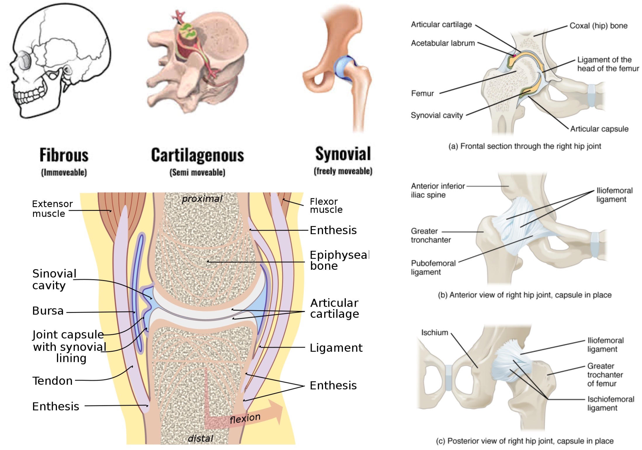
1. synarthroses- immovable joints, two bones fused together, skull is example
2. amphiarthroses- slightly movable joints, vertebral joints are an example
3. diarthroses- freely movable joints, ball and sockets are an example
other terms:
1. ligaments- connect bone to other bones, dense connective tissue
2. tendons- connect bone to muscles, dense connective tissue
3. synovial fluid- lubricate joints, kept within synovial capsule
4. articular cartilage- smoothes bone contact with each other
5. arthritis- joint inflammation, damages articular cartilage
143
bone growth
endochondral ossification- hyaline cartilage is produced and replaced by bone, most bone growth does this
intramembranous ossification- bone growth from mesenchyme (embryonic tissue), only flat bones do this
epiphyseal plate (growth plate)- during childhood, hyaline cartilage is produced by chondrocytes between diaphysis and epiphysis, growth hormones promote ossification, which brings fuse together the diaphysis and epiphyses at 18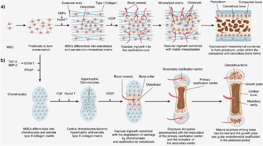
intramembranous ossification- bone growth from mesenchyme (embryonic tissue), only flat bones do this
epiphyseal plate (growth plate)- during childhood, hyaline cartilage is produced by chondrocytes between diaphysis and epiphysis, growth hormones promote ossification, which brings fuse together the diaphysis and epiphyses at 18
144
remodelling
osteoblasts- synthesizes bone around it, eventually becomes osteocytes and retires to its lacuna
osteoclasts- destroy bone by dissolving hydroxyapatite crystals, macrophage
increased ratio of osteoclast/osteoblast activity liberates Ca2+ and phosphate into bloodstream
*parathyroid hormone* (PTH) and *calcitriol*- increase osteoclast activity and increase reabsorption of Ca2+ and phosphate in kidneys
*calcitonin*- inhibits osteoclast activity and decreases reabsorption of Ca2+ in kidneys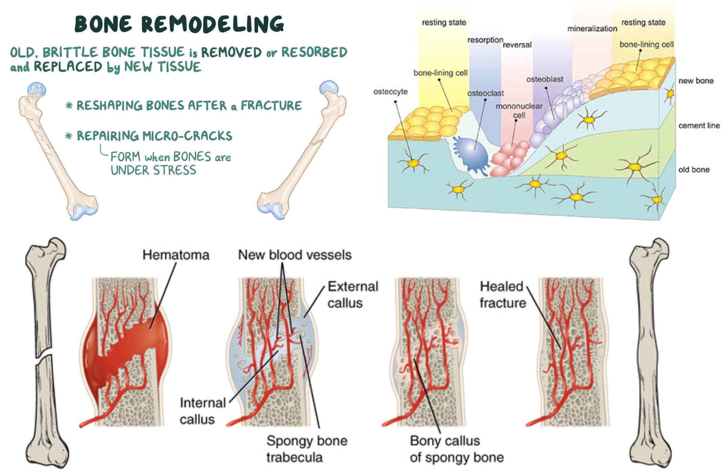
osteoclasts- destroy bone by dissolving hydroxyapatite crystals, macrophage
increased ratio of osteoclast/osteoblast activity liberates Ca2+ and phosphate into bloodstream
*parathyroid hormone* (PTH) and *calcitriol*- increase osteoclast activity and increase reabsorption of Ca2+ and phosphate in kidneys
*calcitonin*- inhibits osteoclast activity and decreases reabsorption of Ca2+ in kidneys
145
respiratory system overview
conduction zone- part of respiratory system that participates in ventilation (moving air in and out)
respiratory zone- part of respiratory system that participates in respiration (exchange of CO2 and O2)
pH regulation- hyperventilation causes respiratory alkalosis, hypoventilation causes respiratory acidosis
thermoregulation- evaporative water loss as water vapor absorbs heat, countercurrent exchange with nasal passages to conserve water and heat
disease protection- mucociliary escalator and alveolar macrophages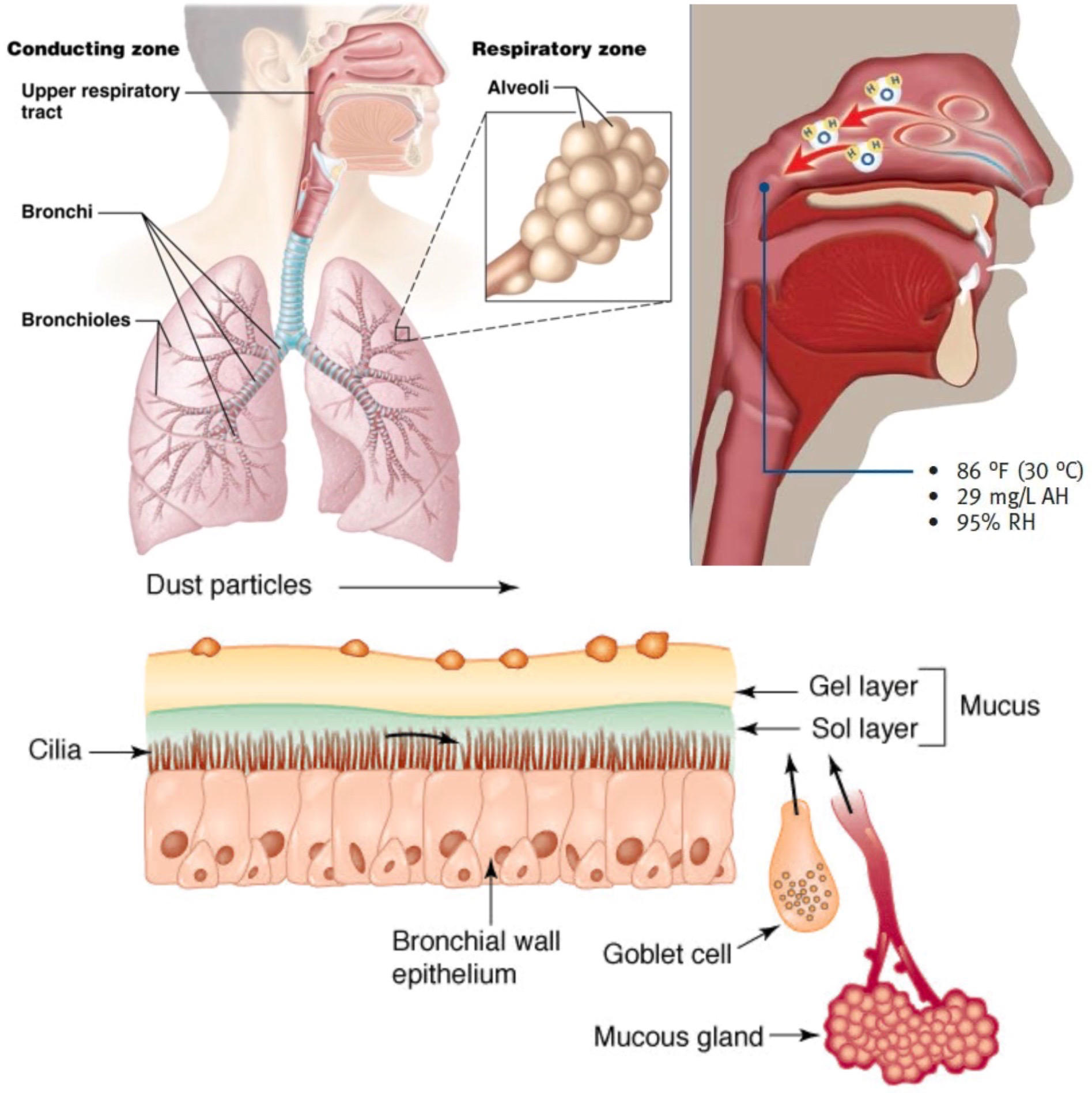
respiratory zone- part of respiratory system that participates in respiration (exchange of CO2 and O2)
pH regulation- hyperventilation causes respiratory alkalosis, hypoventilation causes respiratory acidosis
thermoregulation- evaporative water loss as water vapor absorbs heat, countercurrent exchange with nasal passages to conserve water and heat
disease protection- mucociliary escalator and alveolar macrophages
146
conduction zone
nose- warming, humidifying, filtering air with hair and mucus
nasal cavity- open space with olfactory receptors
pharynx- throat
larynx- epiglottis to seal the trachea, vocal cords to produce sound, passage is made of cartilage
trachea- passage is made of cartilage
bronchi- trachea branches to two primary bronchi, which keep branching to smaller bronchioles (walls are smooth muscle)
terminal bronchioles- smallest and final branches, walls are smooth muscle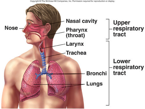
nasal cavity- open space with olfactory receptors
pharynx- throat
larynx- epiglottis to seal the trachea, vocal cords to produce sound, passage is made of cartilage
trachea- passage is made of cartilage
bronchi- trachea branches to two primary bronchi, which keep branching to smaller bronchioles (walls are smooth muscle)
terminal bronchioles- smallest and final branches, walls are smooth muscle
147
respiratory zone
respiratory bronchiole- walls are smooth muscle, has a few alveoli scattered in its walls
alveolar duct- made entirely of alveoli
alveoli- tiny sacs with single cell walls, capillaries surround them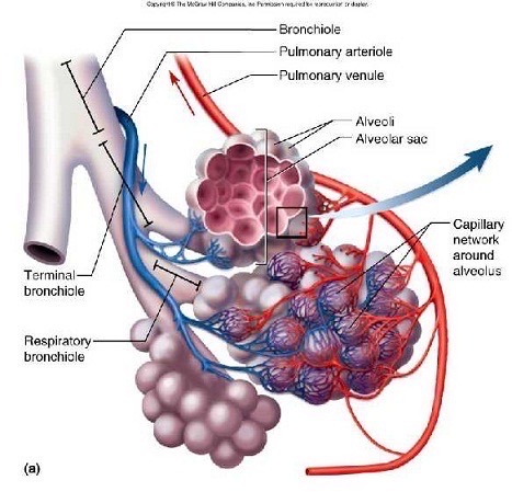
alveolar duct- made entirely of alveoli
alveoli- tiny sacs with single cell walls, capillaries surround them
148
respiratory epithelium
epithelial cells- column shaped cells that line the entire respiratory tract
goblet cells- epithelial cells that secrete mucus
mucociliary escalator- epithelial cells in upper respiratory tract have cilia that sweep mucus towards pharynx, to be coughed out or swallowed (stomach acidity destroys pathogens)
squamous epithelial cells- flat cells in alveoli, no mucus here so alveolar macrophages engulf pathogens
surfactant- surface tension of water would cause alveoli to collapse, so this soapy substance reduces the tension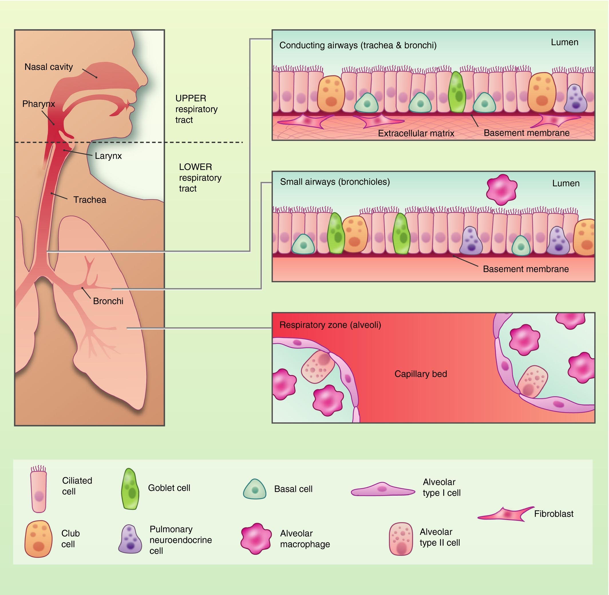
goblet cells- epithelial cells that secrete mucus
mucociliary escalator- epithelial cells in upper respiratory tract have cilia that sweep mucus towards pharynx, to be coughed out or swallowed (stomach acidity destroys pathogens)
squamous epithelial cells- flat cells in alveoli, no mucus here so alveolar macrophages engulf pathogens
surfactant- surface tension of water would cause alveoli to collapse, so this soapy substance reduces the tension
149
pleural space
inspiration- moving air into lungs, diaphragm contraction
expiration- passive, elastic recoil
parietal pleura- lines inside of chest cavity
visceral pleura- lines surface of lungs
pleural space- the space in between, has negative pressure to keep lungs open, also some fluid to hold surfaces together with surface tension
diaphragm- dome contracts downward to force lungs and chest cavity to expand
intercostal muscles- muscles between ribs pulls ribs upward to further expand chest cavity
forced expiration- usually passive, but can be forced by contraction of abdominal muscles to press upward on diaphragm
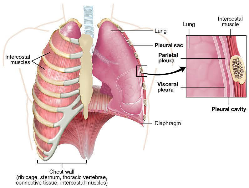
expiration- passive, elastic recoil
parietal pleura- lines inside of chest cavity
visceral pleura- lines surface of lungs
pleural space- the space in between, has negative pressure to keep lungs open, also some fluid to hold surfaces together with surface tension
diaphragm- dome contracts downward to force lungs and chest cavity to expand
intercostal muscles- muscles between ribs pulls ribs upward to further expand chest cavity
forced expiration- usually passive, but can be forced by contraction of abdominal muscles to press upward on diaphragm
150
lung volume
spirometry- measurement of volume of air in lungs during ventilation, uses a spirometer
tidal volume (TV)- amount of air inspired during normal, relaxed breathing.
inspiratory reserve volume (IRV)- additional air that can be forcibly inhaled after the inspiration of a normal tidal volume.
expiratory reserve volume (ERV)- additional air that can be forcibly exhaled after the expiration of a normal tidal volume.
residual volume (RV)- amount of air still remaining in the lungs after the expiratory reserve volume is exhaled.
vital capacity (VC)- total amount of air that can be expired after fully inhaling (VC = TV + IRV + ERV). The value varies according to age and body size.
total lung capacity (TLC)- maximum amount of air that can fill the lungs (TLC = VC + RV).
inspiratory capacity (IC)- maximum amount of air that can be inspired (IC = TV + IRV).
functional residual capacity (FRC)- amount of air remaining in the lungs after a normal expiration (FRC = RV + ERV).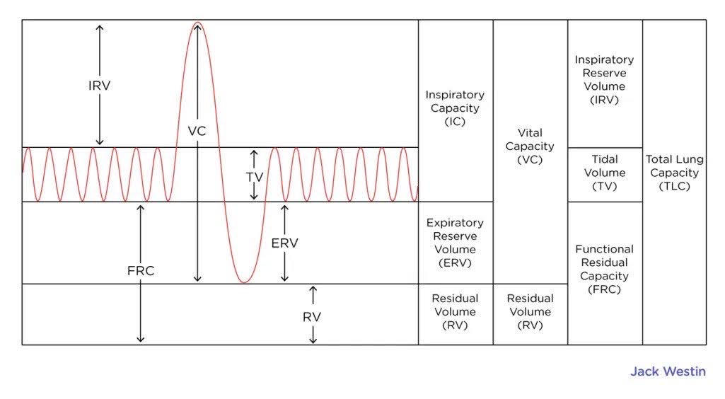
tidal volume (TV)- amount of air inspired during normal, relaxed breathing.
inspiratory reserve volume (IRV)- additional air that can be forcibly inhaled after the inspiration of a normal tidal volume.
expiratory reserve volume (ERV)- additional air that can be forcibly exhaled after the expiration of a normal tidal volume.
residual volume (RV)- amount of air still remaining in the lungs after the expiratory reserve volume is exhaled.
vital capacity (VC)- total amount of air that can be expired after fully inhaling (VC = TV + IRV + ERV). The value varies according to age and body size.
total lung capacity (TLC)- maximum amount of air that can fill the lungs (TLC = VC + RV).
inspiratory capacity (IC)- maximum amount of air that can be inspired (IC = TV + IRV).
functional residual capacity (FRC)- amount of air remaining in the lungs after a normal expiration (FRC = RV + ERV).
151
pulmonary circulation
pulmonary capillaries- alveolar capillaries, branch from pulmonary artery to bring deoxygenated blood to lungs
RBCs pass through capillaries single file to maximize surface area exposed and increase gas exchange
pulmonary edema- usually pulmonary veins can dilate, but too high of pressure will force blood into lung tissue, lymphatic system carries fluid out of lungs
respiratory membrane- alveolar epithelium, interstitial liquid, capillary endothelium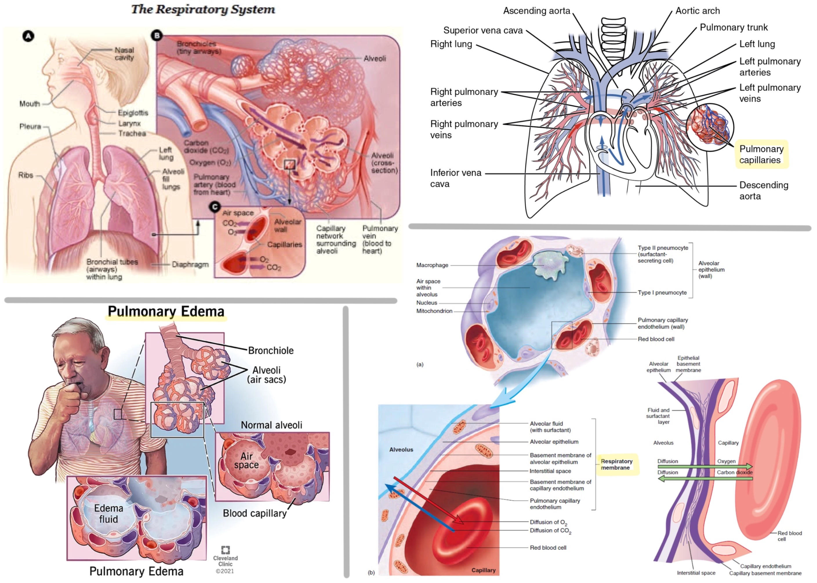
RBCs pass through capillaries single file to maximize surface area exposed and increase gas exchange
pulmonary edema- usually pulmonary veins can dilate, but too high of pressure will force blood into lung tissue, lymphatic system carries fluid out of lungs
respiratory membrane- alveolar epithelium, interstitial liquid, capillary endothelium
152
atmospheric pressure
atmospheric gas- 80% N2, 20% O2, some H2O, CO2, etc.
atmospheric pressure- 760 torr, so partial pressure of O2 is 150 tor
atmospheric pressure- 760 torr, so partial pressure of O2 is 150 tor
153
Henry's Law
amount of gas that dissolves into liquid depends on partial pressure of gas and solubility of gas in liquid
[O2] = P_O2 * S_O2
concentration of dissolved oxygen = partial pressure of oxygen times solubility of oxygen in liquid
gases become less soluble as temperature increases (goldfish gulp for air when water is too warm)
difference in partial pressures between alveolar air and blood drives exchange of gas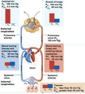
[O2] = P_O2 * S_O2
concentration of dissolved oxygen = partial pressure of oxygen times solubility of oxygen in liquid
gases become less soluble as temperature increases (goldfish gulp for air when water is too warm)
difference in partial pressures between alveolar air and blood drives exchange of gas
154
regulation of ventilation rate
medulla- contains respiratory control center
trigger increased ventilation rate:
1. increased P_CO2
2. decreased P_O2
3. decreased pH
peripheral chemoreceptors- monitor blood for pH, P_CO2, P_O2
central chemoreceptors- monitor cerebrospinal fluid for pH, P_CO2, P_O2
irritant receptors- trigger coughing, bronchoconstriction
stretch receptors- trigger inhibit inspiration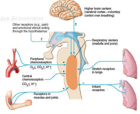
trigger increased ventilation rate:
1. increased P_CO2
2. decreased P_O2
3. decreased pH
peripheral chemoreceptors- monitor blood for pH, P_CO2, P_O2
central chemoreceptors- monitor cerebrospinal fluid for pH, P_CO2, P_O2
irritant receptors- trigger coughing, bronchoconstriction
stretch receptors- trigger inhibit inspiration
155
epidermis
outmost layer of skin
layers of squamous epithelial cells
stratum basale- deepest part that replenishes upper layers with mitosis
keratin- as the cells die, they become filled with keratin, which is tough and hydrophobic and makes skin waterproof
melanin- melanocytes produce melanin, which is a brown pigment that helps absorb UV to prevent damage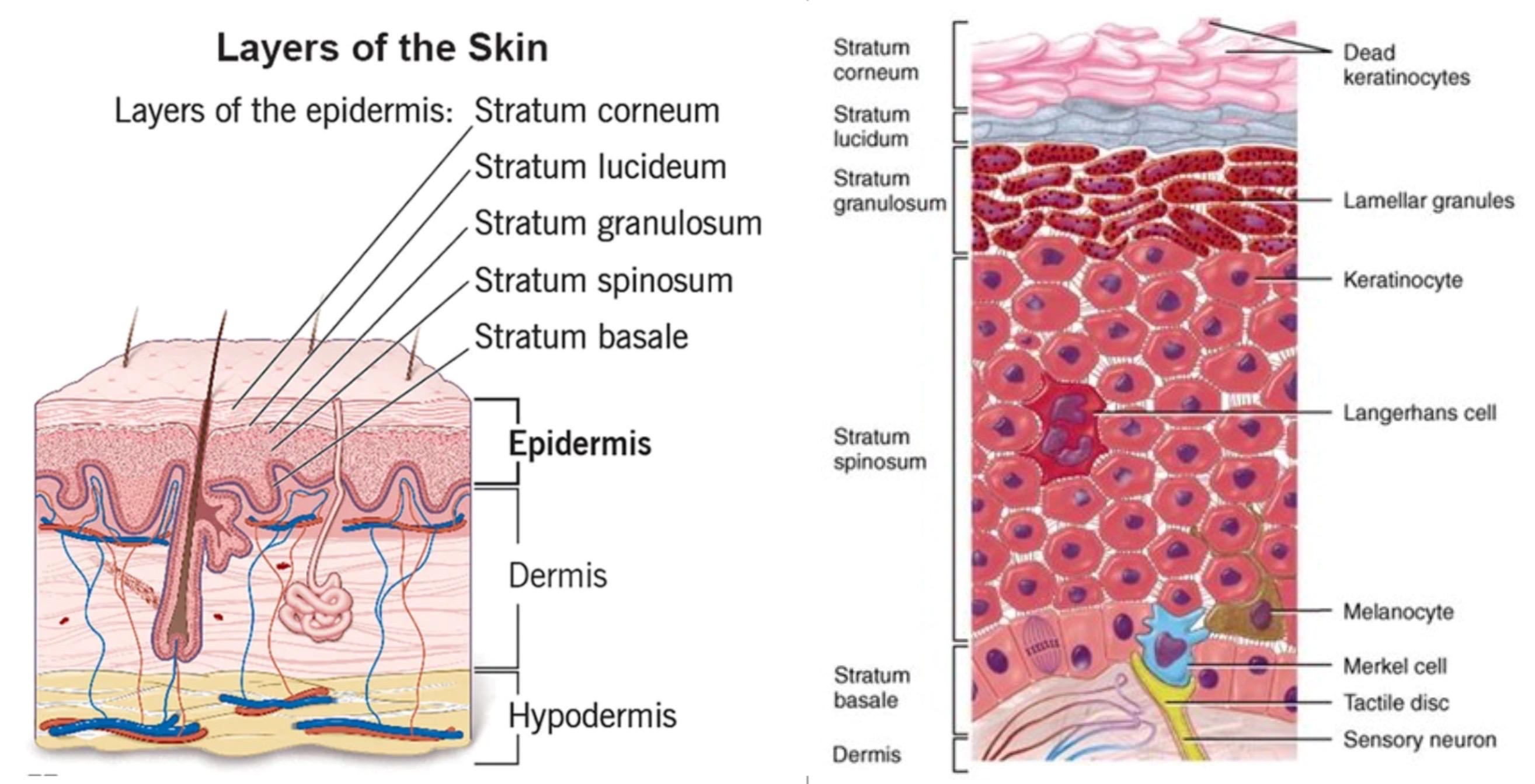
layers of squamous epithelial cells
stratum basale- deepest part that replenishes upper layers with mitosis
keratin- as the cells die, they become filled with keratin, which is tough and hydrophobic and makes skin waterproof
melanin- melanocytes produce melanin, which is a brown pigment that helps absorb UV to prevent damage
156
dermis
middle layer of skin
embedded in connective tissue, has blood vessels
sensory receptors- touch, pressure, pain, temperature
sudoriferous glands- sweat glands, loss of heat by evaporation, signalled by aldosterone, sweat has water, ions, and urea
sebaceous glands- oil glands
ceruminous glands- wax glands, only in ear
hair follicles- hair is made of dead epithelial cells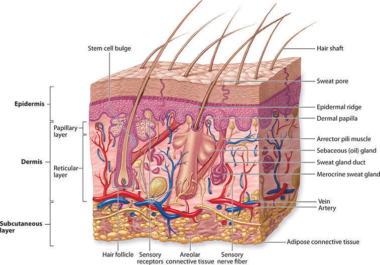
embedded in connective tissue, has blood vessels
sensory receptors- touch, pressure, pain, temperature
sudoriferous glands- sweat glands, loss of heat by evaporation, signalled by aldosterone, sweat has water, ions, and urea
sebaceous glands- oil glands
ceruminous glands- wax glands, only in ear
hair follicles- hair is made of dead epithelial cells
157
hypodermis
subcutaneous tissue
protective, insulating layer of adipose tissue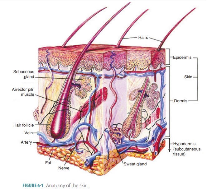
protective, insulating layer of adipose tissue
158
temperature regulation
homeotherms- body temperature kept constant
chemical thermogenesis- bears have brown adipose tissue that has a lot of mitochondria, uncouples ETC to generate heat instead of ATP, humans cannot do this
stay warm:
1. muscle contraction- shivering, moving
2. skin insulation- subcutaneous tissue
3. cutaneous vasoconstriction- cut off blood to skin, activated by sympathetic NS (pale when frightened)
cool down:
1. sweating
2. cutaneous vasodilation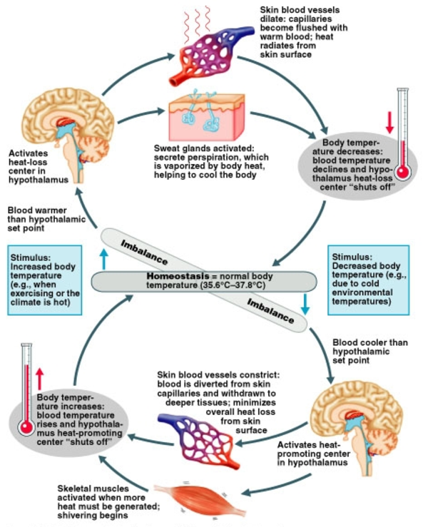
chemical thermogenesis- bears have brown adipose tissue that has a lot of mitochondria, uncouples ETC to generate heat instead of ATP, humans cannot do this
stay warm:
1. muscle contraction- shivering, moving
2. skin insulation- subcutaneous tissue
3. cutaneous vasoconstriction- cut off blood to skin, activated by sympathetic NS (pale when frightened)
cool down:
1. sweating
2. cutaneous vasodilation
159
types of tissue
epithelial tissue- line the surface of all organs, *epithelial cells divide the most and spend the least time in G0*
muscle tissue- muscles
connective tissue- ECM, between tissues and underneath epithelial tissue, fibroblasts become adipocytes, chondrocytes, osteocytes
nervous tissue- nervous system, *neurons are always in G0*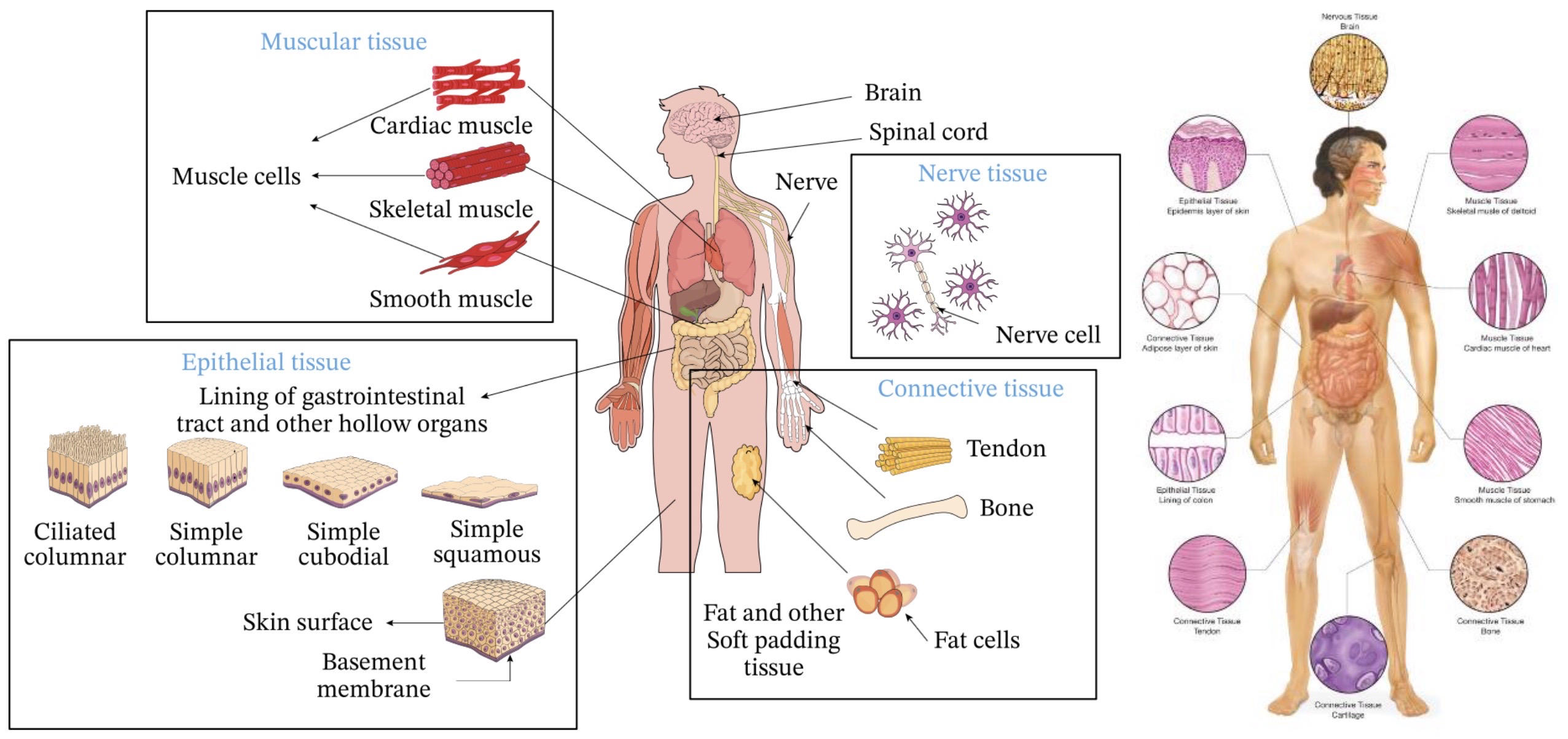
muscle tissue- muscles
connective tissue- ECM, between tissues and underneath epithelial tissue, fibroblasts become adipocytes, chondrocytes, osteocytes
nervous tissue- nervous system, *neurons are always in G0*
160
male reproductive system
1. testes- produce sperm, produce testosterone
2. scrotum- regulates temperature, keeps testes cool
3. epididymis- sperm hang out here to develop their flagella and more mitochondria
4. Vas deferens- sperm travels, within the spermatic cord
5. seminal vesicles- add slightly basic fluid to sperm to form semen, remove acidity
6. prostate gland, bulbourethral gland- make more fluid
7. urethra- semen travels
8. penis- penetrate vagina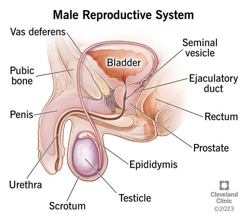
2. scrotum- regulates temperature, keeps testes cool
3. epididymis- sperm hang out here to develop their flagella and more mitochondria
4. Vas deferens- sperm travels, within the spermatic cord
5. seminal vesicles- add slightly basic fluid to sperm to form semen, remove acidity
6. prostate gland, bulbourethral gland- make more fluid
7. urethra- semen travels
8. penis- penetrate vagina
161
spermatogenesis
sperm is produced by the Sertoli cells of the *seminiferous tubules*
1. diploid germ cells become haploid gametes through meiosis
2. undergo more replications, makes mutations more likely on Y chromosome (since it's inherited through sperm)
sperm swims and fuses with the egg to become zygote
1. *acrosome*- contains hydrolytic enzymes to penetrate ovum
2. *bindin*- surface proteins that attaches to receptors on zona pellucida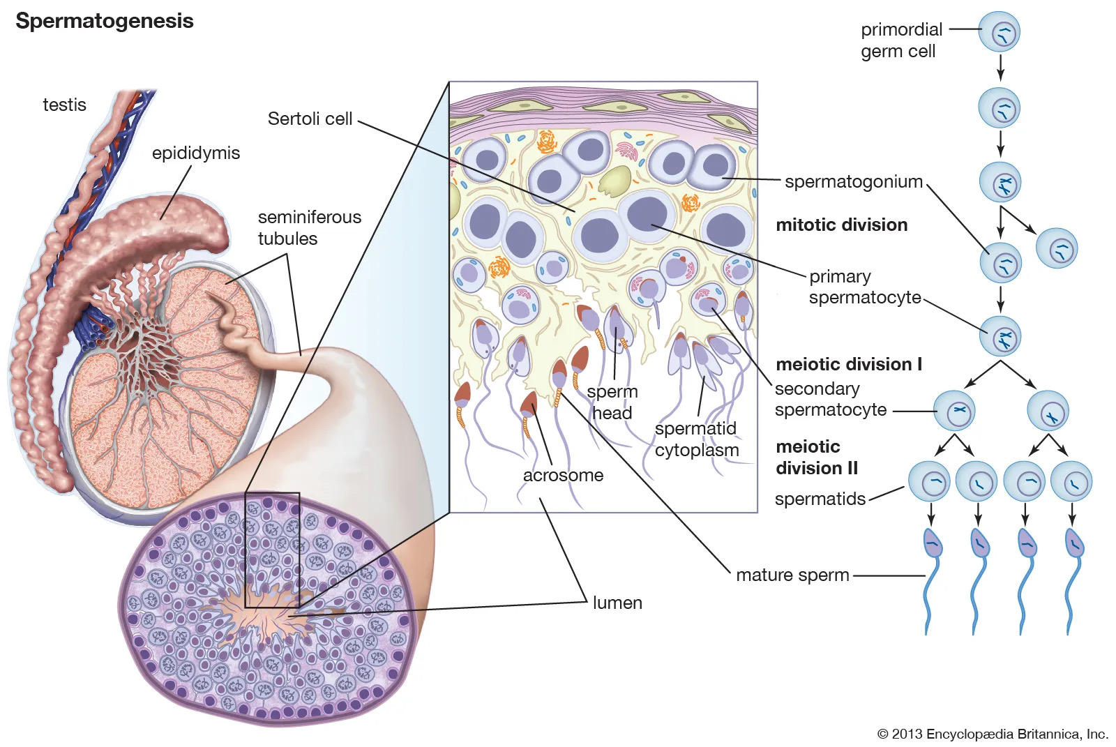
1. diploid germ cells become haploid gametes through meiosis
2. undergo more replications, makes mutations more likely on Y chromosome (since it's inherited through sperm)
sperm swims and fuses with the egg to become zygote
1. *acrosome*- contains hydrolytic enzymes to penetrate ovum
2. *bindin*- surface proteins that attaches to receptors on zona pellucida
162
testosterone
testosterone is produced by Leydig cells in testes, stimulates spermatogenesis, later causes secondary sex characteristics to develop during puberty
androgen- male hormones
LH and FSH stimulate testosterone secretion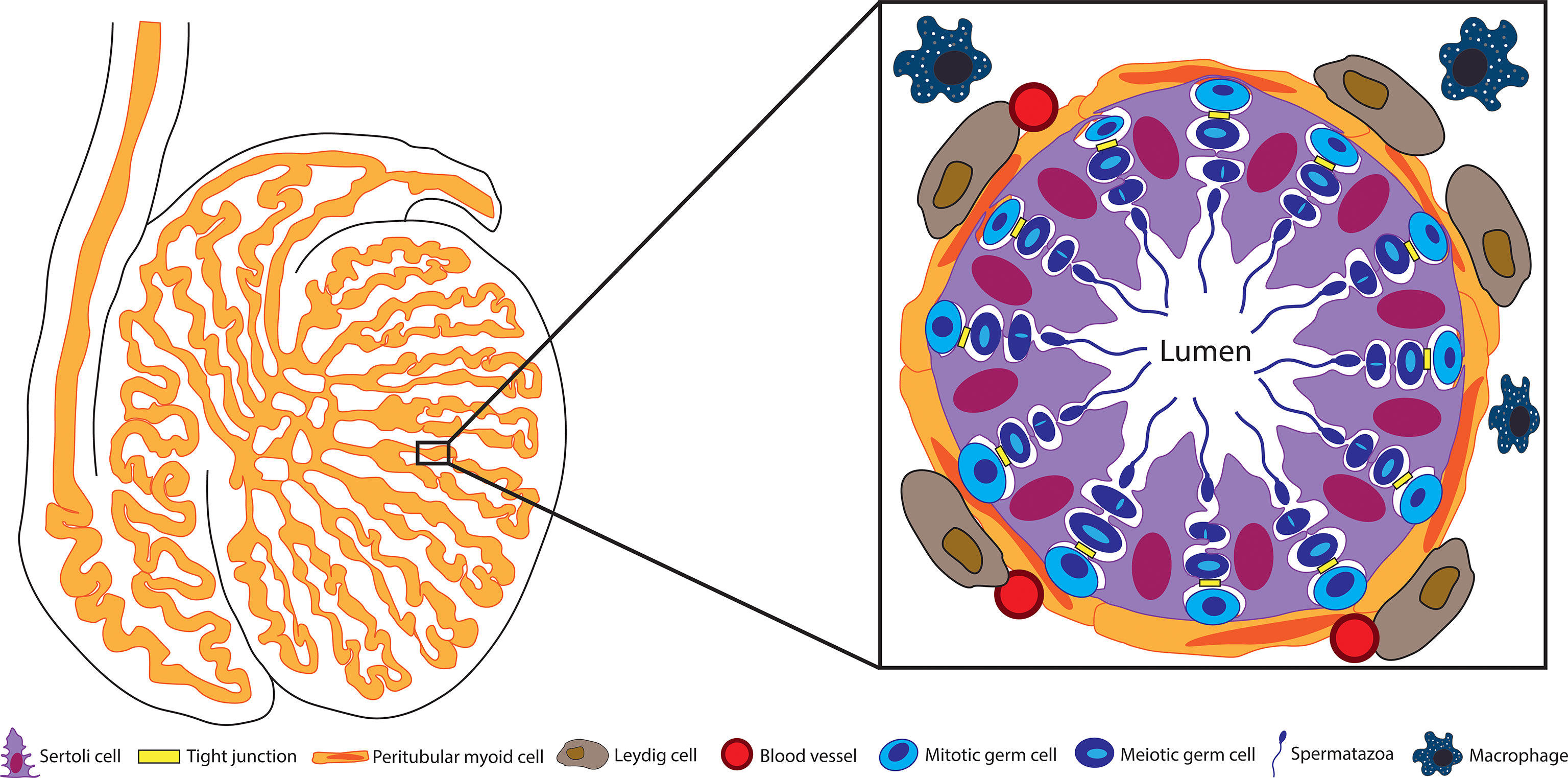
androgen- male hormones
LH and FSH stimulate testosterone secretion
163
sexual act
para does arousal and erection
sympa does orgasm
arousal, orgasm, resolution (takes longer in females)
sympa does orgasm
arousal, orgasm, resolution (takes longer in females)
164
reproductive system development
all embryos possess Wolffian ducts (which become male genitalia) and Mullerian ducts (which become female genitalia)
testes produce Mullerian inhibiting factor (MIF)
analogous structure:
1. testis and ovaries
2. penis and clitoris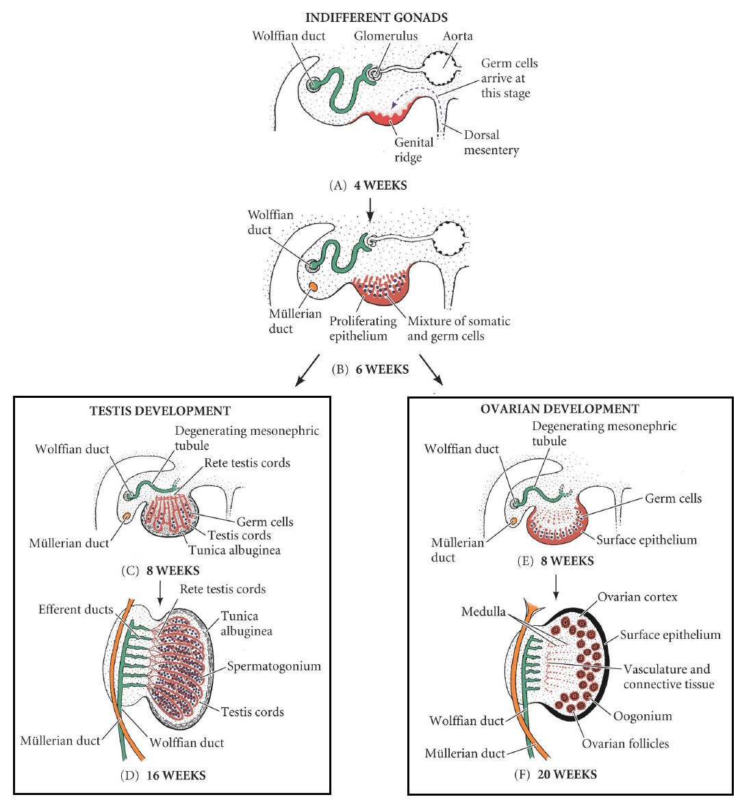
testes produce Mullerian inhibiting factor (MIF)
analogous structure:
1. testis and ovaries
2. penis and clitoris
165
female reproductive system
1. ovaries- produce ovum
2. uterus- carries embryo, cervix is entrance, endometrium is lining that is shed each month for menstrual bleeding, myometrium is smooth muscle
3. vagina- penis enters
4. breasts- lactation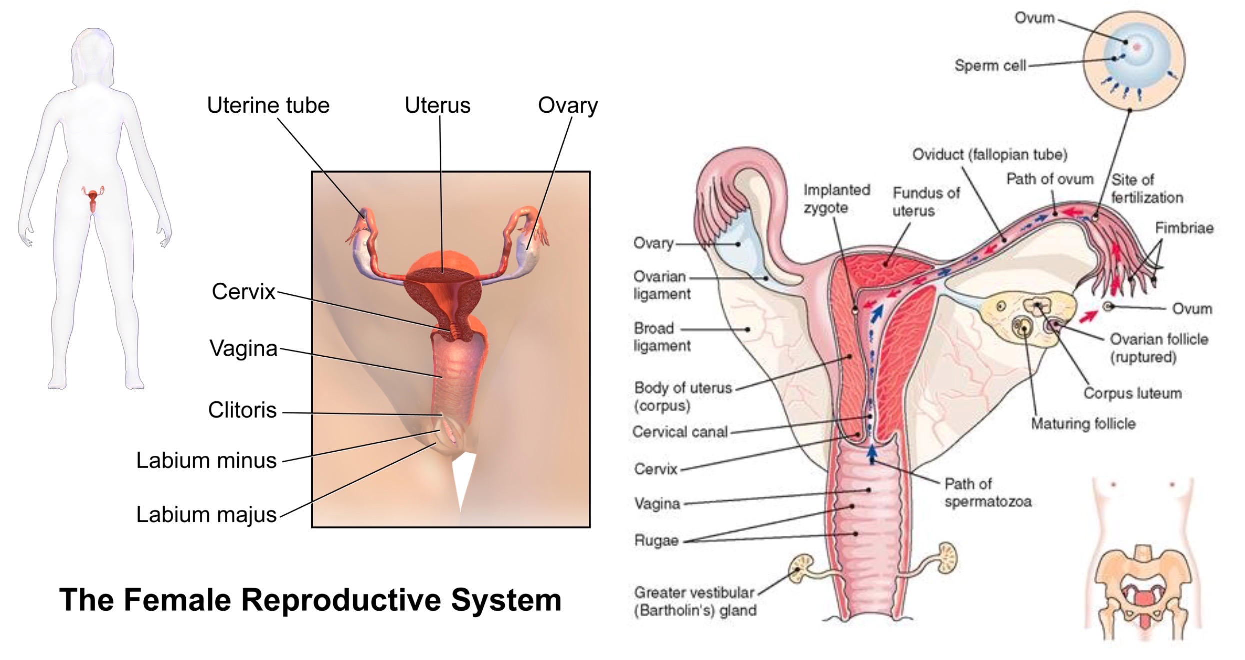
2. uterus- carries embryo, cervix is entrance, endometrium is lining that is shed each month for menstrual bleeding, myometrium is smooth muscle
3. vagina- penis enters
4. breasts- lactation
166
oogenesis
primary oocytes are frozen in prophase I, hormones stimulate completion of first meiotic division
unequal division to become secondary oocyte and small polar body
second meiotic division occurs when sperm arrives, unequal division to large ovum and another polar body
ova and sperm have haploid genome size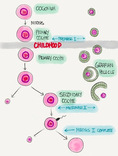
unequal division to become secondary oocyte and small polar body
second meiotic division occurs when sperm arrives, unequal division to large ovum and another polar body
ova and sperm have haploid genome size
167
follicle
follicle- a collection of granulosa cells around the ova that support its development and secrete estrogen
granulosa cells become zona pellucida- protective layer
Graafian (mature) follicle- one follicle matures during ovulation, releases ovum into fallopian tube
corona radiata- protective layer still on egg
corpus luteum- follicle stuff left over, secretes estrogen and progesterone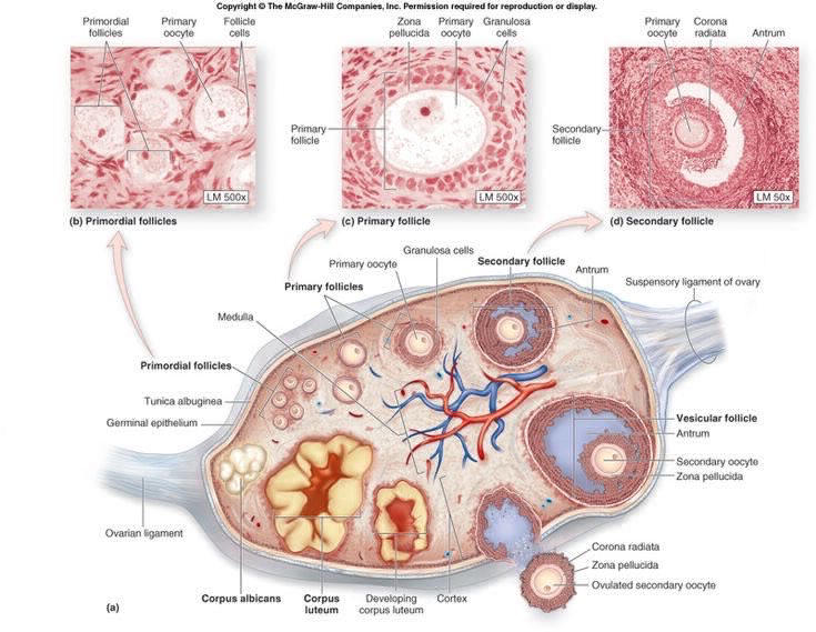
granulosa cells become zona pellucida- protective layer
Graafian (mature) follicle- one follicle matures during ovulation, releases ovum into fallopian tube
corona radiata- protective layer still on egg
corpus luteum- follicle stuff left over, secretes estrogen and progesterone
168
menstrual cycle
ovarian cycle:
1. follicular phase- follicle matures and secretes estrogen
2. ovulatory phase- secondary oocyte released from ovary
3. luteal phase- corpus luteum forms, secretes estrogen and progesterone
uterine cycle:
1. menstruation- corpus luteum degenerates, estrogen and progesterone stops, endometrial lining bleeds out
2. proliferative phase- estrogen from follicle produces new endometrium
3. secretory phase- estrogen and progesterone from corpus luteum increase development of endometrium
menopause- menstrual cycle stops at 50-60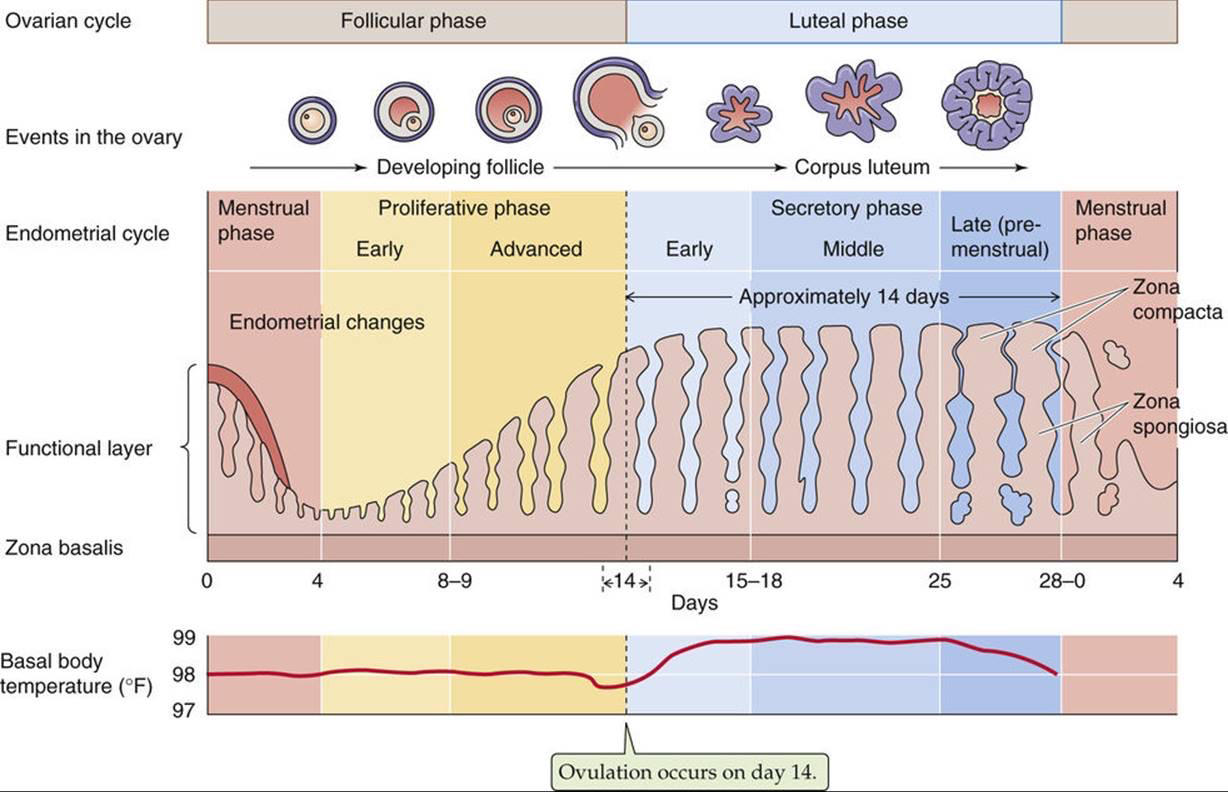
1. follicular phase- follicle matures and secretes estrogen
2. ovulatory phase- secondary oocyte released from ovary
3. luteal phase- corpus luteum forms, secretes estrogen and progesterone
uterine cycle:
1. menstruation- corpus luteum degenerates, estrogen and progesterone stops, endometrial lining bleeds out
2. proliferative phase- estrogen from follicle produces new endometrium
3. secretory phase- estrogen and progesterone from corpus luteum increase development of endometrium
menopause- menstrual cycle stops at 50-60
169
birth control vs. pregnancy
birth control- keep estrogen and progesterone high so menstruation and ovulation does not happen
pregnancy- when embryo implants, placenta develops and secretes hCG which replaces LH, so corpus luteum does not degenerate and estrogen and progesterone stay high
*hCG* is used to test for pregnancy
pregnancy- when embryo implants, placenta develops and secretes hCG which replaces LH, so corpus luteum does not degenerate and estrogen and progesterone stay high
*hCG* is used to test for pregnancy
170
fertilization
acrosome and bindin
Ca2+ influx causes egg activation
polyspermy:
1. fast block- depolarization of ovum plasma membrane by Ca2+ influx
2. slow block- cortical reaction to harden zona pellucida and increase space between zona pellucida and plasma membrane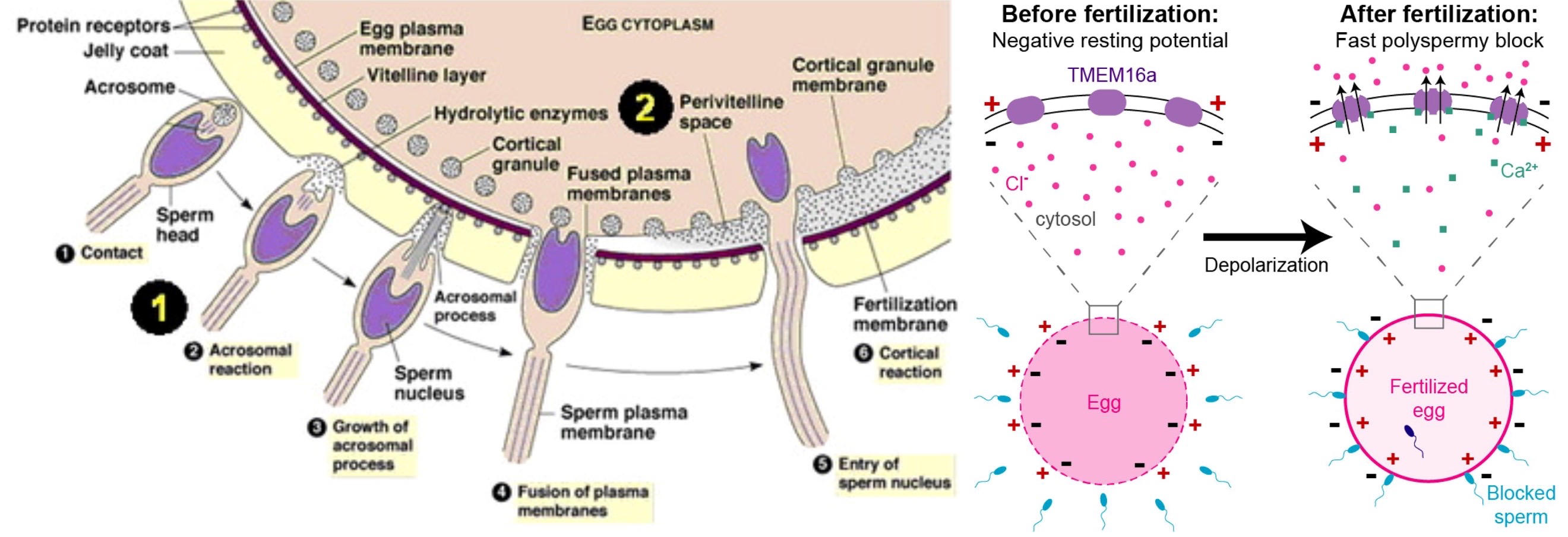
Ca2+ influx causes egg activation
polyspermy:
1. fast block- depolarization of ovum plasma membrane by Ca2+ influx
2. slow block- cortical reaction to harden zona pellucida and increase space between zona pellucida and plasma membrane
171
maternal inheritance
egg provides all the cellular parts of the zygote, including mitochondrial inheritance
172
germ layers
ectoderm- forms skin, facial things, mouth and butt, and nervous system
endoderm- forms epithelial lining inside body (GI, urinary, respiratory, endocrine, auditory)
mesoderm- muscle, bone, kidneys, heart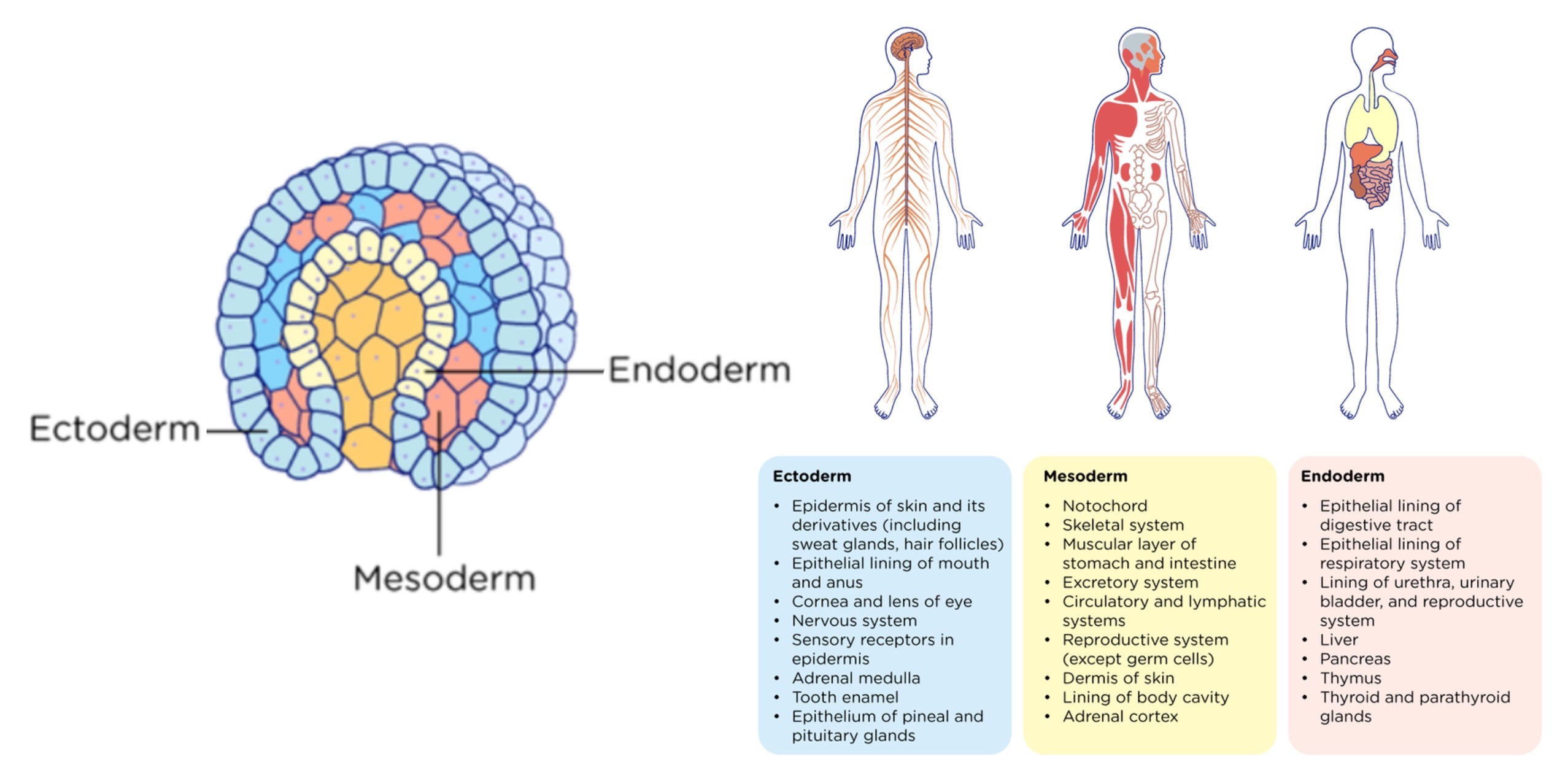
endoderm- forms epithelial lining inside body (GI, urinary, respiratory, endocrine, auditory)
mesoderm- muscle, bone, kidneys, heart
173
embryogenesis
cleavage- zygote produces *morula*, a solid ball of 16 undifferentiated cells.
blastulation- blastocyst forms, consists of:
1. trophoblast- outer cell layer of blastocyst
2. blastocoele- fluid-filled central region in the blastocyst
3. inner cell mass (ICM)- inside of blastocyst, will eventually give rise to the fetus
implantation- blastocyst burrows into endometrium, starts taking nutrients from endometrium, placenta develops 3 months later
inner cell mass also becomes amnion (amniotic fluid), yolk sac (RBC synthesis), allantois (forms umbilical cord)
gastrulation- single-layered blastula becomes the three-layered gastrula, the three germ layers
neurulation- ectoderm differentiates into neural plate, folds into neural tube, which becomes CNS
MBGN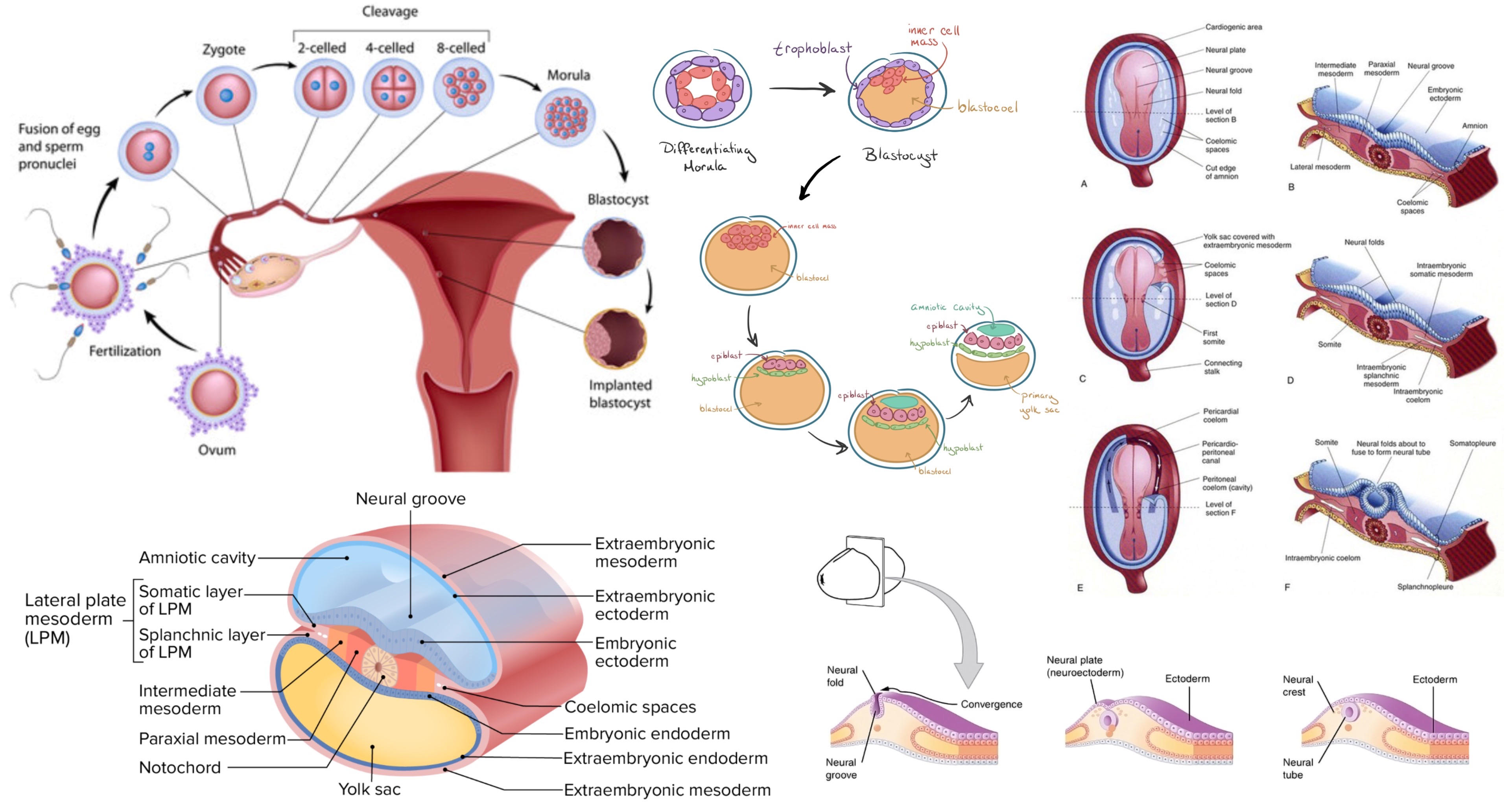
blastulation- blastocyst forms, consists of:
1. trophoblast- outer cell layer of blastocyst
2. blastocoele- fluid-filled central region in the blastocyst
3. inner cell mass (ICM)- inside of blastocyst, will eventually give rise to the fetus
implantation- blastocyst burrows into endometrium, starts taking nutrients from endometrium, placenta develops 3 months later
inner cell mass also becomes amnion (amniotic fluid), yolk sac (RBC synthesis), allantois (forms umbilical cord)
gastrulation- single-layered blastula becomes the three-layered gastrula, the three germ layers
neurulation- ectoderm differentiates into neural plate, folds into neural tube, which becomes CNS
MBGN
174
pregnancy
increased demand for oxygen, GFR, nutrients
oxytocin- causes uterine contraction
parturition- birth
prolactin- released by suckling (anterior pituitary), oxytocin (posterior pituitary) also necessary for milk let-down
oxytocin- causes uterine contraction
parturition- birth
prolactin- released by suckling (anterior pituitary), oxytocin (posterior pituitary) also necessary for milk let-down
175
estrogen
estrogen and progesterone together cause the endometrium to thicken
estrogen later regulates uterine cycle and development of secondary sexual characteristics
estrogen inhibits bone resorption
estrogens- female hormones
estrogen later regulates uterine cycle and development of secondary sexual characteristics
estrogen inhibits bone resorption
estrogens- female hormones
176
follicle stimulating hormone (FSH)
in females, triggers follicle development, estrogen production by granulosa cells and corpus luteum
in males, stimulates Sertoli cells with sperm development
in males, stimulates Sertoli cells with sperm development
177
X inactivation
in females, cells inactivated one X chromosome to become Barr body, creating two populations of cells
so only 50% of cells will have an X-linked trait in females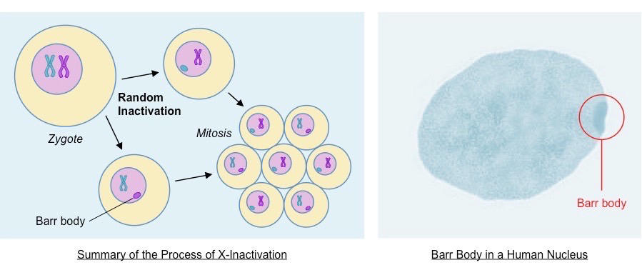
so only 50% of cells will have an X-linked trait in females
178
Y chromosome
smaller, contains less genes
179
cell specialization
development
migration
communication
differentiation- totipotent, pluripotent, multipotent cells
dedifferentiation- cancer, induced pluripotent stem cells
gene regulation
apoptosis
regenerative capacity
senescence and aging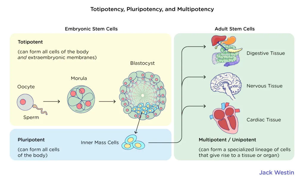
migration
communication
differentiation- totipotent, pluripotent, multipotent cells
dedifferentiation- cancer, induced pluripotent stem cells
gene regulation
apoptosis
regenerative capacity
senescence and aging
180
sex linked traits
XY is male
XX is female
mitochondrial linked traits
XX is female
mitochondrial linked traits