Lecture 8 - Hypersensitivity
Study these flashcardsFree · spaced repetition · iPhone, Android, Mac & web
All cards
1
What is Hypersensitivity?
-An exaggerated response to a typically harmless antigen that results in tissue injury, disease, or even death
- 4 groups - classified based on the immunologic mechanism
- 4 groups - classified based on the immunologic mechanism
2
What are the 4 classes found in hypersensitivity?
• Class I (aka anaphylactic hypersensitivity)
• Class II (aka antibody-mediated cytotoxic hypersensitivity)
• Class III (aka immune complex-mediated hypersensitivity)
• Class IV (aka cell-mediated hypersensitivity)
• Class II (aka antibody-mediated cytotoxic hypersensitivity)
• Class III (aka immune complex-mediated hypersensitivity)
• Class IV (aka cell-mediated hypersensitivity)
3
What is Class I hypersensitivity also known as?
anaphylactic hypersensitivity
4
What is Class II hypersensitivity also known as?
antibody-mediated cytotoxic hypersensitivity
5
What is Class III hypersensitivity also known as?
immune complex-mediated hypersensitivity
6
What is Class IV hypersensitivity also known as?
cell-mediated hypersensitivity
7
What mediates the 4 classes of hypersensitivity?
• I, II and III are antibody-mediated, and called immediate hypersensitivity reactions (happen within few minutes to a few hours after contact with Ag)
• IV is T cell-mediated and called delayed hypersensitivity reaction (takes 24-48h)
• IV is T cell-mediated and called delayed hypersensitivity reaction (takes 24-48h)
8
What is Class I also called?
allergies
9
What are some examples of Class I causes?
hay fever, hives, allergic asthma, systemic anaphylaxis
10
What is the major antibody involved, which is greatly increased in these patients?
IgE
11
What is atopy, which explains Class I at the genetics level?
the inherited tendency to develop classic allergic responses to naturally occurring inhaled or ingested allergens
12
In the Class I, which are the main players and why?
- IgE
- basophils, mast cells, eosinophils,
- they have receptor for IgE
- basophils, mast cells, eosinophils,
- they have receptor for IgE
13
Which is the major cell for Class I hypersensitivity?
mast cells
14
Which Th cells are imbalanced during Class I rxn?
Th1 and Th2 balance is shifted so that Th2 response will predominate
15
What happens once the Th cells are activated? Which cytokines are produced?
Il-4 and Il-13 - cytokines responsible for B cell differentiation and IgE production
16
How long does the immunologic reaction for Class I take?
30-60 minutes
17
What are the Class I hypersensitivity phases?
1. sensitization
2. activation
2. activation
18
What happens in the Class I sensitization phase?
--> APC presents allergen to Th2 cells
--> IgE produced by B cells
--> IgE binds FcR1 on mast cells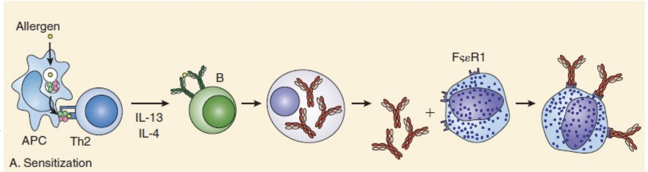
--> IgE produced by B cells
--> IgE binds FcR1 on mast cells
19
What happens in the Class I activation phase?
--> upon IgE binding
--> mast cells will be activated and release their granular contents including:
1. histamine (the most important factor)
2. other substances include heparin
3. Eosinophil chemotactic factor of anaphylaxis (ECF-A)
4. Neutrophil chemotactic factor of anaphylaxis (NCF-A)
5. Proteases (e.g., tryptase, chymase)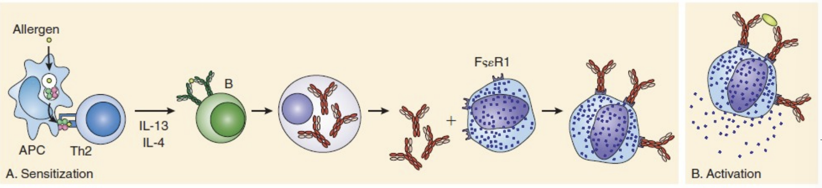
--> mast cells will be activated and release their granular contents including:
1. histamine (the most important factor)
2. other substances include heparin
3. Eosinophil chemotactic factor of anaphylaxis (ECF-A)
4. Neutrophil chemotactic factor of anaphylaxis (NCF-A)
5. Proteases (e.g., tryptase, chymase)
20
What is the outcome of the Class I activation phase?
• Clinical presentation of allergy depending on the affected organ: skin (wheal-and-flare), lung (asthma), etc.
21
What is the late phase activation for Class I?
• 6-8 h after allergen exposure, basophils and mast cells release secondary mediators including platelet-activating factor (PAF); prostaglandin (PG) D2; leukotrienes (LT) B4, C4, D4, and E4; and cytokines. These are more potent than primary (immediate) mediators
• In the late phase, numerous cells, including eosinophils, neutrophils, Th2 cells, mast cells, basophils, and macrophages, infiltrate the allergen-filled tissue. They release additional mediators that prolong hyperactivity and cause further tissue damage
• In the late phase, numerous cells, including eosinophils, neutrophils, Th2 cells, mast cells, basophils, and macrophages, infiltrate the allergen-filled tissue. They release additional mediators that prolong hyperactivity and cause further tissue damage
22
What are some common allergies for Class I?
• Pollen
• Mold spores
• Animal dander
• Dust mites
• Insect venom
• Certain foods (peanuts, shellfish, dairy products)
• Certain drugs (penicillin)
• Latex
• Mold spores
• Animal dander
• Dust mites
• Insect venom
• Certain foods (peanuts, shellfish, dairy products)
• Certain drugs (penicillin)
• Latex
23
What are some clinical manifestations of Class I?
• Rhinitis (“hay fever”)
• Allergic asthma
• Food allergies
• Skin reactions (Urticaria (“hives”), angioedema, eczema) - Wheal and flare
• Systemic anaphylaxis - A potentially fatal reaction, and associated with IgA-deficient patients
• Allergic asthma
• Food allergies
• Skin reactions (Urticaria (“hives”), angioedema, eczema) - Wheal and flare
• Systemic anaphylaxis - A potentially fatal reaction, and associated with IgA-deficient patients
24
What is Urticaria?
- aka hives
- caused by an immediate hypersensitivity rxn to a medication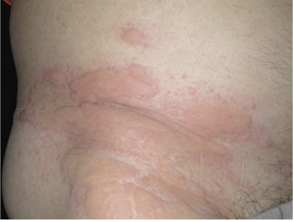
- caused by an immediate hypersensitivity rxn to a medication
25
What is angioedema?
- a condition that causes swelling in the deeper layers of the skin due to fluid leaking from small blood vessels
- the example shown here is caused by a yellow jacket sting on the right hand just above the middle finger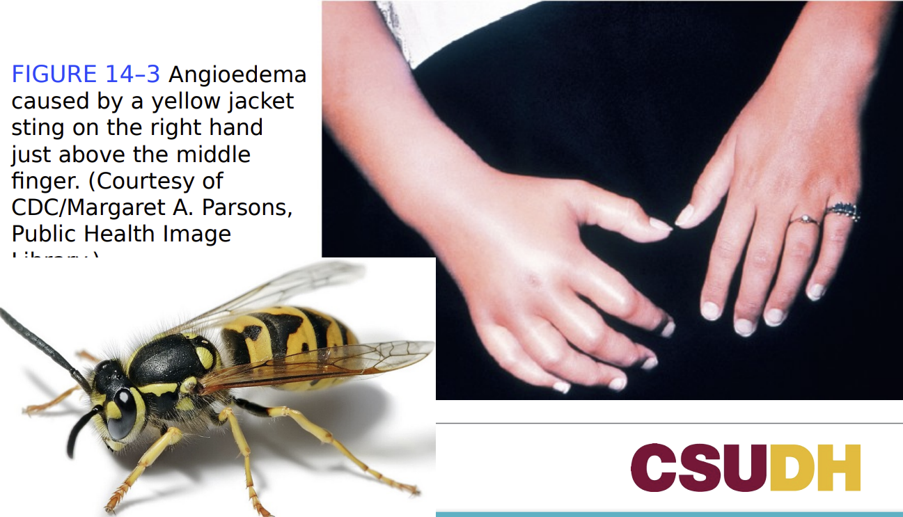
- the example shown here is caused by a yellow jacket sting on the right hand just above the middle finger
26
What are some treatments for Type I hypersensitivity?
• Avoidance of allergens
• Drug therapy
- Antihistamines, bronchodilators, mast cell stabilizers, corticosteroids, epinephrine
• Monoclonal anti-IgE antibody
• Allergy immunotherapy (AIT) - administer gradually, increasing doses of
allergen
• Drug therapy
- Antihistamines, bronchodilators, mast cell stabilizers, corticosteroids, epinephrine
• Monoclonal anti-IgE antibody
• Allergy immunotherapy (AIT) - administer gradually, increasing doses of
allergen
27
What are the in vivo skin tests for Class I hypersensitivity?
- Percutaneous or intradermal
- Process:
-- Apply a panel of allergens to separate sites on the skin.
-- Wait 15 to 20 minutes.
- Positive test = wheal and flare at the site of application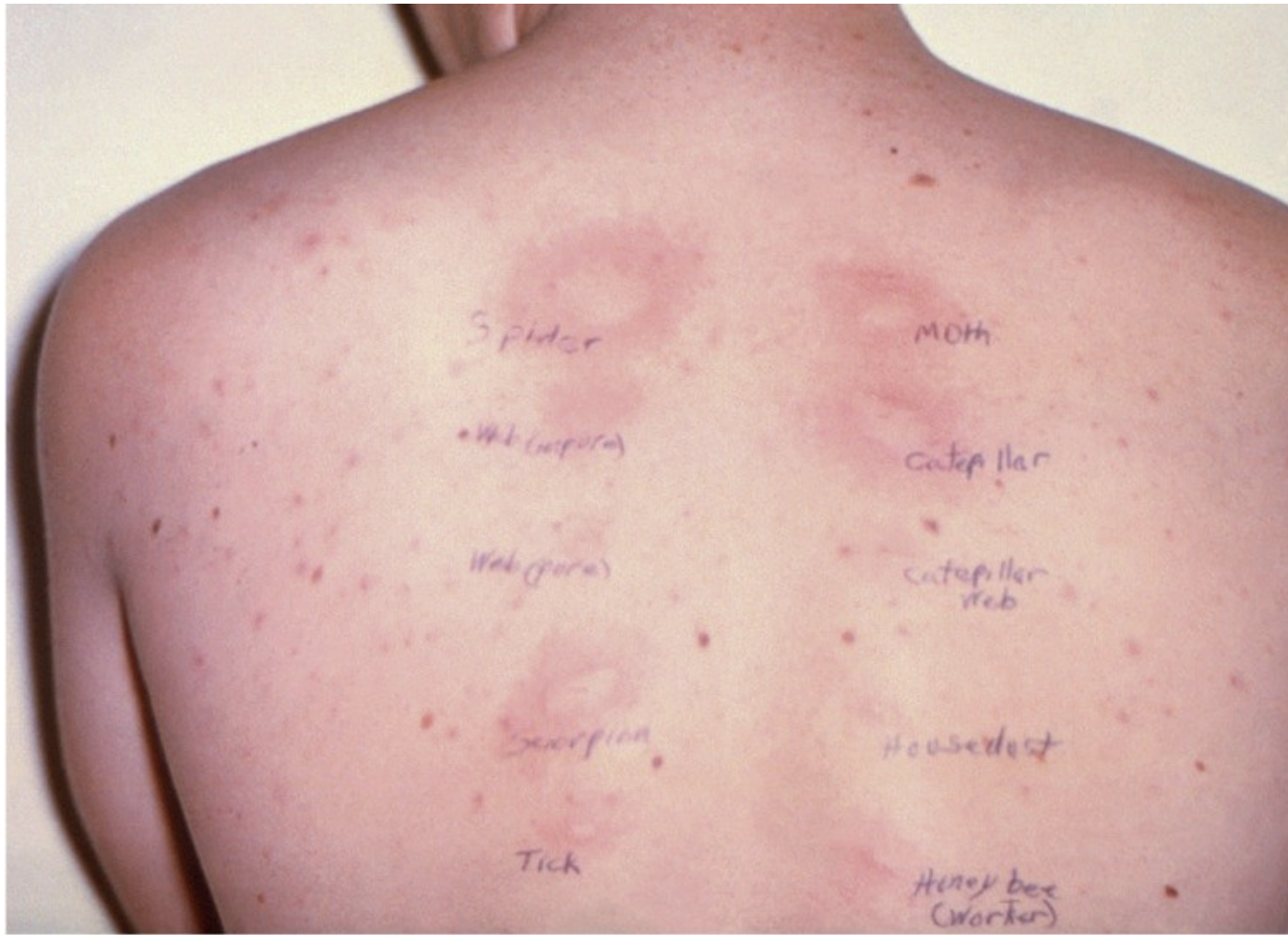
- Process:
-- Apply a panel of allergens to separate sites on the skin.
-- Wait 15 to 20 minutes.
- Positive test = wheal and flare at the site of application
28
What is the Allergen-specific IgE testing?
Used to detect IgE to a specific allergen in patient serum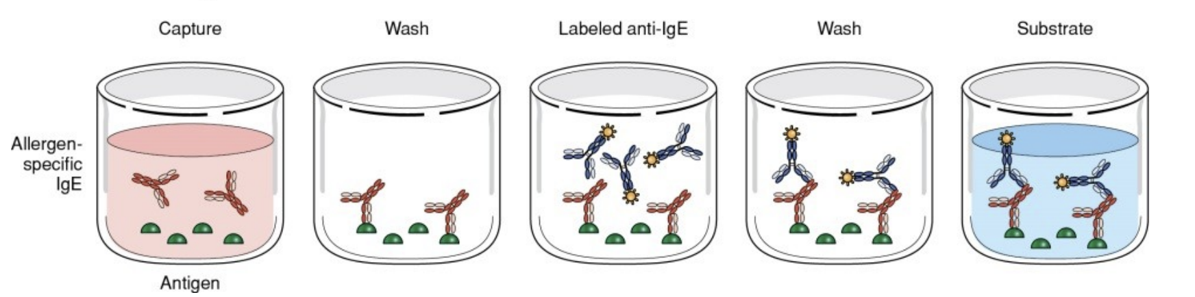
29
What is the Testing For Total IgE?
is used to detect the total concentration of IgE in patient serum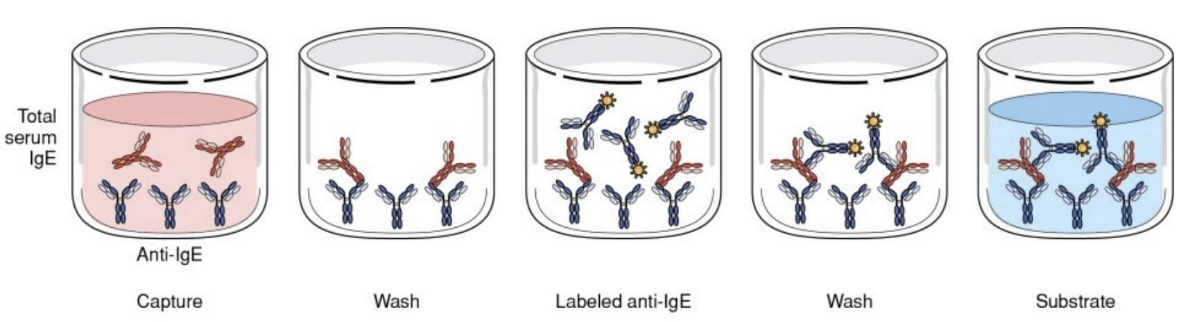
30
What is Type II hypersensitivity?
- aka antibody-mediated cytotoxic hypersensitivity
31
What are the key antigens and their effects in Type II?
- IgG and IgM directed AGAINST a cell surface antigen
- Effects:
• Cell destruction
• Inhibition of cell function
• Increase in cell function
- Effects:
• Cell destruction
• Inhibition of cell function
• Increase in cell function
32
What is the cell damage in Type II?
- activation of classical pathway of complement and cell lysis
- opsonization and phagocytosis of the cell
- antibody-dependent cell-mediated cytotoxicity (ADCC)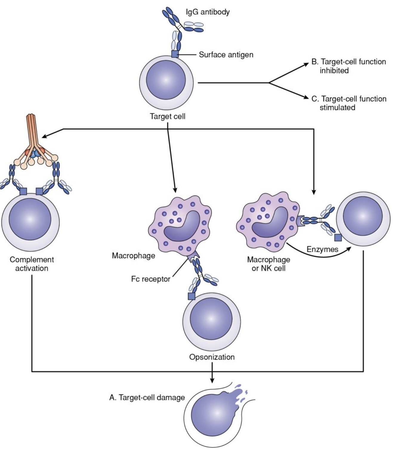
- opsonization and phagocytosis of the cell
- antibody-dependent cell-mediated cytotoxicity (ADCC)
33
What are the clinical examples of Type II?
- transfusion rxns
- hemolytic disease of the newborn (HDN)
- autoimmune hemolytic anemia: warm reactive abs, cold agglutinins, paroxysmal cold hemoglobinuria
- anti-GBM (glomerular basement membrane) disease
- Hashimoto's disease
- hemolytic disease of the newborn (HDN)
- autoimmune hemolytic anemia: warm reactive abs, cold agglutinins, paroxysmal cold hemoglobinuria
- anti-GBM (glomerular basement membrane) disease
- Hashimoto's disease
34
How does Hemolytic disease of the fetus and newborn (HDFN) occur?
- pregnant women produces antibodies to Rh antigens (usually RhD) on fetal RBC's
- usually occurs when the female is Rh- and baby is Rh+
- first exposure may be ok with treatment if caught early, but second exposure is fatal
- there are treatments for this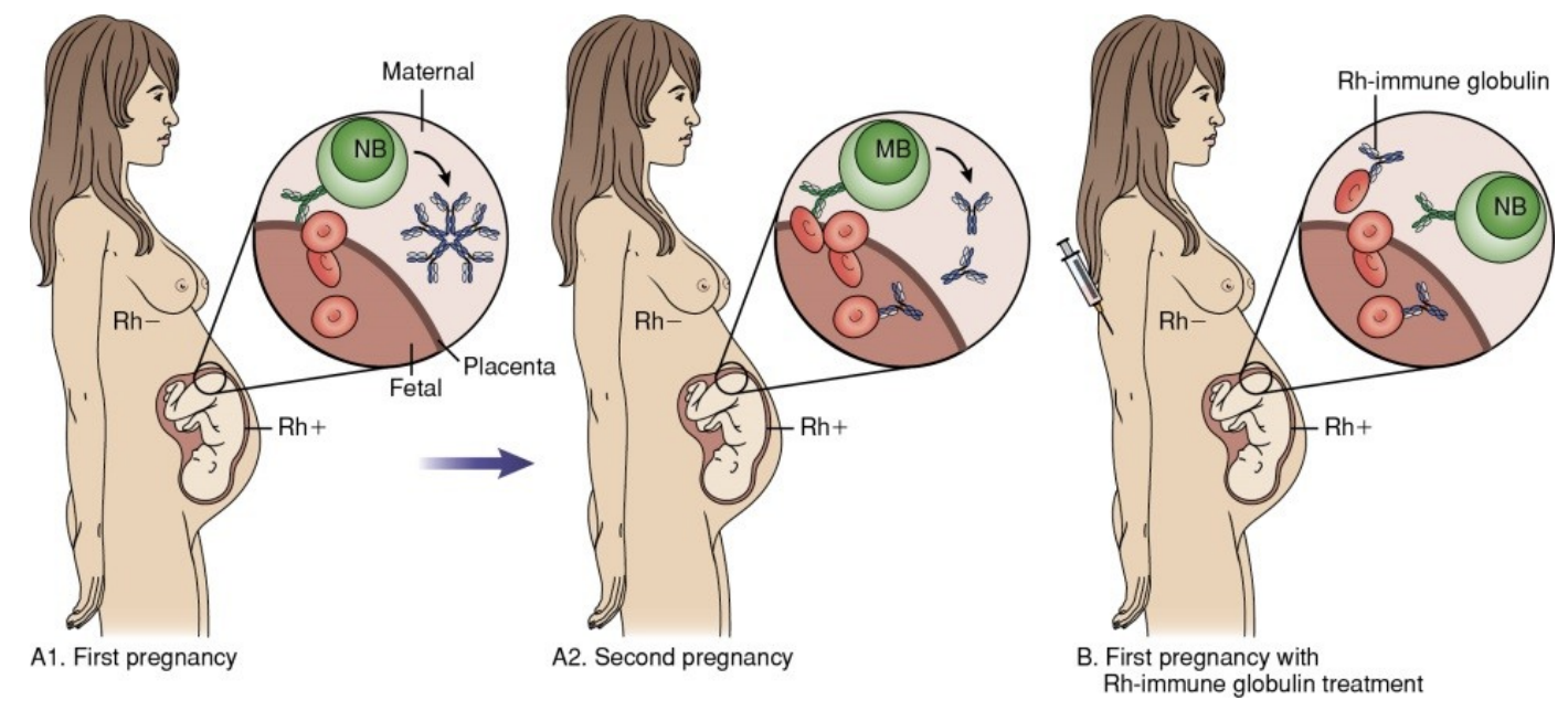
- usually occurs when the female is Rh- and baby is Rh+
- first exposure may be ok with treatment if caught early, but second exposure is fatal
- there are treatments for this
35
What is the DAT test?
- Direct Antiglobulin Test (DAT)
- Detects RBCs coated with complement components or IgG antibody
- Patient RBCs are incubated with a poly-specific anti-human Ig directed against IgG and C’
- If positive (agglutination), the test is repeated with mono-specific anti-IgG, anti-C3b, and anti-C3d
- Detects RBCs coated with complement components or IgG antibody
- Patient RBCs are incubated with a poly-specific anti-human Ig directed against IgG and C’
- If positive (agglutination), the test is repeated with mono-specific anti-IgG, anti-C3b, and anti-C3d
36
What is the Indirect Antiglobulin Test (IAT)?
- Coombs’ test
- Tests patient serum for antibodies to RBC antigens.
- Process
• Incubate reagent RBCs with patient serum at 37C; wash to remove excess.
• Add anti-human globulin.
- Positive test = RBC agglutination
- Tests patient serum for antibodies to RBC antigens.
- Process
• Incubate reagent RBCs with patient serum at 37C; wash to remove excess.
• Add anti-human globulin.
- Positive test = RBC agglutination
37
What is Type III hypersensitivity?
- immune complex-mediated hypersensitivity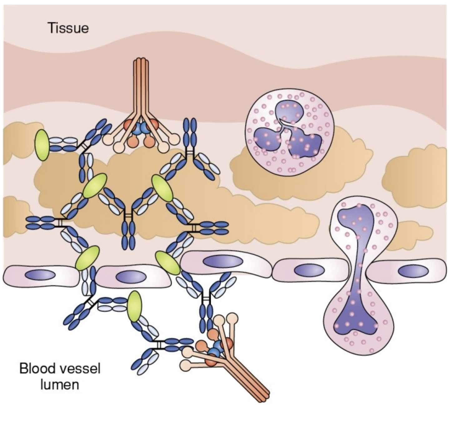
38
What are the key antibodies in Type III?
- IgG and IgM directed AGAINST a soluble antigen
39
What occurs in Type III?
- Small antigen–antibody complexes precipitate out and deposit in tissues.
- Complement binds; vasodilation and vasopermeability increase.
- Macrophages and neutrophils migrate to the affected areas and release their lysosomal enzymes, resulting in tissue damage.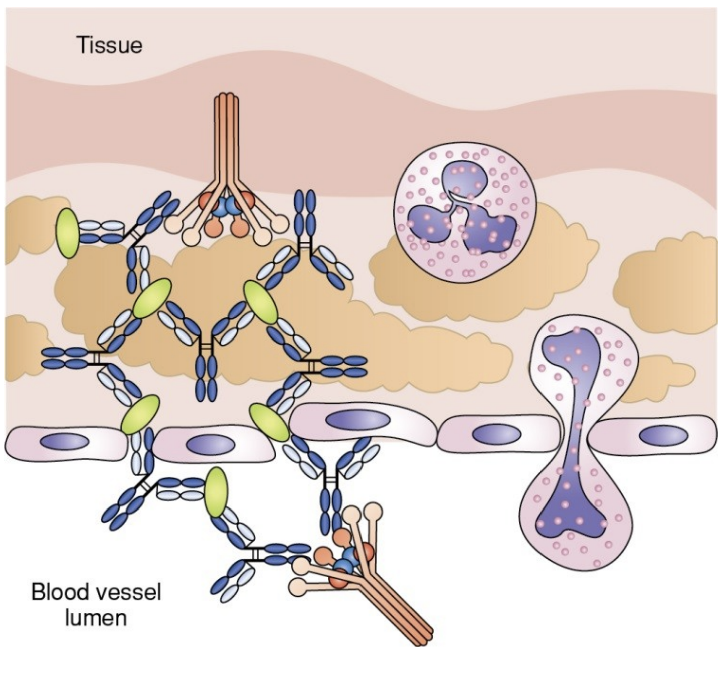
- Complement binds; vasodilation and vasopermeability increase.
- Macrophages and neutrophils migrate to the affected areas and release their lysosomal enzymes, resulting in tissue damage.
40
What are some clinical exampls of Type III?
1. Arthus Reaction
2. Serum Sickness
3. Some autoimmune (SLE and RA)
4. Reactions to bee stings
5. Drug reactions (e.g. penicillin)
6. Sequelae to infections (e.g. post-streptococcal glomerulonephritis)
2. Serum Sickness
3. Some autoimmune (SLE and RA)
4. Reactions to bee stings
5. Drug reactions (e.g. penicillin)
6. Sequelae to infections (e.g. post-streptococcal glomerulonephritis)
41
What occurs in Arthus Reaction?
- Type III skin reaction
- localized inflammation characterized by redness and edema
- peaks at 3-8 hours
- localized inflammation characterized by redness and edema
- peaks at 3-8 hours
42
What occurs in Serum Sickness?
- Generalized Type III reaction
- Caused by passive immunization of humans with animal serum
- Produces antibodies against the foreign animal proteins in patients.
- Causes immune complexes to form and deposit in tissues.
- Symptoms: headache, fever, nausea, joint pain, rashes, and lymphadenopathy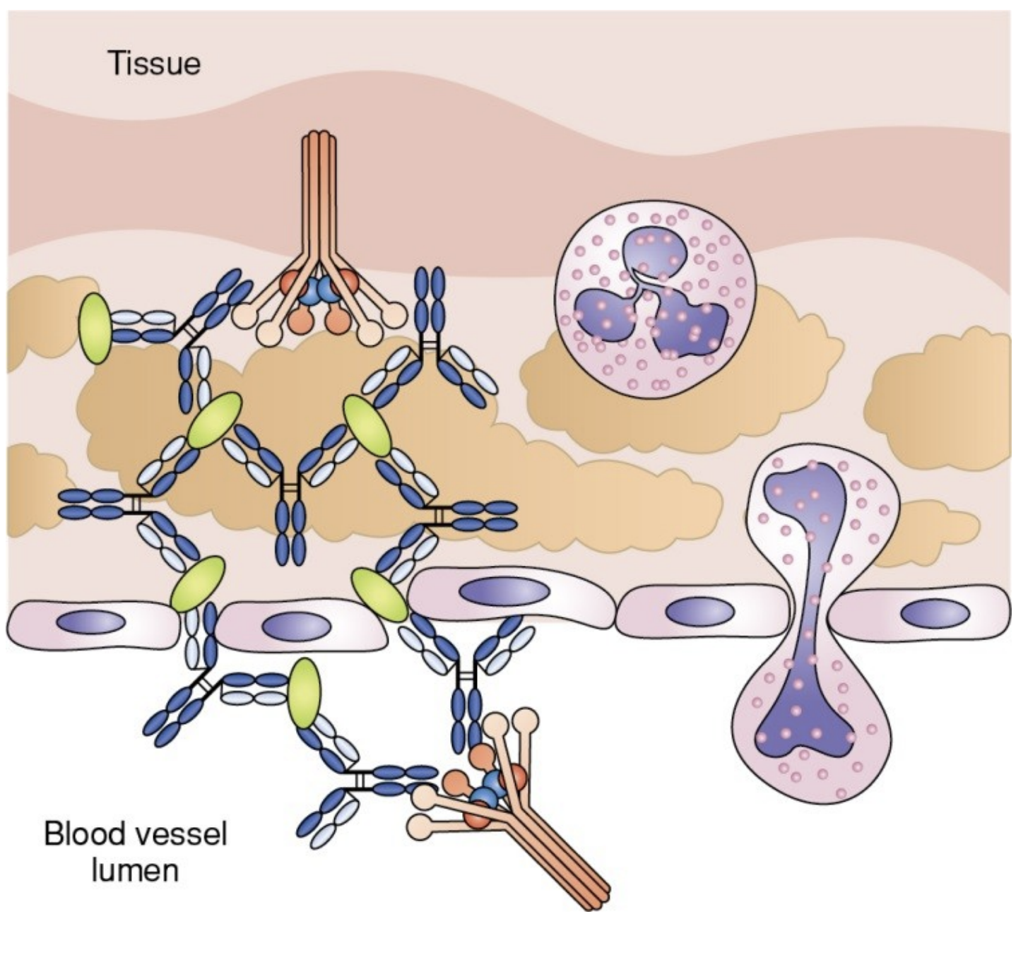
- Caused by passive immunization of humans with animal serum
- Produces antibodies against the foreign animal proteins in patients.
- Causes immune complexes to form and deposit in tissues.
- Symptoms: headache, fever, nausea, joint pain, rashes, and lymphadenopathy
43
What are some Lab Tests for Type III?
• Testing for ANAs
• Fluorescent staining of tissue sections to detect deposited immune complexes
• Testing for rheumatoid factor (an anti-IgG)
• Testing complement levels - may be decreased in the serum during periods of high disease activity.
• Fluorescent staining of tissue sections to detect deposited immune complexes
• Testing for rheumatoid factor (an anti-IgG)
• Testing complement levels - may be decreased in the serum during periods of high disease activity.
44
What is Type IV hypersensitivity?
cell-mediated hypersensitivity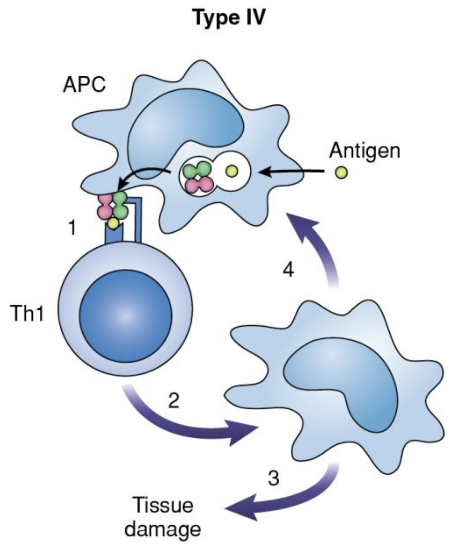
45
What occurs in type IV?
--> APCs present antigen to naïve T helper cells, which differentiate into Th1 cells.
--> Th1 cells release cytokines that attract and activate macrophages.
--> Macrophages induce inflammation.
--> Cytotoxic T lymphocytes are recruited and destroy target cells.
--> Hypersensitivity peaks 48 to 72 hours after antigen exposure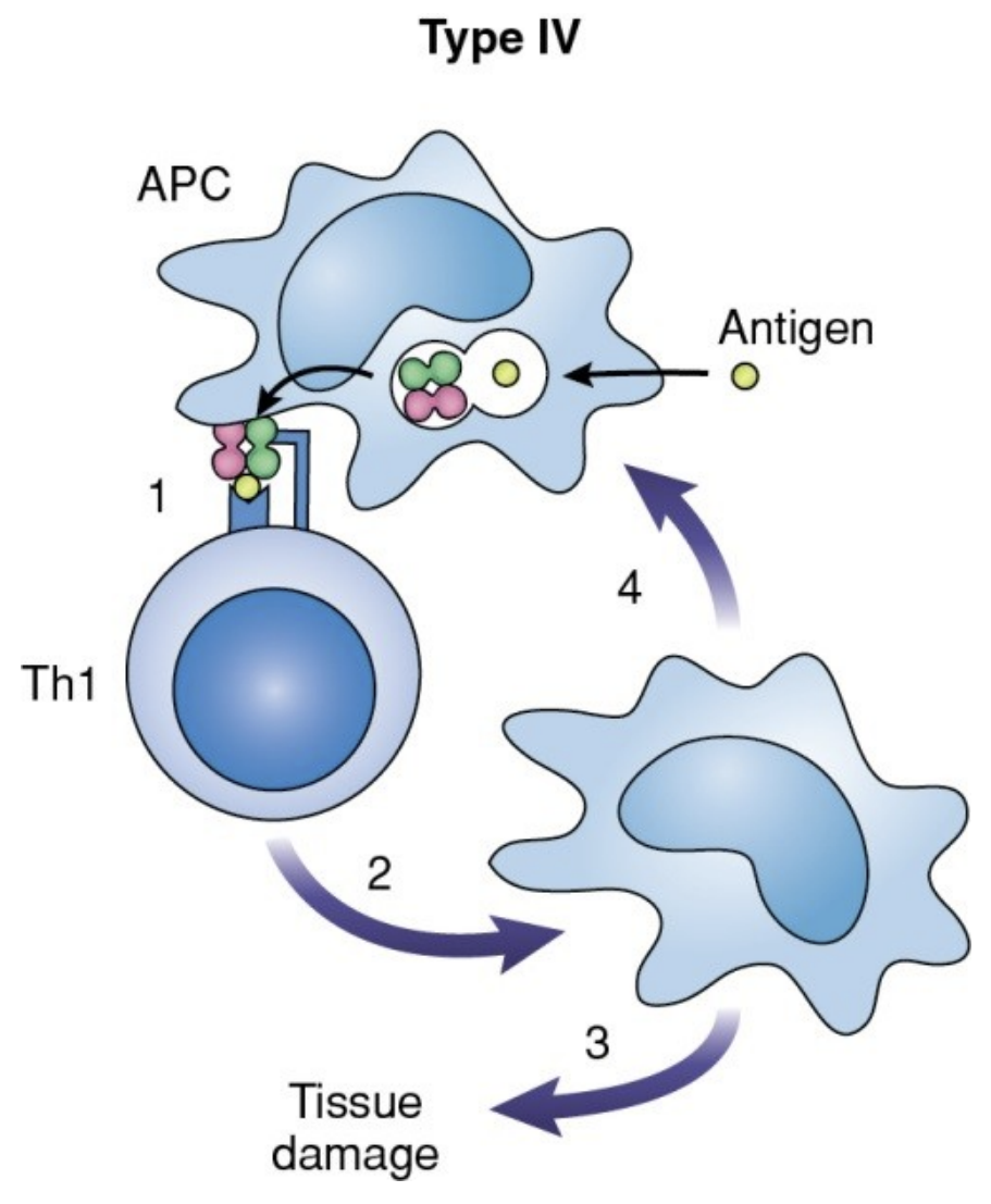
--> Th1 cells release cytokines that attract and activate macrophages.
--> Macrophages induce inflammation.
--> Cytotoxic T lymphocytes are recruited and destroy target cells.
--> Hypersensitivity peaks 48 to 72 hours after antigen exposure
46
What are some Clinical examples of Type IV?
1. Infections with intracellular pathogens
2. Contact dermatitis
3. Hypersensitivity pneumonitis
2. Contact dermatitis
3. Hypersensitivity pneumonitis
47
What are some examples of infections with intracellular pathogens for Type IV?
• Mycobacterium tuberculosis
• Mycobacterium leprae
• Pneumocystis carinii
• Leishmania species
• Herpes simplex virus
• Mycobacterium leprae
• Pneumocystis carinii
• Leishmania species
• Herpes simplex virus
48
What is hypersensitivity pneumonitis for Type IV?
- allergic disease of the lungs
- caused by inhalation of bacterial and fungal spores
- example: Farmer's lung disease, bird breeder's lung disease, and humidifier lung disease
- caused by inhalation of bacterial and fungal spores
- example: Farmer's lung disease, bird breeder's lung disease, and humidifier lung disease
49
What is contact dermatitis for Type IV?
- Low-molecular-weight compounds contact the skin and act as haptens to sensitize Th1 cells.
- Examples include poison ivy, poison oak, nickel salts, materials in cosmetics and hair dyes, and latex.
- Skin eruptions with erythema, swelling, and papules are produced.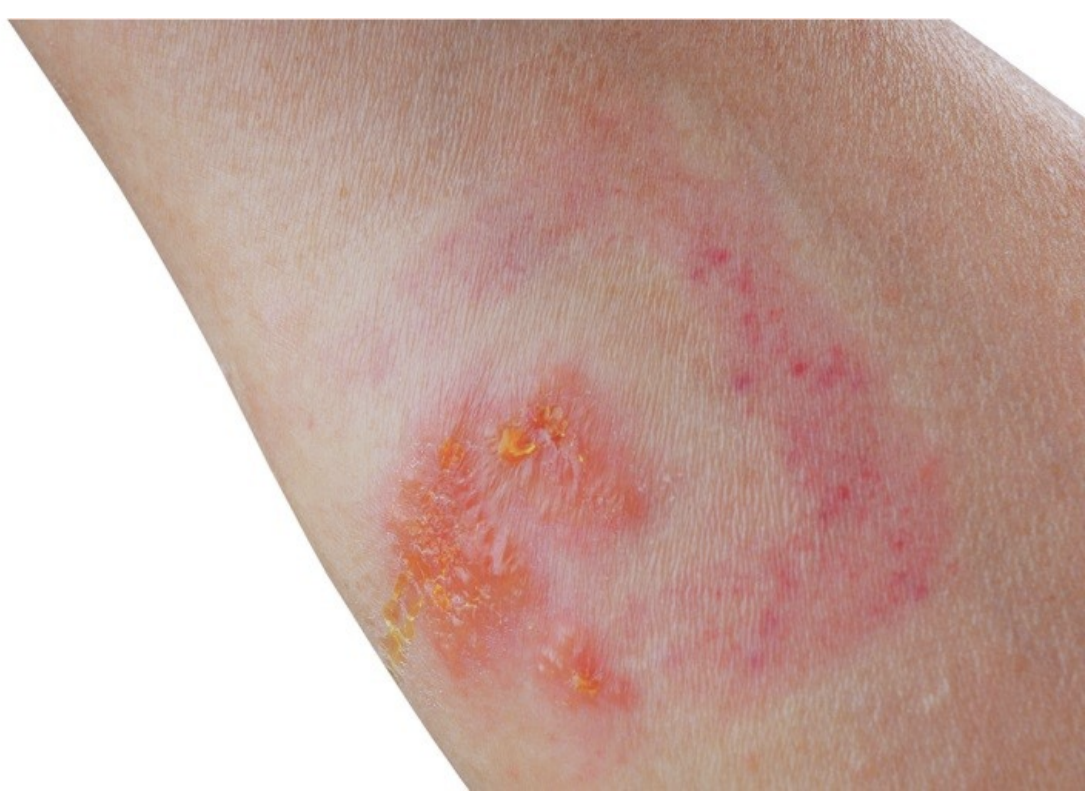
- Examples include poison ivy, poison oak, nickel salts, materials in cosmetics and hair dyes, and latex.
- Skin eruptions with erythema, swelling, and papules are produced.
50
What are some Skin tests for delayed hypersensitivity?
1. Patch test
2. Skin testing for immunodeficiency
3. Mantoux Method
2. Skin testing for immunodeficiency
3. Mantoux Method
51
What is the Patch test?
- antigen applied to skin surface
- test for contact dermatitis
- Positive test = redness with papules or tiny blisters
- test for contact dermatitis
- Positive test = redness with papules or tiny blisters
52
What is the skin testing for immunodeficiency?
inject common antigens intradermally
53
What is the Mantoux method?
- antigen injected intradermally
- test for tubervulosis exposure (PPD) or T cell function
- positive test = induration (thickening and hardening of skin)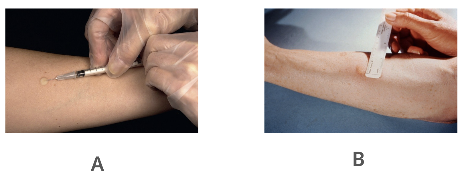
- test for tubervulosis exposure (PPD) or T cell function
- positive test = induration (thickening and hardening of skin)
54
What are the Interferon Gamma Release Assays (IGRA)?
under Type IV
1. Measure production of IFN-gamma
2. Quantiferon TB Gold Plus Assay
3. T-SPOT-TB Test
1. Measure production of IFN-gamma
2. Quantiferon TB Gold Plus Assay
3. T-SPOT-TB Test
55
What occurs when production of IFN-gamma is measured?
- measure production of IFN-gamma by patient T cells stimulated with MTB antigens
56
What occurs in the Quantiferon Gold Plus Assay?
- patient blood is incubated in special tubes with MTB antigens
- plasma is tested for IFN-gamma by ELISA
- plasma is tested for IFN-gamma by ELISA
57
What occurs in the T-SPOT-TB test?
- patient mononuclear cells are incubated with MTB antigens and tested for IFN-gamma by ELISpot