Lecture 1
Study these flashcardsFree · spaced repetition · iPhone, Android, Mac & web
All cards
1
Parasitology
a study of symbiosis especially the form known as parasitism
2
symbiotic relationship
any 2 organisms living in close association (each called a symbiont) is called a symbiotic relationship
they are living basically together and they may or may not benefit from each other
they are living basically together and they may or may not benefit from each other
3
symbiosis can be divided into several categories
a. phoresis
b. mutualism
c. commensalism
d. parasitism
b. mutualism
c. commensalism
d. parasitism
4
phoresis
1.This exists when two symbionts are merely traveling together.
2. Neither is physiologically dependent on the other. Usually one organism is smaller than the other and mechanically carried about by its larger companion.
3. Examples are bacteria on the legs of a fly or fungus spores on the feet of a beetle. Bees & pollen. Cockroach & bacteria known to have transmitted MRSA.
this is a mechanical type of relationship
2. Neither is physiologically dependent on the other. Usually one organism is smaller than the other and mechanically carried about by its larger companion.
3. Examples are bacteria on the legs of a fly or fungus spores on the feet of a beetle. Bees & pollen. Cockroach & bacteria known to have transmitted MRSA.
this is a mechanical type of relationship
5
mutualism
1.In this relationship the partners are called mutuals because both members benefit from the association.
a.Two organisms that have evolved so that one cannot live w/out the other
2. Mutualism is usually obligatory, since in most cases physiological dependence on one another has evolved to such a degree that one mutual can’t survive without the other.
a. Both organisms will not do well w/out the other
3. An example is the termite and its intestinal protozoan fauna. Termites can’t digest cellulose fibers from wood b/c they don’t secrete the enzyme cellulase.
a. Another example: humans have bacteria in the gut that make vitamin K
b.On the bench you have to isolate the normal flora from that that is infectious
c.The normal microbiota keep pathogens out
d.Trichomonas vaginalis – protozoan
4.However, the protozoa in the termite’s gut can.
5.The termite is nourished by the fermentation products excreted by the protozoa.
6.The protozoa in turn benefit by living in a stable, secure environment being constantly supplied with food.
7.Another example of mutualism is the bacteria that live in the human gut.
8.They help us digest our food, make vitamin K and exclude pathogenic organisms from colonizing the gut.
9.We in turn give them nutrients and a stable, secure environment that is constantly supplied with food.
10.Rare orchids in the amazon rely on a flying beetle to pollenate them = one will die without the other // when the relationship is so close that there is no substitute is kind of dangerous
a.Two organisms that have evolved so that one cannot live w/out the other
2. Mutualism is usually obligatory, since in most cases physiological dependence on one another has evolved to such a degree that one mutual can’t survive without the other.
a. Both organisms will not do well w/out the other
3. An example is the termite and its intestinal protozoan fauna. Termites can’t digest cellulose fibers from wood b/c they don’t secrete the enzyme cellulase.
a. Another example: humans have bacteria in the gut that make vitamin K
b.On the bench you have to isolate the normal flora from that that is infectious
c.The normal microbiota keep pathogens out
d.Trichomonas vaginalis – protozoan
4.However, the protozoa in the termite’s gut can.
5.The termite is nourished by the fermentation products excreted by the protozoa.
6.The protozoa in turn benefit by living in a stable, secure environment being constantly supplied with food.
7.Another example of mutualism is the bacteria that live in the human gut.
8.They help us digest our food, make vitamin K and exclude pathogenic organisms from colonizing the gut.
9.We in turn give them nutrients and a stable, secure environment that is constantly supplied with food.
10.Rare orchids in the amazon rely on a flying beetle to pollenate them = one will die without the other // when the relationship is so close that there is no substitute is kind of dangerous
6
commensalism
1.When one organism, the commensal, benefits from its relationship with the host.
2.The host neither benefits or is harmed by the other organism.
3.For example certain amebic protozoans can live in the human gut and do not cause disease.
a.Entamoeba Coli --> get it from drinking water that has not been treated with chlorine
b.Not considered a pathogens
c.Absorbs nutrients in the gut – we are not benefited or harmed by its presence – a safe place for it to live
d.Divide by binary fission – they die if we die
4.The ameba derives nutrients from the gut and a safe place to live from the human host.
5.The human host does not benefit from the presence of the ameba nor is it not harmed.
2.The host neither benefits or is harmed by the other organism.
3.For example certain amebic protozoans can live in the human gut and do not cause disease.
a.Entamoeba Coli --> get it from drinking water that has not been treated with chlorine
b.Not considered a pathogens
c.Absorbs nutrients in the gut – we are not benefited or harmed by its presence – a safe place for it to live
d.Divide by binary fission – they die if we die
4.The ameba derives nutrients from the gut and a safe place to live from the human host.
5.The human host does not benefit from the presence of the ameba nor is it not harmed.
7
parasitism
1.A symbiotic relationship where an organism actually harms the host or in some sense lives at the expense of the host, then it is called a parasite.
2.It may harm its host in a number of ways: by mechanical injury, such as boring a hole into it.
a.Mechanical injury
ex. Nematodoes that bore holes into roots of trees of oranges
even plants get parasites
3.Or by eating or digesting and absorbing its tissues
a.Especially liver flukes – can cause an abscess
b.PMNs can try but sometimes too big
c.Nigleria flaveria
d.Amongst the flukes and ____ are hermaphrodites (have both genders)- does not need to find another of the opposite gender to make eggs
e. Can become egg laying machines and pass it out by fecal matter and those eggs are gonna get into another host
4. Or by poisoning the host with toxic metabolites; or simply by robbing its host of nutrition.
a.Some can be pretty big
b.Verminous intoxication = bc their metabolites can cause harm to the host
5.Most parasites inflict a combination of these conditions on their hosts.
2.It may harm its host in a number of ways: by mechanical injury, such as boring a hole into it.
a.Mechanical injury
ex. Nematodoes that bore holes into roots of trees of oranges
even plants get parasites
3.Or by eating or digesting and absorbing its tissues
a.Especially liver flukes – can cause an abscess
b.PMNs can try but sometimes too big
c.Nigleria flaveria
d.Amongst the flukes and ____ are hermaphrodites (have both genders)- does not need to find another of the opposite gender to make eggs
e. Can become egg laying machines and pass it out by fecal matter and those eggs are gonna get into another host
4. Or by poisoning the host with toxic metabolites; or simply by robbing its host of nutrition.
a.Some can be pretty big
b.Verminous intoxication = bc their metabolites can cause harm to the host
5.Most parasites inflict a combination of these conditions on their hosts.
8
types of parasitism
a. ectoparasites
b. endoparasites
c. obligate parasites
d. faculative parasites
e. accidental / incidental parasite
b. endoparasites
c. obligate parasites
d. faculative parasites
e. accidental / incidental parasite
9
ectoparasites
A parasite that lives on the surface of its host.
Example: lice, fleas, barnacles, sometimes flies but unusual (common to grass eating mammals – botflies = female lands on skin, bites, deposits eggs, pupates, causes pain, after done growing it bores holes out of the host and leaves and pupates on the grass)
Screwworm was eliminated from the USA
Sometimes ppl can be subjected to infestation – babies --> diaper is not changed often – rare occasions but the maggot eats the skin
Example: lice, fleas, barnacles, sometimes flies but unusual (common to grass eating mammals – botflies = female lands on skin, bites, deposits eggs, pupates, causes pain, after done growing it bores holes out of the host and leaves and pupates on the grass)
Screwworm was eliminated from the USA
Sometimes ppl can be subjected to infestation – babies --> diaper is not changed often – rare occasions but the maggot eats the skin
10
endoparasites
A parasite that lives inside its host.
Example: roundworm
Endo are most common
Endo = inside
Example: roundworm
Endo are most common
Endo = inside
11
obligate parasites
Most parasites fit this definition, that is, they spend at least part of their lives as parasites to survive and complete their life-cycles.
a. Need to have a parasitic life
a. Need to have a parasitic life
12
faculative parasites
These are not normally parasitic but can become so, at least for a time, when they are accidentally eaten or enter a wound or other body orifice. Certain species of fly maggots can do this.
- lives a parasitic life when opportunity arises
- lives a parasitic life when opportunity arises
13
accidental / incidental parasite
This is when the parasite enters or attaches to the body of a species of host different from its normal one. Accidental parasites are usually unable to stay living on, or live long in, the wrong host. An example is when the dog hookworm infects a human host.
example: plumber that goes under the house where there is a big space // dogs poop and leaves it there and if a plumber need to get into, hands are exposed to contaminated soil they get “plumbers itch” , larvae never get mature – there can be complications, a wound appears and can see them migrating in the skin
a. Great lakes area = birds that poop that have parasites and burrow a hole into your skin
example: plumber that goes under the house where there is a big space // dogs poop and leaves it there and if a plumber need to get into, hands are exposed to contaminated soil they get “plumbers itch” , larvae never get mature – there can be complications, a wound appears and can see them migrating in the skin
a. Great lakes area = birds that poop that have parasites and burrow a hole into your skin
14
what is a lifecycle
1.Parasites go though various stages of development in what is called a lifecycle.
a.These are the stages of development a parasite goes through before it reaches its sexual maturity
2.The parasite reaches sexual maturity on completion of the lifecycle.
3.The lifecycle frequently starts out in a particular host species, and ends(reaches sexual maturity) in the same species.
a.If it starts in human then it must end in human not necessarily the same one just the same species, goal is to get into a new host of the same species
4.That’s why it’s called a “lifecycle” because it tends to be cyclical in nature.
5.The parasite frequently uses different types of “hosts” in order to complete its lifeycle.
6.Some parasites use more hosts than others.
a.These are the stages of development a parasite goes through before it reaches its sexual maturity
2.The parasite reaches sexual maturity on completion of the lifecycle.
3.The lifecycle frequently starts out in a particular host species, and ends(reaches sexual maturity) in the same species.
a.If it starts in human then it must end in human not necessarily the same one just the same species, goal is to get into a new host of the same species
4.That’s why it’s called a “lifecycle” because it tends to be cyclical in nature.
5.The parasite frequently uses different types of “hosts” in order to complete its lifeycle.
6.Some parasites use more hosts than others.
15
types of hosts
a. definitive host
b. intermediate host
b. intermediate host
16
definitive host
This is the host in which a parasite reaches sexual maturity and reproduction.
i. Example: malaria = mosquito bites human with plasmodium goes through several stages but needs to get back into mosquito before it can reach sexual maturity another mosquito bites get malaria and in her body it will reach sexual maturity and it propagates injects human again
ii. Humans don’t host the sexually active plasmodium
2. If no sexual reproduction occurs in the life of the parasite, such as an ameba or trypanosome, the host believed to be the most important is arbitrarily called the definitive host.
example: mosquito is the definitive host of the malarial parasite. Humans are the definitive host for Taenia saginata
i. Example: malaria = mosquito bites human with plasmodium goes through several stages but needs to get back into mosquito before it can reach sexual maturity another mosquito bites get malaria and in her body it will reach sexual maturity and it propagates injects human again
ii. Humans don’t host the sexually active plasmodium
2. If no sexual reproduction occurs in the life of the parasite, such as an ameba or trypanosome, the host believed to be the most important is arbitrarily called the definitive host.
example: mosquito is the definitive host of the malarial parasite. Humans are the definitive host for Taenia saginata
17
intermediate host
1. This is the host in which some development of the parasite occurs but in which it does not reach maturity.
2. The intermediate host is obligatory. If the parasite doesn’t get into the intermediate host the lifecycle is not completed and the parasite usually dies.
3. Asexual form of parasite takes place. Essential for the completion of the lifecycle of the parasite.
example: cattle are the intermediate host for Taenia saginata Tsetse fly for Trypanosome
4. We shed it and someone else eats it
5. Host that that the parasite must complete must complete part of its development but does not reach sexual maturity in the intermediate host
2. The intermediate host is obligatory. If the parasite doesn’t get into the intermediate host the lifecycle is not completed and the parasite usually dies.
3. Asexual form of parasite takes place. Essential for the completion of the lifecycle of the parasite.
example: cattle are the intermediate host for Taenia saginata Tsetse fly for Trypanosome
4. We shed it and someone else eats it
5. Host that that the parasite must complete must complete part of its development but does not reach sexual maturity in the intermediate host
18
what are the protozoa: the abemas
1. Entamoeba histolytica
2. Entamoeba hartmanni
3.Endolimax nana
4. Iodamoeba buetschlii
2. Entamoeba hartmanni
3.Endolimax nana
4. Iodamoeba buetschlii
19
entamoeba histolytica overview
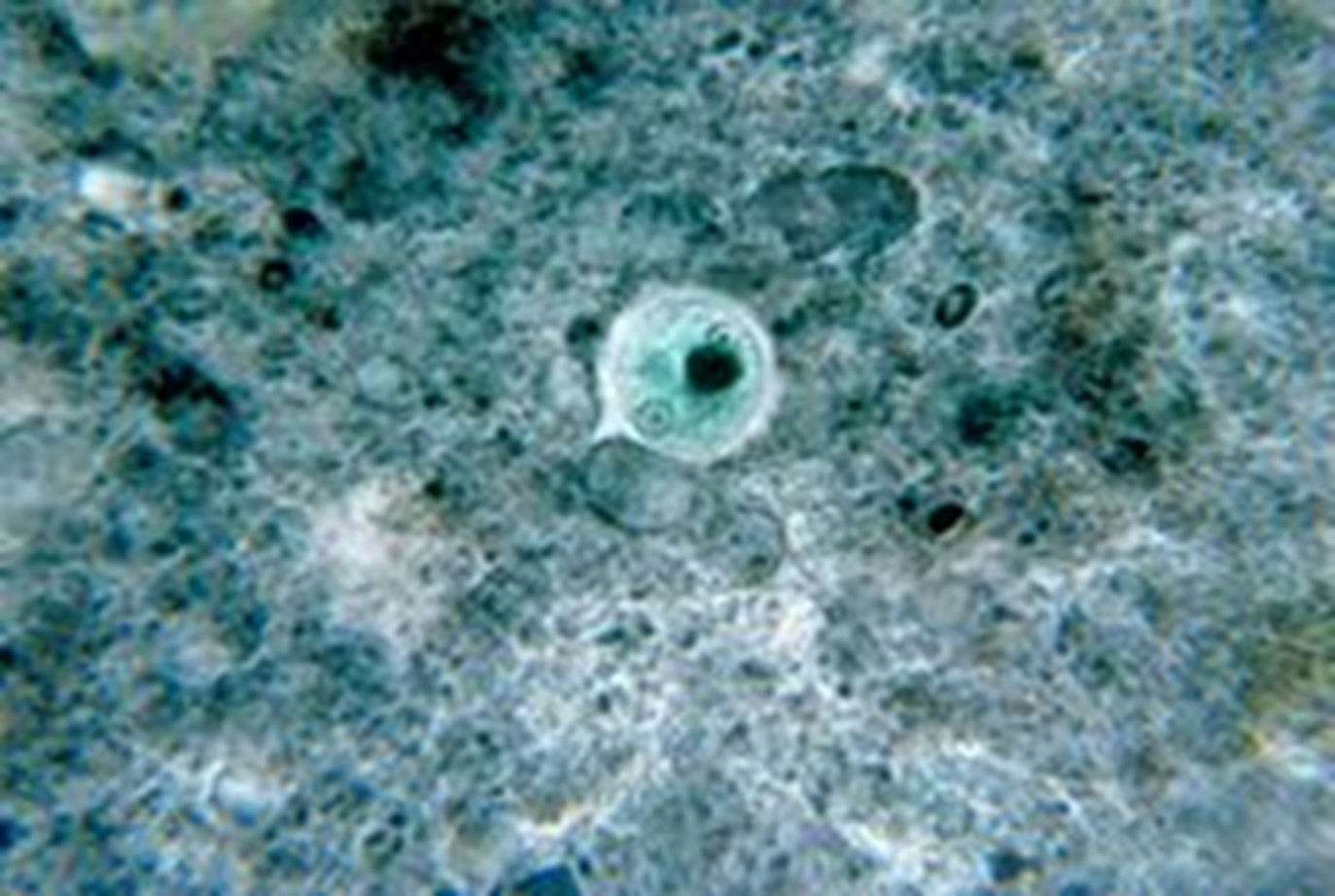
A. This is a parasitic protozoan, part of the genus Entamoeba.
B. It predominantly infects humans and other primates
B. It predominantly infects humans and other primates
20
enatmoeba histolytica transmission
A. In order to become infected one must swallow the cyst or trophozoite stage of the parasite in fecally contaminated food or water.
B. The cysts of E. histolytica can be transmitted via anal-oral sex too.
C. The cysts survive outside the host in water, soils and on foods, especially under moist conditions on the latter.
D. When cysts are swallowed they cause infections by excysting (emerging from the cyst) in the digestive tract.
E. When the cysts “excyst” they become what is called the “trophozoite” stage. It is this stage that causes disease.
F. histolytica trophozoite: note the chromatin is in the center of nucleus: "bulls-eye" appearance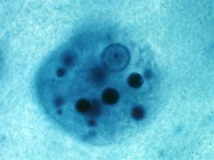
B. The cysts of E. histolytica can be transmitted via anal-oral sex too.
C. The cysts survive outside the host in water, soils and on foods, especially under moist conditions on the latter.
D. When cysts are swallowed they cause infections by excysting (emerging from the cyst) in the digestive tract.
E. When the cysts “excyst” they become what is called the “trophozoite” stage. It is this stage that causes disease.
F. histolytica trophozoite: note the chromatin is in the center of nucleus: "bulls-eye" appearance
21
Entamoeba histolytica pathogenesis "amebic dysentery"
B. Pathogenesis: “Amebic dysentery”
A. E. histolytica, as its name suggests (histo–lytic = tissue destroying), is pathogenic; infection can lead to amebic dysentery or amebic liver abscess.
B. Amebic dysentery is not to be confused with “bacillary dysentery”.
C. Bacillary dysentery is caused by bacteria.
D. Amebic dysentery is caused by amebas.
E. Dysentery is different from diarrhea. Dysentery occurs when blood and mucus enter the stool and indicates invasion of the bowel by a microbe.
F. Dysentery is life-threatening while diarrhea usually is not.
G. With amebic dysentery, symptoms can include fulminating bloody diarrhea, weight loss, fatigue, abdominal pain.
H. The amoeba can actually 'bore' into the intestinal wall, causing lesions and intestinal symptoms, and it may reach the blood stream.
I. From there, it can reach different vital organs of the human body, usually the liver, but sometimes the lungs, brain, spleen, etc.
J. A common outcome of this invasion of tissues is a liver abscess, which can be fatal if untreated.
K. The trophozoite ingests red blood cells and these are sometimes seen in the amoeba cell cytoplasm.
A. E. histolytica, as its name suggests (histo–lytic = tissue destroying), is pathogenic; infection can lead to amebic dysentery or amebic liver abscess.
B. Amebic dysentery is not to be confused with “bacillary dysentery”.
C. Bacillary dysentery is caused by bacteria.
D. Amebic dysentery is caused by amebas.
E. Dysentery is different from diarrhea. Dysentery occurs when blood and mucus enter the stool and indicates invasion of the bowel by a microbe.
F. Dysentery is life-threatening while diarrhea usually is not.
G. With amebic dysentery, symptoms can include fulminating bloody diarrhea, weight loss, fatigue, abdominal pain.
H. The amoeba can actually 'bore' into the intestinal wall, causing lesions and intestinal symptoms, and it may reach the blood stream.
I. From there, it can reach different vital organs of the human body, usually the liver, but sometimes the lungs, brain, spleen, etc.
J. A common outcome of this invasion of tissues is a liver abscess, which can be fatal if untreated.
K. The trophozoite ingests red blood cells and these are sometimes seen in the amoeba cell cytoplasm.
22
Entamoeba histolytica diagnosis
A. Microscopy: It can be diagnosed in stool samples by seeing either the trophozoite or the cyst or both.
* Trophozoites may be seen using iodine concentration technique, Trichrome stain or both in an ordinary stool sample.
*Cysts are more easily seen using iodine concentration technique.
B. Antigen detection kits using stool can be helpful in distinguishing between E. histolytica and nonpathogenic members of Entamoeba species, like E. dispar.
C. Real time PCR is becoming more common as a tool to use when distinguishing E. histolytica from other nonpathogenic members of Entamoeba.
D. Enzyme Linked Immunosorbet Assay (ELISA) can also be useful when diagnosing extra- intestinal disease (liver abscess). Serum is used for detection of antibody.
* Trophozoites may be seen using iodine concentration technique, Trichrome stain or both in an ordinary stool sample.
*Cysts are more easily seen using iodine concentration technique.
B. Antigen detection kits using stool can be helpful in distinguishing between E. histolytica and nonpathogenic members of Entamoeba species, like E. dispar.
C. Real time PCR is becoming more common as a tool to use when distinguishing E. histolytica from other nonpathogenic members of Entamoeba.
D. Enzyme Linked Immunosorbet Assay (ELISA) can also be useful when diagnosing extra- intestinal disease (liver abscess). Serum is used for detection of antibody.
23
Entamoeba hartmanni
A.Does not cause disease and is a commensal.
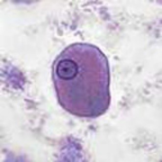
B.The trophozoite looks exactly like E. histolytica except this ameba is a lot smaller.
C.Acquired through consuming fecally contaminated food or water.
E. hartmanni trophozoite (image)
Diagnosis: Finding trophozoites and/or cysts in fecal matter using Trichrome technique.
B.The trophozoite looks exactly like E. histolytica except this ameba is a lot smaller.
C.Acquired through consuming fecally contaminated food or water.
E. hartmanni trophozoite (image)
Diagnosis: Finding trophozoites and/or cysts in fecal matter using Trichrome technique.
24
Endolimax nana
A.This is a genus of ameba that is found in the intestines of various animals, including the species E. nana found in humans.
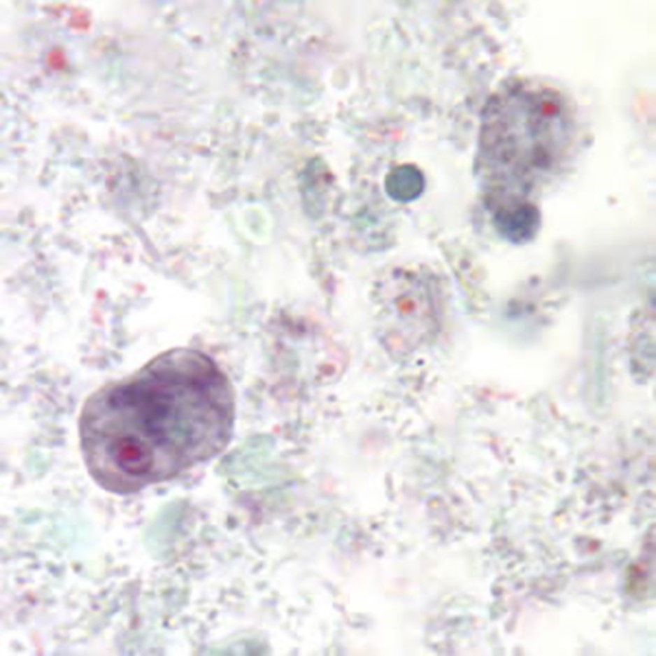
B. It causes no known disease and is a commensal.
C.However, if a patient has this ameba it indicates the consumption of fecally contaminated food or water and could indicate other pathogenic amebas present.
D.The cyst excysts in the small intestine and then migrates to the large intestine where it lives.
Diagnosis: Finding cysts and/or trophozoites in fecal matter.
B. It causes no known disease and is a commensal.
C.However, if a patient has this ameba it indicates the consumption of fecally contaminated food or water and could indicate other pathogenic amebas present.
D.The cyst excysts in the small intestine and then migrates to the large intestine where it lives.
Diagnosis: Finding cysts and/or trophozoites in fecal matter.
25
Iodamoeba buetschlii
A.This ameba infects humans, other primates and pigs.
B.Its distribution is worldwide.
C.It is found most commonly in swine which is probably the original host.
D.It lives in the large intestine where it feeds on intestinal flora.
E.Cysts are swallowed in fecally contaminated food or water.
F.They become trophozoites in the large intestine
G.It’s possible that people living among pigs can become infected by swallowing cyst-containing pig feces.
H. It is a commensal.
Iodamoeba buetschlii trophozoite (image)
Diagnosis: Finding trophozoites or cysts in fecal matter.
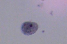
B.Its distribution is worldwide.
C.It is found most commonly in swine which is probably the original host.
D.It lives in the large intestine where it feeds on intestinal flora.
E.Cysts are swallowed in fecally contaminated food or water.
F.They become trophozoites in the large intestine
G.It’s possible that people living among pigs can become infected by swallowing cyst-containing pig feces.
H. It is a commensal.
Iodamoeba buetschlii trophozoite (image)
Diagnosis: Finding trophozoites or cysts in fecal matter.
26
what parasites belong to the family of schizopyrenidae
1. Naegleria fowleri
27
Naegleria fowleri
I. Naegleria fowleri; not common; from 2009-2018( 34) cases have been reported to CDC.
A. This is an opportunistic pathogen that causes disease under special circumstances.
B. It is usually not parasitic. Disease is rare.
C. It causes a disease called Primary Amebic Meningo-encephalitis (PAM).
D. It is an acute, rapidly fatal illness usually affecting children or young adults who have been exposed to water harboring free-living N. fowleri.
E. Most cases have been contracted in warm lakes, or via exposure to un-chlorinated water.
F. The patient goes swimming in water where this ameba lives.
G. The ameba is forced deep into the nasal passages when the victim dives into water (water goes up the nose).
H. Water may be purposefully forced up the nose too. In two cases in Louisiana, irrigation of the nose using unchlorinated water led to disease.
I. After entrance to the nasal passages the ameba migrate along the olfactory nerves and into the cranium.
J. Death from brain destruction is rapid and few cures have been reported. Death can occur in 24 hours.
K. The ameba divides rapidly as water temperature rises and so most bodies of water where this has been contracted have been natural thermal pools or warm lakes/ponds.
L. In 2017 a sixteen year old boy in Florida survived after doctors induced a coma, lowered his body temp to 33 degrees and gave him an anti-parasitic drug. Only four have survived in the last 50 years.
Nasal irrigation using a “neti pot”
A. This is an opportunistic pathogen that causes disease under special circumstances.
B. It is usually not parasitic. Disease is rare.
C. It causes a disease called Primary Amebic Meningo-encephalitis (PAM).
D. It is an acute, rapidly fatal illness usually affecting children or young adults who have been exposed to water harboring free-living N. fowleri.
E. Most cases have been contracted in warm lakes, or via exposure to un-chlorinated water.
F. The patient goes swimming in water where this ameba lives.
G. The ameba is forced deep into the nasal passages when the victim dives into water (water goes up the nose).
H. Water may be purposefully forced up the nose too. In two cases in Louisiana, irrigation of the nose using unchlorinated water led to disease.
I. After entrance to the nasal passages the ameba migrate along the olfactory nerves and into the cranium.
J. Death from brain destruction is rapid and few cures have been reported. Death can occur in 24 hours.
K. The ameba divides rapidly as water temperature rises and so most bodies of water where this has been contracted have been natural thermal pools or warm lakes/ponds.
L. In 2017 a sixteen year old boy in Florida survived after doctors induced a coma, lowered his body temp to 33 degrees and gave him an anti-parasitic drug. Only four have survived in the last 50 years.
Nasal irrigation using a “neti pot”
28
what are the 3 forms of Naegleria fowleri
a. flagellate stage
b. trophozite stage (ameboid)
c. cyst stage
b. trophozite stage (ameboid)
c. cyst stage
29
Naegleria fowleri - flagellate stage
A biflagellate form occurring when trophozites are exposed to a change in ionic concentration such as placement in distilled water. The transformation of trophozoites to flagellate form occurs within a few minutes.
30
Naegleria fowleri - trophozite stage
The reproductive stage of the protozoan organism, which grows fastest at around 42°C and proliferates by binary fission. In their free-living state, trophozoites feed on bacteria. In tissues, trophozoites phagocytize red blood cells and white blood cells and destroy tissue
31
Naegleria fowleri - cyst stage
Trophozoites encyst due to unfavorable conditions. Factors that induce cyst formation can include food deprivation, crowding, desiccation, accumulation of waste products, and cold temperatures. N. fowleri has been found to encyst at temperatures below 10°C.
32
Naegleria fowleri diagnosis
*Culture:
A.N. fowleri can be grown in several kinds of liquid axenic media or on non-nutrient agar plates coated with bacteria.
B. Escherichia coli can be used to overlay the non-nutrient agar plate and a drop of CSF sediment added to it.
C.Plates are then incubated at 37 (warmer temp) degrees Celsius and checked daily for clearing of the agar in thin tracks, which indicate that the trophozoites have fed on the bacteria.
**Microscopy:
Direct observation of ameba in CSF using methylene blue.
***Antigen Detection:
Naegleria fowleri antigen can be detected in CSF, tissue or biopsy specimens using indirect immunofluorescence (IIF) or immunohistochemistry (IHC) kits.
****Molecular Methods:
PCR can be used to amplify DNA from the ameba in CSF or tissue.
Because PAM is rare 75% of diagnosed cases occur at autopsy.
A.N. fowleri can be grown in several kinds of liquid axenic media or on non-nutrient agar plates coated with bacteria.
B. Escherichia coli can be used to overlay the non-nutrient agar plate and a drop of CSF sediment added to it.
C.Plates are then incubated at 37 (warmer temp) degrees Celsius and checked daily for clearing of the agar in thin tracks, which indicate that the trophozoites have fed on the bacteria.
**Microscopy:
Direct observation of ameba in CSF using methylene blue.
***Antigen Detection:
Naegleria fowleri antigen can be detected in CSF, tissue or biopsy specimens using indirect immunofluorescence (IIF) or immunohistochemistry (IHC) kits.
****Molecular Methods:
PCR can be used to amplify DNA from the ameba in CSF or tissue.
Because PAM is rare 75% of diagnosed cases occur at autopsy.
33
FLAGELLATED PROTOZOA: Giardia lamblia
Most common intestinal parasitic infection in the U.S. Two percent of adults and 6-8% of kids in developed countries worldwide have been infected. Thirty three percent of people in developing countries have had giardiasis.
A. It is a flagellated protozoan parasite that colonizes and reproduces in the small intestine, causing the disease giardiasis.
B.The Giardia parasite attaches to the epithelium by a ventral adhesive disc, and reproduces via binary fission.
C. Giardiasis does not spread via the bloodstream, nor does it spread to other parts of the gastro-intestinal tract, but remains confined to the lumen of the small intestine.
D. Giardia trophozoites absorb their nutrients from the lumen of the small intestine, and are anaerobes.
E. If the organism is split and stained, it has a very characteristic pattern that resembles a familiar "smiley face" symbol
A. It is a flagellated protozoan parasite that colonizes and reproduces in the small intestine, causing the disease giardiasis.
B.The Giardia parasite attaches to the epithelium by a ventral adhesive disc, and reproduces via binary fission.
C. Giardiasis does not spread via the bloodstream, nor does it spread to other parts of the gastro-intestinal tract, but remains confined to the lumen of the small intestine.
D. Giardia trophozoites absorb their nutrients from the lumen of the small intestine, and are anaerobes.
E. If the organism is split and stained, it has a very characteristic pattern that resembles a familiar "smiley face" symbol
34
Giardia lamblia Hosts
Giardia infects humans, but is also one of the most common parasites infecting cats, dogs and birds. Mammalian hosts also include cows, beavers, deer, and sheep.
35
Giardia lamblia Epidemiology
1. Giardia infection can occur through ingestion of dormant cysts in contaminated water, food, or sometimes on objects. (through poor hygiene practices).
2. The Giardia cyst can survive for weeks to months in cold water a. It can be found in contaminated wells and water systems,
b. stagnant water sources such as naturally occurring ponds.
c. storm water storage systems,
d. mountain streams contaminated by fecal matter.
3. They may also occur in city reservoirs and persist after water treatment, as the Giardia cysts are resistant to conventional water treatment methods such as chlorination.
4. Zoonotic transmission is also possible, and therefore Giardia infection is a concern for people camping in the wilderness or swimming in contaminated streams or lakes, especially the artificial lakes formed by beaver dams (hence the popular name for giardiasis, "Beaver Fever").
5. In addition to waterborne sources, fecal-oral transmission can also occur, for example in day care centers, where children may have poor hygiene practices.
6. Those who work with children are also at risk of being infected, as are family members of infected individuals.
7. Not all Giardia infections are symptomatic, and many people can unknowingly serve as carriers of the parasite.
2. The Giardia cyst can survive for weeks to months in cold water a. It can be found in contaminated wells and water systems,
b. stagnant water sources such as naturally occurring ponds.
c. storm water storage systems,
d. mountain streams contaminated by fecal matter.
3. They may also occur in city reservoirs and persist after water treatment, as the Giardia cysts are resistant to conventional water treatment methods such as chlorination.
4. Zoonotic transmission is also possible, and therefore Giardia infection is a concern for people camping in the wilderness or swimming in contaminated streams or lakes, especially the artificial lakes formed by beaver dams (hence the popular name for giardiasis, "Beaver Fever").
5. In addition to waterborne sources, fecal-oral transmission can also occur, for example in day care centers, where children may have poor hygiene practices.
6. Those who work with children are also at risk of being infected, as are family members of infected individuals.
7. Not all Giardia infections are symptomatic, and many people can unknowingly serve as carriers of the parasite.
36
Giardia lamblia LIFECYCLE
1. The life cycle begins with a noninfective cyst being excreted with the feces of an infected individual.
2. The cyst is hardy, providing protection from various degrees of heat and cold, desiccation, and infection from other organisms.
3. A distinguishing characteristic of the cyst is four nuclei and a retracted cytoplasm.
4. Once ingested by a host, the trophozoite emerges to an active state of feeding and motility.
5. After the feeding stage, the trophozoite undergoes asexual replication through longitudinal binary fission.
6.The resulting trophozoites and cysts then pass through the digestive system in the feces.
7.While the trophozoites may be found in the faeces, only the cysts are capable of surviving outside of the host.
2. The cyst is hardy, providing protection from various degrees of heat and cold, desiccation, and infection from other organisms.
3. A distinguishing characteristic of the cyst is four nuclei and a retracted cytoplasm.
4. Once ingested by a host, the trophozoite emerges to an active state of feeding and motility.
5. After the feeding stage, the trophozoite undergoes asexual replication through longitudinal binary fission.
6.The resulting trophozoites and cysts then pass through the digestive system in the feces.
7.While the trophozoites may be found in the faeces, only the cysts are capable of surviving outside of the host.
37
Giardia lamblia PATHOLOGY: “Giardiasis”
1. Colonization of the gut results in inflammation and villous atrophy, reducing the gut's absorptive capability.
2. In humans, infection is symptomatic only about 50% of the time, and protocol for treating asymptomatic individuals is controversial.
3. Symptoms of infection include diarrhea, malaise, excessive gas.
4. Steatorrhea (pale, foul smelling, greasy stools)
5. Weight loss and lack of appetite.
6. In healthy individuals, the condition is usually self-limiting, although the infection can be prolonged in patients who are immunocompromised, or who have decreased gastric acid secretion.
2. In humans, infection is symptomatic only about 50% of the time, and protocol for treating asymptomatic individuals is controversial.
3. Symptoms of infection include diarrhea, malaise, excessive gas.
4. Steatorrhea (pale, foul smelling, greasy stools)
5. Weight loss and lack of appetite.
6. In healthy individuals, the condition is usually self-limiting, although the infection can be prolonged in patients who are immunocompromised, or who have decreased gastric acid secretion.
38
Giardia lamblia Laboratory Diagnosis
I. Microscopy:
1. Giardia lamblia infection in humans is frequently misdiagnosed. Accurate diagnosis requires an antigen test or, if that is unavailable, an ova and parasite examination of stool.
II. Antigen Detection:
Stool is tested for G. lamblia antigen using enzyme immunoassay kits.
III.Direct Immunofluorescence Assay:
Stool is tested for the parasite itself using immunofluorescent techniques.
**Multiple stool examinations are recommended for all tests, since the cysts and trophozoites are not shed consistently
1. Giardia lamblia infection in humans is frequently misdiagnosed. Accurate diagnosis requires an antigen test or, if that is unavailable, an ova and parasite examination of stool.
II. Antigen Detection:
Stool is tested for G. lamblia antigen using enzyme immunoassay kits.
III.Direct Immunofluorescence Assay:
Stool is tested for the parasite itself using immunofluorescent techniques.
**Multiple stool examinations are recommended for all tests, since the cysts and trophozoites are not shed consistently
39
Trichomonas vaginalis
A. Is an anaerobic, parasitic flagellated protozoan, is the causative agent of trichomoniasis, and is the most common pathogenic protozoan infection of humans in industrialized countries.
B. It is sexually transmitted.
C. Infection rates between men and women are the same with women showing symptoms.
D. Infections in men are usually asymptomatic.
E. Transmission takes place directly because the trophozoite does not have a cyst. There is no cyst stage.
B. It is sexually transmitted.
C. Infection rates between men and women are the same with women showing symptoms.
D. Infections in men are usually asymptomatic.
E. Transmission takes place directly because the trophozoite does not have a cyst. There is no cyst stage.
40
Trichomonas vaginalis, Pathology "Trichomoniasis"
An estimated 3.7 million people have trichomoniasis in the U.S.
1. Symptoms include inflammation of the vagina (vaginitis). Men usually don’t show symptoms.
2. A greenish-yellow frothy vaginal discharge that smells bad and itching.
3. The acidity of the vagina is shifted from a healthy, semi-acidic pH (3.8 - 4.2) to a much more basic one (5 - 6) that is conducive to T. vaginalis growth.
Some of other complications of T. vaginalis infection include:
1. Preterm delivery
2. Low infant birth weight, and increased mortality.
3. Trichomoniasis is also a predisposing to HIV infection, AIDS, and cervical cancer.
4. T. vaginalis has also been reported in the urinary tract, fallopian tubes, and pelvis.
5. It can cause pneumonia, bronchitis, and oral lesions.
6. Recent research also suggests a link between T. vaginalis infection in males and subsequent aggressive prostate cancer.
1. Symptoms include inflammation of the vagina (vaginitis). Men usually don’t show symptoms.
2. A greenish-yellow frothy vaginal discharge that smells bad and itching.
3. The acidity of the vagina is shifted from a healthy, semi-acidic pH (3.8 - 4.2) to a much more basic one (5 - 6) that is conducive to T. vaginalis growth.
Some of other complications of T. vaginalis infection include:
1. Preterm delivery
2. Low infant birth weight, and increased mortality.
3. Trichomoniasis is also a predisposing to HIV infection, AIDS, and cervical cancer.
4. T. vaginalis has also been reported in the urinary tract, fallopian tubes, and pelvis.
5. It can cause pneumonia, bronchitis, and oral lesions.
6. Recent research also suggests a link between T. vaginalis infection in males and subsequent aggressive prostate cancer.
41
Trichonomas vaginalis Diagnosis
Microscopy: Still the best method of lab diagnosis.
Doing a wet mount of a vaginal smear, and then finding the trophozoites at 40x. They wiggle around using flagella to swim.
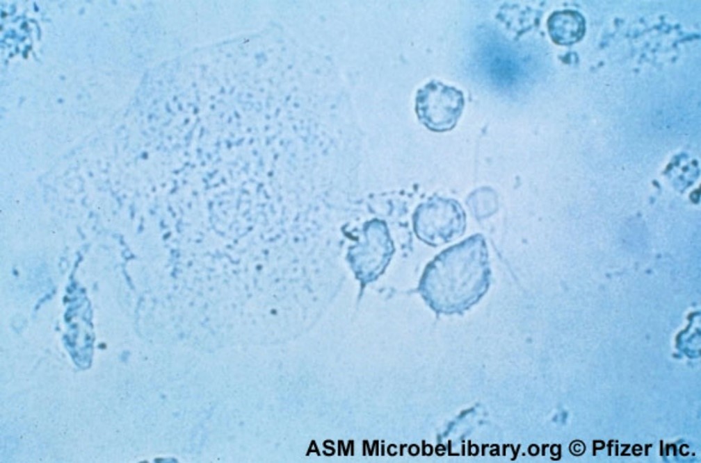
Doing a wet mount of a vaginal smear, and then finding the trophozoites at 40x. They wiggle around using flagella to swim.
42
Pentatrichomonas hominis
A. A flagellated protozoa. There is no cyst stage.
B. It is a commensal of the intestinal tract.
C. It does not cause disease.
D. Next to G. lamblia and C. mesnili, this is the most common intestinal flagellate in humans.
E. It is acquired by swallowing the trophozoite; this could be fecally conataminated food or water or oral/anal sex.
F. It then sets up residence in the large instestine.
G. It is known to exist in other primates and various domestic animals.
B. It is a commensal of the intestinal tract.
C. It does not cause disease.
D. Next to G. lamblia and C. mesnili, this is the most common intestinal flagellate in humans.
E. It is acquired by swallowing the trophozoite; this could be fecally conataminated food or water or oral/anal sex.
F. It then sets up residence in the large instestine.
G. It is known to exist in other primates and various domestic animals.
43
Pentatrichomonas hominis
P. hominis in a stool specimen
Diagnosis: finding the trophozoites in a stool specimen.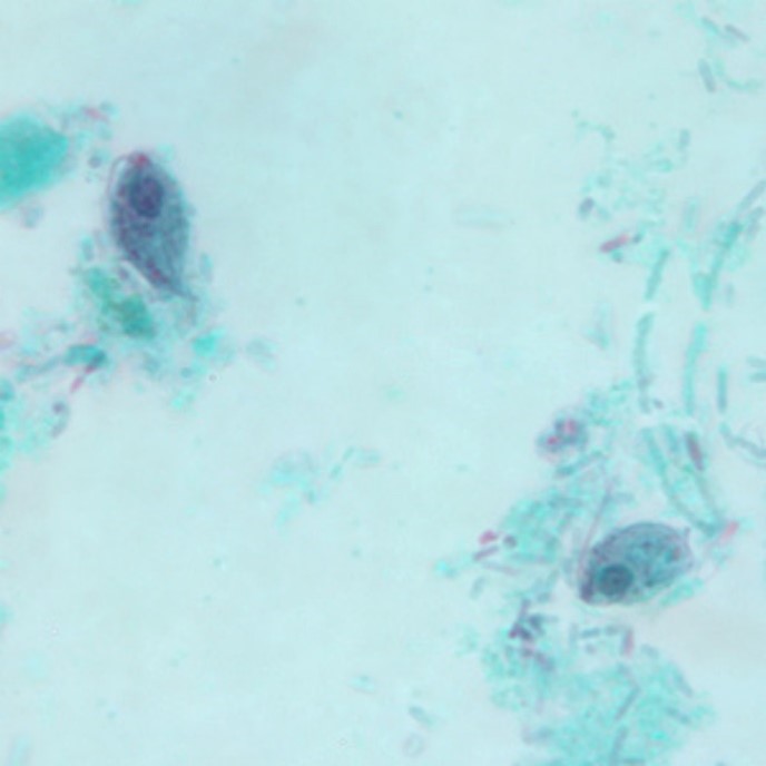
Diagnosis: finding the trophozoites in a stool specimen.
44
Chilomastix mesnili
This is another flagellated protozoan.
B. It infects about 3.5% of the population in the US.
C. It is found in humans, chimpanzees, orangutans, monkeys, and pigs.
D. It lives in the cecum and colon. C. mesnili has a similar life style to Giardia lamblia.
E. It is acquired through ingesting the cysts in fecal contaminated food or water. The cysts become trophozoites in the large intestine.
E. Although Chilomastix mesnili is considered non-pathogenic, it often occurs with other parasite infections.
F. C. mensnili maybe be confused with other pathogenic species during diagnosis.
Diagnosis: finding cysts or trophozoites in the feces.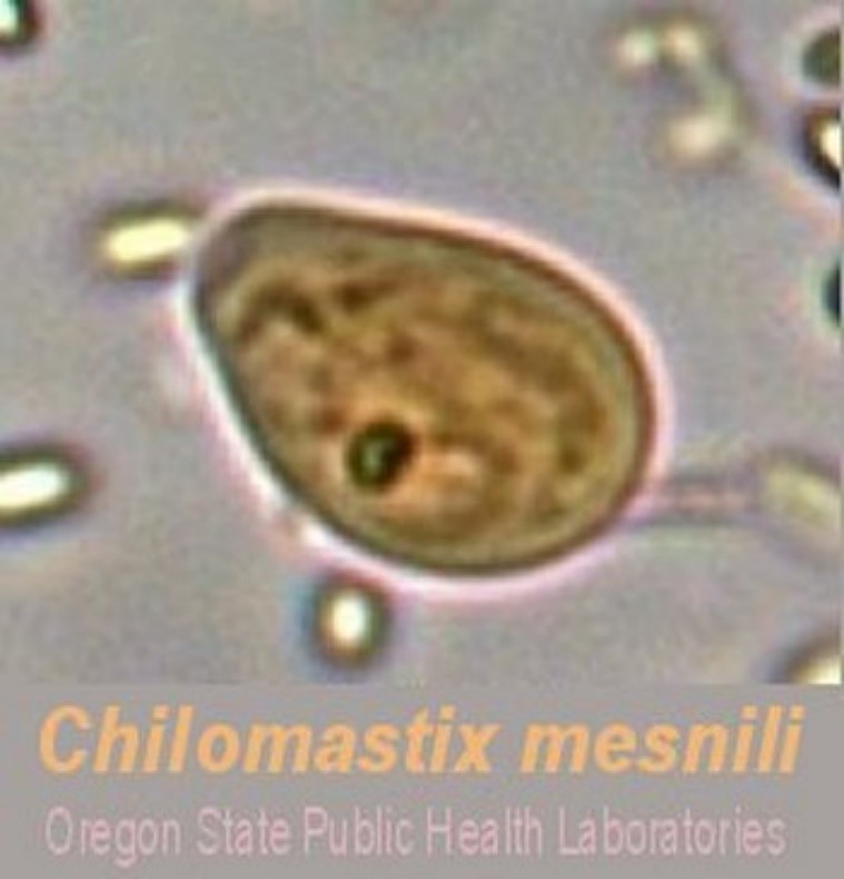
B. It infects about 3.5% of the population in the US.
C. It is found in humans, chimpanzees, orangutans, monkeys, and pigs.
D. It lives in the cecum and colon. C. mesnili has a similar life style to Giardia lamblia.
E. It is acquired through ingesting the cysts in fecal contaminated food or water. The cysts become trophozoites in the large intestine.
E. Although Chilomastix mesnili is considered non-pathogenic, it often occurs with other parasite infections.
F. C. mensnili maybe be confused with other pathogenic species during diagnosis.
Diagnosis: finding cysts or trophozoites in the feces.
45
Dientamoeba fragilis
A. Trophozoites are formed, there are no cysts.
B. The trophozoite is swallowed in fecal contaminated food or water.
C. It lives in the large intestine and passes out with feces.
D. It feeds on intestinal debris and is usually a commensal.
E. It can cause pathology in the form of diarrhea, abdominal pain, abnormal stools and abdominal distress.
F. Diagnosis: finding the trophozoite in the stool.
D.fragilis trophozoite: note the two nuclei.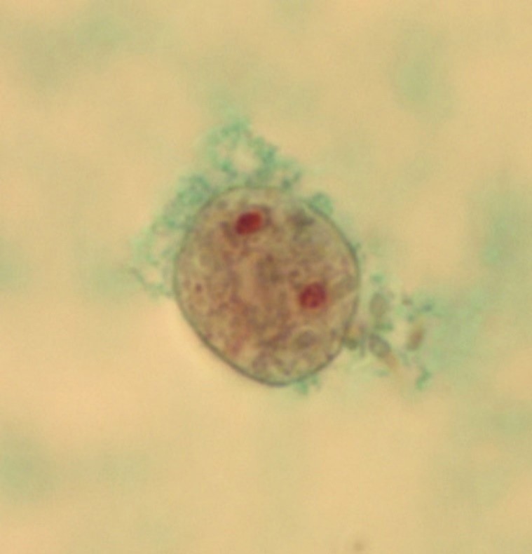
B. The trophozoite is swallowed in fecal contaminated food or water.
C. It lives in the large intestine and passes out with feces.
D. It feeds on intestinal debris and is usually a commensal.
E. It can cause pathology in the form of diarrhea, abdominal pain, abnormal stools and abdominal distress.
F. Diagnosis: finding the trophozoite in the stool.
D.fragilis trophozoite: note the two nuclei.
46
CILIATED PROTOZOA: Balantidium coli
This is a parasitic species of ciliate protozoan that causes the disease “Balantidiasis”. It is the only member of the ciliate phylum known to be pathogenic to humans.
47
CILIATED PROTOZOA: Balantidium coli - lifecycle
1. Humans acquire B. coli from living in close proximity to pigs.
2. The B. coli cyst must be swallowed in food or water contaminated with pig feces.
3. This makes it a zoonotic disease because the pig is the normal host where it is asymptomatic.
4. It then travels to the cecum and colon of humans.
5. It is not readily transmissible from one species of host to another because it requires a period of time to adjust to the symbiotic flora of the new host.
6. It then becomes a trophozoite.
7. Once it has adapted to a host species, the protozoan can become a serious pathogen, especially in humans.
8. Trophozoites multiply and encyst due to the dehydration of feces and pass out with the feces.
2. The B. coli cyst must be swallowed in food or water contaminated with pig feces.
3. This makes it a zoonotic disease because the pig is the normal host where it is asymptomatic.
4. It then travels to the cecum and colon of humans.
5. It is not readily transmissible from one species of host to another because it requires a period of time to adjust to the symbiotic flora of the new host.
6. It then becomes a trophozoite.
7. Once it has adapted to a host species, the protozoan can become a serious pathogen, especially in humans.
8. Trophozoites multiply and encyst due to the dehydration of feces and pass out with the feces.
48
CILIATED PROTOZOA: Balantidium coli - Pathology
1. Infection occurs when the cysts are ingested, usually through contaminated food or water.
2. Balantidium infection in immunocompetent individuals is not unheard of, but it rarely causes a serious disease of the gastrointestinal tract.
3. Infection most likely occurs in people with malnutrition due to the low stomach acidity.
4. Infection can also occur in people with immune compromised systems.
5. In acute disease, explosive diarrhea may occur as often as every twenty minutes.
6. Perforation of the colon may also occur in acute infections which can lead to life-threatening situations.
7. If the protozoan becomes invasive then blood and mucus appears in stools which is dysentery.
2. Balantidium infection in immunocompetent individuals is not unheard of, but it rarely causes a serious disease of the gastrointestinal tract.
3. Infection most likely occurs in people with malnutrition due to the low stomach acidity.
4. Infection can also occur in people with immune compromised systems.
5. In acute disease, explosive diarrhea may occur as often as every twenty minutes.
6. Perforation of the colon may also occur in acute infections which can lead to life-threatening situations.
7. If the protozoan becomes invasive then blood and mucus appears in stools which is dysentery.
49
CILIATED PROTOZOA: Balantidium coli - Epidemiology
1. “Balantidiasis” (the disease caused by B. coli) in humans is common in the Philippines.
2. However it can be found anywhere in the world, especially among those that are in close contact with swine.
3. The disease is considered to be rare and occurs in less than 1% of the human population.
4. The disease poses a problem mostly in developing countries, where water sources may be contaminated with swine or human feces.
Diagnosis: Finding cysts or trophozoites in the feces.
image -> B. coli cyst: note the macronucleus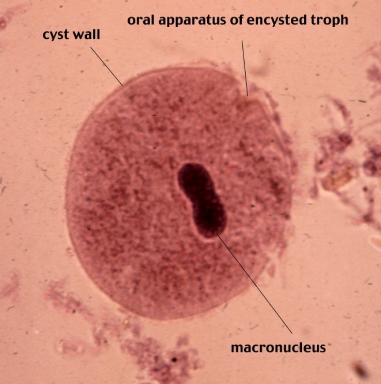
2. However it can be found anywhere in the world, especially among those that are in close contact with swine.
3. The disease is considered to be rare and occurs in less than 1% of the human population.
4. The disease poses a problem mostly in developing countries, where water sources may be contaminated with swine or human feces.
Diagnosis: Finding cysts or trophozoites in the feces.
image -> B. coli cyst: note the macronucleus