All cards
1
What is this picture an example of?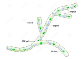
molds
2
What is this picture an example of?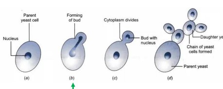
Yeast
3
What are the characteristics of molds?
MULTICELLULAR filaments, with hyphae
4
What are the characteristics of yeast?
single cell that grows / reproduces through budding
5
What is Mycelium?
matlike network of hyphae
6
What is this called?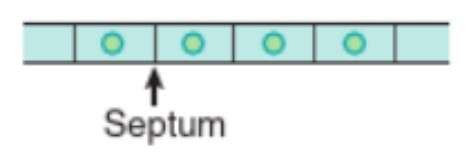
septate hyphae
7
What are the 5 kinds of vegetative hyphae?
favic chandelier / antler hyphae
nodular hyphae
pectinate body hyphae
spiral (coiled) hyphae
racquet hyphae
nodular hyphae
pectinate body hyphae
spiral (coiled) hyphae
racquet hyphae
8
What kind of hyphae is this?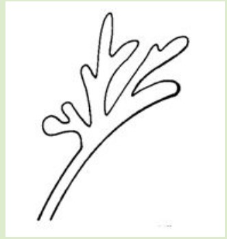
favic chandelier / antler hyphae
9
What kind of hyphae is this?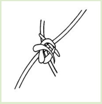
nodular hyphae
10
What kind of hyphae is this?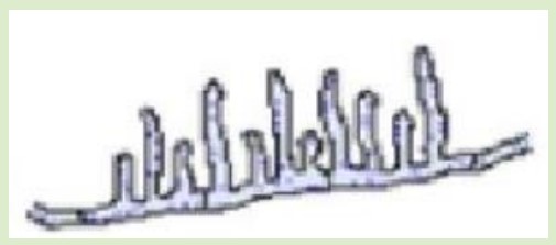
pectinate body hyphae
11
What kind of hyphae is this?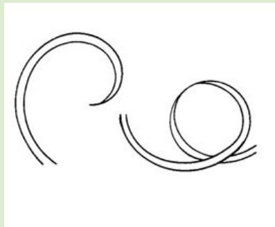
spiral (coiled) hyphae
12
What kind of hyphae is this?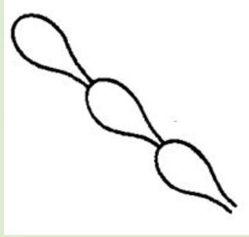
racquet hyphae
13
What kind of hyphae is this?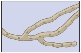
coenocytic (nonseptate) hyphae
14
What are pseudohyphae?
marked by constrictions, not septate
15
What are fungal spores?
means of fungal reproduction;
asexual or sexual
major source of fungal infection
asexual or sexual
major source of fungal infection
16
What are the 3 kinds of sexual spores?
basidiospore
ascospore
zygospore
ascospore
zygospore
17
How are asexual spores formed?
through mitosis
18
What are the 5 kinds of asexual spores?
(SCCAB)
sporangiospore
conidiospore
chlamydospore
arthrospore
blastospore
sporangiospore
conidiospore
chlamydospore
arthrospore
blastospore
19
What kind of spore is this?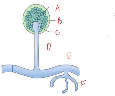
sporangiospore
20
What are 3 examples of sporangiospore?
(RAM)
rhizopus
mucor
absidia (lictheimia)
rhizopus
mucor
absidia (lictheimia)
21
What kind of spore is this?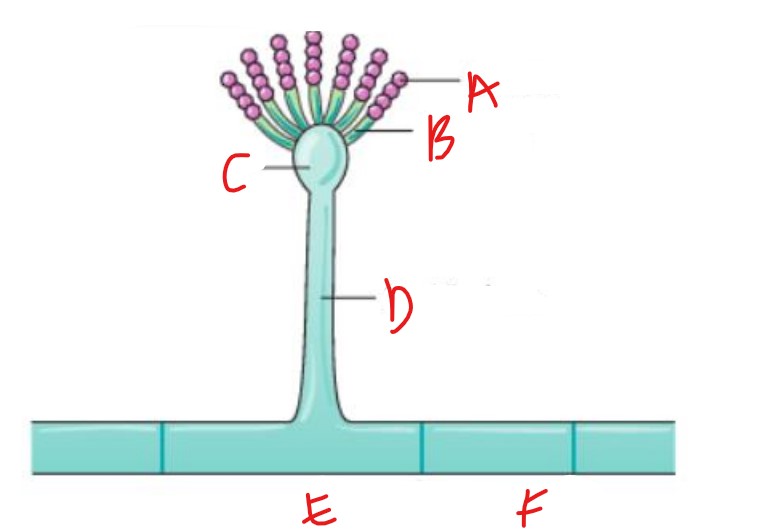
uniserate conidiospore
22
What is letter F?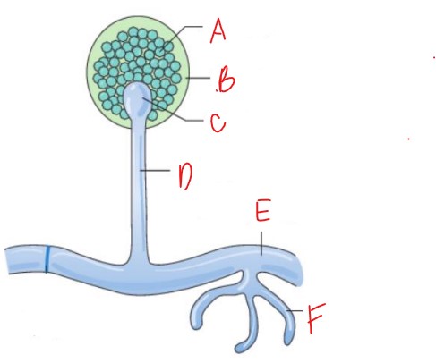
Rhizoids
23
What kind of spore is this?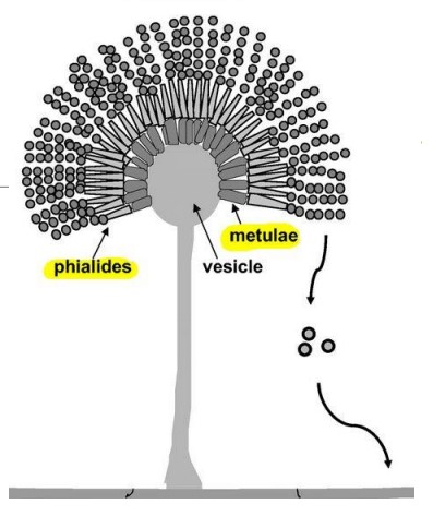
biserate conidiospore
24
What does conidiospore have that sporangiospore usually does not?
septate hyphae
25
What are 2 examples of conidiospores?
penicillum
aspergillus
aspergillus
26
What kind of spore is this?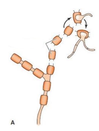
arthrospore
27
What are 3 examples of arthrospores?
trichosporon
geotrichum
coccidioides immitus
geotrichum
coccidioides immitus
28
What kind of spore is this?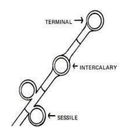
chlamydospore
29
What is an example of chlamydospore?
Candida albicans
30
What kind of spore is this?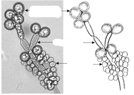
blastospore
31
What are blastospores a result of?
budding of a yeast (Asexual rerproduction)
32
Label A and B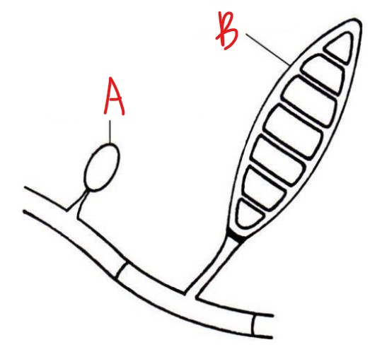
A-microconidium
B-macroconidium
B-macroconidium
33
What are macroconididia?
large asexual structures divided into cells with transverse septa
34
What are microconidia?
small asexual strcutures
35
Label A and B dermatophytes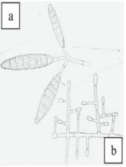
A-macroconidia macrosporum
B-microcondia microsporum
B-microcondia microsporum
36
Label A and B dermatophytes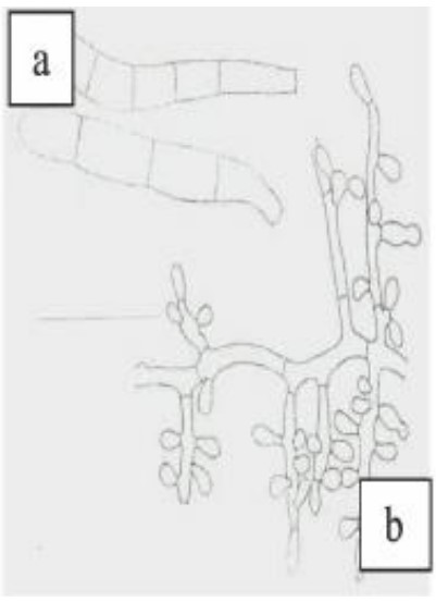
A-macroconidia trichophyton
B-microcondia trichophyton
B-microcondia trichophyton
37
Label this structure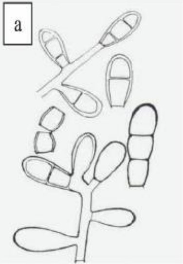
macrocondia epidermophyton
38
What kind of infection does superficial / cutaneous mycoses : dematophytosis do?
outermost layers of skin or hair because they love keratin
39
How does ringworm present?
circular rash
40
What causes ringworm?
dermatophytes
41
What are the dermatophytes that cause ringworm?
trichophyton
microsporum
epidermophyton
microsporum
epidermophyton
42
What is ringworm also known as?
tinea
dermatophytosis
dermatophytosis
43
What are the 4 ways fungi can invade the hair shaft?
ectothrix
ecto-endothrix
endothrix
favus
ecto-endothrix
endothrix
favus
44
What kind of hair invasion is shown?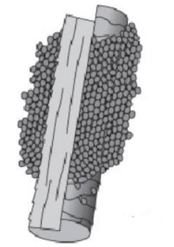
ectothrix
45
What kind of hair invasion is shown?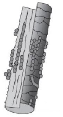
ecto-endothrix
46
What kind of hair invasion is shown?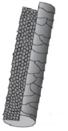
endothrix
47
What kind of hair invasion is shown?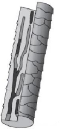
favus
48
What hair invasion would show UV light negative?
endothrix (?)
49
What is favus hair invasion?
honeycomb infection;
clusters of hyphae at teh base with air spaces in hair shaft
yellow crust around hair shaft
clusters of hyphae at teh base with air spaces in hair shaft
yellow crust around hair shaft
50
Where does Tinea capitis affect?
head
(scalp ringworm)
(scalp ringworm)
51
What is a known species of scalp ringworm?
T. tonsurans (most common in US)
52
What causes favus?
T. schoenleinii
53
What is favus?
severe form of scalp ringworm
54
What is ringworm of the feet?
Tinea pedis
55
What is Tinea pedis associated with?
athelete's foot
56
Where does Tinea manuum affect?
hands!
manuum = thumb
manuum = thumb
57
What kind of ringworm is tinea manuum?
ringworm of the hands
58
What species primarily causes Tinea manuum?
trichophyton rubrum
59
What is the common clinical presentation of ringworm of the hands?
two foot, one hand syndrome (acquired when picking at athlete's foot)
60
What does cruris in Tinea cruris refer to?
cruris = leg
61
What is ringowrm of the groin / pubic caused by?
trichophyton rubrum
62
What is another name for Tinea cruris (ringworm of the groin / pubic?
jock itch
63
What does barbae in Tinea barbae mean?
beard
64
What is another name for Tinea barbae (beard ringworm) infection?
Barber's itch
65
What does unguium in Tinea unguium mean?
nails
Tinea unguium = ringworm of the nails
Tinea unguium = ringworm of the nails
66
What is the the major cause of ringworm of the nail (Tinea unguium)?
onychomycosis (dermatophyte)
67
What does corporis in Tinea corporis mean?
corporis = body
Tinea corporis = ringworm of the body
Tinea corporis = ringworm of the body
68
What strain causes ringworm of the body / Tinea corporis?
T. rubrum
69
What organism sporuting resembles 'bird on a wire'?
Trichophyton rubrum
70
What kind of ringworm is trichophyton tonsurans?
scalp ringworm
71
What causes 'black-dot' tinea capitis?
trichoypton tonsurans
due to hair breaking easily
due to hair breaking easily
72
What can be seen in trichophyton tonsurans?
abundant microconidia, chlamydospores
73
What causes microsporum canis?
zoophilic--cats, dogs, others
mostly by domestic cat
mostly by domestic cat
74
Where can Microsporum gypseum be found?
geophillic--in contaminated soils
also in dogs, cats, and other animals exposed to dirt
also in dogs, cats, and other animals exposed to dirt
75
What does epidermophyton floccosum look like?
club shaped (beaver tail)
macroconidia
macroconidia
76
What does Wood's Lamp ID?
ectothrix infections
77
What are the main dermatophytes that fluoresce under UV light?
Microsporum spp.
78
What is piedra translated to?
stone
79
What is piedra?
superficial fungal infection of hair shaft that presents as small nodules stuck onto hair
80
What organism causes Thrush?
Candida spp.
81
What causes Tinea nigra?
superficial mycosis caused by Hortaea wernickii
82
What organism resembles 'spaghetti and meatballs'?
Malassezia furfur
83
What is M. furfur dependent on?
lipid-dependent
84
What kind of pathogen is Malassezia furfur?
opportunistic
85
What does subcutaneous mycoses involve?
muscle, lymph vessels, and connective / neighboring tissue
86
What is another name for Mycetoma?
'Madura Foot'
87
What are the two kinds of mycetoma?
eumycotic
actinomycotic
actinomycotic
88
What causes eumycotic mycetoma?
fungi
89
What causes actinomycotic mycetoma?
bacteria
90
What are the agents of eumycetoma?
Madurella (M. mycetomatis, M. grisea)
Pseudallescheria boydii
Leptosphaeria spp.
Pseudallescheria boydii
Leptosphaeria spp.
91
What are the agents of actinomycetoma?
Nocardia
Streptomyces
Actinomyces
Streptomyces
Actinomyces
92
What should be collected when mycetoma is considered?
grains / granules of the sinus discharge that contain colonies of the causative organism
93
What does black grain eumycetoma indicate?
madurella complex
94
What does white grain mycetoma indicate?
Pseudallescheria boydii
95
Which is more dangerous, white or black grain mycetoma?
white grain
96
What organism uses the partial acid fast stain?
Nocardia
97
What can be seen in Nocardia mycetoma lesions?
sulfur granules
98
What is another name for Lymphocutaneous sporotrichosis infection?
Rose gardener's Disease
99
What kind of fungus is Lymphocutaneous sporotrichosis?
DIMORPHIC fungus: Sporothrix schenckii
100
What are the two forms of lymphocutaneous sporotrichosis?
mold form and yeast form
101
What temperature do you see mold form of L. sporotrichosis?
25C
102
What temp do you see yeast form of L. sporotrichosis?
37C
103
What does 'hyalo' in hyalophomycosis mean?
hyalo=glass
due to colorless hyaline, nonpigmented, or lightly colored mold with septate hyphae in tissue
due to colorless hyaline, nonpigmented, or lightly colored mold with septate hyphae in tissue
104
What are the common causative agents in hyalophomycosis?
acremonium
fusarium (mycotoxins on food)
geotrichum (ceral / dairy products)
paecilomyces (eye infections)
scedosporium
fusarium (mycotoxins on food)
geotrichum (ceral / dairy products)
paecilomyces (eye infections)
scedosporium
105
What does chromo in chromoblastomycosis mean?
chromo = color
106
What causes chromobastomycosis / chromomycosis?
caused by several species of melanized fungi that produce a dark pigment
107
What are the common causative agents of chromobastomycosis?
Fonsecaea spp.
Phialophora spp.
Cladosporium spp.
Phialophora spp.
Cladosporium spp.
108
What does 'phaeo' in phaeohyphomycosis mean?
phaeo = gray
109
What causes phaeohyphomycosis infections?
darkly pigmented dermatiaceous fungi
110
What are the common causative agents of phaeohyphomycosis?
Cladosporium
Exophiala
Wangiella
Cladophialophora
Bipolaris
Exophiala
Wangiella
Cladophialophora
Bipolaris
111
What causes subcutaneous zygomycosis / mycormycosis?
caused by a group pof fungi -- mucormycetes
112
What organisms are related to subcutaenous zygomycosis / mucormycosis?
Mucor
Rhizopus
Absidia (Lictheimia)
Rhizopus
Absidia (Lictheimia)
113
How can you tell the difference between zygomucosis / mucormycosis?
Looking at roots
114
What organinsm is shown?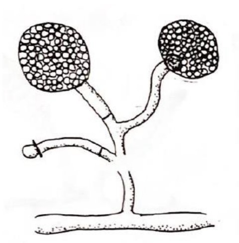
Mucor
115
What organinsm is shown?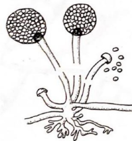
Rhizopus
116
What organinsm is shown?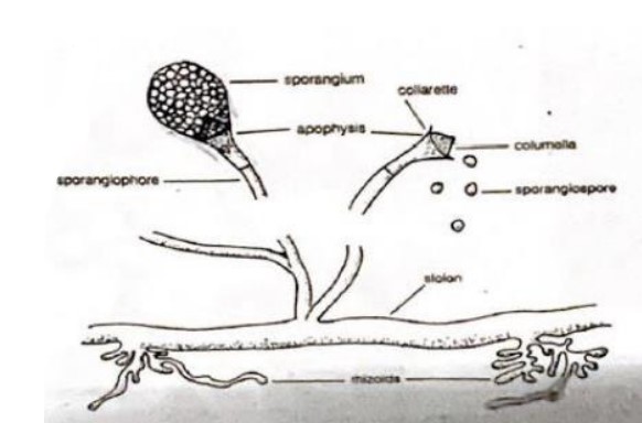
Absidia (Lichtheimia)
117
What kind of pathogens are Dimorphic Fungal Pathogens?
primary systematic pathogens (causing infection in normal and immunocompromised people)
118
What temps do dimorphic species take mold form?
25C-30C
119
What temps do dimorphic species take yeast form?
37C
120
What are the dimorphic fungi?
Histoplasma capsulatum
Blastomyces dermatiditis
Paracoccidiodes braziliensis
Coccidiodes immitis
Penicillum (Talaromyces) marneffei
Sporothrix schenkii
Blastomyces dermatiditis
Paracoccidiodes braziliensis
Coccidiodes immitis
Penicillum (Talaromyces) marneffei
Sporothrix schenkii
121
What dimorphic species is shown?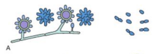
Histoplasma capsulatum
122
What dimorphic species is shown?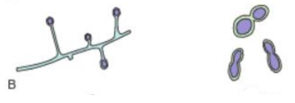
Blastomyces dermatiditis
123
What dimorphic species is shown?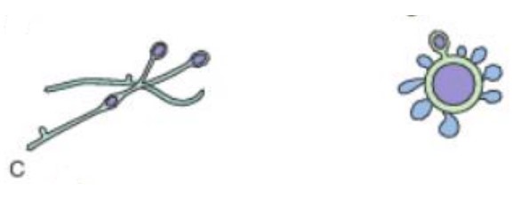
Paracoccidiodes braziliensis
124
What dimorphic species is shown?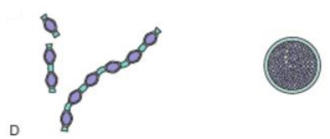
Coccidiodes immitis
125
What dimorphic species is shown?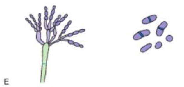
Penicillum (Talaromyces) marneffei
126
What dimorphic species is shown?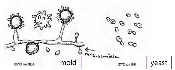
Histoplasma capsulatum
127
Where can you get exposed to Histoplasma capsulatum?
bird droppings and bat guano
128
What dimorphic species is shown?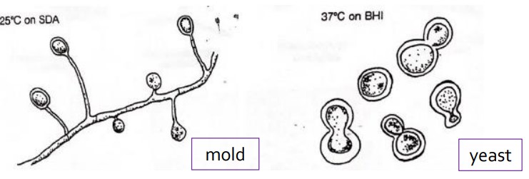
Blastomyces dermatiditis
129
What species has a yeast form that resembles a 'captain's wheel'?
Paracoccidiodes braziliensis
130
What dimorphic species is shown?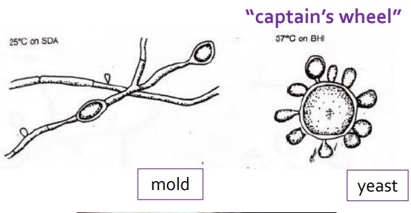
Paracoccidiodes braziliensis
131
What dimorphic species is shown?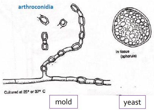
Coccidiodes immitis
132
What kind of form does Coccidioides immitis take?
arthroconidia
133
What infection does coccidioidomycosis lead to?
San Joaquin Valley Fever
VERY DANGEROUS!
VERY DANGEROUS!
134
What species is shown?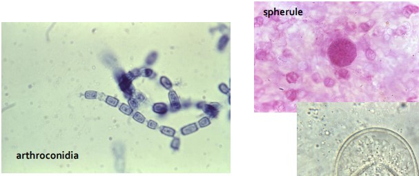
Coccidioides immitis
135
What species is shown?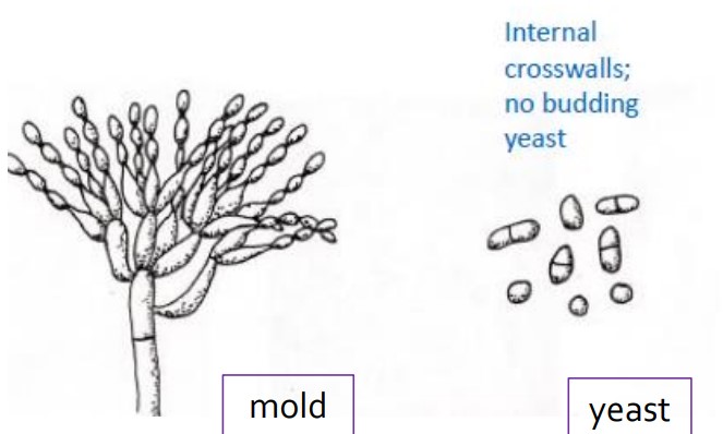
Penicillum marneffei
136
What is unique of the yeast form in Penicillium (talaromyces) marneffei?
Yeast form has internal transverse / septum crosswalls, no budding yeast
137
What is the name of Sprothrix schenkii infection?
Sporotrichosis
"Rose Gardener's Disease"
"Rose Gardener's Disease"
138
What are the 3 kinds of Rose Gardener's Disease?
Cutaneous (skin) sporotrichosis
Pulmonary (lung) sporotrichosis
Disseminated sporotrichosis
Pulmonary (lung) sporotrichosis
Disseminated sporotrichosis
139
What is the organism shown?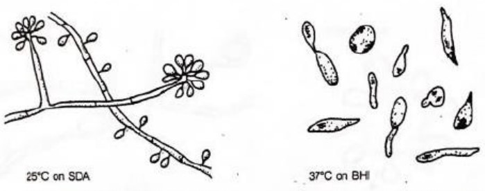
Sporothrix schenkii
140
What are the 4 kinds of opportunistic fungi?
Candida
Aspergillus
Cryptococcus
Mucor
Aspergillus
Cryptococcus
Mucor
141
What are the 2 main candida species?
C. albicans
C. auris
C. auris
142
What is the most common candida isolated?
C. albicans
143
What candida is an emerging public health threat?
C. auris
144
What organism is shown?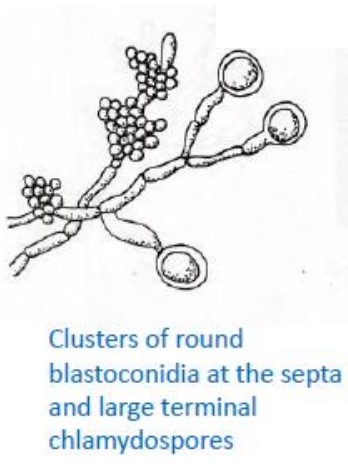
Candida spp.
145
Why is Candida auris an emerging threat?
multidrug resistant yeast with outbreaks in long term care facilities and hospitals worldwide with a high mortality rate
146
What kind of pathogen is Aspergillus spp.?
Opportunistic!
147
What organism is shown?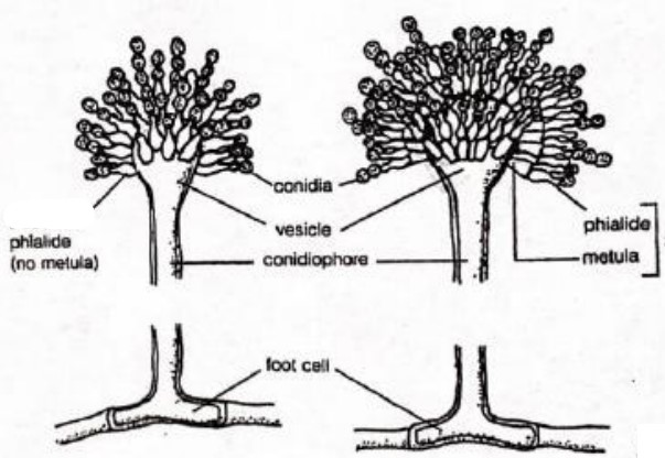
Aspergillus spp.
148
What is the difference between the right and left?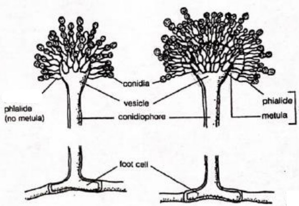
left is uniserate while the right is biserate
149
What does biserate phialide contain?
metula (close to vesicle) and the phialide
(whereas uniseriate only containes phialide and no metula)
(whereas uniseriate only containes phialide and no metula)
150
How can you tell the difference between A. fumigatus, A. niger, and A. flavus
color
Aspergillus fumigatus = green
A. niger = black
A. flavus = brown
Aspergillus fumigatus = green
A. niger = black
A. flavus = brown
151
Where can cryptococcus neoformans be found?
in the lungs and brain
inhaled from the environment
inhaled from the environment
152
What is C. neoformans associated with?
pigeon droppings, decaying wood, soil, tree hollows
153
What is the common specimen used when testing for C. neoformans?
CSF
154
What is the stain for C. neoformans?
india ink (highlights the protective capsule around organism)
155
What organism is shown?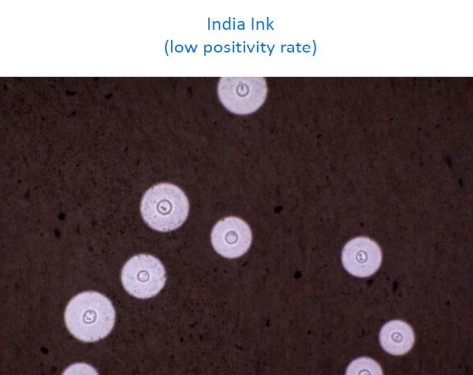
Cryptococcus neoformans
156
Why do physicians want antifungal susceptibility testing?
fungal infection is invasive, when acquired drug resistance is suspected, or when the pt is unexpectedly failing therapy
157
What agar is good for cryptococcus?
bird seed agar
(also remember cryptococcus = india ink stain)
(also remember cryptococcus = india ink stain)
158
What is the purpose of india ink?
to identify encapsulated yeast