Lecture 11 - Synovial fluid
Study these flashcardsFree · spaced repetition · iPhone, Android, Mac & web
All cards
1
What are the functions of synovial fluid?
Lubrication for the moveable joints aka diarthroses (movable joints)
Lessens shock of joint compression
Lessens shock of joint compression
2
What is synovial fluid formed from?
Ultrafiltrate of plasma across synovial membrane.
Synoviocytes
Synoviocytes
3
What do synoviocytes secrete?
hyaluronic acid that makes the fluid viscous (lubrication)
4
What is the normal color of synovial fluid?Corticoster
colorless - pale yellow
5
What are the 4 classifications of disorders (arthritis)?
1. Noninflammatory
2. Inflammatory
3. Septic
4. Hemorrhagic
2. Inflammatory
3. Septic
4. Hemorrhagic
6
What are noninflammatory disorders?
degenerative, osteoarthritis
7
What are inflammatory disorders?
immunologic, lupus erythematosus (LE), rheumatoid arthritis (RA), Lyme disease
Crystal-induced, goutand pseudogout
Crystal-induced, goutand pseudogout
8
What are septic disorders?
microbial infection
9
What are hemorrhagic disorders?
trauma, tumors, coagulation deficiencies
10
What color is the synovial fluid when there is a septic disorder?
yellow-green color
11
What color is the synovial fluid when there is a hemorrhagic disorder?
red-brown color
12
Is the culture (+ or -) for noninflammatory disorders?
Negative
13
What does the viscosity look like in noninflammatory disorders?
HIGH
14
Is the culture (+ or -) for inflammatory disorders?
Negative
15
What does the viscosity look like in inflammatory disorders?
low
16
What are the levels of WBC in noninflammatory disorders?
< 25 %
17
What are the levels of WBC in inflammatory disorders?
> 50 %
18
What causes lyme disease? (which vector)
tick
19
What is the culture, viscosity and neutrophils in septic disorders?
Culture: POSITIVE
Viscosity: low (bc it can break down hydrolic acid)
Neutrophils are > 75%
Viscosity: low (bc it can break down hydrolic acid)
Neutrophils are > 75%
20
What is the culture, viscosity and neutrophils in hemorrhagic disorders?
Culture: neg
Viscosity: low
Neutrophils: < 25%
Viscosity: low
Neutrophils: < 25%
21
What are the normal ranges for synovial fluid:
Volume
Color
Clarity
Viscosity
Leukocyte Count
Neutrophils
Crystals
Glucose: plasma difference
Total protein
Volume
Color
Clarity
Viscosity
Leukocyte Count
Neutrophils
Crystals
Glucose: plasma difference
Total protein
Volume: <3.5 mL
Color: colorless to pale yellow
Clarity: clear
Viscosity: able to form a string 4 - 6 cm long
Leukocyte Count: <200 cells/ uL
Neutrophils: <25% of the differential
Crystals: none present
Glucose: plasma difference: <10 mg/dL lower than the blood glucose level
Total protein: <3 g/dL
Color: colorless to pale yellow
Clarity: clear
Viscosity: able to form a string 4 - 6 cm long
Leukocyte Count: <200 cells/ uL
Neutrophils: <25% of the differential
Crystals: none present
Glucose: plasma difference: <10 mg/dL lower than the blood glucose level
Total protein: <3 g/dL
22
Needle aspiration of synovial fluid is called?
Arthrocentesis
23
How much synovail fluid is collected during arthrocentesis? what about when it is inflammed
Normal knee fluid amount 3.5 mL (>25 mL if inflamed)
24
Does normal synovial fluid clot?
no!!!
25
What does it mean when synovial fluid clots?
Diseased fluid !!!!
26
What is used to collect synovial fluid for microbiology?
sterile heparinized
27
What is used to collect synovial fluid for hematology?
Liquid EDTA
28
What should be done to nonanticoagulated ( no substance to prevent blood from clotting) tubes? what reagent is used to collect glucose?
Centrifuge nonanticoagulated tube and separate
Sodium fluoride for glucose
Sodium fluoride for glucose
29
When is synovial fluid tested? and why?
ASAP to avoid cellular lysis and changes in crystals
30
How many tubes and what kind of tubes are used normally to collect synovial fluid?
Tube #1: no anticoagulant tube for chemical and immunologic studies
Tube #2: anticoagulant tube for microscopic studies
Tubes #3: sterile anticoagulant tube for microbiological studies
Tube #2: anticoagulant tube for microscopic studies
Tubes #3: sterile anticoagulant tube for microbiological studies
31
If the synovial fluid color is deeper yellow with green tinge, what does that mean?
infection
32
when synovial fluid is red, what does that mean?
hemorrhagic or traumatic tap
33
To differentiate between hemorrhagic and traumatic tap, what should you look out for?
look for decreasing blood in tubes
34
Is the synovial fluid is milky, what causes it?
crystal induced
35
If the synovial fluid is turbid, what does that mean?
fluid contains WBCs or cellular debris, fibrin
36
What happens when there's an increase in polymerization of hyaluronic acid?
its viscosity increases significantly due to the formation of longer polymer chains, which entangle more readily with each other, resulting in a thicker, more viscous solution;
37
What is hyaluronic acid essential for?
joint movement
38
A decrease in polymerization can indicate what?
arthritis
39
How do you measure viscosity?
4 to 6 cm string from aspirating needle
40
What are 2 ways to know if it is synovial fluid?
1. 4 - 6 cm strings
2. mucin clot test
2. mucin clot test
41
When is mucin clot test performed?
if there are any concerns that fluid sample is not synovial fluid
42
Does normal synovial fluid contain fibrinogen and does it clot?
no fibrinogen and does not clot.
43
How do you perform the mucin clot test? what is the science behind it.
Dilute fluid with 2% acetic acid: 1 part fluid and 4 parts acid
Hyaluronate from joint fluid wil form a clot in acid, confirming fluid is joint fluid.
Hyaluronate from joint fluid wil form a clot in acid, confirming fluid is joint fluid.
44
What test is used to count the WBCs for synovial fluid?
hemocytometer counting chamber
45
How do you prepare the sample for the hemocytometer cell count?
use normal saline/methylene blue
46
If the synovial fluid is viscous, how should the sample be treating before observing in the hemocytometer?
with hyaluronidase
47
What is the normal count of WBCs/ uL for synovial fluid?
< 200 WBCs/ uL
48
If the synovial fluid has a septic case, what is the WBC count?
> 100,000 WBCs/ uL
49
How do you prepare the sample for differential count?
incubate with hylauronidase, then cytocentrifuge
50
What are normal cells found in a differential count of synovial fluid?
monocytes, macrophages, synovial tissue cells
51
what is the normal count for neutrophils in a differential count for synovial fluid? and if it increases what is the condition?
< 25 %; increase in sepsis
52
what is the normal count for lymphocytes in a differential count for synovial fluid? and if it increases what is the condition?
< 15 %; osteoarthritis
53
What may the cells look like in the differntial count for synovial fluid?
all cells may appear more vacuolated
54
What are other cells seen in a differential count for synovial fluid?
- LE cells
- Eosinophils
- Reiter cells/ neutrophages: vacuolated macrophages with ingested neutrophils... nom nom nom
- Ragocytes (RA cells): neutrophils with small, dark granules containing RA factor (IgM)
- Lipid droplets: crush injuries
- Hemosiderin granules
- Eosinophils
- Reiter cells/ neutrophages: vacuolated macrophages with ingested neutrophils... nom nom nom
- Ragocytes (RA cells): neutrophils with small, dark granules containing RA factor (IgM)
- Lipid droplets: crush injuries
- Hemosiderin granules
55
what is an important diagnositic test; frequently performed ID for synovial fluid?
Crystal Identification
56
What can crystal identification in synovial fluid identify/ AKA WHAT DISORDERS? give 2 examples.
acute and chronic cases:
Metabolic disorders and decreased renal function.
other causes: increased blood levels, degeneration of bone and cartilage, injection of corticosteroids.
Metabolic disorders and decreased renal function.
other causes: increased blood levels, degeneration of bone and cartilage, injection of corticosteroids.
57
What type of crystals is formed in "decreased renal function", which can be identified in the synovial fluid?
oxalate crystals formed
58
What type of crystals is formed in "degeneration of bone and cartilage", which can be identified in the synovial fluid?
calcium pyrophosphate dihydrate
59
What type of crystals is formed in "injection of corticosteroids", which can be identified in the synovial fluid?
corticosteroid crystals
60
What is the primary type of crystal in gout?
monosodium urate (MSU)
61
What is the type of crystal in pseudo-gout?
Calcium pyrophosphate dihydrate
62
what is MSU? and what causes it?
Monosodium urate:
impaired purine metabolism, high purine foods, leukemia chemotherapy, decreased renal excretion of uric acid
impaired purine metabolism, high purine foods, leukemia chemotherapy, decreased renal excretion of uric acid
63
what is CPPD? and what causes it?
Calcium pyrophosphate deposition:
degenerative arthritis, disorders causing elevated calcium levels.
degenerative arthritis, disorders causing elevated calcium levels.
64
What color are monosodium urate (MSU) and Calcium pyrophosphate dihydrate (CPPD) crystals look like under compensator ray?
MSU = yellow
CPPD = blue
CPPD = blue
65
Cholesterol crystals are due to what condition? and what do they appear similar to?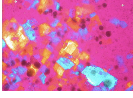
Systemic autoimmune diseases (LE, RA); appear similar to urine
66
Describe cholesterol crystals.
notched corners
67
Corticosteroid crystals look like what?
injections, flat, variable plates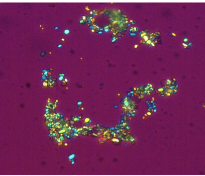
68
Calcium oxalate crystals can be seen in what kind of patients?
renal dialysis patients
69
What types of artifacts can be mistaken with crystals?
starch, powdered anticoagulants, dust, scratches (clean slides, cover slips)
70
For a normal synovial test results, what are the normal values similar to ?
those of plasma
71
Are all chemistry tests important?
no, few are clinically important
72
What is the most frequent chemistry test observed for Synovial fluid comparison?
Glucose
73
What are normal values of glucose in plasma?
not less than 10 mg/dL
74
When is the plasma sample for chemsitry test drawn?
at the same time as synovial fluid is collected
75
If the glucose levels are decreased significantly in plasma, what does this indicate?
inflammatory and septic categories
76
What are 2 chemistry tests that could be done, but not performed routinely, on synovial fluid?
Synovial fluid protein
Synovial fluid lactate
Synovial fluid lactate
77
What is the normal value for synovial fluid protein? and what does it mean if it is increased?
normal is less than 3 g/dL;
increased in inflammatory and hemorrhagic categories
increased in inflammatory and hemorrhagic categories
78
Severity of synovial fluid protein and lactate indicate what coniditon?
rheumatoid arthritis (RA)
79
What tests are performed in microbiology? and what can infections be caused by?
gram stain and cultures. ; infections caused by inflammation, trauma, and systemic infections
80
Which are the most common autoimmune causes of arthritis?
RA and LE
81
What complication is seen in lyme disease? and what test should be done and which sample should be used?
arthritis; test serum for Borrelia burgdorferi antibodies! (PCR needed)