Lecture 5_ applications
Study these flashcardsFree · spaced repetition · iPhone, Android, Mac & web
All cards
1
Q: What is one method used in the lab diagnosis of bacterial infections involving growth of organisms?
A: Culture of the biologic sample.
2
Q: What technique uses visual examination to diagnose bacterial infections?
A: Microscopy.
3
Q: What method detects bacterial antigens in a clinical sample?
A: Detection of bacterial antigens in the clinical sample.
4
Q: What method detects bacterial DNA or RNA?
A: Molecular detection of bacterial DNA or RNA.
5
Q: What technique detects antibodies produced in response to infection?
A: Serology.
6
Q: What proteomic method is used in bacterial infection diagnosis?
A: Proteomics (MALDI-TOF).
7
Q: What type of hemolysis does Group A Streptococcus show on blood agar (BA)?
A: Beta hemolysis on BA.
8
Q: What antibiotic is Group A Streptococcus susceptible to?
A: Bacitracin.
9
Q: What enzyme activity is associated with Group A Streptococcus?
A: PYR activity.
10
Q: What classification method is used for Group A Streptococcus?
A: Lancefield typing.
11
Q: What method is used for antigen detection in Group A Streptococcus?
A: Lateral flow immunochromatographic assays (LFAs).
12
Q: Which antibody can be detected for Group A Streptococcus infection?
A: ASO (streptozyme test).
13
Q: What molecular method can detect Group A Streptococcus?
A: Real-time PCR.
14
Q: What invasive method is used to diagnose Helicobacter pylori?
A: Endoscopy and biopsy.
15
Q: What does the endoscopy biopsy detect for H. pylori diagnosis?
A: Detection of urease on the biopsy sample.
16
Q: What test is used to detect urease in H. pylori biopsy samples?
A: CLOtest.
17
Q: What are the non-invasive methods for diagnosing Helicobacter pylori?
A: Urea breath test and antigen detection in stool.
18
Q: What type of test detects antibodies in serum for H. pylori diagnosis?
A: ELISA or LFA.
19
Q: Which antibody is clinically relevant in H. pylori serology testing?
A: H. pylori-specific IgG.
20
Q: Is IgM clinically significant in H. pylori antibody testing?
A: No, IgM is not clinically significant.
21
Q: Are molecular tests for H. pylori currently FDA-approved?
A: No, molecular tests are not yet FDA-approved.
22
Q: What are the three main categories of traditional laboratory tests for syphilis?
A: Direct detection of spirochetes, nontreponemal serological tests, and treponemal serological tests.
23
Q: What microscopy method is used to directly detect T. pallidum from skin lesions?
A: Dark-Field Microscopy.
24
Q: Which direct detection method uses antibodies to detect T. pallidum?
A: Fluorescent Antibody Testing (FAT), also called Direct Fluorescent Antibody (DFA) test.
25
Q: What do nontreponemal serological tests detect?
A: Anticardiolipin antibody (reagin).
26
Q: Name two nontreponemal tests used for syphilis.
A: Venereal Disease Research Laboratory (VDRL) test and Rapid Plasma Reagin (RPR) test.
27
Q: What testing phenomenon may occur in secondary syphilis with RPR/VDRL?
A: Prozone effect.
28
Q: What do treponemal serological tests detect?
A: Antibodies against T. pallidum.
29
Q: When do treponemal serological tests typically become positive?
A: Before RPR or VDRL, and are usually 100% reactive in secondary and latent syphilis.
30
Q: Which treponemal test is highly specific and used as a confirmatory test?
A: Fluorescent treponemal antibody absorption (FTA-ABS).
31
Q: Name another treponemal test used for syphilis.
A: T. pallidum particle agglutination (TP-PA) test.
32
Q: What type of syphilis test usually remains positive for life?
A: Treponemal antibody test (TP-PA, FTA-ABS, EIA/CLIA).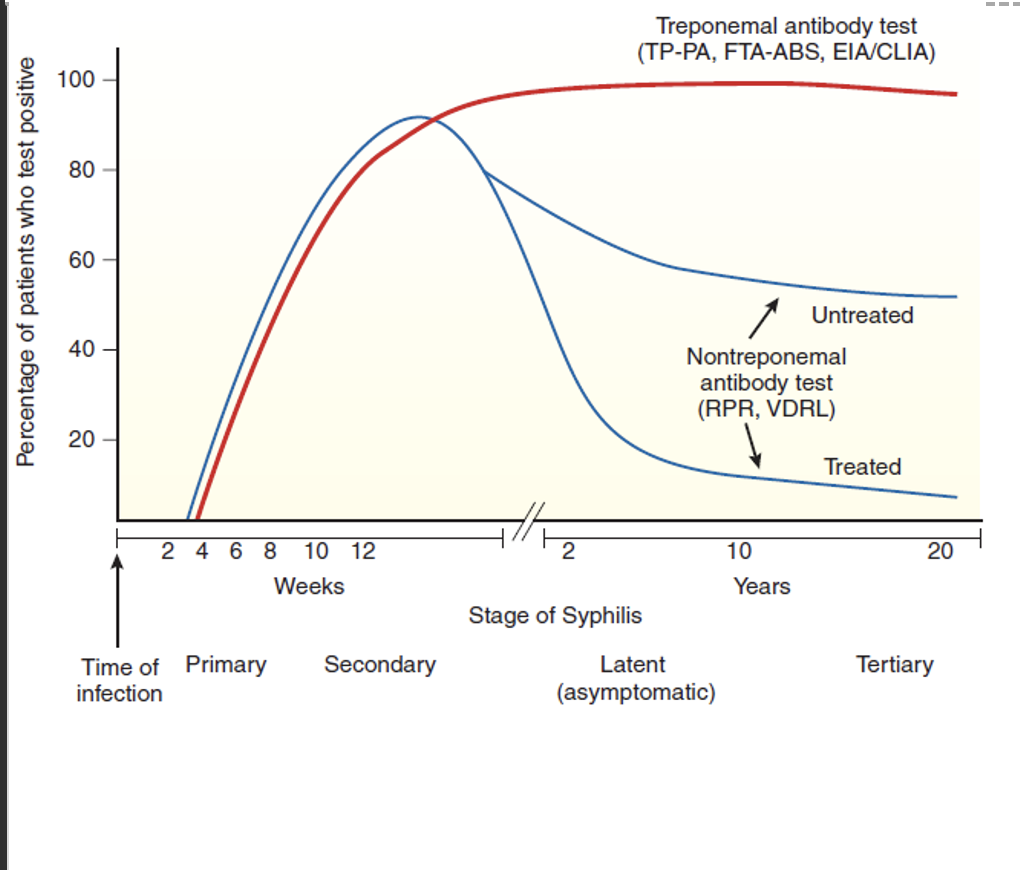
33
Q: What happens to nontreponemal antibody test titers (RPR, VDRL) after treatment?
A: They decrease.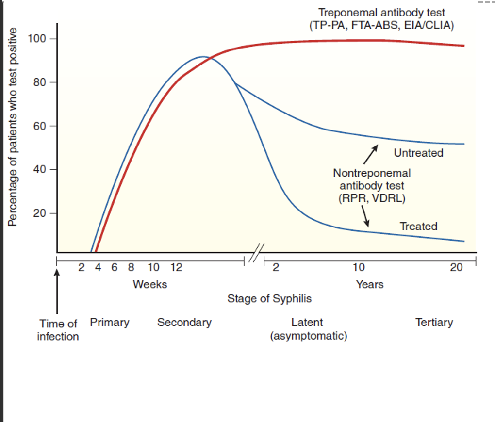
34
Q: In untreated syphilis, what happens to nontreponemal antibody test titers over time?
A: They decline but do not return to zero.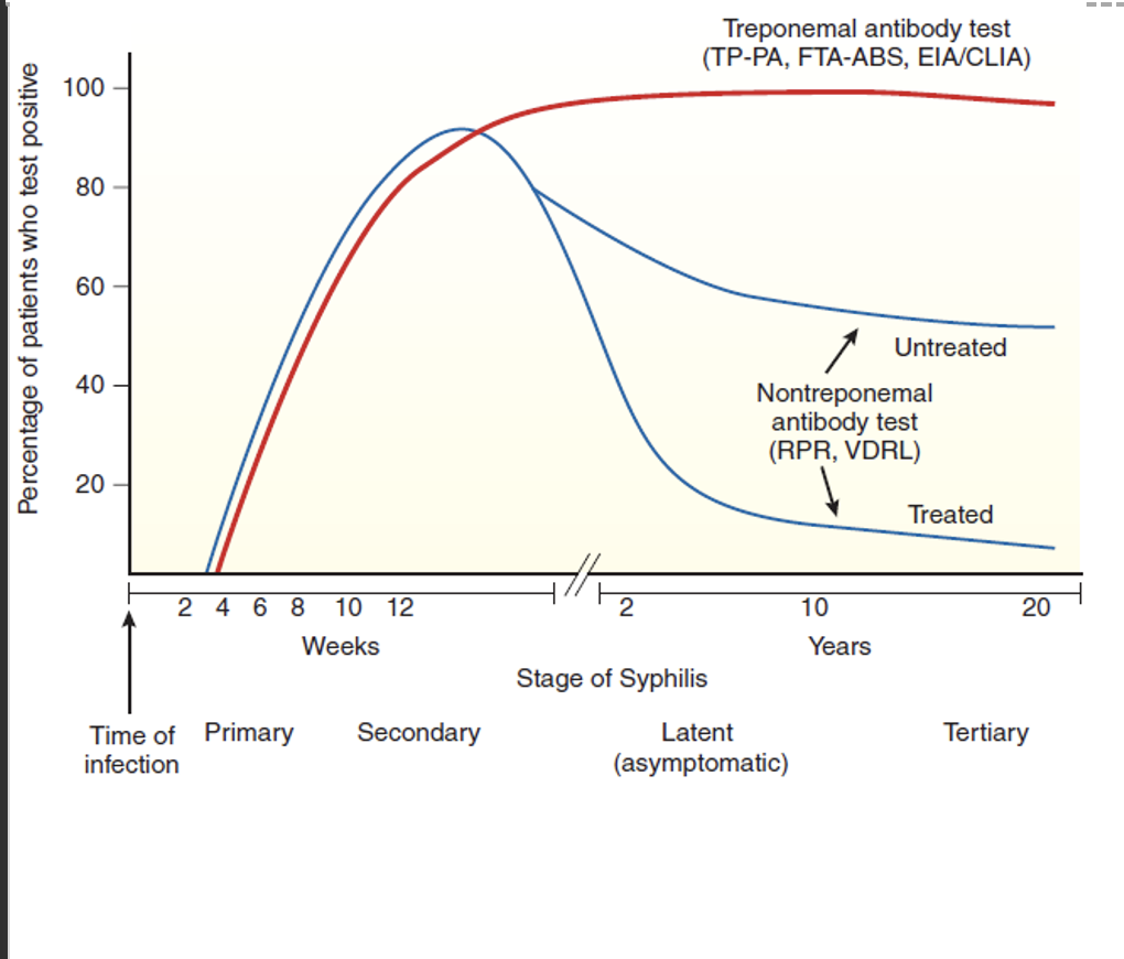
35
Q: Which stage shows the highest serologic response on the graph?
A: Secondary syphilis.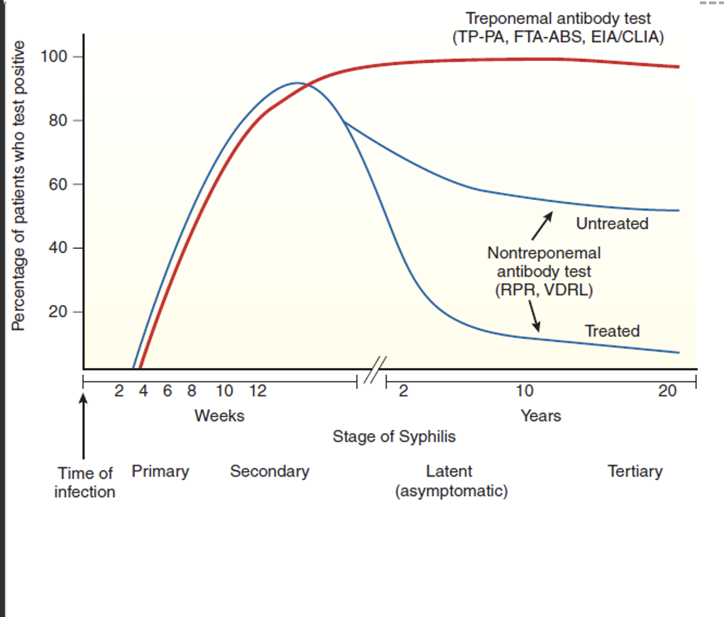
36
Q: What does titer notation such as 1:2, 1:4, 1:8, and 1:16 represent in syphilis testing? (TEST CARD)
A: Serial dilutions of a positive RPR sample.
37
Q: List causes of transient false-positive nontreponemal syphilis test results.
A: Hepatitis, infectious mononucleosis, varicella, herpes, measles, malaria, tuberculosis, and pregnancy.
38
Q: List causes of sustained false-positive nontreponemal syphilis test results.
A: SLE, leprosy, IVDU, autoimmune arthritis, advanced age, and advanced malignancy.
39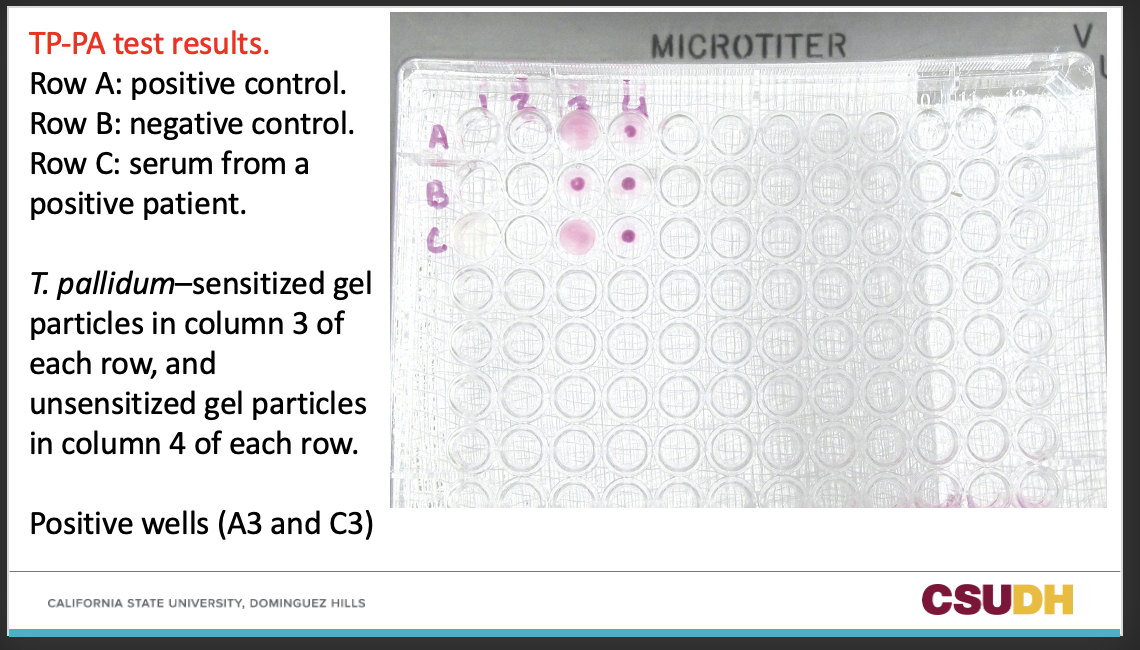
40
Q: What does real-time PCR involve in syphilis diagnosis?
A: Isolating and amplifying a specific sequence of DNA.
41
Q: What specimens can real-time PCR test for treponemes?
A: Whole blood, spinal fluid, amniotic fluid, various tissues, and swab samples from syphilis lesions.
42
Q: How sensitive is real-time PCR for primary syphilis?
A: Extremely sensitive.
43
Q: What is the detection capability of real-time PCR regarding treponemes?
A: Able to detect one treponeme in some clinical samples.
44
Q: In which patients is real-time PCR helpful for detecting treponemes?
A: Neonates with symptoms of congenital syphilis.
45
Q: What clinical fluid is tested using real-time PCR for suspected neurosyphilis?
A: Cerebrospinal fluid (CSF).
46
Q: What is a limitation of real-time PCR in syphilis diagnosis?
A: Low sensitivity in secondary syphilis.
47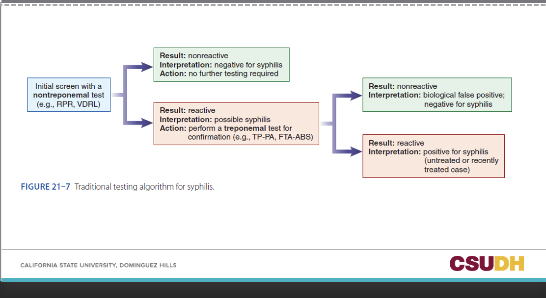
48
Q: When and where was Lyme disease first described in the United States?
A: In 1975 in a geographically clustered rural area around Old Lyme, Connecticut.
49
Q: What unusual event led to the recognition of Lyme disease in 1975?
A: An unusually large number of cases of juvenile arthritis appeared.
50
Q: Who initially brought attention to the early Lyme disease cases?
A: Two mothers recognized this and brought it to the attention of health officials.
51
Q: What suggested that Lyme disease was transmitted by an arthropod vector?
A: The epidemiological features of the newly described “Lyme arthritis.”
52
Q: When was the arthropod vector for Lyme disease identified and what organism was named?
A: In 1982, an arthropod vector was isolated and a new spirochete named Borrelia burgdorferi.
53
Q: Who was Borrelia burgdorferi named after?
A: Willy Burgdorfer (first author in the original description).
54
Q: What systems can Lyme disease affect?
A: It is a multisystem illness involving the skin, nervous system, heart, and joints.
55
Q: What is Lyme disease considered in the United States?
A: The most common vector-borne disease.
56
Q: How many confirmed and probable Lyme disease cases were reported in 2017?
A: More than 42,743.
57
Q: What are the three stages of Lyme disease manifestations?
A: (1) Localized rash, (2) early dissemination to multiple organ systems, and (3) a late disseminated stage that often includes arthritic symptoms.
58
Q: How does Lyme disease resemble syphilis?
A: Manifestations occur in several stages.
59
Q: How is the immune response to Lyme disease described?
A: Highly variable and complex.
60
Q: What types of immune responses occur in Lyme disease?
A: Humoral and cellular responses occur.
61
Q: What triggers production of macrophage-derived cytokines in Lyme disease?
A: Spirochete lipoproteins.
62
Q: Why is the clinical effectiveness of immune responses in Lyme disease considered questionable?
A: Because late Lyme disease occurs despite high levels of circulating antibody and cellular responses.
63
Q: How is Lyme disease primarily diagnosed?
A: Lyme diagnosis is basically clinical; lab results are only supportive.
64
Q: Why is clinical diagnosis of Lyme disease difficult?
A: If rash is present, it could be just presumptive, and 20% of patients may not develop rash or not recognize the rash.
65
Q: When may antibody response become detectable after a tick bite?
A: 3 to 6 weeks.
66
Q: Which antibody response occurs first in Lyme disease?
A: IgM response occurs first, followed by IgG response (but may not be always the case).
67
Q: When may the IgG response peak?
A: The third and fourth weeks of infection.
68
Q: Why is serological testing not helpful in the first 2 weeks of Lyme disease?
A: Serological testing is too insensitive to be diagnostically helpful.
69
Q: In the modified testing algorithm, what is performed first?
A: Perform EIA #1 for B. burgdorferi antibody.
70
Q: What happens if the first EIA is negative?
A: Results indicate negative for Lyme disease (no further testing) or early Lyme disease (may follow up by testing convalescent serum sample after antibiotic treatment).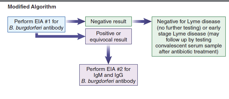
71
Q: What happens if the first EIA result is positive or equivocal?
A: Perform EIA #2 for IgM and IgG B. burgdorferi antibody.
72
Q: When should Lyme testing not be performed?
A: Lyme testing should not be performed in the absence of supporting clinical evidence.
73
Q: What is the predictive value of a positive Lyme test when performed without supporting clinical evidence?
A: It has a low positive predictive value even when done in an endemic area.
74
Q: When does the positive predictive value of Lyme testing rise to nearly 100%?
A: When clinical symptoms and history are present and consistent with Lyme disease.
75
What are the 4 Lyme Testing Procedures?
Immunofluoresence Assay (IFA
Enyzme (EIA)
Western blot
PCR
Enyzme (EIA)
Western blot
PCR
76
Q: What is the first step in performing an Immunofluorescence Assay (IFA) for Lyme testing?
A: Incubate doubling dilutions of patient serum with commercially prepared microscope slides coated with antigen from whole or processed Borrelia spirochetes.
77
Q: What is added after the first wash in the IFA procedure?
A: Anti-human globulin conjugated with a fluorescent tag.
78
Q: What does the fluorescent-tagged anti-human globulin react with?
A: Any specific antibody bound to the spirochetes on the slide.
79
Q: What is done after the second wash in the IFA procedure?
A: The slide is viewed under a fluorescent microscope.
80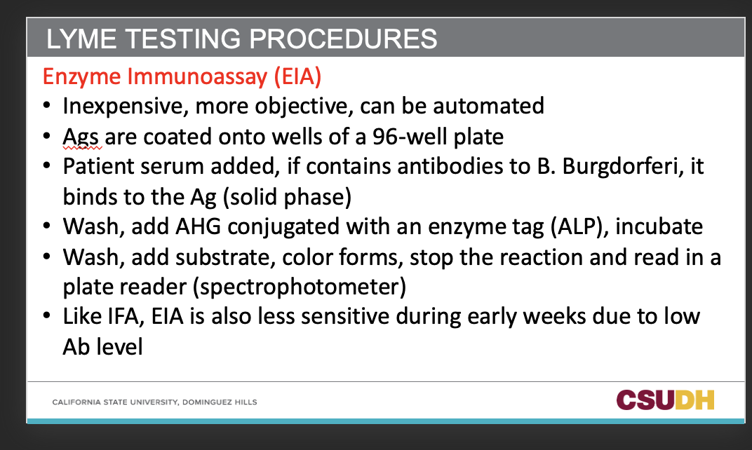
Describe the Enzyme immunoassay (EIA) in lyme testing
81
Q: When is Western blot used as a confirmatory test?
A: For samples that initially test positive or equivocal by EIA or IFA.
82
Q: In which position does Western blot fit in the CDC-recommended testing scheme for Lyme disease?
A: It is employed as the second test.
83
Q: What level of expertise is required for Western blot testing?
A: Very complex, requires expertise in performing and interpretation.
84
Q: What do the nitrocellulose strips used in Western blot contain?
A: Many protein antigens (Ags) of B. burgdorferi.
85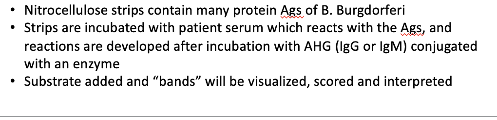
Explain the method of Western Blot in lyme testing
86
Q: How many proteins does the CDC recommend be identified on a Western blot strip for Lyme testing?
A: 10 proteins.
87
Q: What are the molecular weights (MW) of the CDC-recommended Western blot proteins?
A: 18, 23, 28, 30, 39, 41, 45, 58, 66, and 93 kDa.
88
Q: Which bands must be present for a result to be considered positive for specific IgM antibodies?
A: Two of the following: 23 (OSP-C), 39, and 41 (flagellin) kDa.
89
Q: When is an IgG immunoblot considered positive? (in western blot for lyme testing)
A: If any 5 of the 10 bands are positive.
90
Q: Why must only qualified labs interpret Western blot results?
A: Because of complexity in interpretation, and only labs that follow CDC guidelines on WB interpretation can do it.
91
Q: What Lyme testing method is being described?
A: Polymerase Chain Reaction (PCR).
92
Q: What role has PCR found in Lyme diagnosis?
A: It has found a niche in Lyme diagnosis.
93
Q: Why are specimen collection, transport, and preparation of DNA critical in Lyme PCR testing?
A: Because only a few spirochetes are available in samples.
94
Describe the method for PCR in lyme testing.
95
Q: What is the specificity range of PCR in Lyme diagnosis?
A: 93% to 100%.
96
Q: When is PCR on CSF and synovial fluid often used?
A: In difficult diagnostic neurological and arthritic cases.
97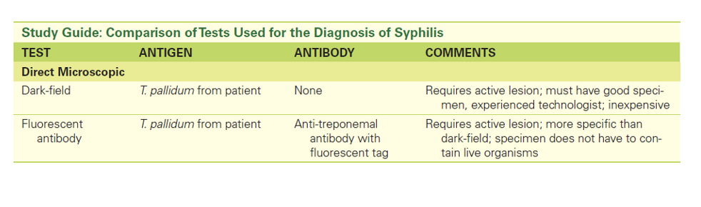
98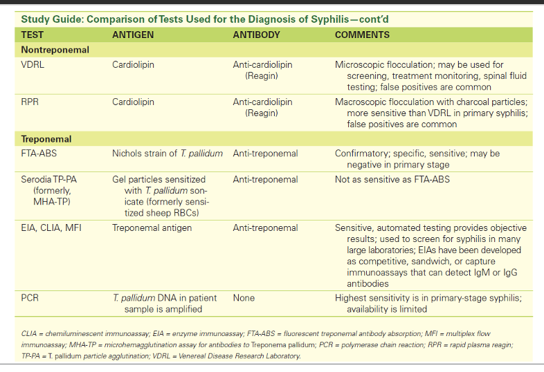
99
Q: What category of tests includes HBs Ag, HBe Ag, HBs Ab, HBc Ab, and HBe Ab?
A: Serologic tests (EIA, CLIA).
100
Q: List the five serologic tests used in Hepatitis B testing.
A: HBs Ag, HBe Ag, HBs Ab, HBc Ab, HBe Ab.
101
Q: What molecular test is used for Hepatitis B diagnosis?
A: HBV DNA real-time PCR.
102
Q: How early can HBV DNA be detected in plasma using PCR?
A: 21 days before HBs Ag.
103
Q: In what situations is molecular testing helpful?
A: Blood donor screening, occupational exposure, therapy monitoring, equivocal HBS Ag test results.
104
Q: What is a hypersensitivity reaction?
A: An exaggerated response to a typically harmless antigen that results in tissue injury, disease, or even death.
105
Q: How many groups are hypersensitivity reactions classified into?
A: Four groups.
106
Q: What is Class I hypersensitivity also called?
A: Anaphylactic hypersensitivity.
107
Q: What is Class II hypersensitivity also called?
A: Antibody-mediated cytotoxic hypersensitivity.
108
Q: What is Class III hypersensitivity also called?
A: Immune complex-mediated hypersensitivity.
109
Q: What is Class IV hypersensitivity also called?
A: Cell-mediated hypersensitivity.
110
Q: Which hypersensitivity classes are antibody-mediated?
A: Classes I, II, and III.
111
Q: What are antibody-mediated hypersensitivity reactions called, and how quickly do they occur?
A: Immediate hypersensitivity reactions; they happen within a few minutes to a few hours after contact with antigen.
112
Q: What type of reaction is Class IV hypersensitivity, and how long does it take?
A: T cell–mediated delayed hypersensitivity reaction; takes 24–48 hours.
113
Q: What is another name for Class I hypersensitivity?
A: Sometimes also called allergies.
114
Q: Give examples of Class I hypersensitivity reactions.
A: Hay fever, hives, allergic asthma, systemic anaphylaxis.
115
Q: What is the major antibody involved in Class I hypersensitivity?
A: IgE (greatly increased in these patients).
116
Q: Who are the main cellular players in Class I hypersensitivity?
A: IgE, basophils, mast cells, eosinophils.
117
Q: What receptor do the main players have in Class I hypersensitivity?
A: They have a receptor for IgE.
118
Q: How soon does Class I hypersensitivity typically occur after antigen exposure?
A: Within 30 to 60 minutes.
119
Q: What are the two phases of Class I hypersensitivity?
A: Sensitization and activation.
120
List common allergens
Pollen
Mold spores
Animal dander
Dust mites
Insect venom
Certain foods (peanuts, shellfish, dairy products)
Certain drugs (penicillin)
Latex
Mold spores
Animal dander
Dust mites
Insect venom
Certain foods (peanuts, shellfish, dairy products)
Certain drugs (penicillin)
Latex
121
Q: What conditions are a clinical manifestation of Type I hypersensitivity?
A:
Rhinitis (“hay fever”)
Allergic asthma
Food allergies
Skin reactions (Urticaria (“hives”), angioedema, eczema)
Wheal and flare
Systemic anaphylaxis
A potentially fatal reaction
IgA-deficient patients
Rhinitis (“hay fever”)
Allergic asthma
Food allergies
Skin reactions (Urticaria (“hives”), angioedema, eczema)
Wheal and flare
Systemic anaphylaxis
A potentially fatal reaction
IgA-deficient patients
122
Q: What chronic airway condition is associated with Type I hypersensitivity?
A: Allergic asthma.
123
Q: What type of skin testing is used for Type I hypersensitivity?
A: Percutaneous or intradermal.
124
Q: What is the first step in the skin testing process for Type I hypersensitivity?
A: Apply a panel of allergens to separate sites on the skin.
125
Q: After applying allergens in a Type I hypersensitivity skin test, how long should you wait?
A: 15 to 20 minutes.
126
Q: What indicates a positive result in Type I hypersensitivity skin testing?
A: Wheal and flare at the site of application.
127
Q: What does RIST stand for?
A: Radioimmunosorbent test.
128
Q: In RIST, what is measured?
A: Total IgE.
129
Q: How is total IgE measured in RIST?
A: By capturing the antibody with an anti-IgE bound to a solid phase.
130
Q: What produces the visible reaction in RIST?
A: A second anti-IgE antibody with an enzyme label.
131
Q: What does RAST stand for?
A: Radioallergosorbent test.
132
Q: In RAST, what is measured?
A: Antigen-specific IgE.
133
Q: How is antigen-specific IgE captured in RAST?
A: Using solid-phase antigen to capture patient antibody.
134
Q: What produces the visible reaction in RAST?
A: A second antibody (enzyme-labeled anti-IgE) combined with substrate.
135
Q: What is another name for Type II hypersensitivity?
A: Antibody-mediated cytotoxic hypersensitivity.
136
Q: What are the key components involved in Type II hypersensitivity?
A: IgG and IgM directed against a cell surface antigen.
137
Q: What are the possible effects of antibodies in Type II hypersensitivity?
Cell destruction
Inhibition of cell function
Increase in cell function
Inhibition of cell function
Increase in cell function
138
Q: What pathway is activated in Type II hypersensitivity that leads to cell lysis?
A: Activation of classical pathway of complement and cell lysis.
139
Q: What process enhances phagocytosis of the cell in Type II hypersensitivity?
A: Opsonization and phagocytosis of the cell.
140
Q: What mechanism involves antibodies directing immune cells to destroy target cells?
A: Antibody-dependent cell-mediated cytotoxicity (ADCC).
141
What are clinical examples of TYPE 2 hypersensitivity?
Transfusion reactions
Hemolytic disease of the newborn (HDN)
Autoimmune hemolytic anemia (Warm reactive antibodies, Cold agglutinins, Paroxysmal cold hemoglobinuria)
Anti-GBM (glomerular basement membrane) disease
Hashimoto’s disease
Hemolytic disease of the newborn (HDN)
Autoimmune hemolytic anemia (Warm reactive antibodies, Cold agglutinins, Paroxysmal cold hemoglobinuria)
Anti-GBM (glomerular basement membrane) disease
Hashimoto’s disease
142
Q: What triggers HDFN?
A: A pregnant woman produces antibodies to Rh antigens (usually RhD) on fetal RBCs.
143
Q: Which Rh antigen is most commonly involved in HDFN?
A: RhD.
144
Q: What should be recalled in relation to HDFN testing?
A: Application of DAT and IAT.
145
Q: What treatment is shown for preventing HDFN?
A: Rh-immune globulin treatment.
146
Q: What is Type III hypersensitivity also called?
A: Immune complex-mediated hypersensitivity.
147
Q: Which antibodies are key components of Type III hypersensitivity?
A: IgG and IgM directed against a soluble antigen.
148
Q: What happens to small antigen–antibody complexes in Type III hypersensitivity?
A: They precipitate out and deposit in tissues.
149
Q: What occurs after complement binds in Type III hypersensitivity?
A: Vasodilation and vasopermeability increase.
150
Q: Which cells migrate to affected areas in Type III hypersensitivity, and what do they do?
A: Macrophages and neutrophils migrate to the affected areas and release lysosomal enzymes, resulting in tissue damage.
151
What are some conditions associated withe Type III hypersensitivity?
Some autoimmune diseases (e.g., systemic lupus erythematosus and rheumatoid arthritis)
Reactions to bee stings
Drug reactions (e.g., penicillin)
Sequelae to infections (e.g., post-streptococcal glomerulonephritis)
Reactions to bee stings
Drug reactions (e.g., penicillin)
Sequelae to infections (e.g., post-streptococcal glomerulonephritis)
152
What are lab testing for type III hypersensitivity?
Testing for ANAs
Fluorescent staining of tissue sections to detect deposited immune complexes
Testing for rheumatoid factor (an anti-IgG)
Testing complement levels (May be decreased in the serum during periods of high disease activity.)
Fluorescent staining of tissue sections to detect deposited immune complexes
Testing for rheumatoid factor (an anti-IgG)
Testing complement levels (May be decreased in the serum during periods of high disease activity.)
153
Q: What type of reaction is Type IV hypersensitivity?
A: Cell-mediated hypersensitivity.
154
Q: Which cells are involved in Type IV hypersensitivity?
A: Th1 cells and macrophages.
155
Q: What do antigen-presenting cells (APCs) do in Type IV hypersensitivity?
A: APCs present antigen to naïve T helper cells, which differentiate into Th1 cells.
156
Q: What do Th1 cells release and what is the effect?
A: Th1 cells release cytokines that attract and activate macrophages.
157
Q: What do macrophages do in Type IV hypersensitivity?
A: Macrophages induce inflammation.
158
Q: What is the role of cytotoxic T lymphocytes in Type IV hypersensitivity?
A: They are recruited and destroy target cells.
159
Q: When does Type IV hypersensitivity peak after antigen exposure?
A: 48 to 72 hours.
160
what are clinical examples of type IV hypersensitivity?
Infections with intracellular pathogens
(Mycobacterium tuberculosis, Mycobacterium leprae, Pneumocystis carinii, Leishmania species, herpes simplex virus)
Contact dermatitis
Hypersensitivity pneumonitis
(Allergic diseases of the lungs,
Caused by inhalation of bacterial and fungal spores
Examples: Farmer’s lung disease, bird breeder’s lung disease)
(Mycobacterium tuberculosis, Mycobacterium leprae, Pneumocystis carinii, Leishmania species, herpes simplex virus)
Contact dermatitis
Hypersensitivity pneumonitis
(Allergic diseases of the lungs,
Caused by inhalation of bacterial and fungal spores
Examples: Farmer’s lung disease, bird breeder’s lung disease)
161
Q: What type of compounds trigger contact dermatitis?
A: Low-molecular-weight compounds.
162
Q: How do low-molecular-weight compounds trigger the immune response in contact dermatitis?
A: They contact the skin and act as haptens to sensitize Th1 cells.
163
Q: What are examples of substances that can cause contact dermatitis?
A: Poison ivy, poison oak, nickel salts, materials in cosmetics and hair dyes, and latex.
164
Q: What skin symptoms are associated with contact dermatitis?
A: Erythema, swelling, and papules.
165
Front: What is the Patch Test used for?
Back: It is used to test for contact dermatitis.
166
Front: How is the Patch Test performed?
Back: Antigen is applied to the skin surface.
167
Front: What indicates a positive Patch Test?
Back: Redness with papules or tiny blisters.
168
Front: What is the purpose of skin testing for immunodeficiency?
Back: To inject common antigens intradermally to evaluate immune response.
169
Front: What is the Mantoux method used for?
Back: Testing for tuberculosis exposure (PPD) or T-cell function.
170
Front: How is the antigen administered in the Mantoux method?
Back: Injected intradermally.
171
Front: What indicates a positive Mantoux test?
Back: Induration.
172
Front: What do Interferon Gamma Release Assays (IGRA) measure?
Back: Production of IFN-γ by patient T cells stimulated with MTB antigens.
173
Front: What is the Quantiferon TB Gold Plus assay?
Back: A test where patient blood is incubated in special tubes with MTB antigens, and plasma is tested for IFN-γ by ELISA.
174
Front: How is IFN-γ detected in the Quantiferon TB Gold Plus test?
Back: By ELISA.
175
Front: What is the T-SPOT TB test?
Back: A test where patient mononuclear cells are incubated with MTB antigens and tested for IFN-γ by ELISpot.
176
Front: How is IFN-γ detected in the T-SPOT TB test?
Back: By ELISpot.