Lecture 2_Immunodeficiency_pt2: disease & lab diagnosis
Study these flashcardsFree · spaced repetition · iPhone, Android, Mac & web
All cards
1
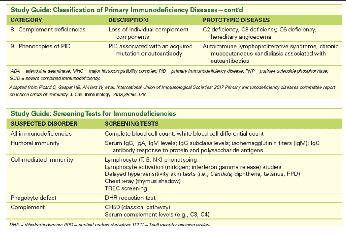
Q: What are the nine categories of primary immunodeficiencies (PIDs)?
1. Combined Immunodeficiencies
2. Combined Immunodeficiencies with Associated or Syndromic Features
3. Predominantly Antibody Deficiencies
4. Diseases of Immune Dysregulation
5. Congenital Defects of Phagocyte Number, Function, or Both
6. Defects in Innate Immunity
7. Autoinflammatory Disorders
8. Complement Deficiencies
9. Phenocopies of Primary Immunodeficiencies
2. Combined Immunodeficiencies with Associated or Syndromic Features
3. Predominantly Antibody Deficiencies
4. Diseases of Immune Dysregulation
5. Congenital Defects of Phagocyte Number, Function, or Both
6. Defects in Innate Immunity
7. Autoinflammatory Disorders
8. Complement Deficiencies
9. Phenocopies of Primary Immunodeficiencies
2
Q: What is Purine-Nucleoside Phosphorylase (PNP) deficiency?
A: A rare autosomal recessive trait affecting an enzyme involved in purine metabolism.
3
Q: When does PNP deficiency usually present?
A: In infancy, with symptoms such as pulmonary infections and urinary tract infections (UTI).
4
Q: What type of immune defect occurs in PNP deficiency?
A: Moderate to severe defect in cell-mediated immunity (CMI), with normal or only mildly impaired humoral immunity (HMI).
5
Q: What causes the progressive decline in T cell number in PNP deficiency?
A: Accumulation of toxic dGTP.
6
Q: What condition can PNP deficiency be confused with?
A: Neonatal HIV — it must be differentiated using specific tests.
7
Q: What are combined immunodeficiencies with associated or syndromic features?
A: They are immunodeficiencies that have nonimmunologic features in addition to the combined immunodeficiency.
8
Q: What part of the immune system is affected in Category 2 PIDs?
A: They affect cell-mediated immunity (CMI) involving T cells, and indirectly other branches of the immune response.
9
Q: What is a complication associated with Category 2 PIDs?
A: Increased risk of graft-versus-host disease (GVHD).
10
Q: What conditions are included in Category 2 PIDs?
Wiskott-Aldrich syndrome (WAS)
22q11.2 Deletion Syndrome (DiGeorge Syndrome)
Ataxia-Telangiectasia (AT)
22q11.2 Deletion Syndrome (DiGeorge Syndrome)
Ataxia-Telangiectasia (AT)
11
Q: What is Wiskott-Aldrich Syndrome (WAS)?
A: A rare X-linked recessive syndrome defined by the triad of immunodeficiency, eczema, and thrombocytopenia.
12
Q: Why is Wiskott-Aldrich Syndrome usually fatal in childhood?
A: Because of infection, hemorrhage, or malignancy.
13
Q: What are the key lab findings in Wiskott-Aldrich Syndrome?
Thrombocytopenia
Prolonged bleeding time (BT, PFA-100)
Very low isohemagglutinins (especially IgM, diagnostic)
Increased serum AFT (diagnostic)
Shortened half-life of platelets (PLTs)
T lymphocytes affected, though B lymphocytes appear to function normally
Prolonged bleeding time (BT, PFA-100)
Very low isohemagglutinins (especially IgM, diagnostic)
Increased serum AFT (diagnostic)
Shortened half-life of platelets (PLTs)
T lymphocytes affected, though B lymphocytes appear to function normally
14
Q: What gene is mutated in Wiskott-Aldrich Syndrome?
A: The WAS gene, which encodes WASp, required for actin polymerization in hematopoietic cells.
15
Q: What is the molecular outcome of the WAS gene mutation?
A: Defective signal transduction in lymphocytes and platelets.
16
Q: What abnormal membrane protein is associated with Wiskott-Aldrich Syndrome?
A: CD43, which becomes abnormal due to the mutated WAS gene on the X chromosome.
17
Q: What are the key features, lab findings, and molecular defects of Wiskott-Aldrich Syndrome (WAS)?
Thrombocytopenia → increased bleeding time
Small platelets
Eczema
Infections (e.g., pneumonia and others)
Autoimmunity and/or malignancies (e.g., B-cell lymphoma)
Small platelets
Eczema
Infections (e.g., pneumonia and others)
Autoimmunity and/or malignancies (e.g., B-cell lymphoma)
18
Q: What type of inheritance pattern does 22q11.2 Deletion Syndrome follow?
A: Autosomal dominant.
19
Q: What chromosome is affected in 22q11.2 Deletion Syndrome?
A: Deletion of q11.2 on chromosome 22.
20
Q: What do the deleted genes in 22q11.2 Deletion Syndrome affect?
A: Thymus development in the embryo.
21
Q: What happens if the 22q11.2 deletion is partial?
A: The person may have near normal immune function.
22
Q: What percentage of children with 22q11.2 Deletion Syndrome have severe T cell deficiency?
A: Around 20% of children.
23
Q: What types of infections are common in 22q11.2 Deletion Syndrome?
A: Severe, recurrent viral and fungal infections.
24
Q: What causes tetany in 22q11.2 Deletion Syndrome?
A: Hypocalcemia resulting from hypoparathyroidism.
25
Q: What type of inheritance does Ataxia-Telangiectasia (AT) have?
A: It is a complex, autosomal recessive syndrome.
26
Q: What are the main characteristics of Ataxia-Telangiectasia?
A: Cerebellar ataxia (involuntary muscle movements) and telangiectasias (capillary swelling causing red blotches on the skin), especially on the earlobes and conjunctiva.
27
Q: What lab finding is increased in Ataxia-Telangiectasia?
A: Serum alpha-fetoprotein (AFP) level.
28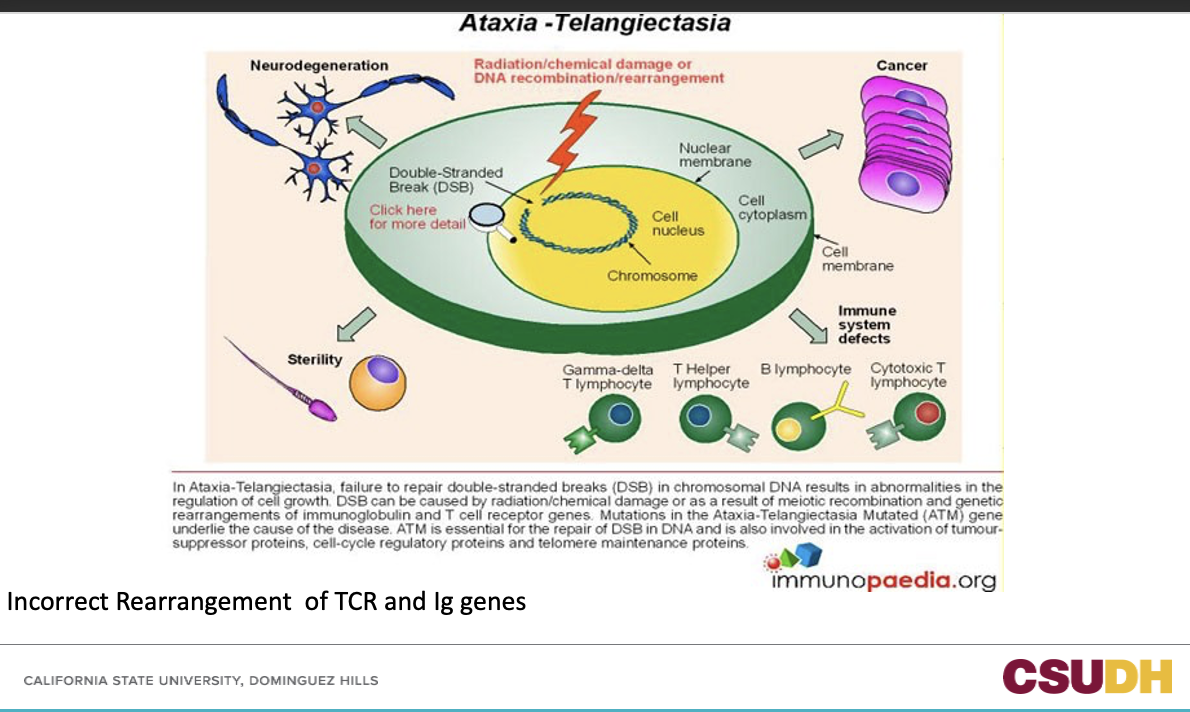
29
Q: What does “ataxia” mean?
A: Loss of motor control.
30
Q: What does “telangiectasia” mean?
A: Spider veins.
31
Q: What causes Ataxia Telangiectasia?
A: It is a neurodegenerative, inherited disease caused by a mutation in the ATM gene.
32
Q: What are three effects of the ATM gene mutation in Ataxia Telangiectasia?
Causes tumor formation
Impairs the cerebellum
Prevents repair of broken DNA
Impairs the cerebellum
Prevents repair of broken DNA
33
Q: What are some symptoms of Ataxia Telangiectasia?
Nystagmus (rapid involuntary eye movement)
Ocular telangiectasia
Ocular telangiectasia
34
Q: What is Category 4 of Primary Immunodeficiencies?
A: Diseases of Immune Dysregulation.
35
Q: What is the main characteristic of diseases in Category 4?
A: They have a normal number of T or B cells but reduced control over their functions.
36
Q: What common feature do many Category 4 diseases share?
A: Autoimmunity.
37
Q: What causes Autoimmune Lymphoproliferative Syndrome (ALPS)?
A: Mutations in genes controlling apoptosis in the thymus, leading to autoreactive cells in circulation.
38
Q: What happens in CD25 deficiency or FOXP3 gene mutation?
A: There is a lack of T regulatory (Treg) cells, causing lymphoproliferation and autoimmunity.
39
Q: What causes Chediak-Higashi Syndrome?
A mutation in the LYST gene, leading to:
- Reduced number of NK cells and neutrophils
- Increased production of inflammatory proteins
- Reduced number of NK cells and neutrophils
- Increased production of inflammatory proteins
40
Q: What category does Chronic Granulomatous Disease (CGD) fall under?
A: Category 5: Congenital Defects of Phagocyte Number, Function, or Both.
41
Q: What causes Chronic Granulomatous Disease (CGD)?
A: It is a group of X-linked recessive or autosomal recessive disorders involving an abnormality in neutrophil microbicidal function.
42
Q: What happens to phagocytic cells in CGD?
A: Phagocytic cells ingest but do not kill bacteria due to failure to form oxygen radicals (no NADPH oxidase activity).
43
Q: What percentage of CGD cases are X-linked?
A: 70%, and these are more severe.
44
Q: What are the symptoms of Chronic Granulomatous Disease?
A: Recurrent suppurative infections, pneumonia, osteomyelitis, draining adenopathy, liver abscesses, dermatitis, and hypergammaglobulinemia.
45
Q: What organisms commonly cause infections in CGD?
A: Catalase-positive organisms like Staphylococcus aureus and fungi such as Aspergillus and Nocardia.
46
Q: When do infections usually begin in CGD, and what is the prognosis?
A: Before 1 year of age, and the syndrome is often fatal in childhood.
47
Q: What causes Chronic Granulomatous Disease (CGD)?
A: Mutations in NADPH oxidase components responsible for generating the superoxide anion.
48
Q: What proteins make up the cytochrome b558 complex in the phagocyte membrane?
A: p22^phox and p91^phox.
49
Q: Which cytosolic proteins associate with cytochrome b558 to form the active NADPH oxidase complex?
A: p47^phox, p67^phox, p40^phox, and the small GTP-binding protein rac2.
50
Q: What is the function of the NADPH oxidase complex?
A: It forms the active oxidase that generates superoxide anions to kill pathogens.
51
Q: What is the most common form of CGD?
A: The X-linked form, caused by mutations in the gp91^phox gene.
52
Q: What causes the autosomal recessive forms of CGD?
A: Mutations in genes encoding other components of the NADPH oxidase complex.
53
Q: Which five components of NADPH oxidase can be mutated in CGD?
p22^phox (chromosome 16q24)
p47^phox (chromosome 7q11.23)
p67^phox (chromosome 1q25)
p40^phox (chromosome 22q13.1)
gp91^phox (chromosome Xp21.1)
p47^phox (chromosome 7q11.23)
p67^phox (chromosome 1q25)
p40^phox (chromosome 22q13.1)
gp91^phox (chromosome Xp21.1)
54
Q: What statement summarizes the genetic cause of CGD?
A: Mutations in any of the five components of NADPH oxidase can cause Chronic Granulomatous Disease.
55
Q: What test is used for diagnosing Chronic Granulomatous Disease (CGD)?
A: Flow cytometry using dihydrorhodamine (DHR).
56
Q: When does DHR fluoresce in the CGD test?
A: DHR fluoresces when it is reduced.
57
Q: How are neutrophils activated in the DHR test?
A: Using phorbol myristate acetate (PMA), which is mitogenic for neutrophils.
58
Q: What happens to DHR during a normal oxidative burst?
A: It is reduced, producing fluorescence that can be measured on a flow cytometer.
59
Q: How do neutrophils from CGD patients differ in the DHR test?
A: They cannot undergo the oxidative burst and show less fluorescence than normal neutrophils.
60
Q: What test is used to measure neutrophil function in CGD?
A: Nitroblue Tetrazolium (NBT) dye test.
61
Q: What does the NBT test measure?
A: The ability of a patient’s neutrophils to reduce the yellow dye nitroblue tetrazolium (NBT) to a deep blue product.
62
Q: What is the deep blue product formed in the NBT test called?
A: Formazan.
63
Q: Where does formazan precipitate, and how is it detected?
A: It precipitates in neutrophils and can be detected microscopically in a PBS.
64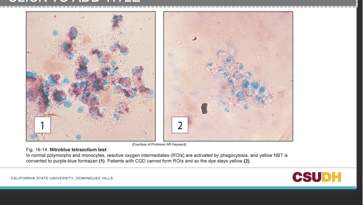
65
Q: What is missing in Leukocyte Adhesion Deficiency (LAD)?
A: The β-subunit (CD18) on neutrophils, which is part of three cell surface integrin complexes.
66
Q: What gene and chromosome are affected in LAD?
A: The ITGB2 gene on chromosome 21q22.3, which encodes CD18, the common β₂ subunit of β₂ integrins.
67
Q: What is the result of the CD18 defect in LAD?
A: Ineffective adhesion due to defective β₂ integrins.
68
Q: Which integrin complexes involve CD18?
A: CD11a/CD18, CD11b/CD18, and CD11c/CD18.
69
Q: What is the functional defect in neutrophils with LAD?
A: Neutrophils cannot migrate toward inflammatory stimuli or adhere to vascular endothelium.
70
Q: What clinical findings suggest Leukocyte Adhesion Deficiency?
Recurrent soft tissue and periodontal infections
Delayed wound healing
Delayed umbilical cord separation
No pus formation despite high white blood cell counts
Delayed wound healing
Delayed umbilical cord separation
No pus formation despite high white blood cell counts
71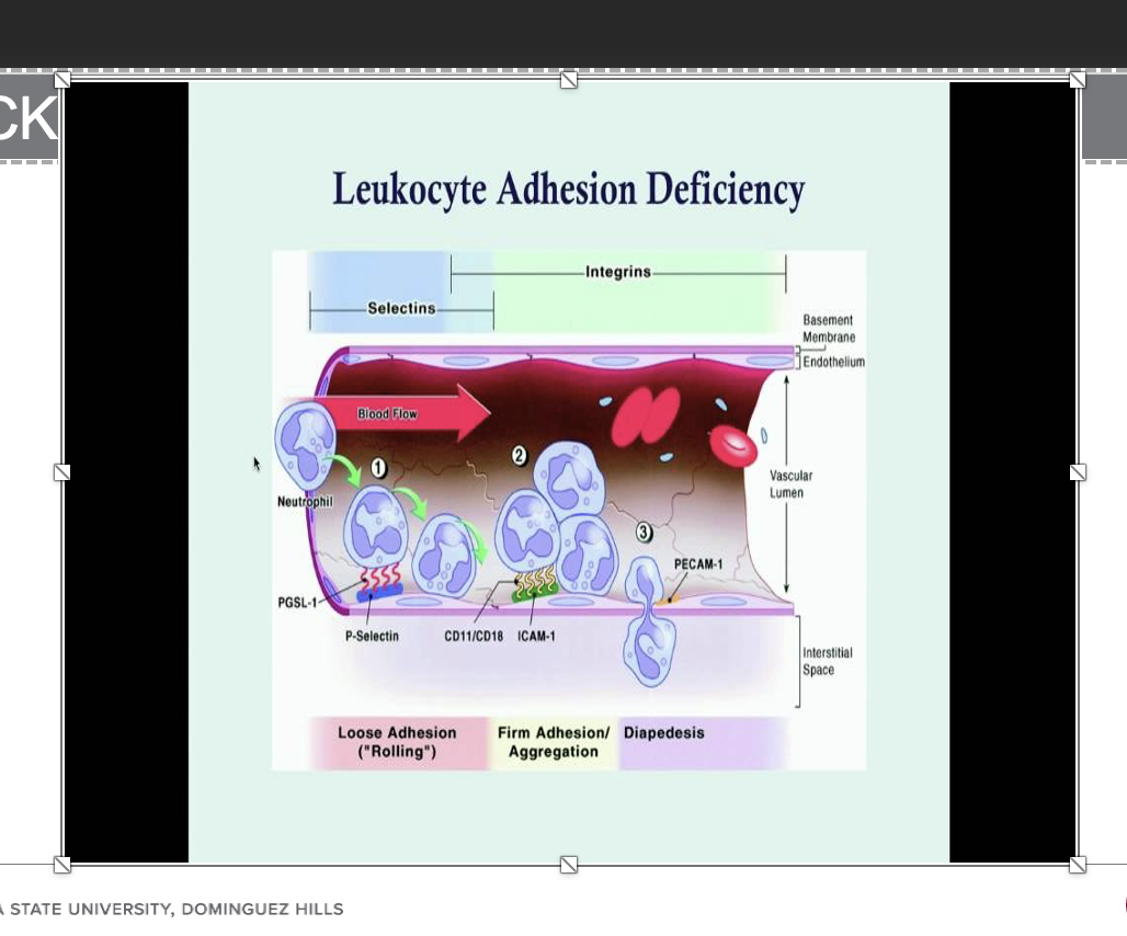
72
Q: What is Category 8 of Primary Immunodeficiencies?
A: Complement Deficiencies.
73
Q: What are early complement component deficiencies (especially C2 and C4) associated with?
A: Recurrent pyogenic infections and connective tissue disease.
74
Q: Deficiency of which complement components is associated with recurrent Neisseria species infections?
A: Components 5 through 8.
75
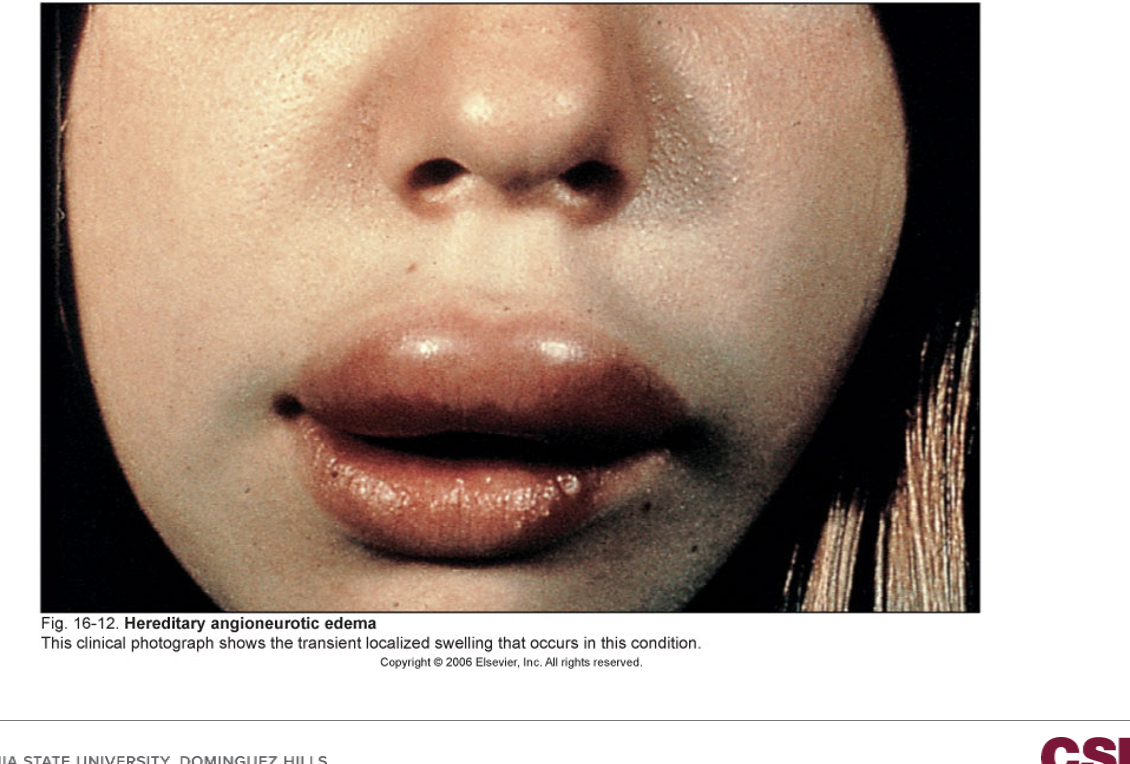
Q: What condition is caused by deficiency of the complement regulatory protein C1 esterase inhibitor?
A: Angioedema (hereditary angioedema).
76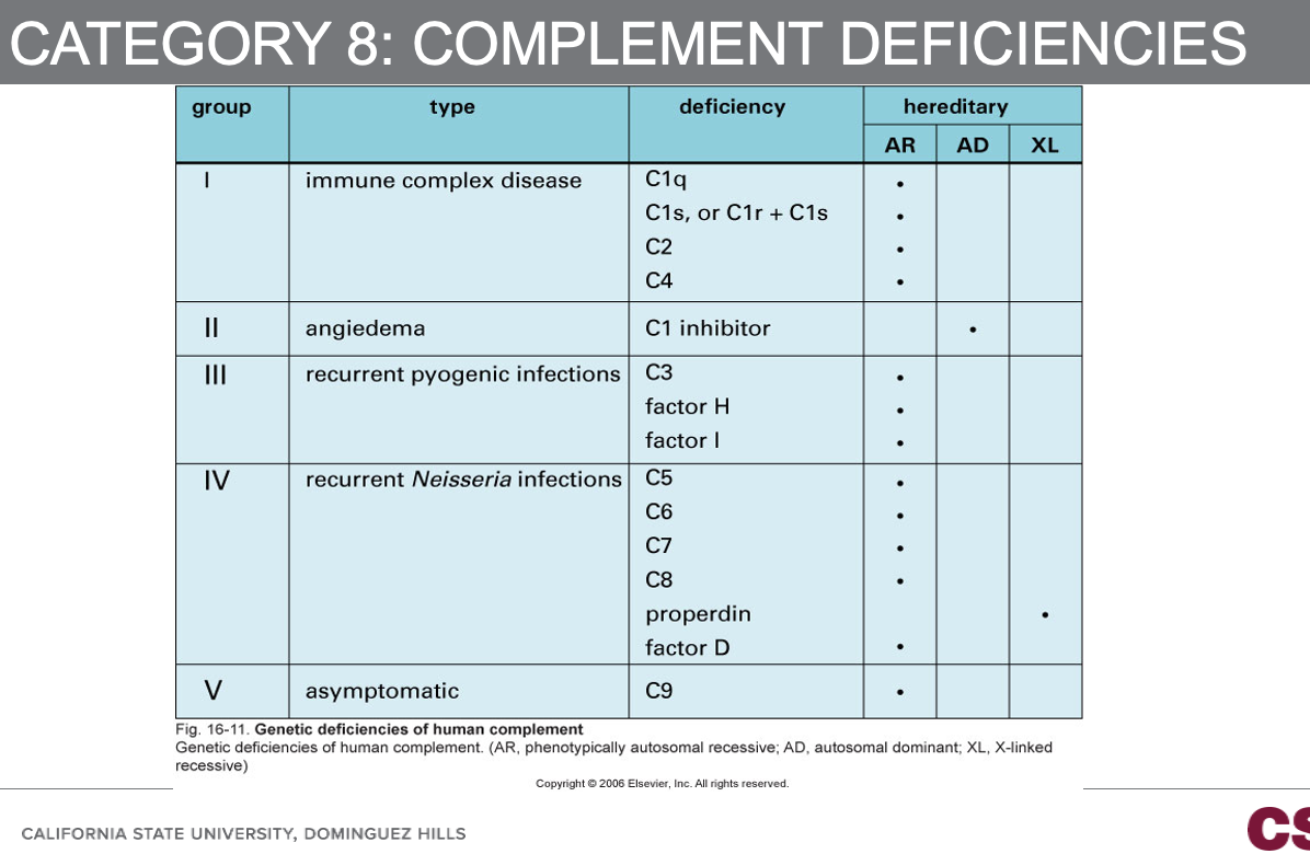
77
Q: When performing diagnostic testing for immunodeficiency, what must laboratorians do?
A: Compare the patient’s results with appropriate age-matched controls.
78
Q: In tests of cellular function, how should a patient’s cells be tested?
A: In parallel with cells from a normal control.
79
Q: What should be done if an abnormal test result is obtained during immune testing?
A: It should be confirmed by repeat testing.
80
Q: What is the purpose of screening tests in immunodeficiency evaluation?
A: They are used for the initial evaluation of a suspected immunodeficiency state.
81
Q: Where can most immunodeficiency screening tests be performed?
A: Routinely in any hospital laboratory.
82
Q: What steps are involved in initial immunodeficiency testing?
Patient history
Complete blood count (CBC)
White blood cell (WBC) differential
Complete blood count (CBC)
White blood cell (WBC) differential
83
Q: What might a CBC and WBC differential reveal in immunodeficiency testing?
A: A reduced lymphocyte count.
84
Q: What condition is associated with thrombocytopenia and small platelets?
A: Wiskott-Aldrich Syndrome (WAS).
85
Q: What measurements are used to screen for defects in antibody production?
A: Serum IgG, IgM, and IgA concentrations and levels of the IgG subclasses.
86
Q: What is tested when screening for isohemagglutinins?
A: The presence of naturally occurring IgM antibodies against ABO blood group antigens.
87
Q: By what age should a child have naturally occurring IgM antibodies against ABO antigens?
A: By the age of 2 years.
88
Q: What does the absence of isohemagglutinins indicate?
A: An abnormal IgM response.
89
Q: How can overall assessment of antibody-mediated immunity be made?
A: By measuring antibody responses to antigens to which the population is exposed normally or following vaccination.
90
Q: Give an example of a test used to assess antibody-mediated immunity.
A: Measuring the titer of specific antibodies produced in response to immunization with vaccines such as diphtheria or tetanus.
91
Q: When is antibody development checked in an unimmunized child after vaccination?
A: Two weeks after immunization.
92
Q: What condition can this testing technique help evaluate?
A: A possible IgG subclass deficiency.
93
Q: Which IgG isotypes respond to protein antigens?
A: IgG1 and IgG3, such as those found in tetanus and diphtheria vaccines.
94
Q: Which IgG isotype responds to polysaccharide antigens?
A: IgG2, such as those in H. influenzae and S. pneumoniae vaccines.
95
Q: What test is usually used to screen for complement deficiencies?
A: The CH50 assay, which determines the functional complement level in an individual.
96
Q: What can undetectable CH50 levels indicate?
A: A deficiency in a specific component of the classical complement pathway, or consumption of complement components.
97
Q: How is the CH50 assay performed?
Serum is serially diluted and added to sensitized sheep red blood cells.
Tubes are incubated at 37°C and then centrifuged to pellet unlysed cells.
Tubes are incubated at 37°C and then centrifuged to pellet unlysed cells.
98
Q: How is CH50 defined?
A: As the reciprocal of the dilution that causes lysis of 50% of the red blood cells used in the assay.
99
Front: What is the best confirmatory test for immune dysfunction?
Back: Flow cytometry.
100
Front: Which immune cell types are assessed using flow cytometry?
Back:
B cells (CD19+)
T cells (CD3+)
T helper (Th) cells (CD3+/CD4+)
Cytotoxic T cells (CD3+/CD8+)
NK cells (CD16+ or CD56+)
B cells (CD19+)
T cells (CD3+)
T helper (Th) cells (CD3+/CD4+)
Cytotoxic T cells (CD3+/CD8+)
NK cells (CD16+ or CD56+)
101
Front: What condition is suggested by an absence or profound decrease of CD3+ cells?
Back: DiGeorge syndrome.
102
Front: What does an absence of CD19+ B cells suggest?
Back: Btk deficiency.
103
Front: What do molecular tests detect in immune dysfunction evaluation?
Back: Mutations.
104
Front: Which mutations may be detected by molecular tests in immune dysfunction assessment?
Back:
22q11.2 deletion syndrome
Wiskott-Aldrich syndrome
SCID
22q11.2 deletion syndrome
Wiskott-Aldrich syndrome
SCID
105
Front: How can T-cell function be measured?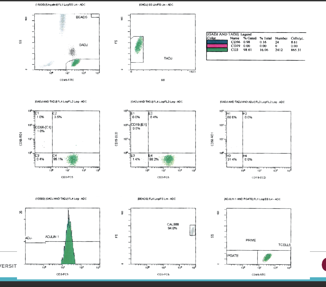
Back: By assessing the ability of isolated T cells to proliferate in response to an antigenic stimulus or to T-cell mitogens in culture.
106
Front: What are examples of T-cell mitogens used in culture?
Back: Phytohemagglutinin (PHA) and Concanavalin A (Con A).
107
Front: What is a mitogen?
Back: A substance that stimulates mitosis in all T cells or all B cells, regardless of antigen specificity
108
Front: What do recent assays such as QuantiFERON-TB and T-SPOT measure?
Back: An individual’s response to Mycobacterium tuberculosis antigens.
109
Front: What happens during the QuantiFERON-TB or T-SPOT assay after overnight incubation with TB antigens?
Back: IFNγ secreted by activated Th1 cells is quantitated.
110
Front: Which methods are used to quantify IFNγ in recent assays?
Back: Enzyme-linked immunosorbent assay (ELISA) or ELISPOT procedure.
111
Front: What do all U.S. states include in newborn screening programs?
Back: PID testing.
112
Front: What do newborn screening tests for immunodeficiencies look for?
Back: The presence of TCR excision circles (TRECs) in newborn blood.
113
Front: How are TRECs identified?
Back: By quantitative PCR.
114
Front: In which cells are TRECs present?
Back: In T cells that have undergone alpha-beta receptor gene rearrangements.
115
Front: What are TRECs?
Back: Genetic material removed from the germline DNA during alpha V-J and beta V-D-J recombination.
116
Front: What does the absence of TRECs indicate?
Back: A lack of functional T cells and early identification of T-cell–related defects leading to SCID.
117
Front: What additional sequencing methods are being explored for newborn screening?
Back: Whole exome sequencing (WES) and whole genome sequencing (WGS).
118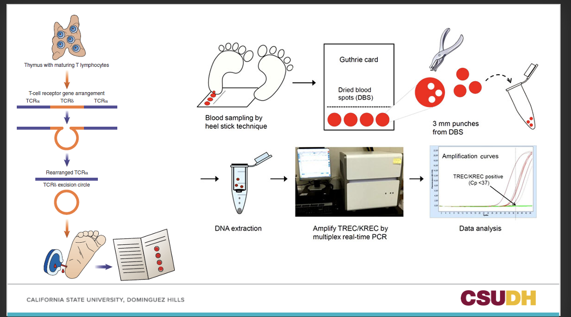
119
Front: How is evaluation of immunoglobulins usually performed?
Back: By automated assays involving nephelometry or immunoturbidimetry.
120
Front: What is an alternative method for evaluating immunoglobulins aside from automated assays?
Back: Radial immunodiffusion (RID).
121
Front: When can serum protein electrophoresis (SPE) be quantitative?
Back: When the total serum protein is determined and the results are read using a densitometer.
122
Front: Which method of electrophoresis is more sensitive?
Back: Capillary electrophoresis.
123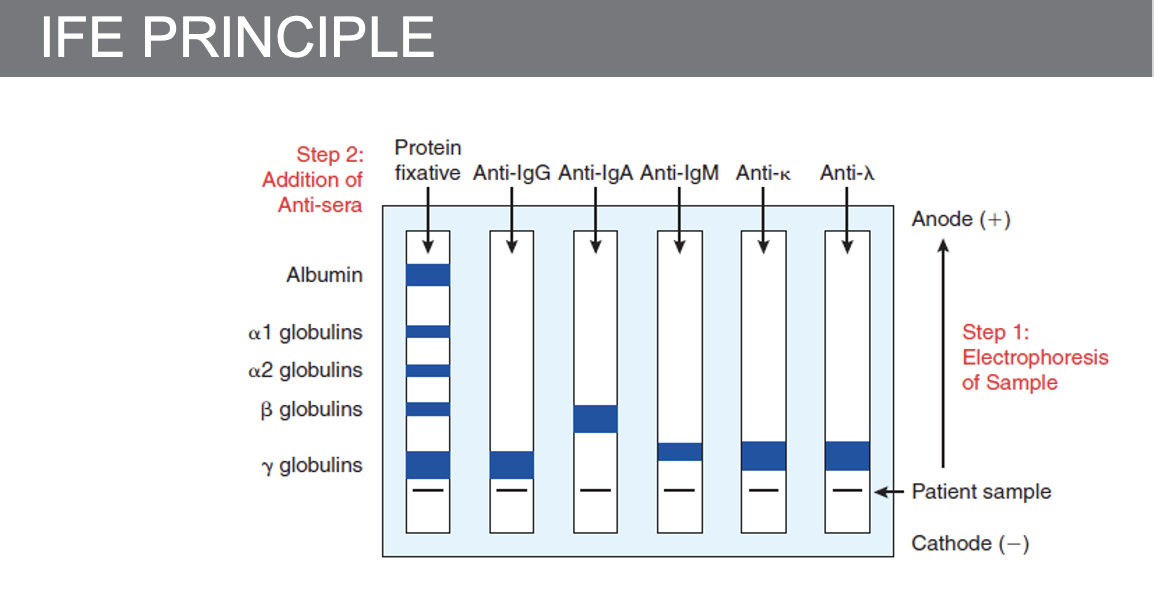
124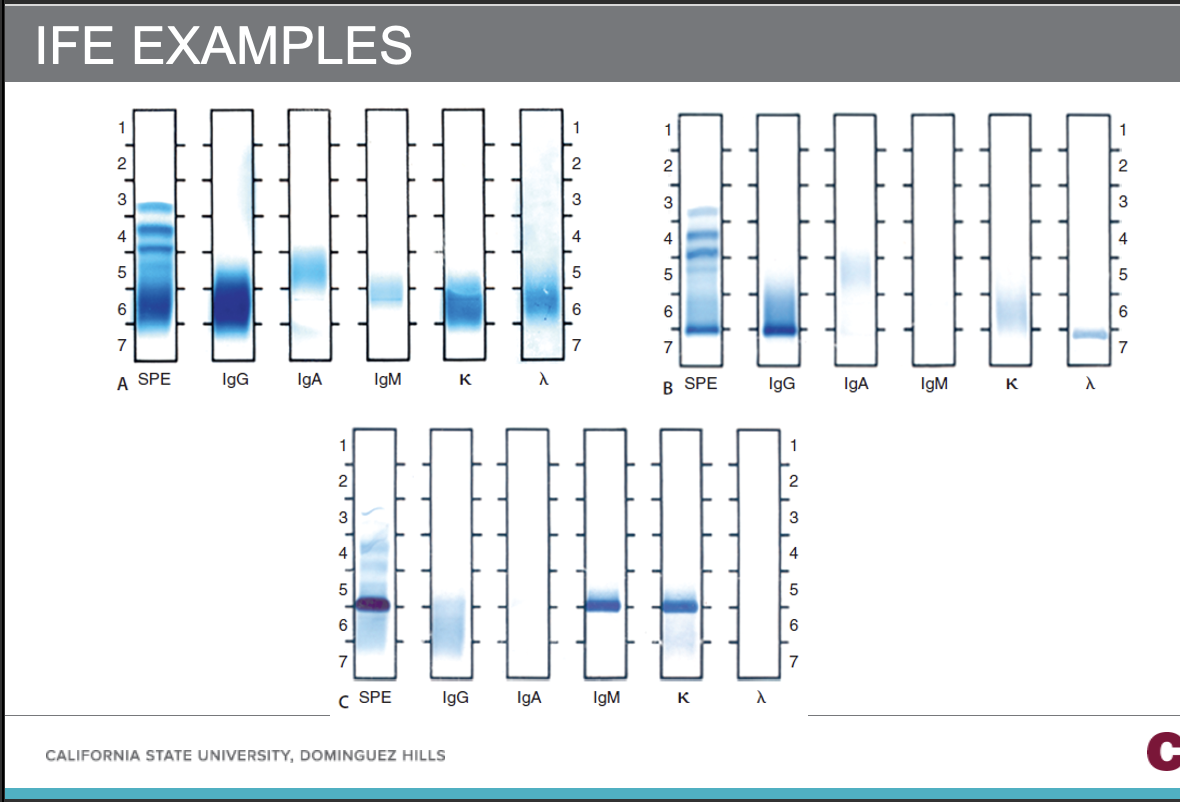
125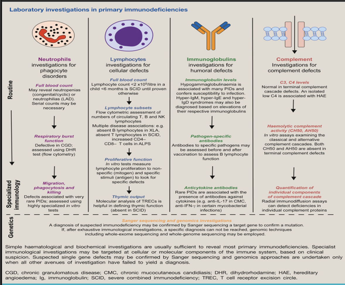
126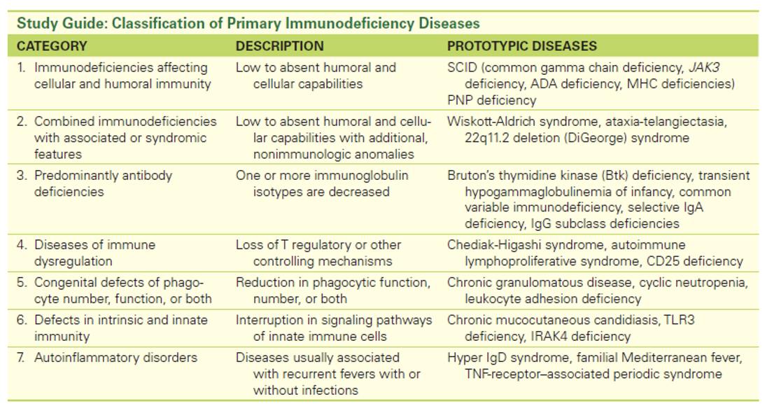
127