Lecture 4 - Subcutaneous Mycoses
Study these flashcardsFree · spaced repetition · iPhone, Android, Mac & web
All cards
1
What do subcutaneous mycoses involve?
muscle, lymphatic vessels, and connective/neighboring (contiguous) tissue
2
Do subcutaneous mycoses spread to other organs?
NO! they do not disseminate in blood
3
What are common characteristics of subcutaneous mycoses?
Progressive, non-healing ulcers
Presence of draining sinus tracts
Presence of draining sinus tracts
4
Which is the subcutaneous layer?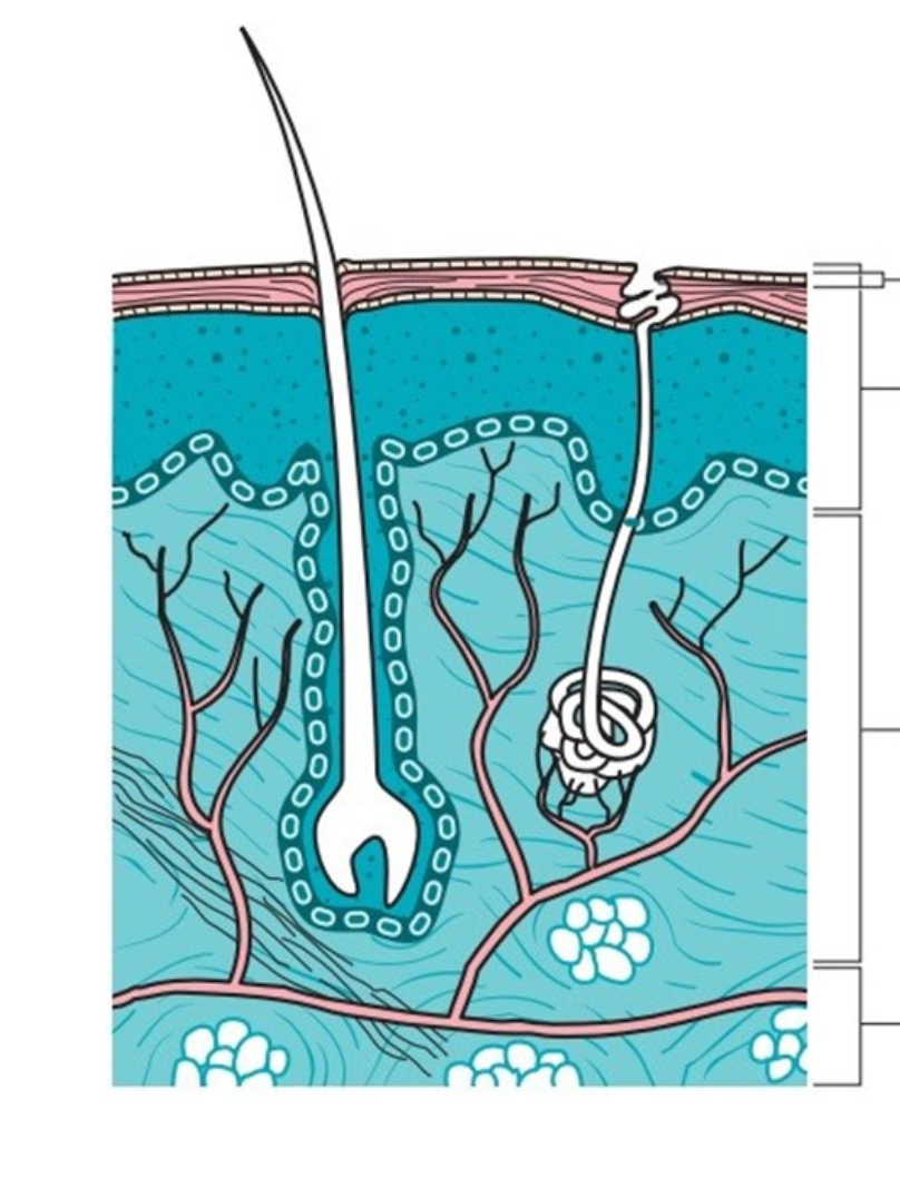
bottom
5
What is a mycetoma?
Chronic skin/soft tissue infection
6
What does mycetoma stand for? (another name for it)
Madura foot
7
What is the clinical presentation of a mycetoma? (what does is look like/features?)
Mass (where inoculation happens), sinus formation, and discahrge with grains (white and black)
8
What happens if a mycetoma is not treated?
Results in disability and deformity.
9
What is the treatment for Mycetoma?
Antifungals, but often unable to eradicate fungus; need to be given for long periods (sometimes years) and are expensive.
Amputation and recurrences are common
Amputation and recurrences are common
10
What body part usually contracts mycetoma?
Foot (sometimes hand - second common), but can affect any area of body.
11
What is the common means of infection in mycetoma?
Thorn prick, other lesions of the skin (mild trauma) and exposure to contaminated environemtn
12
According to WHO, which countries are affects and what is is considered?
Considered a tropical disease and poor countries are affected. Especially children in remote, rural areas; farm workers/herdsman direct contact with soil
13
What is the ratio of infection between male to femal for mycetoma?
3:1 male to female
14
What is Eumycotic?
mycetoma caused by a fungi
15
What is Actinomycotic?
mycetoma caused by bacteria
16
What is the main genus for Eumycetoma?
Madurella
17
What are other organisms that fall under Madurella?
M. mycetomatis
M. grisea
M. grisea
18
What are other organisms that fall under eumycetoma?
Pseudallescheria boydii - white grain
Leptosphaeria spp.
Leptosphaeria spp.
19
What species fall under agents of actinomycetoma?
Nocardia
Streptomyces
Actinomyces
Streptomyces
Actinomyces
20
What is this?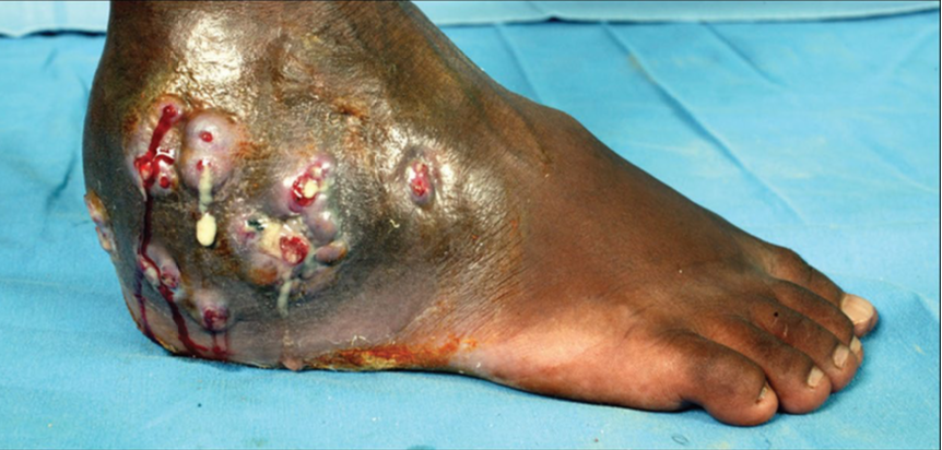
Mycetoma
21
How does a mycetoma develop?
1. Presents as a slowly progressive, painless subcutaneous swelling
2. multiple secondary nodules evolve that may drain through multiple sinus tracts
3. The sinuses often discharge grains or granules containing colonies of the causative organism
2. multiple secondary nodules evolve that may drain through multiple sinus tracts
3. The sinuses often discharge grains or granules containing colonies of the causative organism
22
What do 'white grain' mycetoma look like?
Grains are colorless
23
What do ' black grain' mycetoma look like?
Grains are black 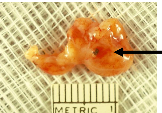
24
What is another name for Madurella grisea?
Trematosphaeria grisea
25
What the commone cause of black grain eumycetoma in South America?
M.grisea - now called Trematosphaeria grisea
26
What is this?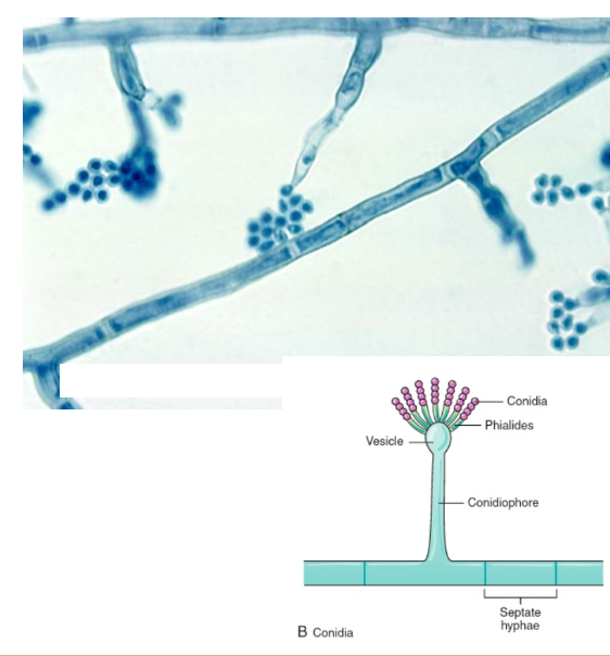
Madurella mycetomatis
27
What is the most common cause of eumycetoma in US?
Pseudallescheria boydii
28
What is the exposure for Pseudallescheria boydii?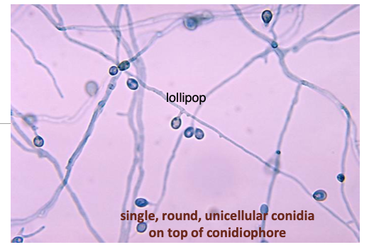
soil, polluted and stagnant water and manure - via inhallation/ near drowing or traumatic inoculation (ex: thorn)
29
What does Pseudallescheria boydii cause?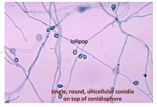
Mycetoma (99% cases)
Sytemic pseudallescheriasis (1%) <- THIS ONE CAN SPREAD! ALWAYS REPORT THIS ONE.
Sytemic pseudallescheriasis (1%) <- THIS ONE CAN SPREAD! ALWAYS REPORT THIS ONE.
30
What is Systemic pseudallescheriasis?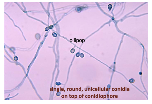
a condition afflicting the immunocompromised (opportunist); associated with cystic fibrosis patients.
31
What is the wide clinical spectrum for Pseudallescheria boydii?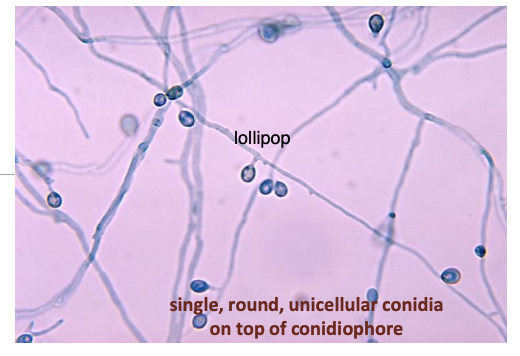
Lung, CNS, systemic spread (sinusitis, arthritis, meningitis, brain abscess, lung abscess, lung fungus ball)
32
What is the treatment for Pseudallescheria boydii?
very difficult; resistant to Amphotericin B and many antifungal drugs
33
What is the aasexual form of Pseudallescheria boydii called?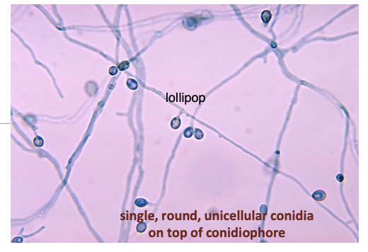
Scedosporium apiospermum
34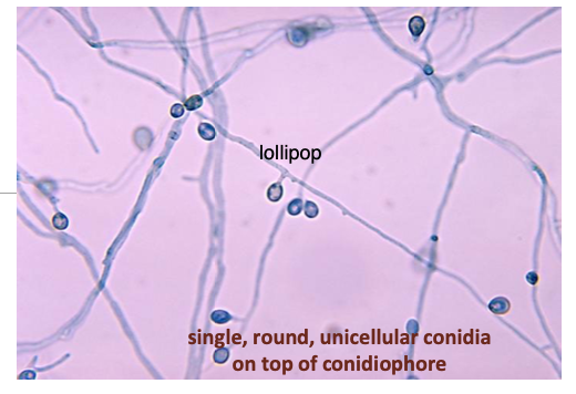
Pseudallescheria boydii
35
what is this?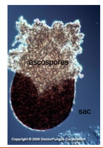
cleistothecium: multicellular, sexual fruiting body, with asci and ascospores inside.
36
Is cleistothecium common or rare?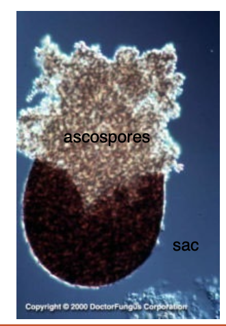
Rarely seen with clinical specimens; requires 2-3 weeks of incubation.
37
where is Leptosphaeria spp. found?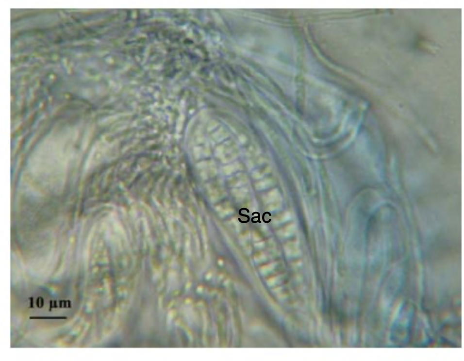
Found in the soil
38
What can Leptosphaeria spp. cause?
Eumycetoma and Phaeohyphomycosis
39
What does the eumycetomalook like caused by Leptosphaeria spp.?
Black grains with pale centers
Common cause of mycetoma in West Africa
Common cause of mycetoma in West Africa
40
what is seen micrscopy in Leptosphaeria spp. (class: Ascomycota)?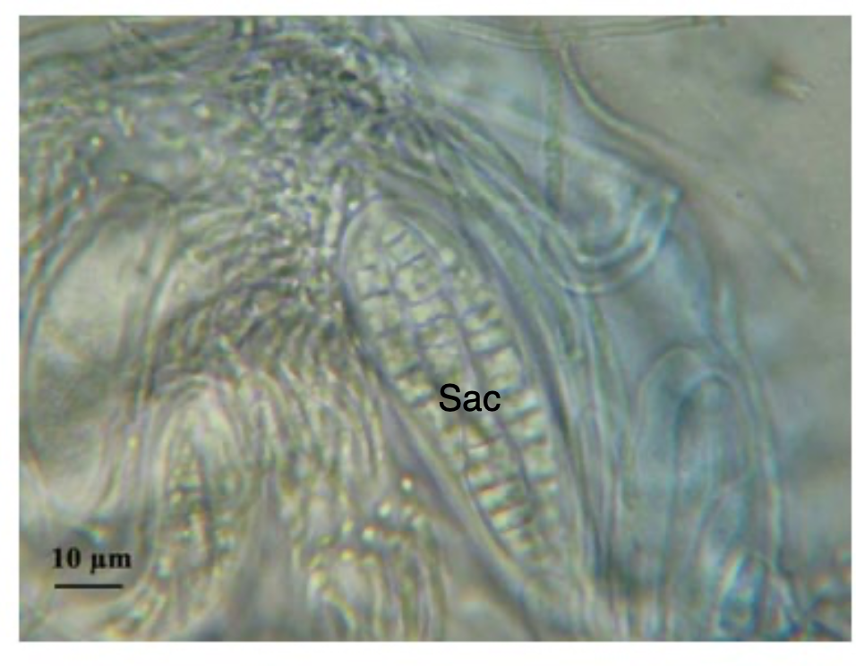
Hyphae, ascomata (cleistothecia), asci, and ascospores [sexual]
41
what is this associated with? (know this for the exam) 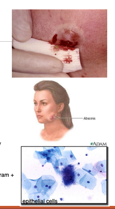
Actinomyces
42
Is Actinomyces aerobic or anaerobic? (know this for the exam) 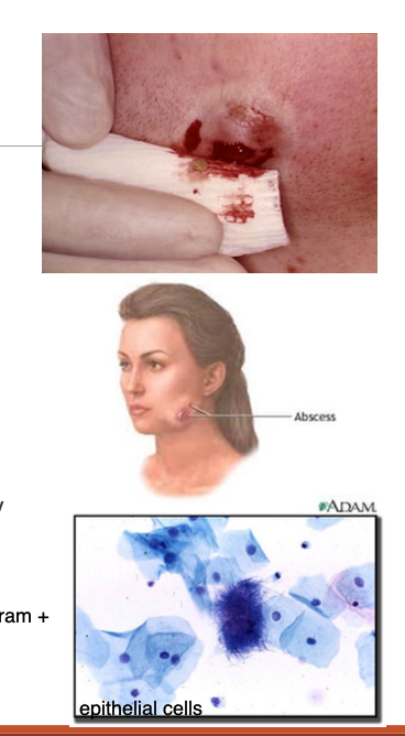
both! May be aerobic or anaerobic
43
Where are aerobic actinomycetes usually found?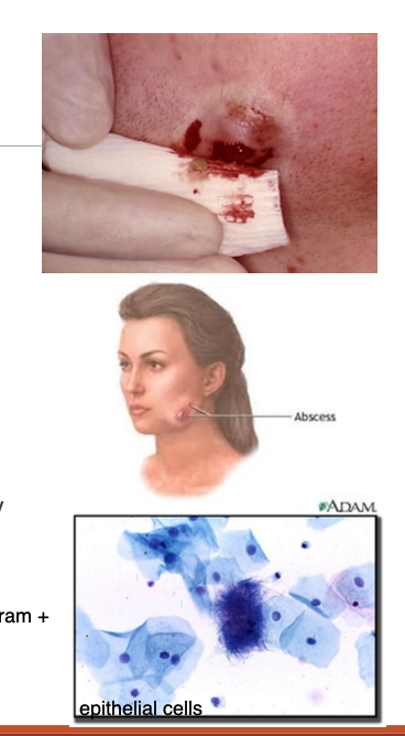
soil and water
44
where are anaerobic/microaerophilic (;ow oxygen concentrations) actinomycetes found?
Normal inhabitants of GI tract humans/animals (from oropharynx -> lower bowel)
45
What is found in the microscopy of Actinomyces?
GPR, filamentous that tend to branch and may fragment in coccbacilliary or coccoid forms (irregular staining) 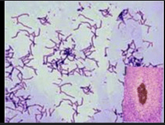
46
What is found in the macroscopy of Actinomyces?
colony appearance varies: waxy, dry, chalky, bumpy, crumbly, adherent; white, tan, orange, yellow, pink, red
47
What is found in the pus of an abcess caused by actinomyces? (know this for the exam) 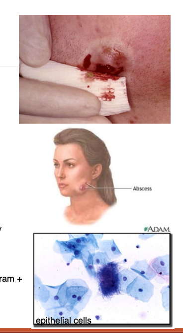
Sulfur granule
48
Describe a sulfur granule from Actinomyces.
Yellow-orange granule of intertwined Actinomyces filaments solidified with elements of tissue exudate
49
What is the most common human actinomyces? (know this for the exam)
A.israelii
50
When do Actinomyces cause disease?
when is transgressed the epithelial barrier under sufficiently low oxygen tension.
51
What is Nocardia? (know this for the exam)
Known bacteria that can cause mycetoma and lung infections (in immunocompromised patients)
52
Which Nocardia bacteria is usually involved in causing a Mycetoma? (know this for the exam)
N. brasiliensis.
53
Pus from mycetoma lesions in Nocardia many contain what?
Sulfur granules
54
What type of STAIN is used for Nocardia species? (know this for the exam)
partial acid fast stain
55
Nocardia can cause chronic diseases. What is usually afffected?
respiratory tract
56
Chronic diseases associated with Nocardia are usually caused by ? (know this for the exam)
N.asteroides
57
Is steptomyces spp. gram positive or gram negative? (know this for the exam)
Gram positive, filamentous bacteria
58
what is this?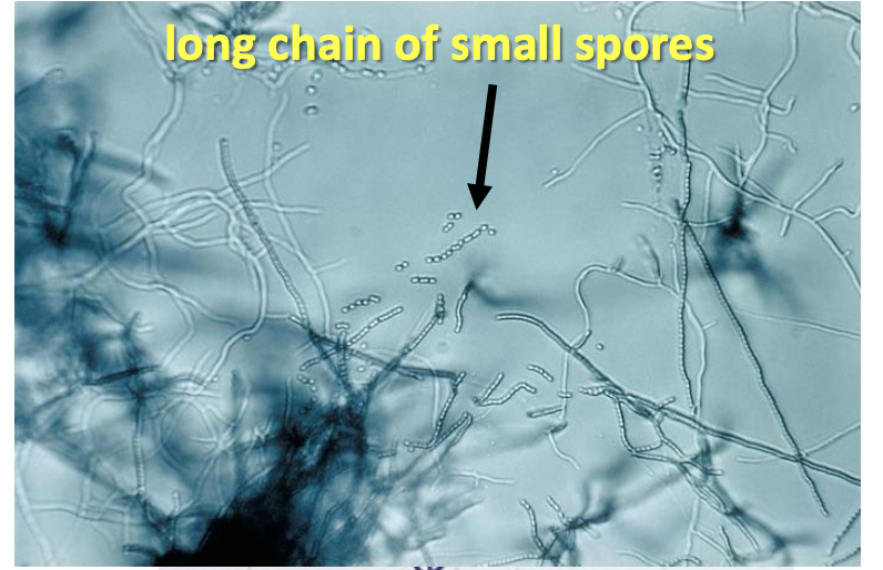
Streptomyces spp.
59
what is streptomyces spp, associated with? (know this for the exam)
Mycetoma
60
What is Lymphocutaneous Sporotrichosis? (know this for the exam)
rose gardener's disease caused by DIMORPHIC FUNGUS
61
What is the name of the DIMORPHIC fungus that causes rose gardener's disease?
Sporothrix schenckii
62
Where do the superficial ccuaneous lesions progress to in Rose Gardener's disease?
along dermal and subcutaneous lymphatics <- majority of cases
63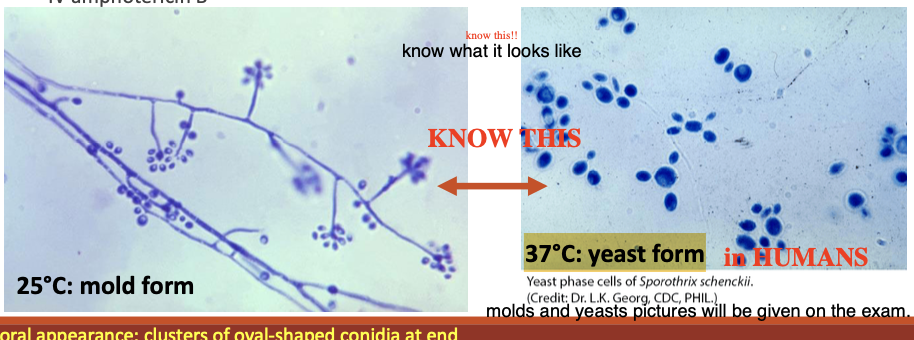
Sporothrix schenckii
64
What is Hyalohyphomycosis?
Fungal infection 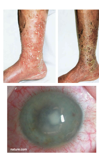
65
What does hyalo stand for?(know this for the exam)
glass
66
Describe hyalohyphomycosis. (know this for the exam) 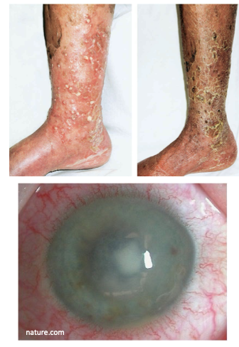
colorless (hyaline), nonpigmented, or lightly color mold with septate hyphae in tissue
67
what is Chromoblastomycosis (chromomycosis)? (know this for the exam)
Fungi that produces a dark pigment
68
what does chromo stand for?
color
69
What is found in the tissue (fibrous scar where fungal infection was)for Chromoblastomycosis? (know this for the exam) 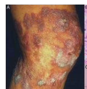
sclerotic bodies
70
What are the most common causative agents for chromoblastomycosis? (know this for the exam) 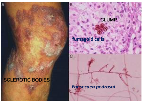
Fonsecaea spp., Phialophora spp., Cladosporium spp.
71
what is phaeohyphomycosis? (know this for the exam)
infections caused by darkly pigmented fungi. 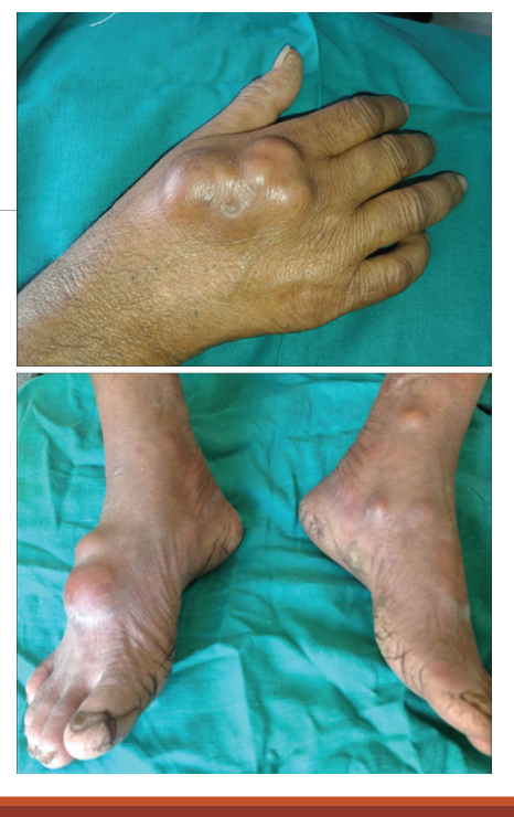
72
What does phaeo stand for?(know this for the exam)
gray
73
How can you tell the difference between Phaeohyphomycosis and Chromoblastomycosis? (know this for the exam)
Phaeohyphomycosis has NO SCLEROTIC BODIES!!
74
What are common causative agents for Phaeohyphomycosis?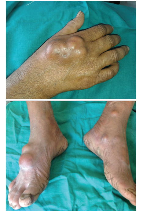
Cladosporium, Exophiala, Wangiella, Cladophialophora, and Bipolaris
75
What is subcutanous zygomycosis?
Also known as Mucormycosis; can be an opprotunistic infection caused by a group of fungi, mucormycetes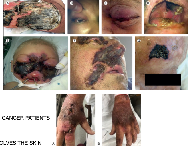
76
Whom does Mucormycosis mainly affect?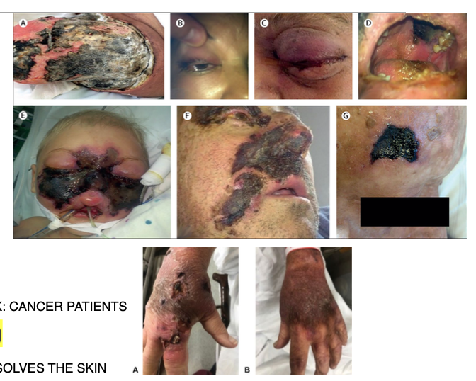
Immunocompromised
77
Which organisms are involved with Subcutaneous Zygomycosis? know this for exam!!
Mucor, Rhizopus, Absidia (Lichtheimia)
78
What type of sporangiospore is this? (know this for exam) 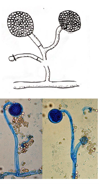
Mucor
79
What type of sporangiospore is this? (know this for exam) 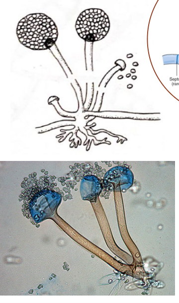
Rhizopus
80
What type of sporangiospore is this? (know this for exam) 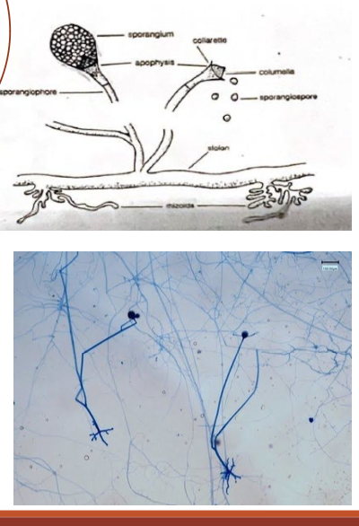
Absidia (Lichtheimia)