WBC: Lecture 4 Myelodysplastic syndromes, acute leukemias
Study these flashcardsFree · spaced repetition · iPhone, Android, Mac & web
All cards
1
Q: What does MAA stand for in aplastic anemia?
A: Moderate Aplastic Anemia.
2
Q: What does SAA stand for in aplastic anemia?
A: Severe Aplastic Anemia.
3
Q: What does VSAA stand for in aplastic anemia?
A: Very Severe Aplastic Anemia.
4
Q: What are the bone marrow criteria for Severe Aplastic Anemia (SAA)?
A: Bone marrow cellularity < 25% plus at least two of the following:
Neutrophils 0.2–0.5 × 10⁹/L
Platelets < 20 × 10⁹/L
Reticulocytes < 20 × 10⁹/L or < 1% corrected for Hct
Neutrophils 0.2–0.5 × 10⁹/L
Platelets < 20 × 10⁹/L
Reticulocytes < 20 × 10⁹/L or < 1% corrected for Hct
5
Q: What are the bone marrow criteria for Very Severe Aplastic Anemia (VSAA)?
A: Same as SAA, but neutrophils < 0.2 × 10⁹/L.
6
Q: How does the severity of aplastic anemia correlate with neutrophil count?
A: The lower the neutrophil count, the more severe the aplastic anemia (MAA > SAA > VSAA).
7
Q: What does the peripheral smear typically show in aplastic anemia?
A: Pancytopenia — reduced RBCs, WBCs, and platelets.
8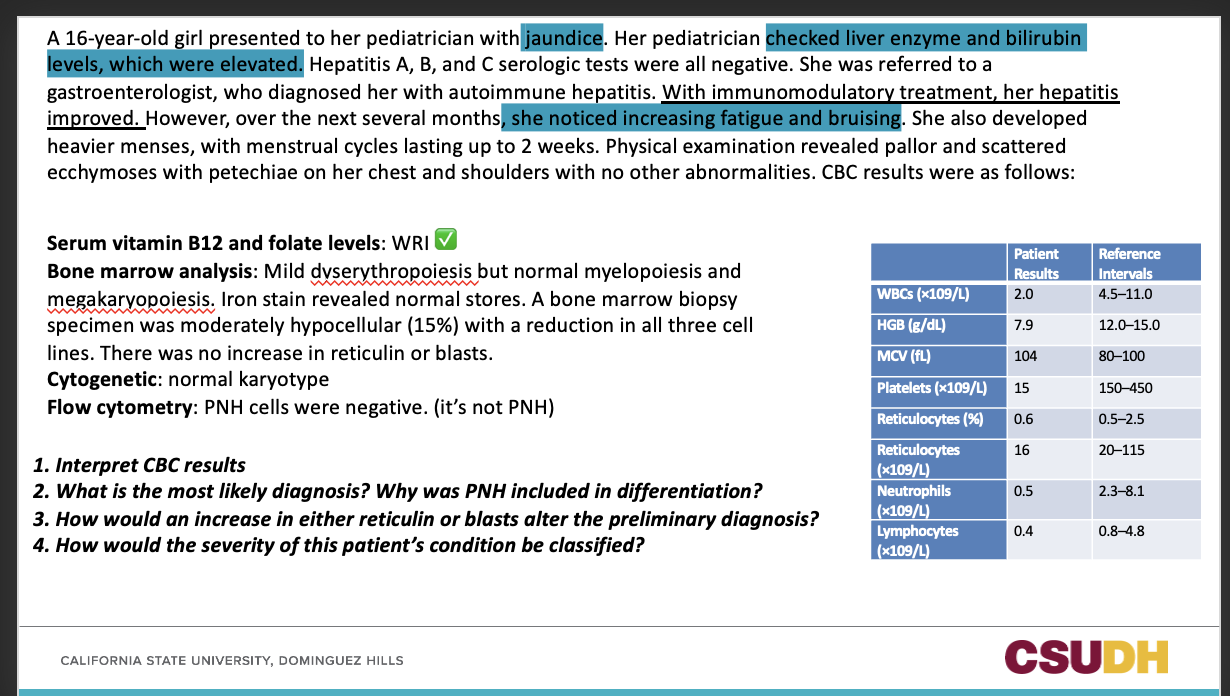
Very typical case on aplastic anemia
Low WBCs
Low HGB - ANEMIA
MCV - macrocytosis.
Platelets - low!!!! KEY severe thrombocytopenia
WBCs (neutrophils and lymphocytes) are affects. LOW in count.
Reticulocytes – absolute value is low but it can be inaccurate sooooo calculate for RPI.
Low WBCs
Low HGB - ANEMIA
MCV - macrocytosis.
Platelets - low!!!! KEY severe thrombocytopenia
WBCs (neutrophils and lymphocytes) are affects. LOW in count.
Reticulocytes – absolute value is low but it can be inaccurate sooooo calculate for RPI.
9
2. What is the most likely diagnosis? Why was PNH included in differentiation? 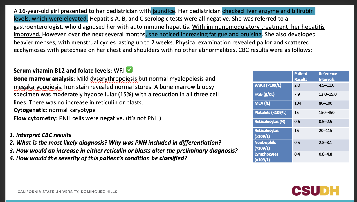
APLASTIC ANEMIA;
STEM CELL DISORDER can develop aplastic anemia ( there is a link to these diagnosis)
PNH can also present with pancytopenia and hemolysis, often overlapping with aplastic anemia. PNH screening is part of the differential diagnosis for acquired bone marrow failure syndromes.
STEM CELL DISORDER can develop aplastic anemia ( there is a link to these diagnosis)
PNH can also present with pancytopenia and hemolysis, often overlapping with aplastic anemia. PNH screening is part of the differential diagnosis for acquired bone marrow failure syndromes.
10
3. How would an increase in either reticulin or blasts alter the preliminary diagnosis?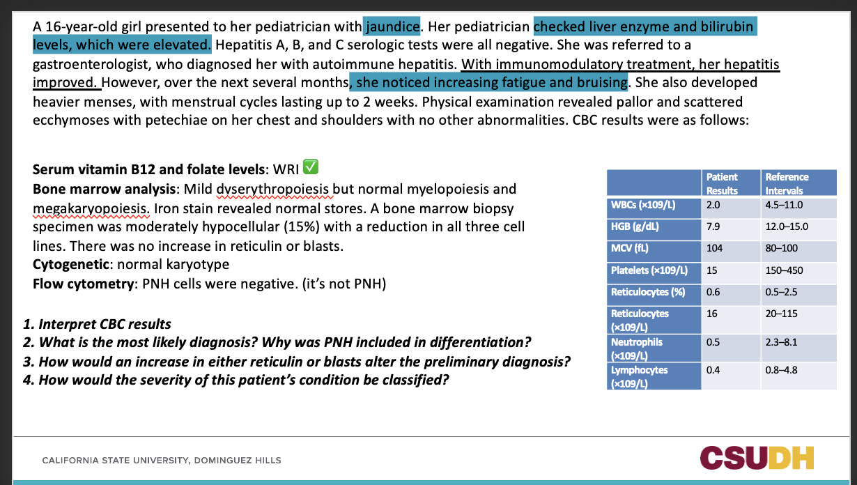
If we detect blasts, it pushes towards AML !!!
11
4. How would the severity of this patient’s condition be classified? 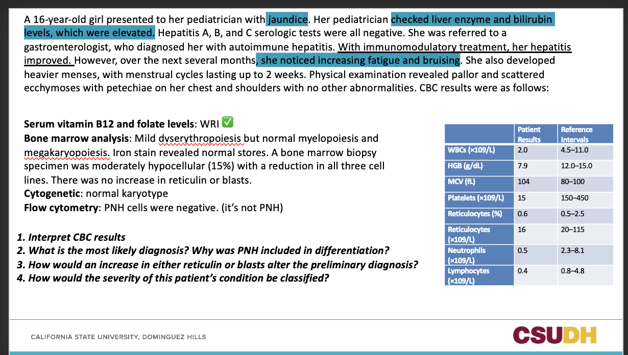
SAA
12
Q: What does MDS stand for?
A: Myelodysplastic Syndromes.
13
What type of disorder are myelodyplastic syndromes? and what is mutated?
acquired clonal hematopoietic disorders;
somatic gene mutation at the HSC level !
somatic gene mutation at the HSC level !
14
Q: What characterizes MDS in the peripheral blood?
A: Progressive cytopenias due to defective maturation of erythroid, myeloid, AND megakaryocytic cell lines.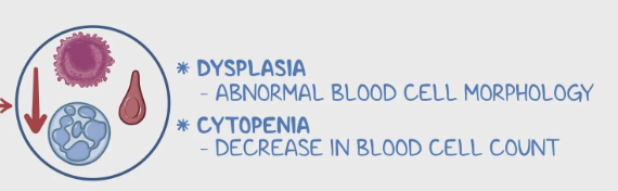
15
Q: What does dyspoiesis mean in the context of MDS?
A: Abnormal or defective cell development in each hematopoietic cell line.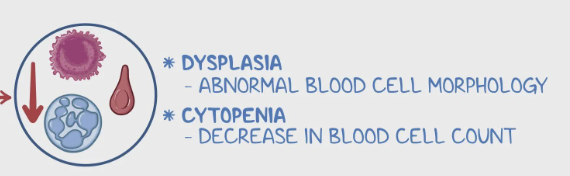
16
Q: What is a potential risk for patients with MDS?
A: Development into acute myeloid leukemia (AML).
17
Q: What is the median age for patients diagnosed with MDS?
A: 76 years old.
18
Q: What are the major risk factors for developing MDS?
A:
Older age (above 65)
Inherited bone marrow failure syndromes
Older age (above 65)
Inherited bone marrow failure syndromes
19
Q: Name some inherited bone marrow failure syndromes associated with MDS.
Fanconi anemia, Diamond-Blackfan anemia, and Shwachman-Diamond syndrome
20
Q: What does dyserythropoiesis mean?
A: Abnormal development or maturation of red blood cells (RBCs) in the bone marrow.
21
Q: What is the most common RBC abnormality seen in the peripheral blood (PB) in dyserythropoiesis?
A: Oval macrocytes.
22
Q: What are hypochromic microcytes, and where are they found in dyserythropoiesis?
A: Small RBCs with decreased hemoglobin (pale color), found in peripheral blood.
23
Q: What does “dimorphic RBC population” mean?
A: The presence of two distinct RBC populations in the same sample — one normal and one abnormal in size or color.
24
What can be seen in bone marrow when a patient has dyserythropoiesis?
- RBC precursors with more than one nucleus
- RBC precursors with abnormal nuclear shapes
- RBC precursors with uneven cytoplasmic staining
- Ring sideroblasts
- RBC precursors with abnormal nuclear shapes
- RBC precursors with uneven cytoplasmic staining
- Ring sideroblasts
25
Q: In the bone marrow (BM), what does it mean when RBC precursors have more than one nucleus?
A: It indicates abnormal erythroid maturation — a hallmark of dyserythropoiesis.
26
Q: What does it mean when RBC precursors have abnormal nuclear shapes?
A: It shows defective maturation of the nucleus during erythropoiesis.
27
Q: What does uneven cytoplasmic staining in RBC precursors suggest?
A: Abnormal hemoglobin synthesis or uneven maturation of the cytoplasm.
28
Q: What are ring sideroblasts, and where are they found?
A: RBC precursors in the bone marrow that contain iron-loaded mitochondria forming a ring around the nucleus (visible with Prussian blue stain).
29
Q: What do ring sideroblasts indicate in the context of MDS?
A: Ineffective erythropoiesis and abnormal iron utilization within developing RBCs.
30
Q: What does dysmyelopoiesis mean?
A: Abnormal development or maturation of myeloid cells (granulocytes) in the bone marrow or peripheral blood.
31
What is seen on the PBS for a patient with Dysmyelopoiesis?
- Persistence of basophilia in the cytoplasm of otherwise mature white blood cells (WBCs), indicating nuclear-cytoplasmic asynchrony
- Agranular or hypogranular neuts
- Hyposegmentation, hypersegmentation, nuclear rings
- Agranular or hypogranular neuts
- Hyposegmentation, hypersegmentation, nuclear rings
32
Q: What does persistence of cytoplasmic basophilia in mature WBCs indicate?
A: Nuclear-cytoplasmic asynchrony — meaning the nucleus and cytoplasm are maturing at different rates.
33
What is seen on the Bone marrow for a patient with Dysmyelopoiesis?
- N:C asynchrony
- Uneven basophilia of the cytoplasm
- Abnormal cytoplasmic granulation (pro or myelo or both may lack granules)
- Agranular pro may be mistaken for a blast
- Hyposegmentation, hypersegmentation
- Uneven basophilia of the cytoplasm
- Abnormal cytoplasmic granulation (pro or myelo or both may lack granules)
- Agranular pro may be mistaken for a blast
- Hyposegmentation, hypersegmentation
34
What is seen in the PB and BM of a patient with dysmegakaryopoiesis?
PB
- Giant PLTs
- Micromegakaryocytes
- Agranular or hypogranular PLTs
BM
- Large mononuclear megakaryocytes
- Bilobed nuclei, or multiple small, separated nuclei
- Giant PLTs
- Micromegakaryocytes
- Agranular or hypogranular PLTs
BM
- Large mononuclear megakaryocytes
- Bilobed nuclei, or multiple small, separated nuclei
35
What does this tell you?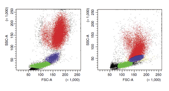
The decrease in neutrophil side scatter shows that when neutrophils lose their granules, they start to overlap with the monocyte area. In the normal (control) sample, neutrophils and monocytes are clearly separated.
36
Q: What does the CD14/CD64 plot analyze?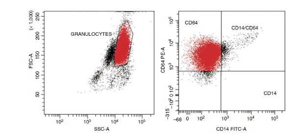
A: It analyzes surface markers on neutrophils to assess for abnormal antigen expression.
37
Q: What abnormal finding is seen on the CD14/CD64 plot in this sample?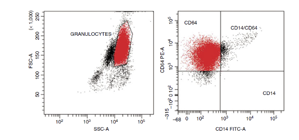
A: A large proportion of granulocytes (neutrophils) abnormally express CD64.
38
Q: What is CD64 normally associated with?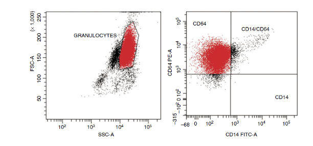
A: CD64 is an antigen typically found on immature myeloid or monocyte cells.
39
Q: What does the expression of CD64 on neutrophils indicate?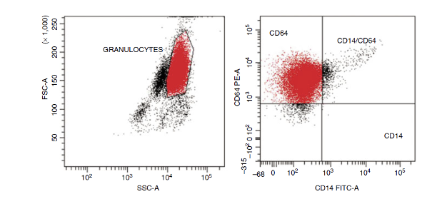
A: It suggests abnormal or immature granulocyte maturation — a sign of dysplasia or myelodysplastic changes.
40
Q: What are the lab features of MDS seen in the blood?
Anemia in >85% cases,
high MCV, poikilo (oval, elliptical, teardrop, spheres, and fragmented cells),
neutropenia, neutrophil hypogranularity, acquired PH (pelger huit), Ring-shaped nuclei in neuts, mild to moderate thrombocytopenia, large PLTs with poor granulation,
prolonged PFA-100.
high MCV, poikilo (oval, elliptical, teardrop, spheres, and fragmented cells),
neutropenia, neutrophil hypogranularity, acquired PH (pelger huit), Ring-shaped nuclei in neuts, mild to moderate thrombocytopenia, large PLTs with poor granulation,
prolonged PFA-100.
41
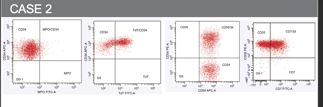
Q: What are the plasma findings in MDS?
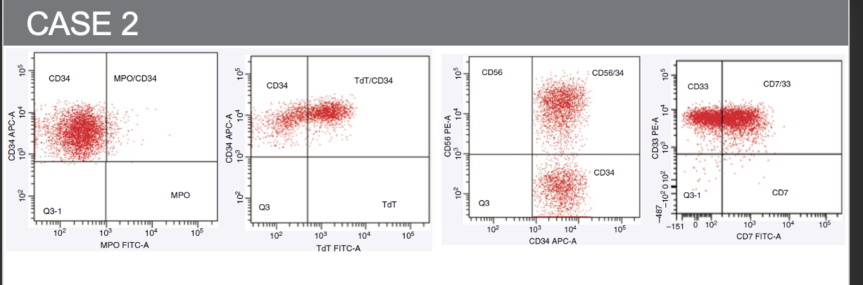
Serum iron/transferrin/ferritin/LDH and uric acid elevated.
42
Q: What are the bone marrow findings in MDS?
Mostly hypercellular in one or more lines, with dysplasia.
43
What are the 6 different WHO classifications of MDS?
MDS with single lineage dysplasia (MDS-SLD):
MDS with multilineage dysplasia (MDS-MLD):
MDS with ring sideroblasts (MDS-RS):
MDS with excess blasts (MDS-EB):
MDS with isolated del(5q) (5q– syndrome):
MDS, unclassifiable
MDS with multilineage dysplasia (MDS-MLD):
MDS with ring sideroblasts (MDS-RS):
MDS with excess blasts (MDS-EB):
MDS with isolated del(5q) (5q– syndrome):
MDS, unclassifiable
44
Q: What is MDS with single lineage dysplasia (MDS-SLD)?
A: Anemia, or infection, or petechiae (depending on the lineage involved), <1% blasts in PB and <5% in BM.
45
Q: What is MDS with multilineage dysplasia (MDS-MLD)?
A: More than one lineage is dyspoietic, <1% blasts in PB and <5% in BM.
46
Q: What is MDS with ring sideroblasts (MDS-RS)?
A: SF3B1 gene (spliceosome) mutation, this mutation + at least 5% ring sideroblasts, dimorphic RBC picture on PBS (combination of hypochromic and normochromic cells).
47
Q: What is MDS with excess blasts (MDS-EB)?
A: Trilineage cytopenia, two types according to WHO.
MDS-EB1: 5% to 9% blasts in the bone marrow or 2% to 4% blasts in the peripheral blood.
MDS-EB2: 10% to 19% blasts in the bone marrow and 5% to 19% blasts in the peripheral blood, Auer rods, more aggressive and higher risk of transformation into AML
MDS-EB1: 5% to 9% blasts in the bone marrow or 2% to 4% blasts in the peripheral blood.
MDS-EB2: 10% to 19% blasts in the bone marrow and 5% to 19% blasts in the peripheral blood, Auer rods, more aggressive and higher risk of transformation into AML
48
Q: What is MDS with isolated del(5q) (5q– syndrome)?
A: Affects predominantly women and occurring at a median age of 70. Patients have anemia without other cytopenias.
49
Q: What is MDS, unclassifiable?
A: MDS that does not fit into other defined subtypes.
50
1-What is a major indication of MDS in the peripheral blood and bone marrow?
a. Dyspoiesis
b. Leukocytosis with left shift
c. Normal bone marrow with abnormal peripheral blood features
d. Thrombocytosis
a. Dyspoiesis
b. Leukocytosis with left shift
c. Normal bone marrow with abnormal peripheral blood features
d. Thrombocytosis
a. Dyspoiesis
51
2-An alert hematologist should recognize all of the following peripheral blood abnormalities as diagnostic clues in MDS EXCEPT:
a. Oval macrocytes
b. Target cells
c. Agranular neutrophils
d. Circulating micromegakaryocytes
a. Oval macrocytes
b. Target cells
c. Agranular neutrophils
d. Circulating micromegakaryocytes
b. Target cells
52
3-A patient has anemia, oval macrocytes, and hypersegmented neutrophils. Which of the following tests would be most efficient in differential diagnosis of this disorder?
a. Serum iron and ferritin levels
b. Erythropoietin level
c. Vitamin B12 and folate levels
d. Chromosome analysis
a. Serum iron and ferritin levels
b. Erythropoietin level
c. Vitamin B12 and folate levels
d. Chromosome analysis
c. Vitamin B12 and folate levels
53
1. What should be included in the differential diagnosis of patients with pancytopenia and elevated mean cell volume?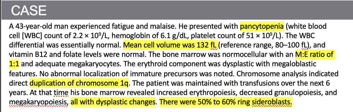
- Vitamin B12 and folate level testing. (megaloblastic picture)
54
2. Given the normal vitamin B12 and folate levels, what is the patient’s probable diagnosis?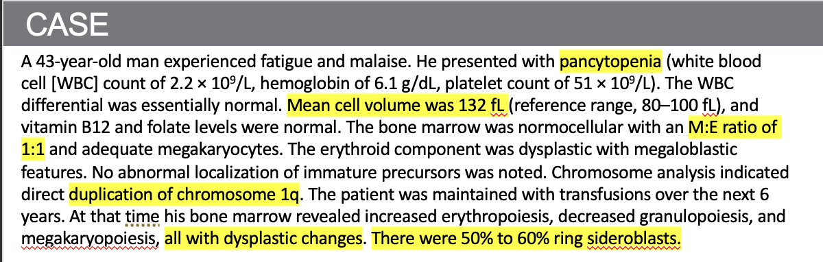
- Myelodysplastic anemia (1:1 = erythropoiesis )
55
3. In which World Health Organization 2016 classification does this disorder belong?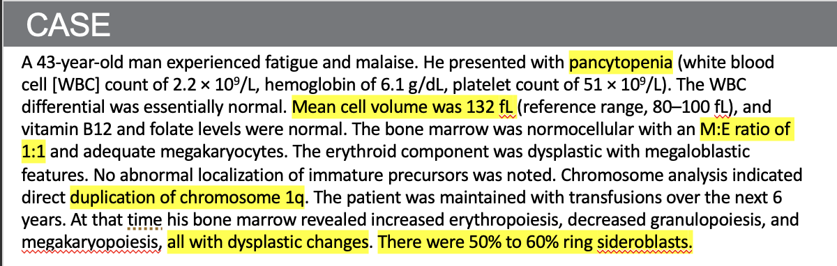
- MDS with ring sideroblasts (MDS-RS)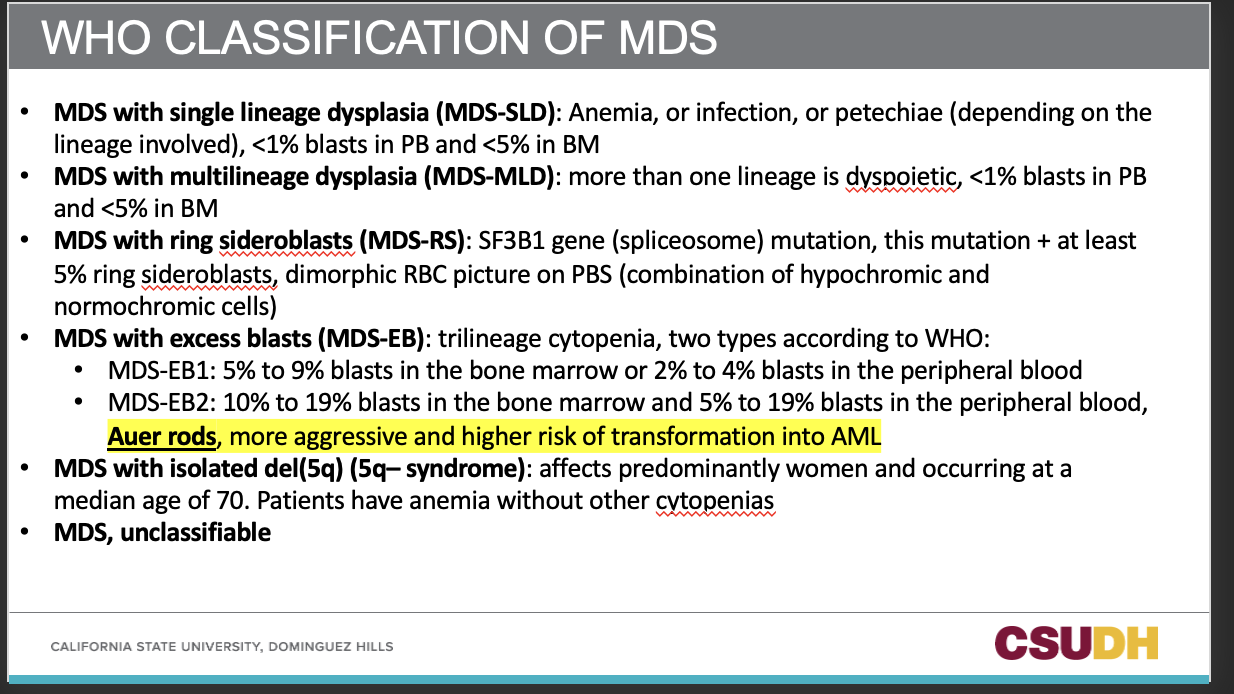
56
What is the classification for Acute Leukemias based on FAB?
considered only morphology and cytochemical stains, required at least 30% blasts for diagnosis
57
What is the classification for Acute Leukemias based on WHO?
more informative, considers clinical features, morphology, immunophenotyping, cytogenetics, and molecular genetics
58
What are 5 WHO classifications of B-ALL?
B-lymphoblastic leukemia/lymphoma with t(9;22) -> BCR-ABL1
B-lymphoblastic leukemia/lymphoma with t(v;11q23.3) -> KMT2A (MLL) rearranged
B-lymphoblastic leukemia/lymphoma with t(12;21) -> TEL-AML1 (ETV6-RUNX1)
B-lymphoblastic leukemia/lymphoma with hyperdiploidy
B-lymphoblastic leukemia/lymphoma with hypodiploidy
B-lymphoblastic leukemia/lymphoma with t(v;11q23.3) -> KMT2A (MLL) rearranged
B-lymphoblastic leukemia/lymphoma with t(12;21) -> TEL-AML1 (ETV6-RUNX1)
B-lymphoblastic leukemia/lymphoma with hyperdiploidy
B-lymphoblastic leukemia/lymphoma with hypodiploidy
59
Why is it important to know B-ALL classifications?
Knowing this helps with treatment and patient outcome of the treatment.
60
What is the difference between hyperdioploidy and hypodiploidy?
Hyperdiploidy is the presence of more than the normal number of chromosomes in a cell, while hypodiploidy is the presence of fewer than the normal number of chromosomes
61
In T-ALL, how do cytogenetic alterations compare to B-ALL in terms of prognosis and treatment correlation?
They show less specificity and less correlation with prognosis and treatment outcome than in B-ALL.
62
Which B-lymphoblastic leukemia/lymphoma has the worst prognosis?
t(9;22) -> BCR-ABL1 — Philadelphia chromosome positive, more common in adults, responds to imatinib (Gleevec).
63
Which B-lymphoblastic leukemia/lymphoma is more common in very young infants and may occur in utero?
t(v;11q23.3); KMT2A (MLL) rearranged — has a very poor prognosis.
64
Which B-lymphoblastic leukemia/lymphoma has an excellent prognosis with a cure rate over 90% in children?
t(12;21) -> TEL-AML1 (ETV6-RUNX1) — rare in adults, excellent prognosis in children.
65
Which B-lymphoblastic leukemia/lymphoma shows hyperdiploidy, and what is its prognosis?
B-lymphoblastic leukemia/lymphoma with hyperdiploidy — less common in adults, favorable prognosis in children.
66
Which B-lymphoblastic leukemia/lymphoma shows hypodiploidy, and what is its prognosis?
B-lymphoblastic leukemia/lymphoma with hypodiploidy — poor prognosis in both children and adults.
67
Which one is ALL and which one is AML?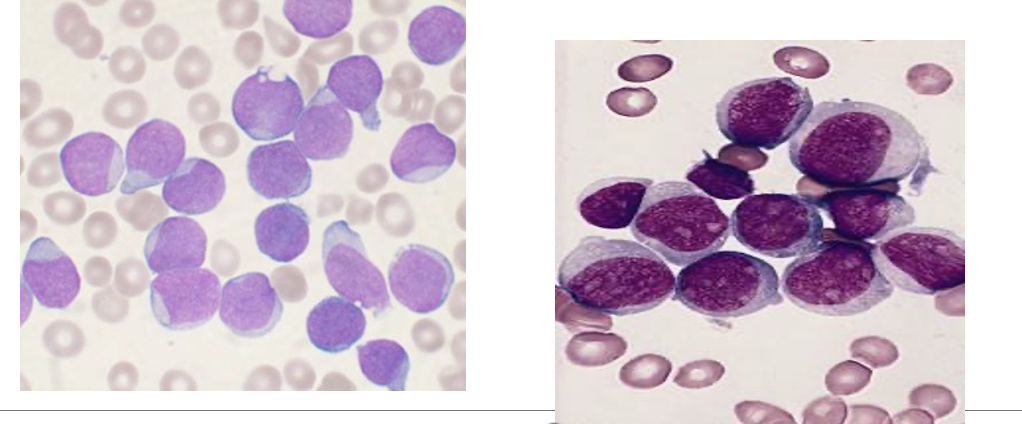
Left: ALL
Right: AML
Right: AML
68
What are the characteristic cell features of Pre-B ALL (L1)?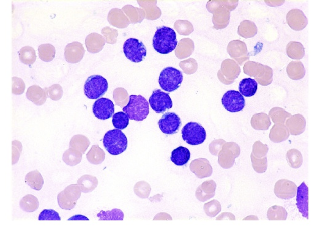
Small blasts with a thin rim of cytoplasm.
69
What are the characteristic cell features of T-Cell ALL (L2)?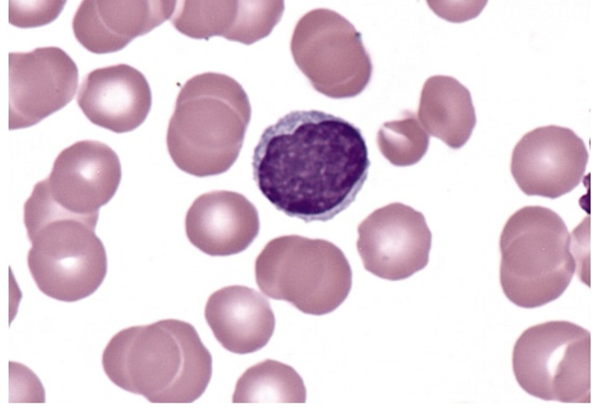
Blasts show irregular clefts or indentations of the nucleus.
70
What are the characteristic cell features of Burkitt leukemia – Mature B Cell ALL (L3)?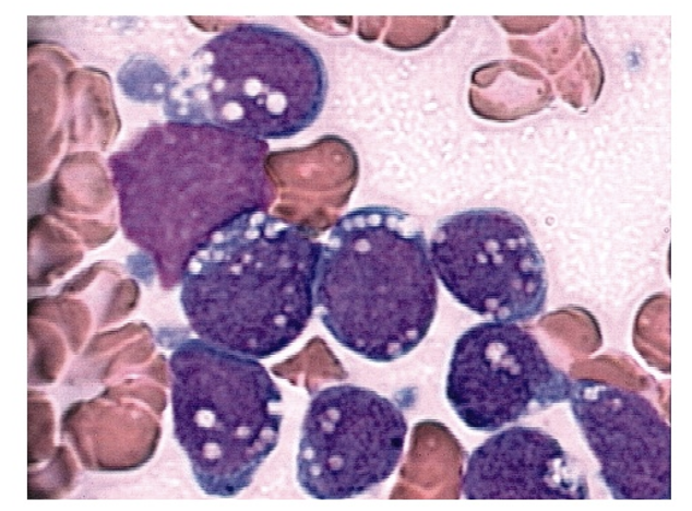
Blasts show prominent cytoplasmic vacuolations - which are lipid contained vaculoes caused by EBV.
71
What are the immunophenotypic markers for Early (pro/pre) B-ALL?
CD34, CD19, cytoplasmic CD22, TdT
72
What are the immunophenotypic markers for Intermediate (common) B-ALL?
CD34, CD19, CD10, cytoplasmic CD22, TdT
73
What are the immunophenotypic markers for Pre-B ALL?
CD19, cytoplasmic CD22, cytoplasmic μ, variable TdT
74
What are the immunophenotypic markers for T-ALL?
CD2, CD3, CD4, CD5, CD7, CD8, TdT
75
Which population is T-ALL most often seen in, and what are its common features?
Most often in teenaged males
Presents with mediastinal mass, elevated peripheral blast counts, meningeal involvement, and extra marrow infiltration
Presents with mediastinal mass, elevated peripheral blast counts, meningeal involvement, and extra marrow infiltration
76
What are the common T-cell markers in T-ALL?
CD2, CD3, CD4, CD5, CD7, CD8
77
What enzyme do most T-ALL cases express, and what does it stand for?
TdT – Terminal deoxynucleotidyl transferase
78
A nine-year-old boy presents with bone pain. Bone marrow aspirate shows large pleomorphic lymphoblasts with prominent nucleoli and cytoplasmic projections (“hand mirror forms”). What is this characteristic of?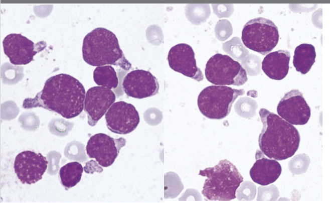
Acute Lymphoblastic Leukemia (ALL) — blasts are large and pleomorphic with prominent nucleoli and cytoplasmic projections (hand mirror forms).
79
What immunophenotypic markers are shown in this flow cytometry plot, and what diagnosis do they support?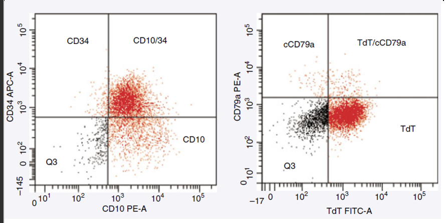
The plots show blasts positive for CD34, CD10, and TdT, but negative for cytoplasmic CD79a.
Intermediate (common) B-ALL: CD34, CD19, CD10, cytoplasmic CD22, TdT
Intermediate (common) B-ALL: CD34, CD19, CD10, cytoplasmic CD22, TdT
80
What immunophenotypic markers are positive in this flow cytometry plot? and what is the diagnosis?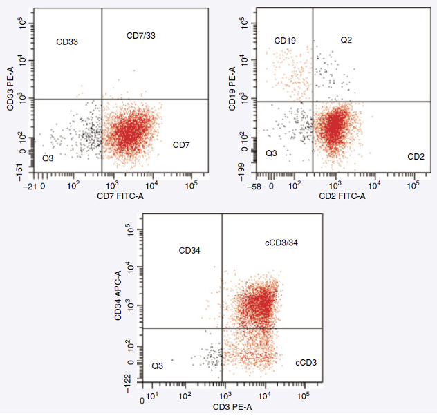
Blasts are positive for CD2, CD3 (cytoplasmic), CD7, and CD34.
➡️ This immunophenotype supports T-lymphoblastic leukemia/lymphoma (T-ALL).
➡️ This immunophenotype supports T-lymphoblastic leukemia/lymphoma (T-ALL).
81
Which cell lineage does cytoplasmic CD3 (cCD3) confirm?
T-cell lineage — cCD3 is a specific marker for immature T-cells.
82
What does co-expression of CD34 indicate in T-ALL?
CD34 expression indicates blast immaturity (an early precursor T-cell stage).
83
What is the most common type of leukemia in adults?
Acute Myeloid Leukemia (AML) — most common in adults, less common in children.
84
What is the FAB classification for AML based on?
Morphology of the blasts.
85
What is the WHO classification for AML based on?
Molecular genetics and cytogenetics.
86
Is the clinical picture of AML specific?
No, the clinical picture is not specific.
Common signs: pallor, fever, easy bruising, splenomegaly (in ~50% of patients), risk of DIC, skin and gum infiltration.
Lymphadenopathy is rare.
Common signs: pallor, fever, easy bruising, splenomegaly (in ~50% of patients), risk of DIC, skin and gum infiltration.
Lymphadenopathy is rare.
87
What percentage of AML patients show blasts in the peripheral blood?
Over 90% of patients show blasts in PB.
88
What is the typical total WBC count in AML?
Usually 5–30 × 10³/µL, but it can vary.
89
What lab abnormalities are commonly seen in AML?
Anemia and thrombocytopenia (always present)
Neutropenia (WBC may be normal)
Bone marrow hypercellular with >20% blasts (diagnostic threshold with supporting genetics)
Neutropenia (WBC may be normal)
Bone marrow hypercellular with >20% blasts (diagnostic threshold with supporting genetics)
90
What serum chemistry findings are common in AML?
Hyperuricemia
Hyperphosphatemia
Hypocalcemia
Risk of acute renal failure and tumor lysis syndrome (especially with very high WBC counts)
Hyperphosphatemia
Hypocalcemia
Risk of acute renal failure and tumor lysis syndrome (especially with very high WBC counts)
91
What are the 7 types of AML WHO classifications?
AML with t(8;21) -> RUNX1-RUNX1T1
AML with inv(16)or t(16;16)-> CBFB-MYH11
APL with PML-RARA
AML with t(9;11) -> KMT2A(MLL)-MLLT3
AML with mutated NPM1
AML with biallelic mutation of CEBPA
AML with mutated RUNX1
AML with inv(16)or t(16;16)-> CBFB-MYH11
APL with PML-RARA
AML with t(9;11) -> KMT2A(MLL)-MLLT3
AML with mutated NPM1
AML with biallelic mutation of CEBPA
AML with mutated RUNX1
92
Which AML mutation is mostly seen in monocytic leukemias and has a good prognosis?
AML with mutated NPM1 (nucleophosmin).
93
Which AML mutation confers a good prognosis when both alleles are affected?
AML with biallelic mutation of CEBPA.
94
Which AML mutation is associated with worse overall survival?
AML with mutated RUNX1.
95
What do the following abbreviations stand for?
RUNX1
MYH11
CBFB
MLL/KMT2A
RARA
PML
RUNX1
MYH11
CBFB
MLL/KMT2A
RARA
PML
RUNX1: Runt-related transcription factor 1
MYH11: Smooth muscle myosin heavy chain 11
CBFB: Core binding factor beta
MLL/KMT2A: Lysine methyltransferase 2A (multi-lineage leukemia gene)
RARA: Retinoic acid receptor alpha
PML: Promyelocytic leukemia gene
MYH11: Smooth muscle myosin heavy chain 11
CBFB: Core binding factor beta
MLL/KMT2A: Lysine methyltransferase 2A (multi-lineage leukemia gene)
RARA: Retinoic acid receptor alpha
PML: Promyelocytic leukemia gene
96
What is AML subtype M0?
Acute myeloid leukemia, minimally differentiated
97
What is AML subtype M1?
Acute myeloid leukemia without maturation
98
What is AML subtype M2?
Acute myeloid leukemia with maturation
99
What is AML subtype M3?
Acute promyelocytic leukemia (APL)
100
What is AML subtype M4?
Acute myelomonocytic leukemia
101
What is AML subtype M4eo?
Acute myelomonocytic leukemia with eosinophilia
102
What is AML subtype M5a?
Acute monocytic leukemia, poorly differentiated
103
What is AML subtype M5b?
Acute monocytic leukemia, well differentiated
104
What is AML subtype M6?
Acute erythroleukemia
105
What is AML subtype M7?
Acute megakaryocytic leukemia
106
What percentage of AML cases show t(8;21); RUNX1-RUNX1T1?
About 5% of AML cases.
107
Which age group is most commonly affected by AML with t(8;21)?
Children and young adults — this subtype is most common in these age groups.
108
What are the characteristic features of the myeloblasts in AML with t(8;21)?
Dysplastic granular cytoplasm
Auer rods
Some maturation (similar to FAB M2 phenotype)
Auer rods
Some maturation (similar to FAB M2 phenotype)
109
What distinctive cells and features are often seen in AML with t(8;21)?
Pseudo–Pelger-Huët cells
Hypogranulation
Possible eosinophilia
Hypogranulation
Possible eosinophilia
110
What is the diagnostic criterion for AML with t(8;21)?
Diagnosis is based on the genetic abnormality (t(8;21)), regardless of blast count.
111
What percentage of AML cases show inv(16) or t(16;16)?
Occurs in 5–8% of AML cases.
112
What types of cells are seen in AML with inv(16)/t(16;16)?
Myeloblasts, monoblasts, and promyelocytes in both peripheral blood and bone marrow.
113
What bone marrow feature is commonly seen in AML with inv(16)/t(16;16)?
Eosinophilia with dysplastic changes in the bone marrow.
114
What complication may occur during relapse in AML with inv(16)/t(16;16)?
CNS involvement (central nervous system relapse).
115
How is the diagnosis of AML with inv(16)/t(16;16) confirmed?
The genetic abnormality itself is sufficient for diagnosis, regardless of blast count.
116
Which genetic abnormality defines Acute Promyelocytic Leukemia (APL)?
t(15;17) translocation involving PML-RARA.
117
What percentage of AML cases are APL?
5–10% of AML cases.
118
At what stage is differentiation blocked in APL?
At the promyelocyte (pro) stage.
119
What are the characteristic cells in APL?
Abnormal hypergranular promyelocytes, some containing Auer rods (often seen in bundles called “faggot cells”).
120
What major complication is associated with APL?
Disseminated Intravascular Coagulation (DIC) — caused by release of procoagulant from promyelocytes.
121
What is the microgranular variant of APL?
Seen in 30–40% of APL cases; the cells have fewer granules and may be mistaken for other AML types.
122
What finding confirms the diagnosis of APL?
Detection of the t(15;17) translocation — sufficient for diagnosis regardless of blast count.
123
What is the unique treatment for APL and its mechanism?
All-trans-retinoic acid (ATRA) and arsenic trioxide;
Mechanism: ATRA induces differentiation of promyelocytes.
Mechanism: ATRA induces differentiation of promyelocytes.
124
What percentage of AML cases involve t(9;11)?
About 6% of AML cases.
125
What type of cells are increased in AML with t(9;11)?
Monoblasts and immature monocytes are increased.
126
Describe the appearance of blasts in AML with t(9;11).
Blasts are large with abundant cytoplasm and fine nuclear chromatin.
127
What distinctive features may the blasts show in AML with t(9;11)?
Blasts may have pseudopodia, granules, and vacuoles.
128
What age group and additional findings are commonly associated with AML t(9;11)?
Occurs typically in children and may involve skin or gum infiltration.
129
What is the FAB subtype M0 in AML?
Acute Myeloid Leukemia, Minimally Differentiated
130
What are the common immunophenotypic markers for AML M0?
CD13+, CD33+, CD34+, CD117+
131
Are MPO and SBB positive or negative in AML M0?
Both MPO and SBB are negative.
132
What percentage of AML cases are M0 subtype?
Less than 5% of AML cases.
133
Which age groups are typically affected by AML M0?
Infants or older patients.
134
What does AML M0 indicate about blast differentiation?
It shows minimal differentiation — blasts lack clear evidence of maturation.
135
What is the FAB subtype M1 in AML?
Acute Myeloid Leukemia Without Maturation
136
What are the common immunophenotypic markers for AML M1?
CD13+, CD33+, CD34+, CD117+
137
Are MPO and SBB positive or negative in AML M1?
MPO and SBB are positive in about 3% of blasts.
138
What percentage of bone marrow non-erythroid cells are blasts in AML M1?
About 90% of non-erythroid cells in the bone marrow are blasts.
139
What percentage of cells show maturation in AML M1, and up to what stage?
Less than 10% of cells show maturation, up to the promyelocyte stage.
140
How does AML M1 differ from AML M0?
AML M1 shows some evidence of myeloid differentiation (MPO/SBB positive), while AML M0 is minimally differentiated (MPO/SBB negative).
141
What is the FAB subtype M2 in AML?
Acute Myeloid Leukemia with Maturation
142
What percentage of blasts are present in the bone marrow in AML M2?
More than 20% blasts in the bone marrow.
143
What percentage of maturing neutrophil lineage cells is seen in AML M2?
More than 10% of cells show neutrophilic maturation.
144
What percentage of blasts belong to the monocytic lineage in AML M2?
Less than 10% of blasts are of monocytic lineage.
145
Which markers are positive in AML M2?
CD34+, CD13+, CD15+, CD11b+, HLA-DR+
146
Which monocytic markers are negative in AML M2?
CD36, CD14, CD64
147
What is the FAB subtype M3 in AML?
Acute Promyelocytic Leukemia (APL)
148
At what stage is differentiation blocked in AML M3?
At the promyelocyte (pro) stage.
149
Which genetic translocation is specific to AML M3?
t(15;17) — the PML-RARA translocation.
150
What is a major complication associated with AML M3?
Disseminated Intravascular Coagulation (DIC) — caused by release of procoagulant material from abnormal promyelocytes.
151
Which markers are positive in AML M3?
CD13+, CD33+, CD64+
152
Which markers are negative in AML M3?
CD34-, CD11b-, HLA-DR-
153
What is a distinctive microscopic feature of AML M3?
Hypergranular promyelocytes and “faggot cells” (bundles of Auer rods).
154
What is the FAB subtype M4 in AML?
Acute Myelomonocytic Leukemia (AMMoL)
155
What is the typical WBC count in AML M4?
Very high WBC count.
156
Which types of cells are present in AML M4?
Monocytic cells (monoblasts and promonocytes) and neutrophilic cells.
157
Which markers are positive in AML M4?
CD13+, CD33+, CD64+, CD14+, CD4+, CD11c+
158
Which two main lineages are involved in AML M4?
Myeloid and monocytic lineages.
159
What stain is positive in AML M4 for monocytic differentiation?
specific esterase (napthol AS-D chloroacetate esterase) — positive for monocytic cells.
160
What is a distinguishing feature of AML M4 under the microscope?
Presence of monoblasts and promonocytes alongside neutrophilic precursors.
161
What is the FAB subtype M5 in AML?
Acute Monoblastic/Monocytic Leukemia (AMoL)
162
What percentage of cells in AML M5 are of monocytic origin?
More than 80% of cells are of monocytic origin.
163
What are the two subtypes of AML M5?
M5a (Acute Monoblastic Leukemia): monoblasts predominate
M5b (Acute Monocytic Leukemia): promonocytes predominate
M5b (Acute Monocytic Leukemia): promonocytes predominate
164
Which markers are positive in AML M5?
CD14+, CD4+, CD64+, CD11c+, CD11b+
165
Which cytochemical stain is positive in AML M5?
α-naphthyl butyrate esterase (nonspecific esterase) — positive in monocytic cells.
166
What is the FAB subtype M6 in AML?
Acute Erythroid Leukemia (Pure Erythroid Type)
167
What percentage of cells are of erythroid origin in AML M6?
More than 80% of cells are erythroid in origin.
168
What percentage of cells are pro-normoblasts in AML M6?
More than 30% are pro-normoblasts.
169
What dysplastic changes are seen in erythroid precursors in AML M6?
Multinucleation
Nuclear-cytoplasmic asynchrony
Vacuolization
Nuclear-cytoplasmic asynchrony
Vacuolization
170
What percentage of nucleated cells on peripheral smear are nRBCs in AML M6?
More than 50% are nucleated red blood cells (nRBCs).
171
What special cell features (aka inclusions) are seen in AML M6?
Ring sideroblasts
Howell–Jolly bodies
Howell–Jolly bodies
172
What stain is positive in AML M6?
Periodic Acid–Schiff (PAS) — positive in erythroid precursors.
173
What is the clinical course of AML M6?
Aggressive and rapid progression.
174
What is the FAB subtype M7 in AML?
Acute Megakaryocytic Leukemia (AMKL)
175
What percentage of blasts must be of megakaryocytic origin in AML M7?
More than 50% of blasts must be of megakaryocytic origin.
176
Which markers are positive in AML M7 megakaryoblasts?
CD41+, CD42b+, CD61+, and cytoplasmic vWF (von Willebrand factor) — positive by immunostaining.
177
What are the characteristic cells seen in AML M7?
Megakaryoblasts and micromegakaryocytes (small, immature megakaryocytes).
178
What blood findings may be seen in AML M7?
Giant or dysplastic platelets
Cytoplasmic blebs on blasts
May show pancytopenia
Cytoplasmic blebs on blasts
May show pancytopenia
179
What is the diagnosis?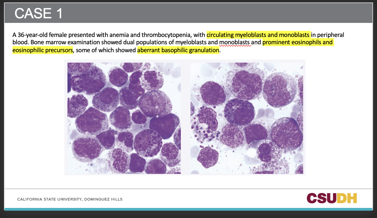
AMML
180
What is being identified in the right plot?
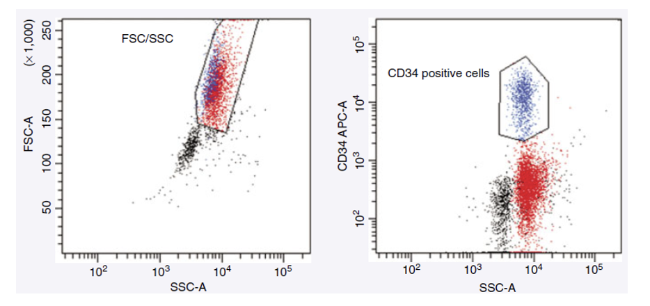
CD34 positive cells — representing hematopoietic blasts or immature precursor cells.
181
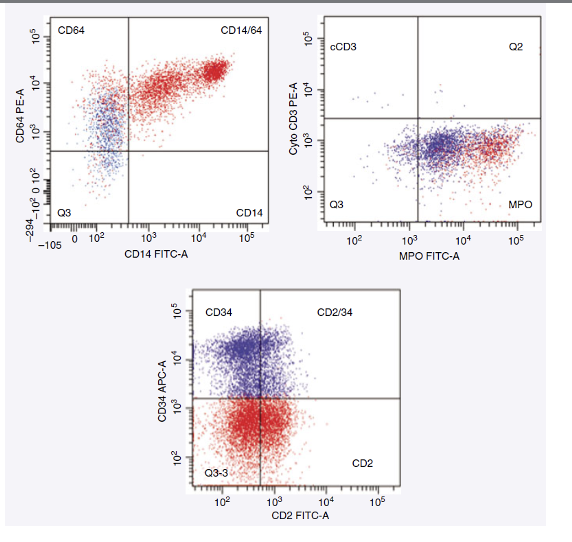
What two cell populations are seen in this immunophenotyping study?
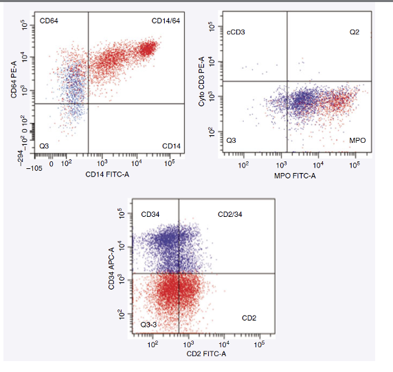
Myeloblasts (blue)
Monoblasts (red)
Monoblasts (red)
182
Which markers are expressed by myeloblasts (blue) in this case?
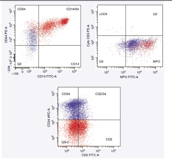
CD34+, MPO+, CD2+
183
Which markers are expressed by monoblasts (red) in this case?
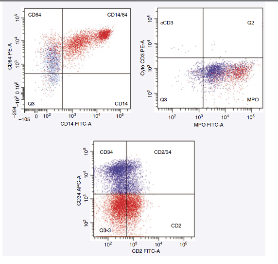
CD14+, CD64+, CD2+
184
What does co-expression of CD2 indicate in this case?
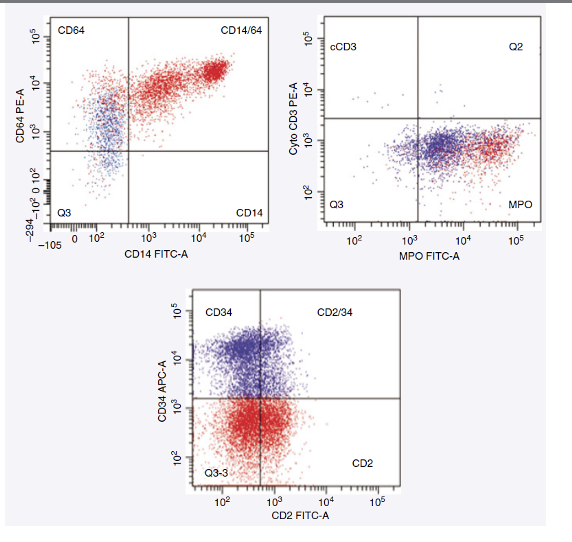
Both myeloblasts and monoblasts express CD2, a T-cell–associated marker that can appear aberrantly (or abnormally) in AML M4, Eo
185
What is the diagnosis for this case?
M4 Eosinophilic AML (M4 Eo)
186
What is the significance of MPO positivity?
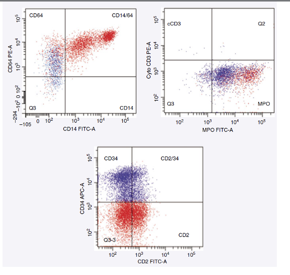
MPO (Myeloperoxidase) confirms myeloid lineage of the blasts.
187
What type of leukemia is shown in Case 2?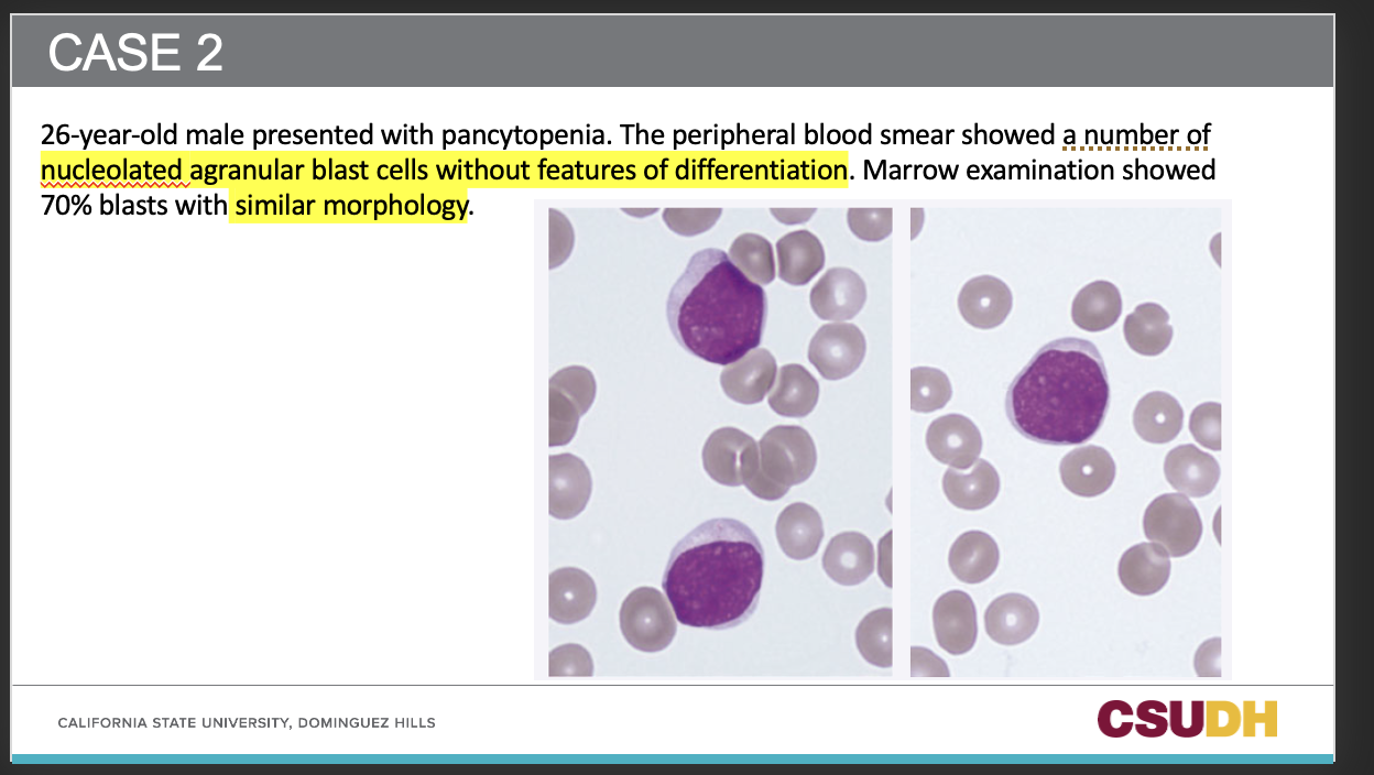
AML
188
What does flow cytometry reveal about the blasts in M0 AML?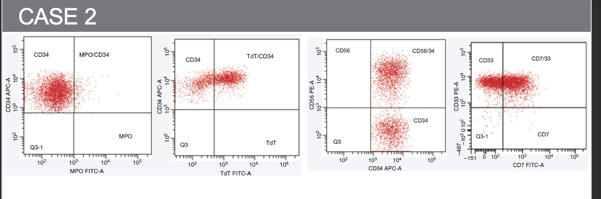
A primitive MPO– (myeloperoxidase negative) myeloblast population.
189
Which markers are positive in this M0 AML case?
CD34+, TdT+, CD38+, with a subpopulation positive for CD56.
190
Which aberrant marker is expressed in this case?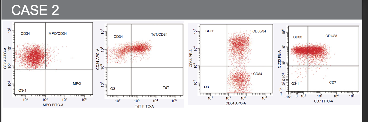
CD7, a T-cell marker aberrantly expressed in AML blasts.
191
Why is this case considered M0 AML?
Because the blasts are MPO negative, indicating minimal myeloid differentiation despite myeloid antigen expression.
192
What is the clinical significance of aberrant CD7 expression in AML?
It’s associated with poor prognosis and increased blast immaturity.
193
What does TdT positivity indicate in AML?
Immature (primitive) blast cells, as TdT is a marker of early progenitor cells.