Lecture 2: Anemias hypoprolliferative (role of iron)
Study these flashcardsFree · spaced repetition · iPhone, Android, Mac & web
All cards
1
Q1: What protein is responsible for transporting iron out of cells such as enterocytes, macrophages, and hepatocytes?
A1: Ferroportin.
2
Q2: When ferroportin is able to transport iron out, what happens to iron absorption and release from storage?
A2: Iron absorption in the intestines and iron release from storage increase.
3
Q3: What is the effect of increased body iron on hepcidin production?
A3: Increased body iron stimulates the liver to produce more hepcidin.
4
Q4: What is the effect of increased hepcidin on ferroportin?
A4: Increased hepcidin blocks ferroportin from transporting iron out of cells.
5
Q5: When ferroportin is blocked, what happens to iron absorption and release from storage?
A5: Iron absorption in the intestines and iron release from storage decrease.
6
Q6: What is the effect of decreased body iron on hepcidin production?
A6: Decreased body iron reduces hepcidin production.
7
Q7: Which cells are shown as locations of ferroportin in this diagram?
A7: Enterocytes, macrophages, and hepatocytes.
8
What is the etiology in IDA?
Inadequate intake
Increased need (stores not sufficient)
Impaired absorption
Chronic blood loss
Increased need (stores not sufficient)
Impaired absorption
Chronic blood loss
9
Chronic blood loss is common in males why?
GI problem colon cancer
10
How many stages are there in IDA?
3
11
Q1: Which IDA pathogenesis stage has no evidence of anemia, normal RBC production, and storage iron depletion?
A1: Stage 1.
12
Q2: Which IDA pathogenesis stage is described as the “common stage”?
A1: Stage 1.
13
Q3: In which stage is iron deficiency latent and the body relies on transport iron?
A3: Stage 2.
14
Q4: In Stage 2, what happens to erythropoiesis?
A4: Restricted erythropoiesis starts.
15
Q5: In Stage 2, what happens to FEP (free erythrocyte protoporphyrin)?
A5: FEP is increased.
16
Q6: In Stage 2, what is the CBC status?
A6: CBC is still normal.
17
Q7: What stain/test is referenced in Stage 2 for marrow iron stores?
A7: Prussian blue result on bone marrow (BM) biopsy.
18
Q8: Which stage shows overt anemia with depleted storage and low functional iron?
A8: Stage 3.
19
Q9: In Stage 3, which indices are low?
A9: Hemoglobin (Hb) and hematocrit (HCT) are low.
20
Q10: What RBC picture is seen in Stage 3?
A10: Hypochromic, microcytic picture.
21
Q11: What is the first PBS abnormality to appear in Stage 3?
A11: Anisocytosis.
22
Q12: What additional PBS features can accompany Stage 3 anisocytosis?
A12: Usually mild ovalocytosis with high RDW.
23
Q13: Which characteristic cell shape may be seen in Stage 3?
A13: Pencil cells (“pencils”).
24
Why is stage 1 most commonly missed?
routine doesn’t test sTfR
25
Q1: What is the screening test panel listed for IDA?
A1: CBC (Hb, HCT, RBC indices), reticulocyte count, and PBS examination.
26
What are diagnostic tests for IDA?(6)
Serum iron (SI)
Total iron-binding capacity (TIBC)
Transferrin saturation (SI/TIBC*100)
Serum ferritin level
Soluble transferrin receptor (sTfR) level
Hemoglobin content of reticulocytes
Total iron-binding capacity (TIBC)
Transferrin saturation (SI/TIBC*100)
Serum ferritin level
Soluble transferrin receptor (sTfR) level
Hemoglobin content of reticulocytes
27
What are specialized tests for IDA? (2)
Bone marrow or liver biopsy with prussian blue staining
RBC free-zinc protophorphyrin level (FEP/ZPP)
RBC free-zinc protophorphyrin level (FEP/ZPP)
28
Q2: Name a diagnostic test that measures circulating iron.
A2: Serum iron (SI) level.
29
Q3: Which test measures iron-binding capacity?
A3: Total iron-binding capacity (TIBC).
30
Q4: How is transferrin saturation calculated?
A4: (Serum iron ÷ TIBC) × 100.
31
Q5: Which test reflects body iron stores?
A5: Serum ferritin level.
32
Q6: Which marker measures the soluble form of the transferrin receptor?
A6: Soluble transferrin receptor (sTfR) level.
33
Q7: Which test assesses recent iron incorporation into young RBCs?
A7: Hemoglobin content of reticulocytes.
34
Q8: Which specialized test stains tissue iron stores?
A8: Bone marrow or liver biopsy with Prussian blue staining.
35
Q9: Which specialized test measures protoporphyrin forms?
A9: RBC free/zinc protoporphyrin level (FEP/ZPP).
36
Q10: Which listed item is a specialized (not routine diagnostic) test?
A10: Bone marrow or liver biopsy with Prussian blue staining.
37
Q11: Which screening component specifically evaluates immature RBCs?
A11: Reticulocyte count.
38
Q12: Which screening step examines morphology directly?
A12: Peripheral blood smear (PBS) examination.
39
what happens when a patient received their first iron therapy?
increase in Hb content of recticulocytes, within 2 days post-treatment
40
what happens within 5-10 days post iron therapy?
increase in reticulocyte number
41
What happens within 2-3 weeks after iron therapy?
increase in Hb; full normalization by 2 months
42
What is the morphology of the PBS? and how long does it usually stay like that?
microcytic and may stay for several months, dimorphism apparent.
43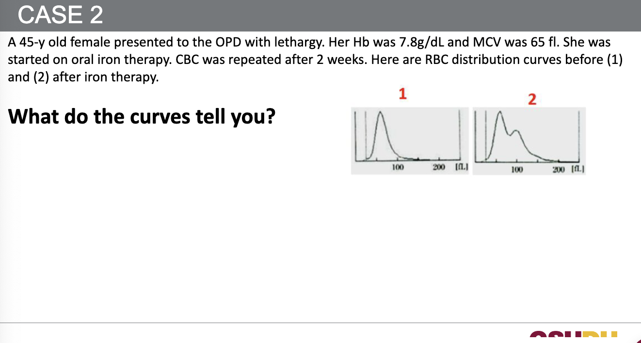
(1) before iron therapy: the RBC histogram is left-shifted with narrow peak at low volume -> unform microcytosis consistent with IDA (low MCV, low Hb)
(2)the histogram becomes bimodal/dimorphic—a new peak appears toward normal cell size while the old microcytic peak remains. This means the marrow has responded to iron with production of larger, normocytic/ reticulocytic cells, so RDW rises.
(2)the histogram becomes bimodal/dimorphic—a new peak appears toward normal cell size while the old microcytic peak remains. This means the marrow has responded to iron with production of larger, normocytic/ reticulocytic cells, so RDW rises.
44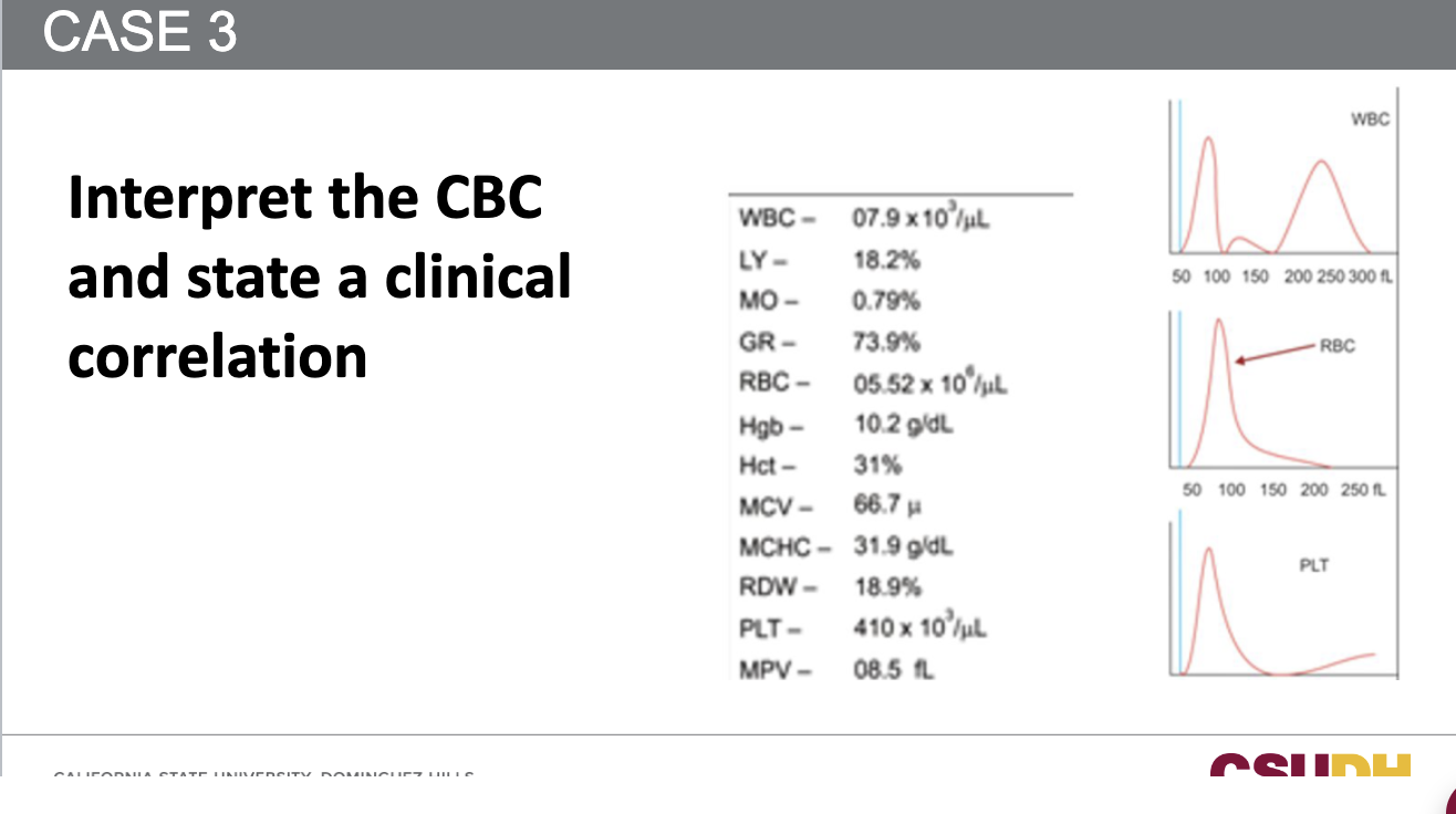
WBC: 7.9 × 10³/µL → Normal
Differential: LY 18.2%, MO 0.79%, GR 73.9% → Normal distribution
RBC count: 5.52 × 10⁶/µL → Slightly high-normal
Hemoglobin (Hb): 10.2 g/dL → Low (anemia)
Hematocrit (Hct): 31% → Low
MCV: 66.7 fL → Low (microcytic)
MCHC: 31.9 g/dL → Normal
RDW: 18.9% → High (anisocytosis present)
Platelets (PLT): 410 × 10³/µL → Mild thrombocytosis
MPV: 8.5 fL → Normal
Histogram Findings:
- RBC curve: Left-shifted with widened base → microcytic anemia with variation in size (↑ RDW).
- WBC and PLT histograms: Normal.
Differential: LY 18.2%, MO 0.79%, GR 73.9% → Normal distribution
RBC count: 5.52 × 10⁶/µL → Slightly high-normal
Hemoglobin (Hb): 10.2 g/dL → Low (anemia)
Hematocrit (Hct): 31% → Low
MCV: 66.7 fL → Low (microcytic)
MCHC: 31.9 g/dL → Normal
RDW: 18.9% → High (anisocytosis present)
Platelets (PLT): 410 × 10³/µL → Mild thrombocytosis
MPV: 8.5 fL → Normal
Histogram Findings:
- RBC curve: Left-shifted with widened base → microcytic anemia with variation in size (↑ RDW).
- WBC and PLT histograms: Normal.
45
Q1: What do the patient’s HGB 3.0 g/dL, MCV 66.3 fL, MCH 18.1 pg, and MCHC 27.3 g/dL indicate?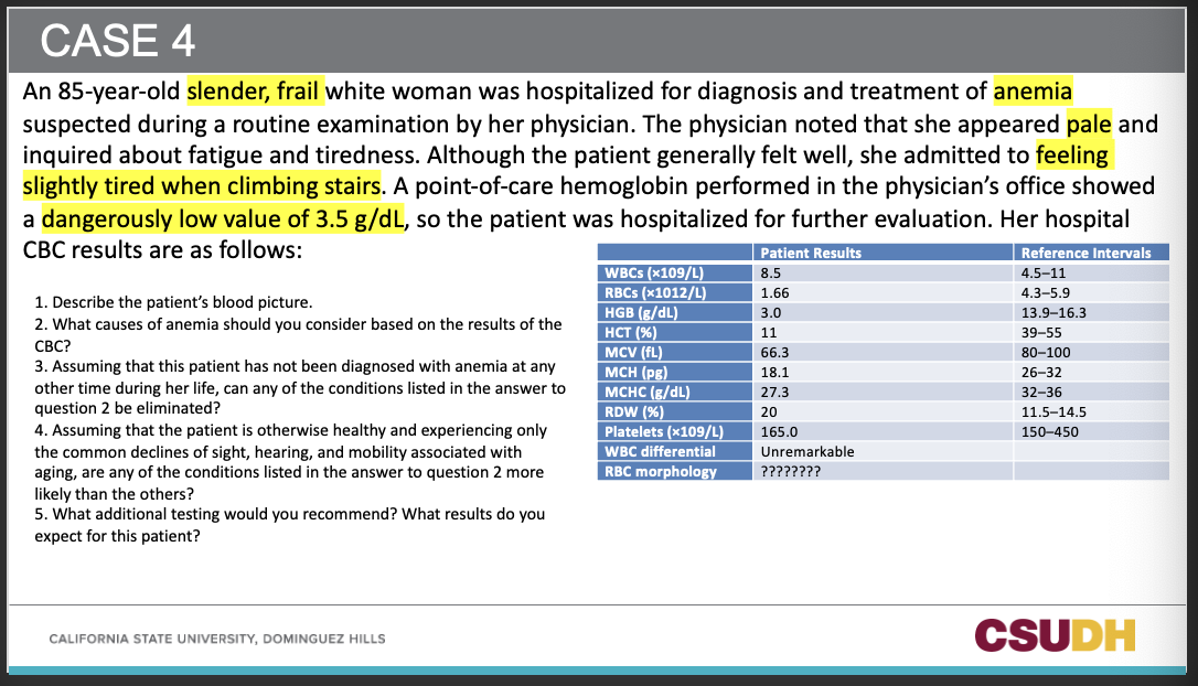
A1: Severe microcytic, hypochromic anemia.
46
Q2: What does an RDW of 20% indicate in this context?
A2: Marked anisocytosis (wide size variation in RBCs).
47
Q3: Are platelets of 165 ×10^9/L abnormal here?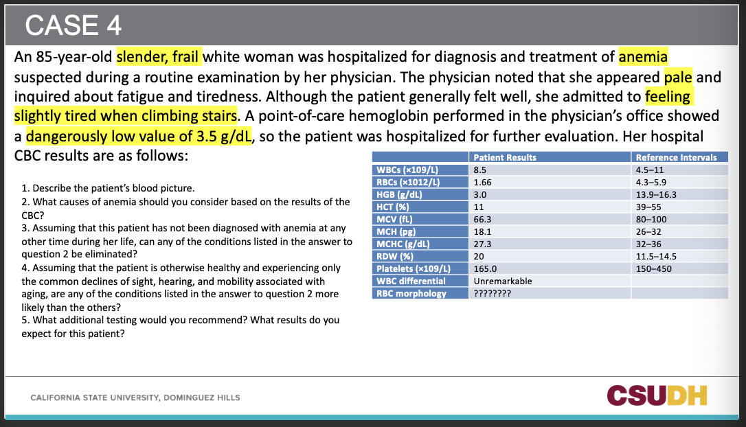
A3: No—within reference range (150–450 ×10^9/L).
48
Q4: Given the CBC pattern, what is the most likely cause of anemia?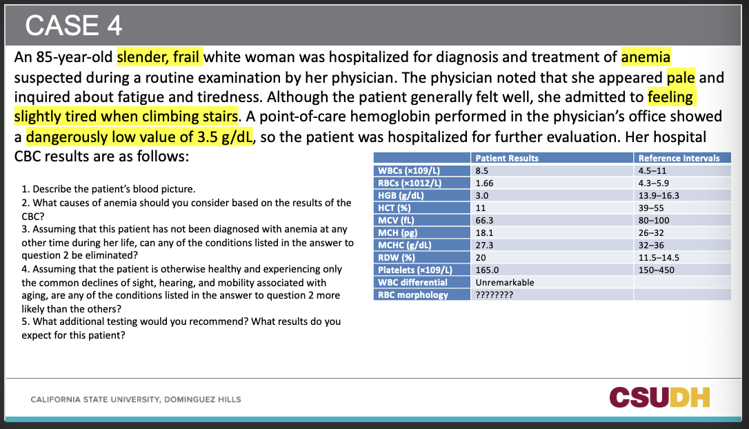
A4: Iron deficiency anemia (IDA), likely from chronic blood loss.
49
Q5: Which condition is least likely if the patient has no prior history of anemia?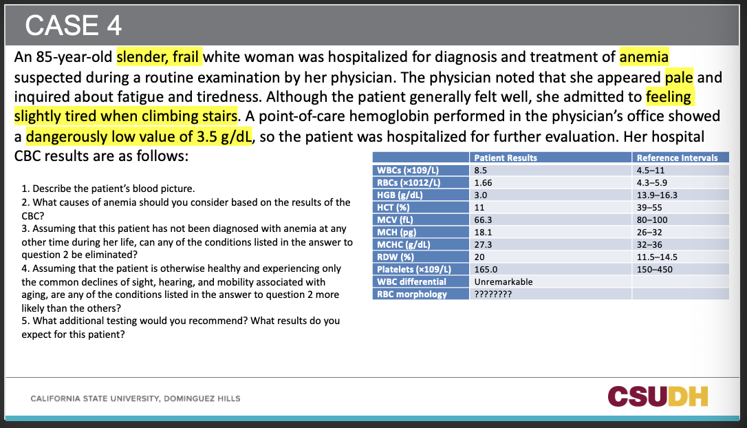
A5: Thalassemia trait (hereditary, typically lifelong microcytosis and often normal/elevated RBC count).
50
Q6: What initial confirmatory testing should be ordered?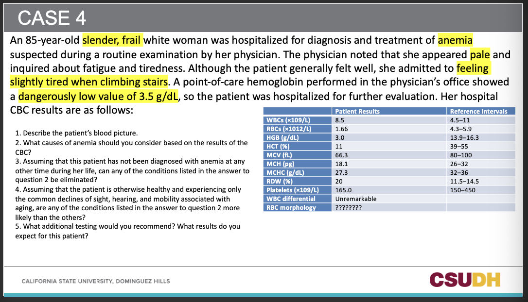
A6: Iron studies—serum ferritin, serum iron (SI), TIBC, transferrin saturation, ± sTfR, and RBC FEP/ZPP.
51
Q7: What iron study pattern is expected in IDA?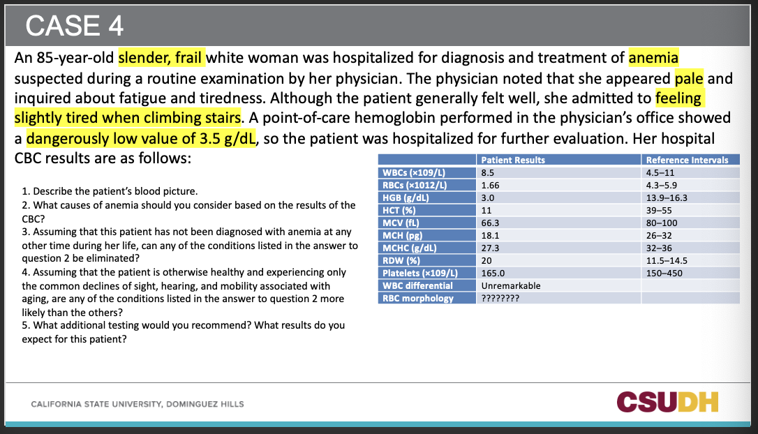
A7: Low ferritin, low SI, high TIBC, low transferrin saturation, ↑ sTfR, ↑ FEP/ZPP.
52
Q8: What next clinical workup is important in an elderly patient with suspected IDA?
A8: Evaluate for occult gastrointestinal bleeding (e.g., fecal occult blood testing and endoscopic evaluation).
53
Q9: What RBC morphology would you expect on the smear?
A9: Microcytosis, hypochromia, anisopoikilocytosis with pencil cells; high RDW.
54
Q10: Is the WBC count (8.5 ×10^9/L) and differential concerning?
A10: No—unremarkable; anemia is the primary issue here.
55
CAN YOU SEE POLYCHROMASIA BEFORE IRON THERAPY?
Short answer: usually not before treatment.
56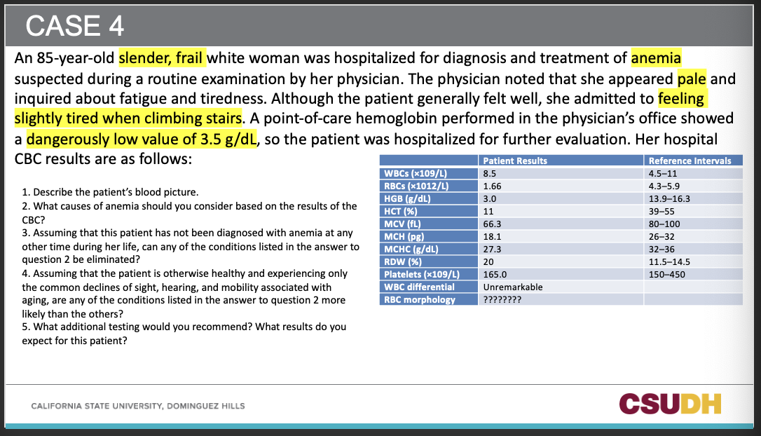
Q1: Describe the patient’s blood picture in Case 4.
A1: Severe microcytic, hypochromic anemia (Hgb 3.0, Hct 11%, MCV 66.3 fL, MCHC 27.3 g/dL) with high RDW (20%); WBC/platelets normal; smear expected to show microcytosis, hypochromia, anisopoikilocytosis and pencil cells.
Q2: Based on the CBC pattern, which causes should be considered?
A2: Iron deficiency anemia (most likely), anemia of chronic inflammation, sideroblastic anemia, and thalassemia trait.
Q3: With no prior history of anemia, which condition can be eliminated?
A3: Thalassemia trait (lifelong microcytosis; often normal/high RBC count).
Q4: If otherwise healthy, which cause is most likely?
A4: Iron deficiency anemia from occult gastrointestinal blood loss.
Q5: What additional tests are recommended and what results are expected?
A5: Iron studies showing ↓ferritin, ↓serum iron, ↑TIBC, ↓transferrin saturation, ↑sTfR, ↑FEP/ZPP; low reticulocyte Hb; evaluate for occult GI bleeding with FOBT and endoscopy/colonoscopy.; Calculate Retic quickly (if below 3) bone marrow is ineffective, if above 3 BM is good
A1: Severe microcytic, hypochromic anemia (Hgb 3.0, Hct 11%, MCV 66.3 fL, MCHC 27.3 g/dL) with high RDW (20%); WBC/platelets normal; smear expected to show microcytosis, hypochromia, anisopoikilocytosis and pencil cells.
Q2: Based on the CBC pattern, which causes should be considered?
A2: Iron deficiency anemia (most likely), anemia of chronic inflammation, sideroblastic anemia, and thalassemia trait.
Q3: With no prior history of anemia, which condition can be eliminated?
A3: Thalassemia trait (lifelong microcytosis; often normal/high RBC count).
Q4: If otherwise healthy, which cause is most likely?
A4: Iron deficiency anemia from occult gastrointestinal blood loss.
Q5: What additional tests are recommended and what results are expected?
A5: Iron studies showing ↓ferritin, ↓serum iron, ↑TIBC, ↓transferrin saturation, ↑sTfR, ↑FEP/ZPP; low reticulocyte Hb; evaluate for occult GI bleeding with FOBT and endoscopy/colonoscopy.; Calculate Retic quickly (if below 3) bone marrow is ineffective, if above 3 BM is good
57
What morphologic alterations are seen in this blood smear field?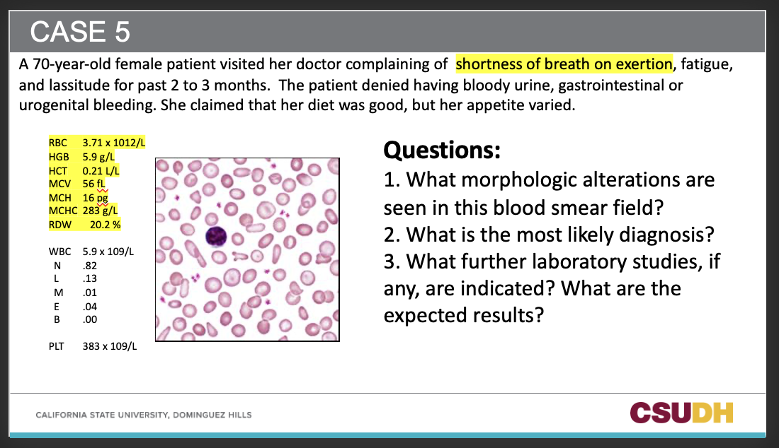
Marked microcytosis and hypochromia with anisocytosis (high RDW 20.2%); poikilocytosis including ovalocytes/pencil cells (and occasional target forms).
58
2. What is the most likely diagnosis?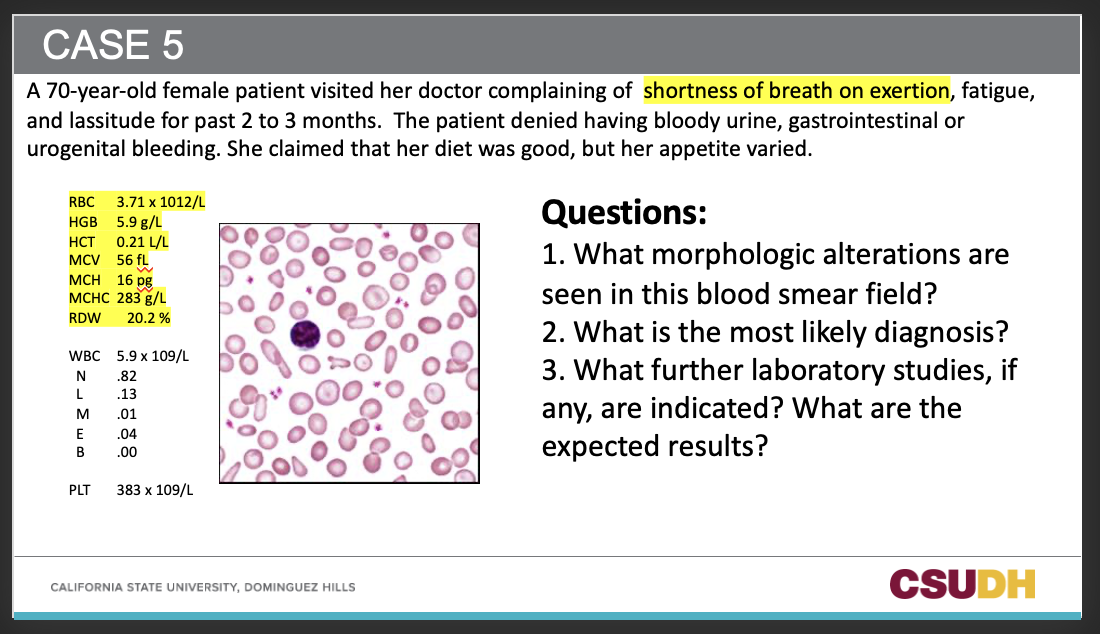
Iron deficiency anemia (IDA)—severe microcytic, hypochromic anemia (MCV 56 fL, MCH 16 pg, low Hgb/Hct) with ↑RDW; platelets high-normal (383 ×10⁹/L) consistent with reactive thrombocytosis often seen in IDA.
59
3. What further laboratory studies, if any, are indicated? What are the expected results?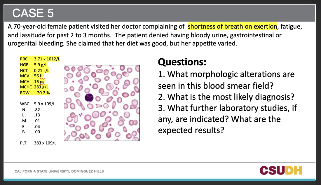
Iron studies: ↓Serum iron, ↑TIBC, ↓Transferrin saturation, ↓Ferritin, ↑sTfR, ↑FEP/ZPP; ↓reticulocyte Hb content.
Evaluate source: Check occult GI blood loss (FOBT, consider endoscopy/colonoscopy) given age/history.
Evaluate source: Check occult GI blood loss (FOBT, consider endoscopy/colonoscopy) given age/history.
60
Q1: Which indices define the anemia in Case 5?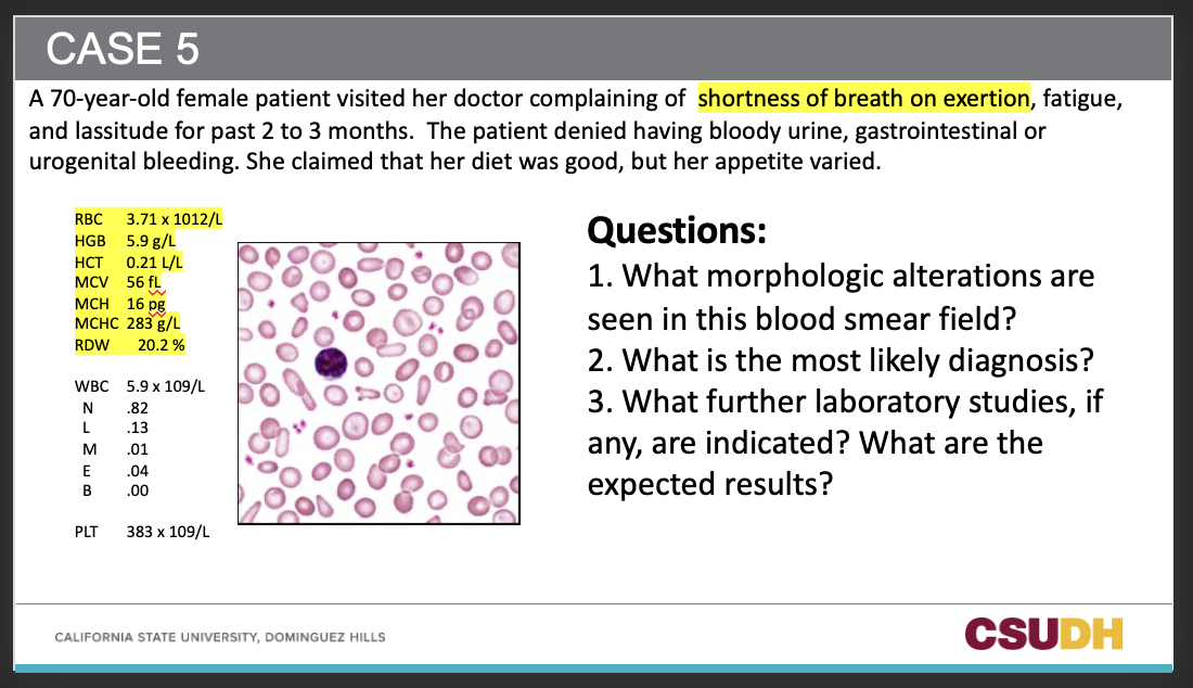
A1: Hgb 5.9, Hct 0.21, MCV 56 fL, MCH 16 pg, MCHC 283 g/L, RDW 20.2%.
61
Q2: What RBC morphology is seen on the smear?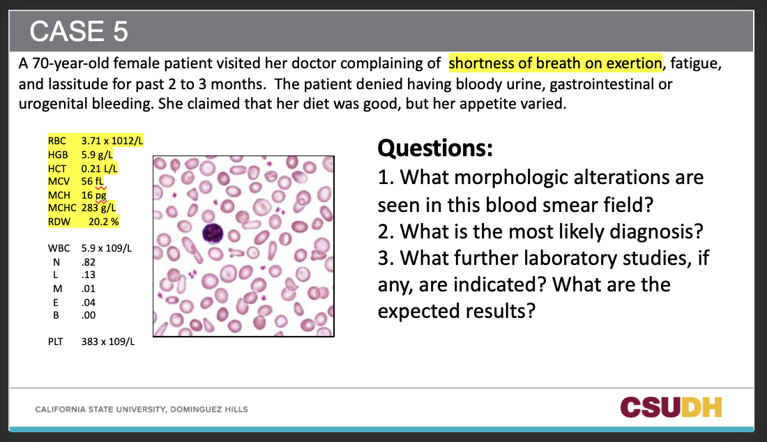
A2: Microcytosis, hypochromia, anisocytosis, poikilocytosis with ovalocytes/pencil cells.
62
Q4: Which CBC feature supports anisocytosis?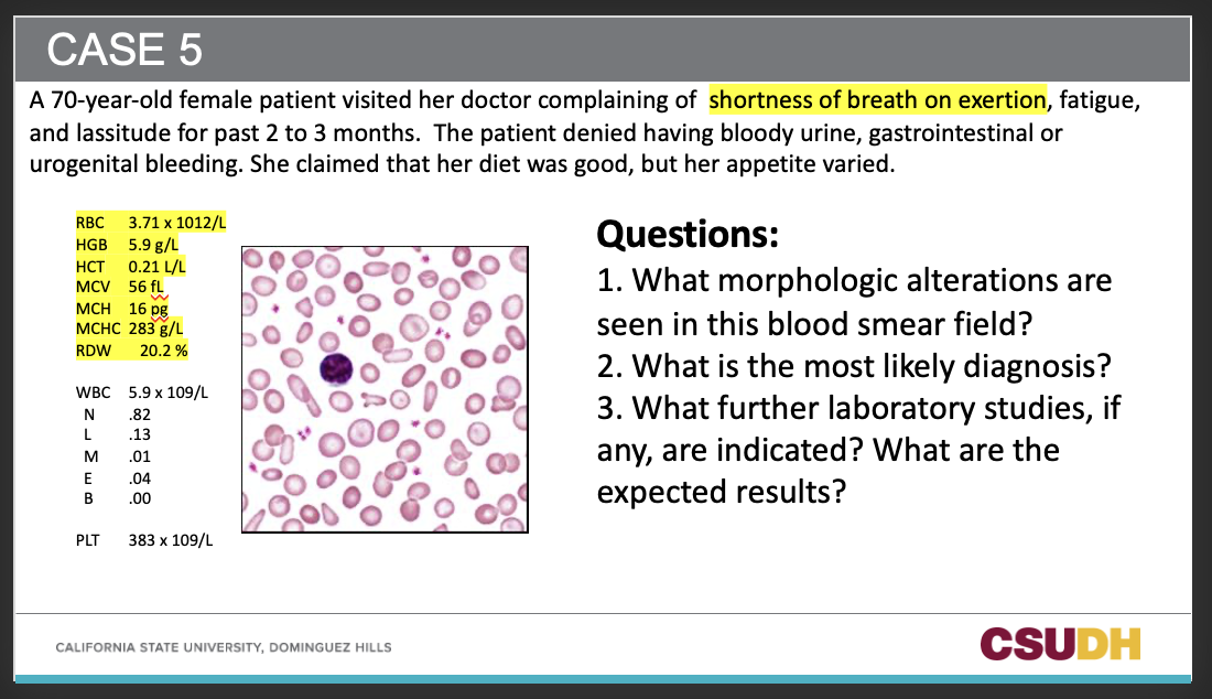
A4: RDW 20.2% (elevated).
63
Q5: What does the platelet count of 383 ×10⁹/L suggest in IDA?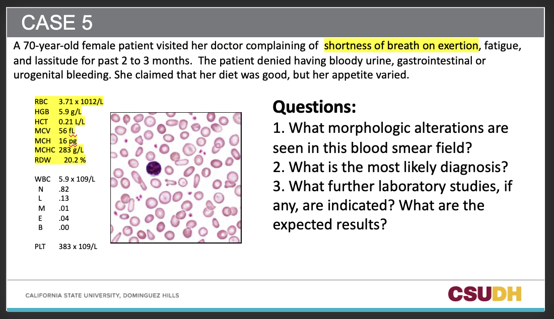
A5: Reactive thrombocytosis/high-normal platelets.
64
Q8: What clinical symptoms did the patient report?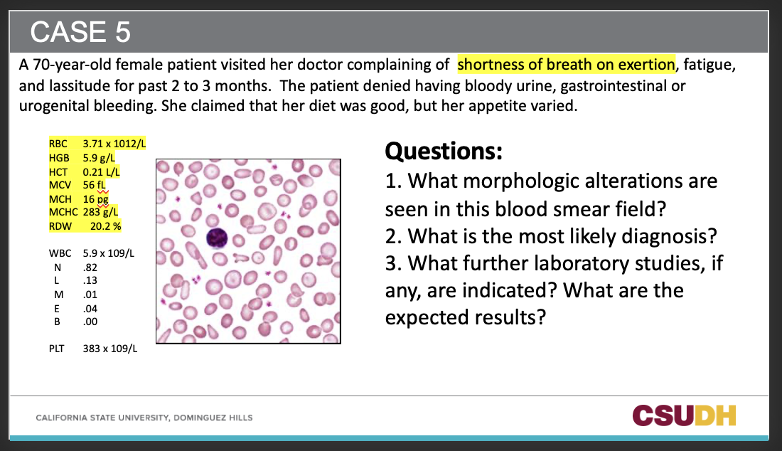
A8: Shortness of breath on exertion, fatigue, lassitude for 2–3 months.
65
Q10: Why are pencil cells common in this case?
A10: They are characteristic of advanced IDA with marked hypochromic microcytosis.
66
Q11: How does high RDW help distinguish IDA from thalassemia trait?
A11: IDA usually has high RDW; thalassemia trait often has normal RDW with high/normal RBC count.
67
Q12: What reticulocyte change is expected after starting iron therapy?
A12: Reticulocyte count rises in 5–10 days; Hb begins to rise by 2–3 weeks.
68
A, B, C, OR D?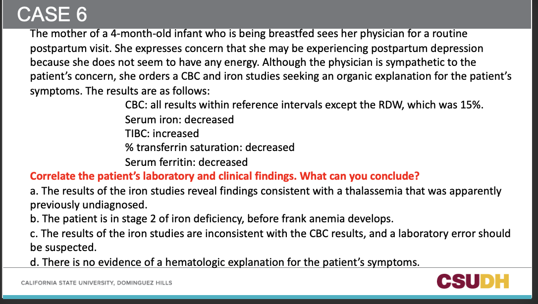
Conclusion: b) The patient is in Stage 2 (latent) iron deficiency, before frank anemia develops.
Why: CBC indices are essentially normal (only RDW 15%), while iron studies show SI↓, TIBC↑, transferrin saturation↓, ferritin↓—the classic pre-anemia ID pattern. Symptoms (low energy) fit iron-restricted erythropoiesis.
Why: CBC indices are essentially normal (only RDW 15%), while iron studies show SI↓, TIBC↑, transferrin saturation↓, ferritin↓—the classic pre-anemia ID pattern. Symptoms (low energy) fit iron-restricted erythropoiesis.
69
Q5: Which finding argues against thalassemia trait in this case?
A5: Abnormal iron studies (ferritin low, SI low, TIBC high); thalassemia trait usually has normal iron studies.
70
Q7: Why might a breastfeeding mother develop iron deficiency?
A7: Increased iron needs can outpace stores (consistent with IDA etiology in the lecture).
71
Anemia of chronic inflammation is associated with what underlying chronic systemic disease?
- Infections (e.g., HIV, TB)
- Autoimmune diseases (e.g, RA, SLE)
- Malignancies
- Autoimmune diseases (e.g, RA, SLE)
- Malignancies
72
Q2: Name infectious examples linked to anemia of chronic inflammation.
A2: HIV and TB.
73
Q3: Name autoimmune examples linked to anemia of chronic inflammation.
A3: Rheumatoid arthritis (RA) and systemic lupus erythematosus (SLE).
74
Q5: What is the main mechanism of anemia of chronic inflammation?
The main mechanism is impaired ferrokinetics: iron-restricted erythropoiesis in spite of abundant iron in the body, increased hepcidin inhibits iron absorption from intestines and iron release from macrophages/hepatocytes
75
Q6: Which hormone is increased, and what does it do? (in anemia of chronic inflammation)
A6: Hepcidin; it inhibits intestinal iron absorption and iron release from macrophages/hepatocytes.
76
Q7: What are the usual PBS (smear) findings?
in anemia of chronic inflammation
in anemia of chronic inflammation
A7: Normochromic, normocytic RBCs.
77
Q8: When would a hypo/micro picture be seen on PBS?
in anemia of chronic inflammation
in anemia of chronic inflammation
A8: If there is coexisting iron deficiency.
78
Q9: In anemia of chronic inflammation, what happens to serum iron (SI)?
A9: Decreased.
79
Q10: In anemia of chronic inflammation, what happens to TIBC?
A10: Decreased.
80
Q11: In anemia of chronic inflammation, what happens to serum ferritin?
A11: normal or Increased.
81
Q12: In anemia of chronic inflammation, what happens to transferrin saturation?
A12: Normal or decreased.
82
Q13: In anemia of chronic inflammation, what happens to soluble transferrin receptor (sTfR)?
A13: Typically normal.
83
Q14: In anemia of chronic inflammation, what happens to the hemoglobin content of reticulocytes?
A14: Decreased.
84
A, B, C, D? and predict the iron study results for this patient with Hodgkin lymphoma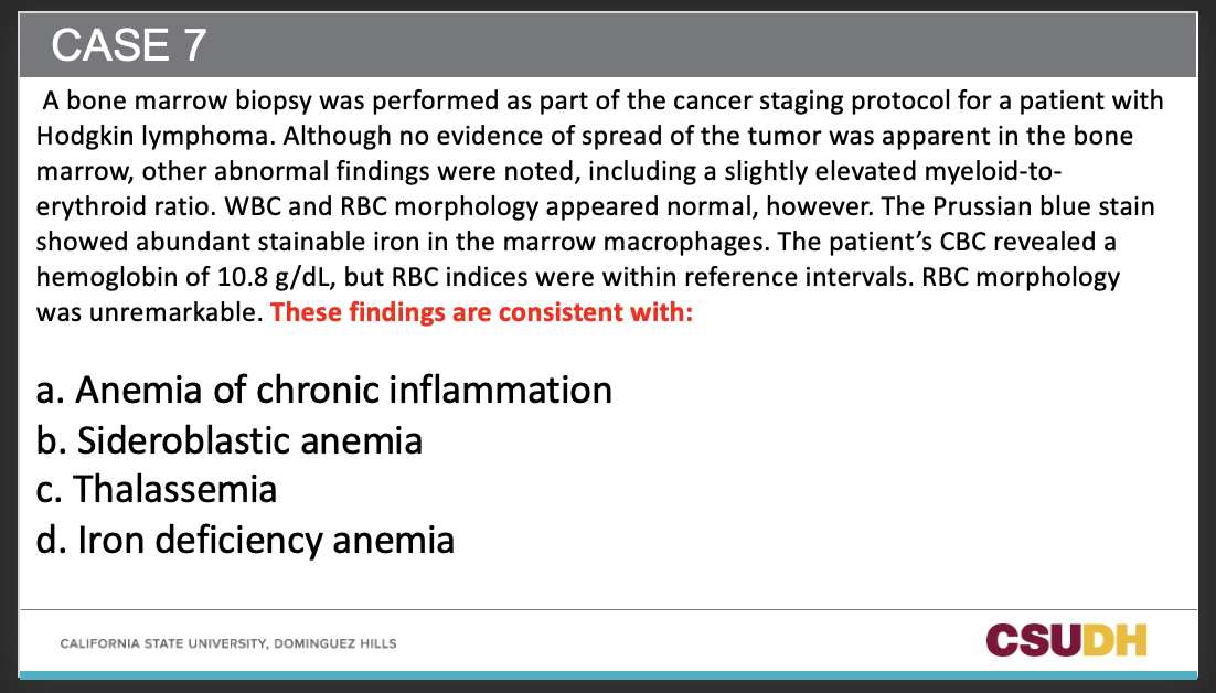
A. Anemia of Chronic Inflammation
Iron studies: D.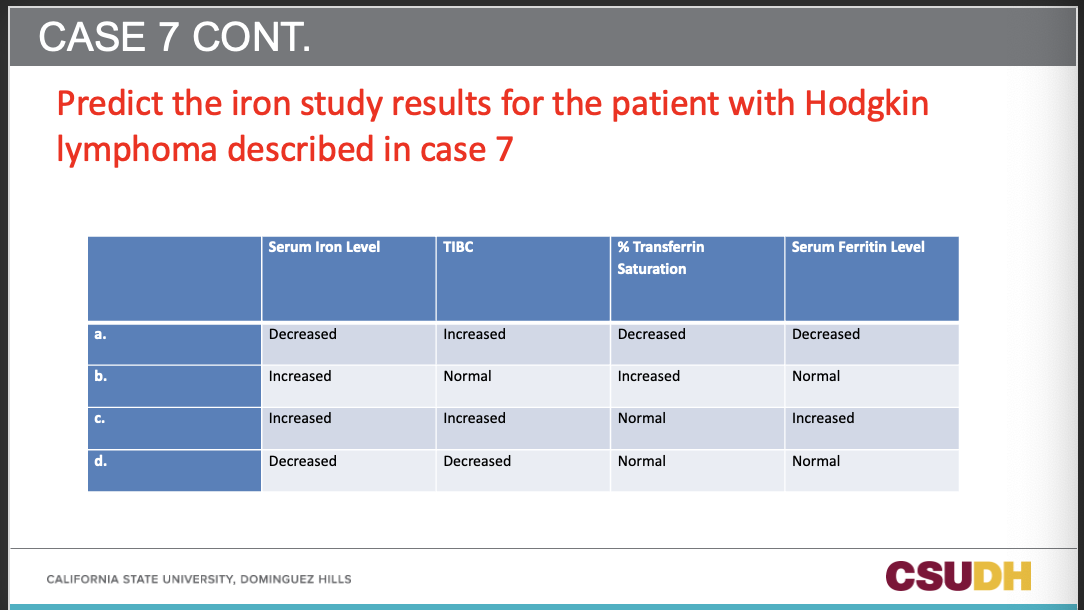
Iron studies: D.
85
Q1: What type of production disorder is sideroblastic anemia?
A1: A “heme” production disorder.
86
Q2: Which pathway is disrupted in sideroblastic anemia?
A2: The protoporphyrin production pathway.
87
Q3: How are bone marrow iron stores in sideroblastic anemia?
A3: Iron is abundant in the bone marrow.
88
Q4: What does Prussian blue staining show in sideroblastic anemia?
A4: “Ringed” sideroblasts.
89
Q5: Where are the iron deposits located in ringed sideroblasts?
A5: In the mitochondria surrounding the nucleus.
90
Q6: What hereditary condition listed is associated with sideroblastic anemia?
A6: Porphyrias.
91
Q7: Name an acquired cause listed for sideroblastic anemia.
A7: Lead poisoning.
92
Sideroblastic anemia: lead poision is a major health concern especially with what type of individuals? why?
in growing children because it impairs mental development with anemia
93
What are the effects of lead on heme synthesis pathway?
Increased FEP OR ZPP
It can also inhibit ALA dehydratase leading to acumulation of ALA, which can be measured in urine
It can also inhibit ALA dehydratase leading to acumulation of ALA, which can be measured in urine
94
Q1: What is the usual anemia pattern in lead poisoning?
A1: Mostly normochromic, normocytic.
95
Q2: How can the indices change with chronic lead exposure?
A2: They can become hypochromic and microcytic.
96
Q3: In whom is anemia from lead poisoning typically more severe?
A3: Children.
97
Q4: What is the classic peripheral smear finding in lead poisoning?
A4: Coarse basophilic stippling (punctate basophilia).
98
Q5: What causes coarse basophilic stippling in lead poisoning?
A5: Lead inhibition of pyrimidine 5′-nucleotidase.
99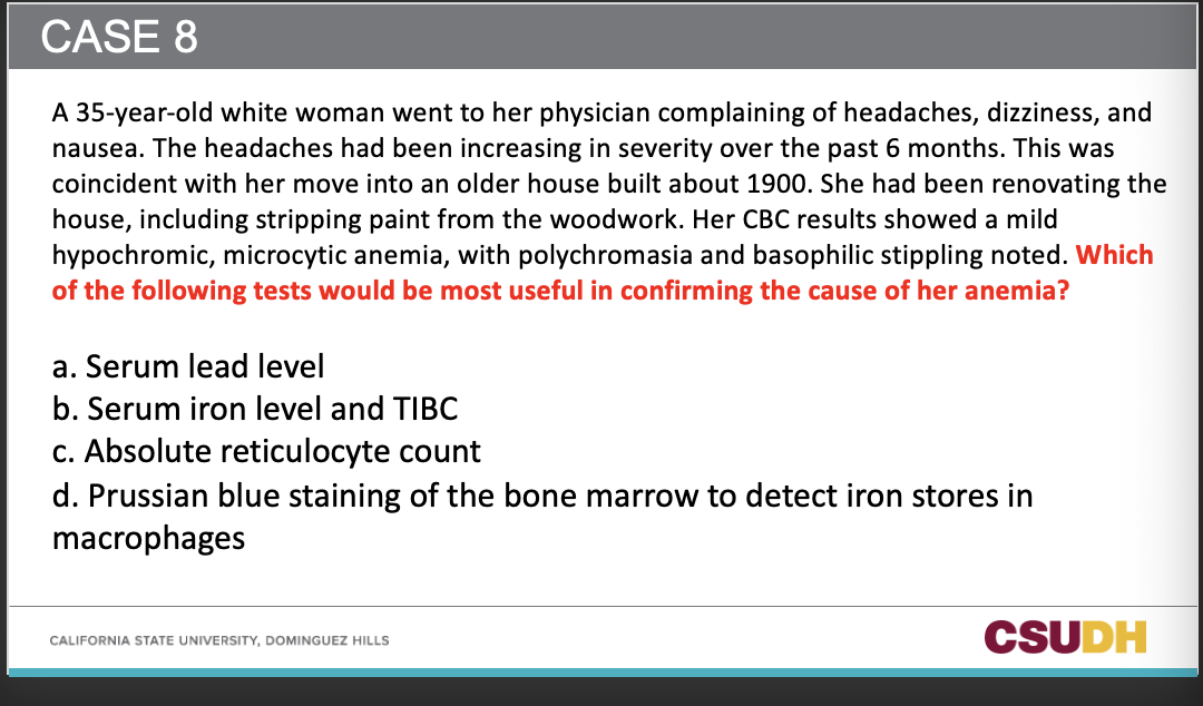
A
100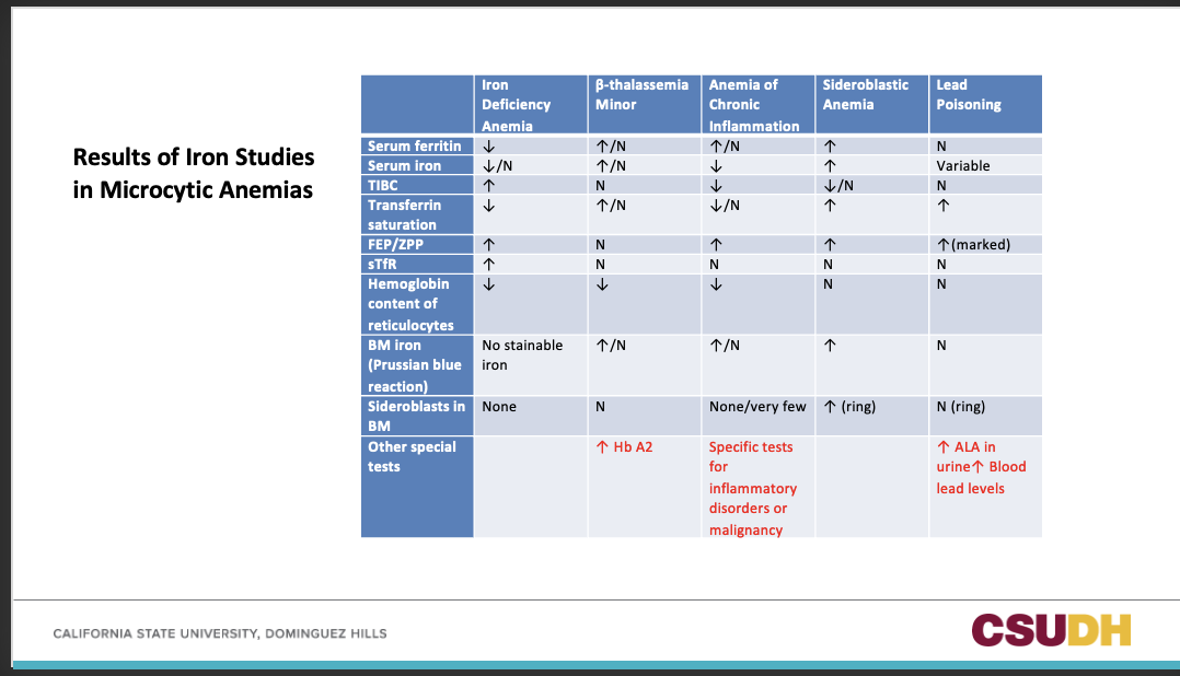
101
What is most likely explanation of this patient's anemia?
a. hereditary hemochromatosis
b. lead poisioning
c. iron-deficiency anemia
d. sideroblastic anemia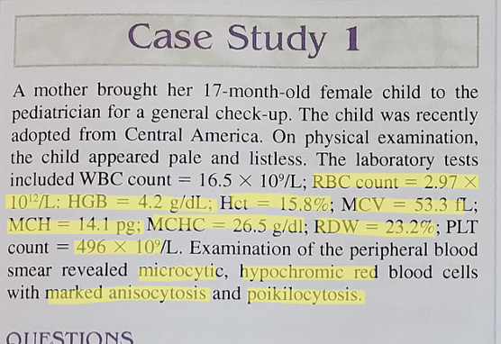
a. hereditary hemochromatosis
b. lead poisioning
c. iron-deficiency anemia
d. sideroblastic anemia
c. Iron-deficiency anemia.
Severe microcytic, hypochromic anemia (MCV 53.3 fL, MCHC 26.5 g/dL) with high RDW (23.2%) and reactive thrombocytosis—classic IDA pattern
Severe microcytic, hypochromic anemia (MCV 53.3 fL, MCHC 26.5 g/dL) with high RDW (23.2%) and reactive thrombocytosis—classic IDA pattern
102
What additional test may useful in confirming the diagnosis?
a. hemoglobin electrophoresis
b. vitamin b12 and folate levels
c. measurement of serum iron, ferritin, and TIBC
d. chromosome analysis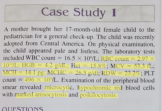
a. hemoglobin electrophoresis
b. vitamin b12 and folate levels
c. measurement of serum iron, ferritin, and TIBC
d. chromosome analysis
c. Measurement of serum iron, ferritin, and TIBC.
These confirm IDA: ↓serum iron, ↓ferritin, ↑TIBC, ↓transferrin saturation.
These confirm IDA: ↓serum iron, ↓ferritin, ↑TIBC, ↓transferrin saturation.
103
Iron-deficiency anemia is associated with:
a. decreased hemoglobin, increased MCV, increased serum ferritin, and decreased serum iron
b. decreased hemoglobin, decreased hematocrit, decreased serum ferritin, and increased stainable bone marrow iron
c. decreased hemoglobin, decreased heamtocrit, increased serum iron, increased serum ferritin, and marked anisocytosis and poikilocytosis.
d. decreased hemoglobin, decreased serum iron, decreased ferritin, increased TIBC, and decreased transferrin saturation.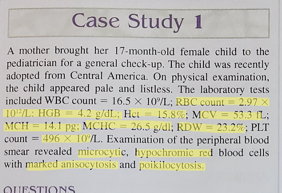
a. decreased hemoglobin, increased MCV, increased serum ferritin, and decreased serum iron
b. decreased hemoglobin, decreased hematocrit, decreased serum ferritin, and increased stainable bone marrow iron
c. decreased hemoglobin, decreased heamtocrit, increased serum iron, increased serum ferritin, and marked anisocytosis and poikilocytosis.
d. decreased hemoglobin, decreased serum iron, decreased ferritin, increased TIBC, and decreased transferrin saturation.
d. Decreased hemoglobin, decreased serum iron, decreased ferritin, increased TIBC, and decreased transferrin saturation.
This is the characteristic lab profile for IDA.
This is the characteristic lab profile for IDA.
104
Assuming the patient has normal dietary iron absorption the minimum dietary requirement of iron for this patient would be:
a. 3 mg/day
b. 0.5 mg/day
c. 2 mg/day
d. 1 mg/day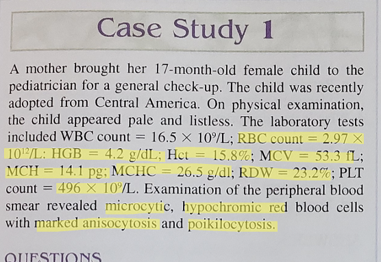
a. 3 mg/day
b. 0.5 mg/day
c. 2 mg/day
d. 1 mg/day
b. 0.5 mg/day
105
Prussian blue staining of the bone marrow in this patient would reveal:
a. increased storage and increased sideroblastic iron
b. increased storage and decreased sideroblastic iron
c. decreased storage and decreased sideroblastic iron
d. increased ringed sideroblasts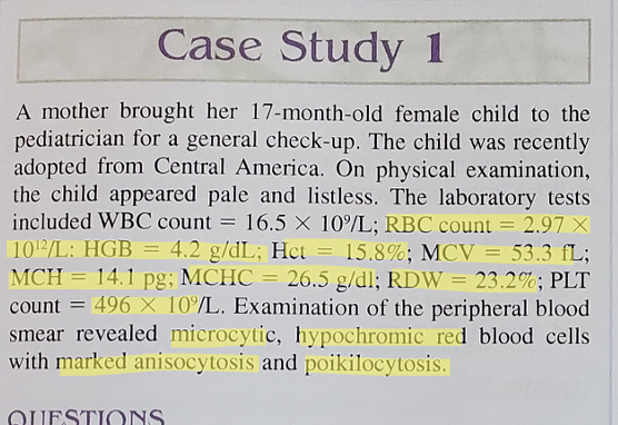
a. increased storage and increased sideroblastic iron
b. increased storage and decreased sideroblastic iron
c. decreased storage and decreased sideroblastic iron
d. increased ringed sideroblasts
c.
106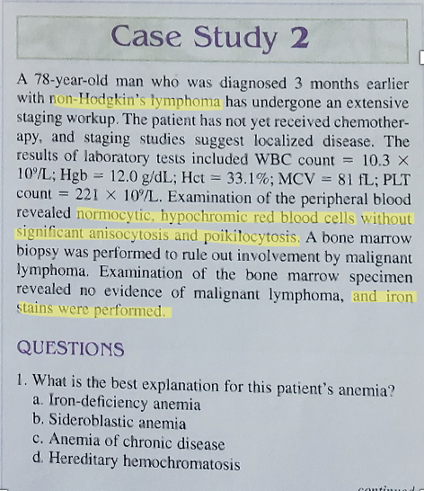
Answer: c. Anemia of chronic disease.
Why: Mild normocytic, hypochromic anemia with no significant anisopoikilocytosis in a patient with non-Hodgkin’s lymphoma fits anemia of chronic inflammation. Expected iron studies: low serum iron & TIBC, normal/high ferritin, normal sTfR; marrow iron present/abundant.
Why: Mild normocytic, hypochromic anemia with no significant anisopoikilocytosis in a patient with non-Hodgkin’s lymphoma fits anemia of chronic inflammation. Expected iron studies: low serum iron & TIBC, normal/high ferritin, normal sTfR; marrow iron present/abundant.
107
If serum iron studies were performed on this patient, they would reveal:
a. decreased serum iroon, decreased serum ferritin, and increased TIBC
b. decreased serum iron, normal to increased serum ferritin, and decreased TIBC
c. increased serum iron, increased serum ferritin, and decreased or normal TIBC
d. increased serum iron, increased serum ferritin, and increased TIBC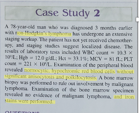
a. decreased serum iroon, decreased serum ferritin, and increased TIBC
b. decreased serum iron, normal to increased serum ferritin, and decreased TIBC
c. increased serum iron, increased serum ferritin, and decreased or normal TIBC
d. increased serum iron, increased serum ferritin, and increased TIBC
b. decreased serum iron, normal to increased serum ferritin, and decreased TIBC
108
Prussian blue staining of a bone marrow aspirate specimen from this patient would reveal:
a. decreased stainable bone marrow iron and decreased ringed sideroblasts
b. decreased stinable bone marrow iron and increased ringed sideroblasts
c. increased stainable bone marrow iron and decreased marrow sideroblasts
d. increased stainable bone marrow iron and increased ringed sideroblasts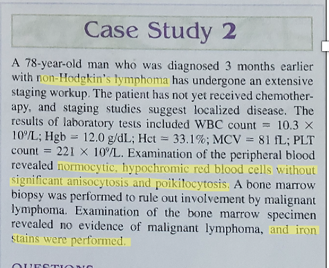
a. decreased stainable bone marrow iron and decreased ringed sideroblasts
b. decreased stinable bone marrow iron and increased ringed sideroblasts
c. increased stainable bone marrow iron and decreased marrow sideroblasts
d. increased stainable bone marrow iron and increased ringed sideroblasts
c. increased stainable bone marrow iron and decreased marrow sideroblasts
109
What is the most likely cause of this patient's disorder?
a. iron deficiency anemia
b. anemia of chronic disease
c. aquired sideroblastic anemia
d. hereditary hemochromatosis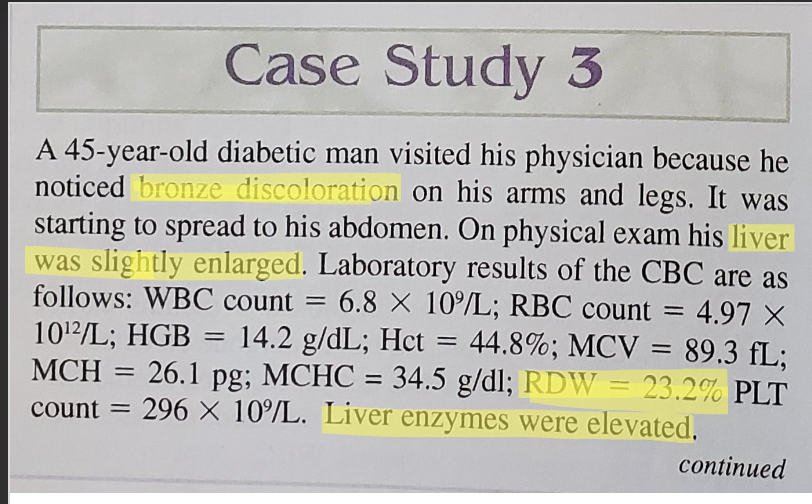
a. iron deficiency anemia
b. anemia of chronic disease
c. aquired sideroblastic anemia
d. hereditary hemochromatosis
d. hereditary hemochromatosis
110
The serum iron studies in this patient would reveal:
a. increased serum iron, increased serum ferritin, and increawsed percent transferrin saturation
b. decreased serum iron, normal serum ferritin, and decreased percent transferrin saturation
c. decreased serum iron, decreased serum ferritin, and decreased percent transferrin saturation
d. increased sumer iron, decreased serum ferritin, and increased percent transferrin saturation.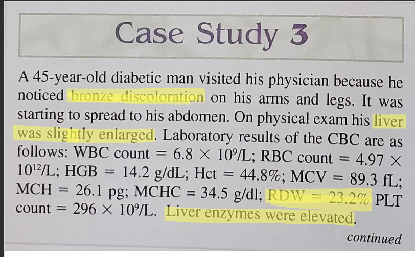
a. increased serum iron, increased serum ferritin, and increawsed percent transferrin saturation
b. decreased serum iron, normal serum ferritin, and decreased percent transferrin saturation
c. decreased serum iron, decreased serum ferritin, and decreased percent transferrin saturation
d. increased sumer iron, decreased serum ferritin, and increased percent transferrin saturation.
a. increased serum iron, increased serum ferritin, and increased percent transferrin saturation
111
What additional test would be most useful in confirming the diagnosis?
a. hemoglobin eletrophoresis
b. vitain b12 and folate levels
c. reticulocyte count
d. measurement of transferrin saturation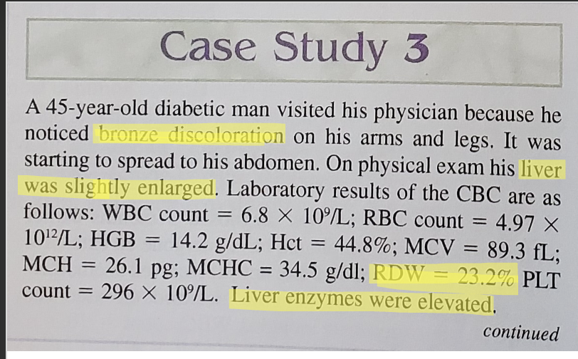
a. hemoglobin eletrophoresis
b. vitain b12 and folate levels
c. reticulocyte count
d. measurement of transferrin saturation
d. measurement of transferrin saturation
112
What is the appropriate therapy or treatment for this disorder?
a. blood transfusion
b. chelation therapy
c. phlebotomy
d. iron supplement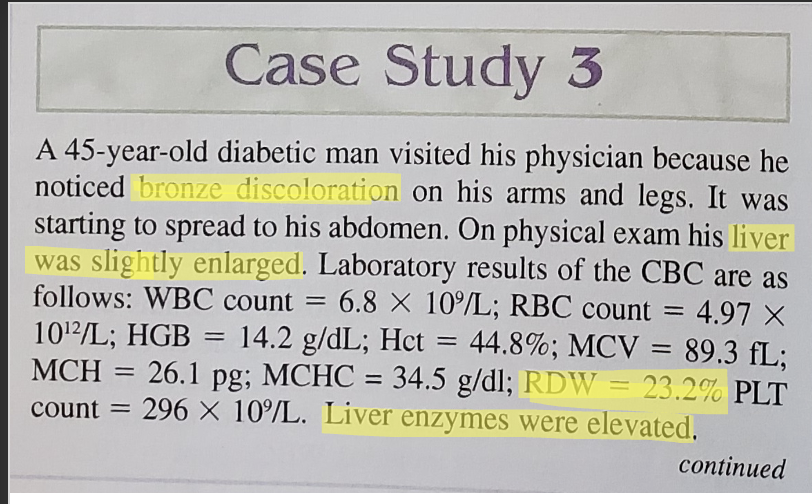
a. blood transfusion
b. chelation therapy
c. phlebotomy
d. iron supplement
c. phlebotomy