Urine Cultures (2)
Study these flashcardsFree · spaced repetition · iPhone, Android, Mac & web
All cards
1
Q: What structures make up the urinary tract?
A: Kidneys, ureters, bladder, urethra
2
Q: What does UTI stand for?
A: Urinary tract infection
3
Q: What organs are involved in an Upper UTI?
A: Kidneys (pyelonephritis) and ureter (ureteritis)
4
Q: What organs are involved in a Lower UTI?
A: Bladder (cystitis), urethra (urethritis), prostate in males (prostatitis)
5
Q: Why are females more prone to UTIs?
A: The female urethra is shorter and in close proximity to the perirectal region, allowing organisms to easily reach the bladder
6
Q: How does sexual activity affect UTI risk in females?
A: Sexual activity can increase contamination of the female urethra
7
Q: Is urine normally sterile?
A: Yes, urine is typically sterile
8
Q: From where can urine become contaminated?
A: microbiota from Perineum, vagina, or urethra
9
Q: Why are quantitative cultures used in UTI diagnosis?
A: To discriminate between contamination, colonization, and infection
10
Q: What are the routes of infection listed for UTIs?
A: Ascending, hematogenous, lymphatic
11
Q: Which route of infection is most common?
A: Ascending
12
Q: What does “ascending” mean in community-acquired UTIs?
A: Bacteria moves up the urinary tract from urethra to bladder and potentially to kidney
13
Q: What healthcare-associated factors contribute to ascending infection?
A: Catheterization, cystoscopy
14
Q: How do organisms enter the bladder in healthcare-associated ascending infections?
A: They are pushed along the urethra into the bladder or migrate along the tract between the catheter and urethral mucosa
15
Q: What is the hematogenous route of infection?
A: Systemic infection can lead to seeding of kidneys
16
Q: Which organisms are examples of hematogenous spread to kidneys?
A: Staphylococcus aureus, Salmonella species
17
Q: What is the lymphatic route of infection?
A: Pressure on bladder causes lymphatic flow into kidneys
18
Q: Why is urine inhibitory to organisms? (4)
A: Low pH, high or low osmolality, high urea, high organic acid content
19
Q: How does constant flushing help prevent infection?
A: It eliminates bacteria
20
Q: What can lead to UTI by interfering with normal voiding?
A: Kidney stone or strictures
21
Q: What property does the bladder mucosal surface have?
A: Antibacterial properties
22
Q: What does the valve-like mechanism at the junction of the ureter and bladder prevent?
A: Reflux from bladder to upper UT
23
Q: What is uromodulin also known as?
A: Tamm-Horsfall protein
24
Q: What is the function of uromodulin (Tamm-Horsfall protein)?
A: Anti-adherence protein
25
Q: What do organisms adhere to in the urinary tract?
A: Vaginal and uroepithelial cells
26
Q: What happens during hydrolysis of urea?
A: Increase pH is toxic to kidney cells and stimulates formation of kidney stones
27
Q: What is the effect of increased pH on kidney cells?
A: It is toxic to kidney cells
28
Q: What does increased pH stimulate?
A: Formation of kidney stones
29
Q: What can uropathogens do to bladder epithelial cells?
A: Invade them and become intracellular bacteria that can shed as a result of exfoliation
30
Q: What are examples of other virulence factors?
A: Necrotizing factor, protease
31
Q: What are struvite crystals also known as? and what are they associated with?
A: Triple phosphates; High pH on urine microscopy. 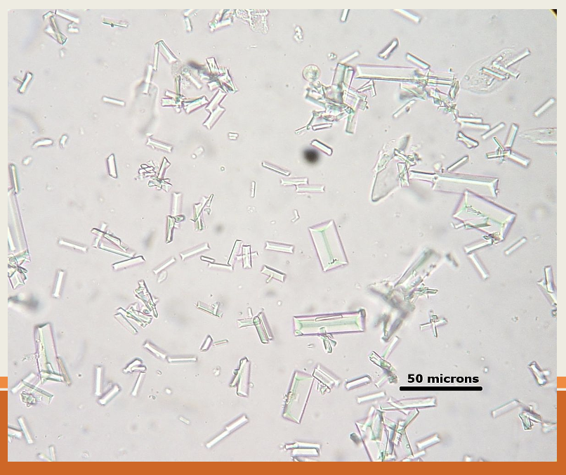
32
Q: What urine specimens are unacceptable?
A:
Urine catheter tips (i.e. Foley catheters)
Pooled 24-hour urine
Unrefrigerated or unpreserved urine more than 2 hours old
More than 1 specimen/day
Non-sterile urines for anaerobic culture
Urine catheter tips (i.e. Foley catheters)
Pooled 24-hour urine
Unrefrigerated or unpreserved urine more than 2 hours old
More than 1 specimen/day
Non-sterile urines for anaerobic culture
33
Q: How long is refrigerated urine acceptable?
A: 24 hours
34
Q: What must be done if urine is unrefrigerated?
A: Specimen must be transported in preservation (0.5 mL boric acid)
35
Q: Why must manufacturer minimum volume be followed when using preservative?
A: To avoid inhibition of uropathogens
36
Q: What type of urine collection is discouraged for neonates, infants, and young children?
A: Pedibag urines, but can be analyzed if that’s the only way to obtain specimen
37
Q: How can a urine culture be ordered?
A: Stand-alone or as urinalysis (UA) with reflex to culture
38
Q: Under UCLA criteria, when can a UA reflex to culture be performed?
A: If no urine culture was collected in the past 24 hours and any one of the criteria is met on UA
39
Q: What UA findings meet criteria for reflex to urine culture?
A:
Leukocyte esterase is positive (positive, 1+, 2+, 3+) – evidence of host response
Nitrite is positive
WBC is >52/µL (9 WBC/HPF) – pyuria
Leukocyte esterase is positive (positive, 1+, 2+, 3+) – evidence of host response
Nitrite is positive
WBC is >52/µL (9 WBC/HPF) – pyuria
40
Q: Is Gram stain typically performed for urine cultures?
A: No, typically not performed (available per request)
41
Q: What suggests contamination on Gram stain?
A: Squamous epithelial cells and many different bacterial morphotypes
42
Q: What media are used for urine cultures?
A: BAP and MAC
43
Q: Why may CNA agar be added to urine cultures?
A: To visualize gram positive microbiota (typically, gram negative will overgrow)
44
Q: What media combination is commonly used by hospitals for urine cultures?
A: CNA/MAC biplate
45
Q: Why are chromogenic media such as CHROMagar Orientation used?
A: To help distinguish uropathogens
46
Q: What is Columbia CNA agar used for?
A: Selective media for isolation of gram positive organisms
47
Q: Which agents in CNA agar inhibit gram negatives?
A: Colistin and nalidixic acid
48
Q: What can be observed about colonial morphology on CNA agar?
A: Media may exhibit multiple colonial morphologies from the same strain
49
Q: Which gram negatives may grow on CNA agar? examples?
A: Gram negatives that are resistant to colistin and nalidixic acid;
A: Proteus, Morganella
A: Proteus, Morganella
50
Q: What is another selective media for gram positives mentioned?
A: Phenylethylalcohol (PEA)
51
Q: How does Staph grow on PEA compared to CNA?
A: Better growth of Staph on PEA than CNA
52
Q: What does PEA inhibit?
A: Swarming Proteus
53
Q: What is done for sterile urine with no colony count?
A: Centrifuge urine, inoculate BAP and MAC with 1 drop of sediment and streak for isolation
54
Q: How are urological reconstructions with no colony count processed?
A: One drop of urine on each agar plate (BAP, CNA, MAC) and streak for isolation
55
Q: How is routine/non-sterile urine with colony count inoculated?
A: Inoculate media with 0.001 mL calibrated loop on BAP and MAC
56
Q: What does 1 colony equal in urine culture quantitation?
A: 1,000 CFU/mL
57
Q: At what temperature and conditions are urine cultures incubated?
A: 35–37°C at ambient air for minimum of 18 hours
58
Q: How long are sterile urines incubated?
A: 48 hours
59
Q: When can incubation be extended or plates incubated in CO₂?
A: Upon physician request
60
Q: Why might plates be incubated in the CO₂ incubator?
A: To enhance gram positive growth
61
Q: What pathogens are commonly recovered in urine cultures?
A:
Escherichia coli
Other Enterobacterales (i.e. Klebsiella species, Proteus species, Enterobacter species)
Pseudomonas aeruginosa (and other nonfermenters)
Staphylococcus aureus
Staphylococcus saprophyticus
Enterococcus species
Streptococcus agalactiae
Candida species (yeast)
Escherichia coli
Other Enterobacterales (i.e. Klebsiella species, Proteus species, Enterobacter species)
Pseudomonas aeruginosa (and other nonfermenters)
Staphylococcus aureus
Staphylococcus saprophyticus
Enterococcus species
Streptococcus agalactiae
Candida species (yeast)
62
Q: What are typical urogenital microbiota?
A:
Lactobacilli
Viridans Group Streptococcus
Coryneform bacteria
Coagulase negative Staphylococci
Lactobacilli
Viridans Group Streptococcus
Coryneform bacteria
Coagulase negative Staphylococci
63
Q: According to the Clinical Microbiology Procedures Handbook, how is urogenital or skin microbiota handled in workup?
A: “Urogenital or skin microbiota that is at least 10 times less than the uropathogens is ignored for purposes of workup”
64
Q: When is antimicrobial susceptibility testing performed?
A: Once pathogens are identified on bacterial cultures
65
Q: What is antimicrobial susceptibility testing also called?
A: Susceptibility testing, sensitivity testing
66
Q: What is the purpose of antimicrobial susceptibility testing?
A: To determine whether the bacterial isolates can express resistance to antimicrobial agents selected for treatment
67
Q: Why is antimicrobial susceptibility testing a key component of the laboratory report?
A: It can help guide therapy for pathogens that have unpredictable susceptibility pattern
68
Q: What methods does the UCLA bacteriology lab use for susceptibility testing?
A: Broth microdilution, disk diffusion testing, and FDA cleared automated susceptibility testing (Vitek 2)
69
Q: When will procedures and methods for antimicrobial susceptibility testing be covered?
A: During AML training
70
Q: What will bacteriology section training focus on?
A: Understanding which agents to report for each organism and body site
71
Q: What is generally the criteria for a significant isolate?
A: ≥10⁵ (100,000) CFU/mL
72
Q: What is noted about protocol for significant isolates?
A: Protocol varies among different institutions
73
Q: What lower colony counts may be significant in certain cases?
A: 10⁴ or even 10²
74
Q: What is done for sterile urines?
A: ID and susceptibility testing on all isolates
75
Q: What is done for reconstruction urines?
A: ID and susceptibility testing on up to 2 organisms
76
Q: What is done if more than 2 organisms are isolated in reconstruction urines?
A: Report each organism using minimal/descriptive ID only
77
Q: What are examples of descriptive ID?
A:
Pseudomonas-like
Lactose positive/negative gram negative rod
Probable Staphylococcus aureus
Coryneform
Viridans strep-like
Coagulase negative Staphylococcus-like
Enterococcus-like
Pseudomonas-like
Lactose positive/negative gram negative rod
Probable Staphylococcus aureus
Coryneform
Viridans strep-like
Coagulase negative Staphylococcus-like
Enterococcus-like
78
Q: What type of specimens are covered in this section?
A: Routine/Non-sterile urines
79
Q: When is ID and susceptibility testing performed for Staphylococcus aureus or gram negative rods?
A: If ≥50,000 CFU/mL of pure isolate
80
Q: When is ID and susceptibility testing performed for up to 2 organisms?
A: If ≥100,000 CFU/mL each
81
Q: What does “No Significant Growth” mean?
A:
<50,000 CFU/mL each of >2 organisms
<10,000 CFU/mL of non-pathogens
<50,000 CFU/mL each of >2 organisms
<10,000 CFU/mL of non-pathogens
82
Q: When is “Contamination Suspected; Submit new specimen” reported?
A: ≥50,000 CFU/mL each of >2 organisms
83
Q: What is done for all other cultures that do not fit criteria?
A: Report quantitation with descriptive ID
84
Q: What is the example given for reporting quantitation with descriptive ID?
A: SA and EC 20,000 CFU/mL each – report 20,000 CFU/mL Probable Staphylococcus aureus and 20,000 CFU/mL Lactose positive gram negative rod
85
Q: What organisms are not routinely tested for susceptibility on urine cultures?
A:
Lactobacillus species
Staphylococcus saprophyticus
Aerococcus species
Beta Streptococcus
Corynebacterium, not urealyticum
Viridans group Streptococcus
Yeast
Lactobacillus species
Staphylococcus saprophyticus
Aerococcus species
Beta Streptococcus
Corynebacterium, not urealyticum
Viridans group Streptococcus
Yeast
86
Q: Why is Staphylococcus saprophyticus not routinely tested for susceptibility?
A: Susceptible to commonly prescribed oral antibiotics for UTI
87
Q: What is noted about Beta Streptococcus susceptibility?
A: Routinely susceptible to Penicillin
88
Q: What type of hemolysis does E. coli show on BAP?
A: Beta or non-beta hemolytic colonies on BAP
89
Q: How is E. coli described on lactose media?
A: Flat lactose positive gram negative rod (some strains lactose negative)
90
Q: What is the most commonly isolated organism from urine cultures?
A: Escherichia coli
91
Q: What is the indole, MR, VP, LDC, and Motility reaction of E. coli?c
A: Positive (+) - INDOLE
A: Positive (+) - MR
A: Negative (−) - VP
A: Positive (+) - LDC
A: Positive (+) - MOTILITY
A: Positive (+) - MR
A: Negative (−) - VP
A: Positive (+) - LDC
A: Positive (+) - MOTILITY
92
Q: How do Klebsiella colonies generally appear?
A: Generally raised, can be mucoid
93
Q: How is Klebsiella described on lactose media?
A: Raised lactose positive gram negative rod
94
Q: What are the key biochemical characteristics for the genus Klebsiella?
A:
VP +
ODC −
Motility −
VP +
ODC −
Motility −
95
Q: What test is used to differentiate between Klebsiella pneumoniae and Klebsiella oxytoca?
A: Indole
96
Q: What is the indole, VP, Motility, Gel, ADH, LDC, ODC reaction of Klebsiella pneumoniae?
Negative (-) - Indole
Positive (+) - VP
Negative (-) -Motility
Negative (-) - Gel
Negative (-) - ADH
Positive (+) - LDC
Negative (-) -ODC
Positive (+) - VP
Negative (-) -Motility
Negative (-) - Gel
Negative (-) - ADH
Positive (+) - LDC
Negative (-) -ODC
97
Q: What is the indole, VP, Motility, Gel, ADH, LDC, ODC reaction of Klebsiella oxytoca?
Positive (+) - Indole
Positive (+) - VP
Negative (-) - Motility
Negative (-) - Gel
Negative (-) - ADH
Positive (+) - LDC
Negative (-) - ODC
Positive (+) - VP
Negative (-) - Motility
Negative (-) - Gel
Negative (-) - ADH
Positive (+) - LDC
Negative (-) - ODC
98
Q: How can Enterobacter species appear on lactose media?
A: Can be lactose positive or lactose negative
99
Q: What are the key biochemical characteristics for the genus Enterobacter?
VP +
ODC + (differentiates from Klebsiella)
DNase −, Gel − (differentiates from Serratia)
ODC + (differentiates from Klebsiella)
DNase −, Gel − (differentiates from Serratia)
100
Q: What is the LDC reaction of Enterobacter cloacae
A: Negative (−)
101
Q: What is the ADH reaction of Enterobacter aerogenes?
A: Negative (−)
102
Q: What is Enterobacter aerogenes currently reclassified as?
A: Klebsiella aerogenes
103
Q: What are the biochemical reactions of Enterobacter cloacae?
A:
Indole −
VP +
ADH +
ODC +
LDC −
Motility +
Indole −
VP +
ADH +
ODC +
LDC −
Motility +
104
Q: What are the biochemical reactions of Enterobacter aerogenes?
A:
Indole −
VP +
ADH −
ODC +
LDC +
Motility +
Indole −
VP +
ADH −
ODC +
LDC +
Motility +
105
Q: How is Serratia marcescens described on lactose media?
A: Lactose negative gram negative rod
106
Q: What type of pathogen is Serratia marcescens?
A: Significant nosocomial opportunist
107
Q: What pigmentation can some strains of Serratia marcescens exhibit?
A: Red/pink pigmentation
108
Q: How should red/pink pigmentation in Serratia be interpreted in relation to lactose?
A: Avoid interpreting this as lactose positive
109
Q: How can pigmentation be enhanced in Serratia cultures?
A: By holding cultures longer and leaving at room temperature
110
Q: Which other Serratia species are also pigmented?
A: Serratia rubidaea
111
Q: What are the key biochemical characteristics of the genus Serratia?
A:
VP +
Gel + / DNase +
VP +
Gel + / DNase +
112
Q: What test is used to differentiate Serratia marcescens from Serratia liquefaciens?
A: RAX (Raffinose, Arabinose, Xylose)
113
Q: What is the RAX reaction of Serratia marcescens?
A: RAX −
114
Q: What is the RAX reaction of Serratia liquefaciens?
A: RAX +
115
Q: What is characteristic of Serratia odorifera?
A: Pungent odor of potatoes/crushed vegetables
116
Q: What are the biochemical reactions of Serratia marcescens?
A:
Indole −
VP +
Gel +
DNase +
ADH −
ODC +
LDC +
Motility +
Indole −
VP +
Gel +
DNase +
ADH −
ODC +
LDC +
Motility +
117
Q: On which media may Proteus swarm?
A: BAP/CHOC (rarely CNA)
118
Q: How is Proteus described on lactose media?
A: Lactose negative gram negative rod
119
Q: What is true of all members of the genus Proteus?
A: All members are TDA positive
120
Q: What smell may strains of Proteus mirabilis have?
A: “Chocolate” or devil’s food cake smell
121
Q: What are the ODC and indole reactions of Proteus mirabilis?
A: ODC +, indole −
122
Q: What are the ODC and indole reactions of Proteus vulgaris?
A: ODC −, indole +
123
Q: What are the ODC and indole reactions of Proteus penneri?
A: ODC −, indole −
124
Q: What are the biochemical reactions of Proteus mirabilis?
A:
Indole −
Urea +
H₂S +
TDA +
ODC +
Gelatin +
Indole −
Urea +
H₂S +
TDA +
ODC +
Gelatin +
125
Q: What are the biochemical reactions of Proteus vulgaris?
A:
Indole +
Urea +
H₂S +
TDA +
ODC −
Gelatin +
Indole +
Urea +
H₂S +
TDA +
ODC −
Gelatin +
126
Q: Is Pseudomonas aeruginosa a fermenter or nonfermenter?
A: Nonfermenter
127
Q: How is Pseudomonas aeruginosa described on lactose media?
A: Lactose negative gram negative rod
128
Q: What odors are associated with Pseudomonas aeruginosa colonies?
A: Grape/corn tortilla odor
129
Q: What sheen may Pseudomonas aeruginosa colonies exhibit?
A: Metallic sheen
130
Q: What pigment is associated with Pseudomonas aeruginosa?
A: Diffuse green pigment
131
Q: What is the oxidase reaction when Pseudomonas aeruginosa has a metallic sheen?
A: Oxidase (+)
132
Q: What are the biochemical reactions when Pseudomonas aeruginosa does not have a metallic sheen?
A: Oxidase (+) and acetamide (+)
133
Q: What type of hemolysis do Enterococcus colonies show?
A: Gamma colonies (can also be beta or alpha)
134
Q: What are the biochemical characteristics used to identify Enterococcus to the genus?
A:
Catalase (−)
PYR (+)
6.5% NaCl (+)
Bile-esculin (+) (non-Enterococcus group D are bile-esculin −)
Catalase (−)
PYR (+)
6.5% NaCl (+)
Bile-esculin (+) (non-Enterococcus group D are bile-esculin −)
135
Q: What is the treatment of choice for Enterococcus in urine cultures?
A: Ampicillin (concentrates well in the bladder)
136
Q: When is susceptibility testing performed for outpatient cultures?
A: Only upon request
137
Q: When is susceptibility testing automatically performed?
A: For inpatient cultures
138
Q: What method is used for full speciation (for Enterococcus) if needed?
A: API Strep
139
Q: How do Staphylococcus saprophyticus colonies appear?
A: Opaque, flat white colonies
140
Q: What are the biochemical reactions of Staphylococcus saprophyticus?
A:
Catalase (+)
Coagulase (−)
Novobiocin R (≤16 mm)
Catalase (+)
Coagulase (−)
Novobiocin R (≤16 mm)
141
Q: Is routine susceptibility testing performed for Staphylococcus saprophyticus?
A: No, routine susceptibility not performed
142
Q: What antibiotics are commonly used to treat uncomplicated UTI caused by Staphylococcus saprophyticus?
A: Bactrim (SXT), fluoroquinolone, nitrofurantoin
143
Q: Staphylococcus saprophyticus is associated with which patient population?
A: Young, sexually active females and IUD
144
Q: What type of hemolysis does Streptococcus agalactiae show on BAP?
A: Narrow zone of beta-hemolysis on BAP
145
Q: What are the key biochemical characteristics of Streptococcus agalactiae?
A:
Catalase (−)
Lancefield group B (ID by latex agglutination)
Catalase (−)
Lancefield group B (ID by latex agglutination)
146
Q: What are the other biochemical reactions of Streptococcus agalactiae?
A:
CAMP (+)
Hippurate (+)
6.5% NaCl (+)
Bile-esculin (−)
Bacitracin and SXT (R)
CAMP (+)
Hippurate (+)
6.5% NaCl (+)
Bile-esculin (−)
Bacitracin and SXT (R)
147
Q: In which patients should Streptococcus agalactiae be reported in any quantity?
A: Females of childbearing age (14–55 yo per protocol)
148
Q: Why is Streptococcus agalactiae significant in females of childbearing age?
A: Organism is implicated in neonatal infections
149
Q: Which organism can cross react with group B latex?
A: Streptococcus pseudoporcinus
150
Q: How can Streptococcus pseudoporcinus be identified?
A: MALDI-TOF
151
Q: What is the clinical implication of Streptococcus pseudoporcinus?
A: Premature rupture of membranes or spontaneous birth
152
Q: In which population is Streptococcus pseudoporcinus more prevalent?
A: African American
153
Q: How does Streptococcus pseudoporcinus differ from GBS (Group B streptococcus) on blood agar?
A: Larger zone of beta-hemolysis than GBS and LAP (+)
154
Q: How do most coryneform bacteria relate to the urogenital tract?
A: Most coryneform bacteria are urogenital microbiota, except for Corynebacterium urealyticum
155
Q: What type of infection does Corynebacterium urealyticum cause?
A: UTI with alkaline encrusted cystitis (stones)
156
Q: What urinalysis findings are associated with Corynebacterium urealyticum infection?
A:
Urinalysis pH 8–9
Struvite crystals present on UA microscopy
Urinalysis pH 8–9
Struvite crystals present on UA microscopy
157
Q: What is the antibiotic resistance profile of Corynebacterium urealyticum?
A: Highly resistant to gram positive antibiotic, but susceptible to vancomycin (similar to Corynebacterium jeikeium)
158
Q: How does Corynebacterium urealyticum appear on Gram stain?
A: Small gram positive coccobacilli, PLGPR
159
Q: How do colonies of Corynebacterium urealyticum appear on BAP?
A: Slow-growing, non-hemolytic pinpoint colonies at 24 hour
160
Q: What is the growth requirement of Corynebacterium urealyticum regarding lipids?
A: Lipophile (will not grow if plated on CHOC)
161
Q: What are the biochemical reactions of Corynebacterium urealyticum?
A:
Catalase +
Urea: Rapid (+), <10 min (key characteristic)
Glucose (−)
Nitrate (−)
Esculin (−)
Catalase +
Urea: Rapid (+), <10 min (key characteristic)
Glucose (−)
Nitrate (−)
Esculin (−)
162
Q: How does Aerococcus relate to urogenital flora and Viridans group Streptococcus?
A: Aerococcus can be a member of the urogenital flora; differentiate from Viridans group Streptococcus if numbers are significant or pure
163
Q: How does Aerococcus urinae appear on Gram stain?
A: Gram positive cocci in clusters (Staph-like)
164
Q: What are the colony characteristics of Aerococcus urinae?
A: Small alpha hemolytic colonies (looks like Str. viridans)
165
Q: What are the biochemical reactions of Aerococcus urinae?
A:
Catalase (−)
6.5% NaCl (+)
Catalase (−)
6.5% NaCl (+)
166
Q: What are the Bacticard Strep reactions for Aerococcus urinae?
A: LAP +, PYR −, ESC v
167
Q: What is the availability status of Bacticard Strep?
A: No longer available
168
Q: How can full identification of Aerococcus urinae be performed?
A: Using API Strep kit
169
Q: What disease is caused by Salmonella typhi?
A: Typhoid Fever
170
Q: How common is Salmonella typhi in the USA and what is it often related to?
A: Rare in USA, often related to foreign travel
171
Q: What symptoms occur in the first 1–2 weeks of Salmonella typhi infection?
A: Fever, headache, and constipation
172
Q: What are the culture results in the first 1–2 weeks of illness?
A:
Blood cultures (+)
Stool and urine cultures (−)
Blood cultures (+)
Stool and urine cultures (−)
173
Q: What symptom appears after 2 weeks of Salmonella typhi infection?
A: Diarrhea
174
Q: What are the culture results after 2 weeks of illness?
A:
Blood cultures (−)
Stool and urine cultures (+)
Blood cultures (−)
Stool and urine cultures (+)
175
Q: What is the biochemical characteristic of Salmonella typhi on TSI?
A: Weak H₂S+ (mustache-look streak of TSI)
176
Q: What must be done if an organism is identified as Salmonella species?
A: Must serologically type
177
Q: What is the O antigen?
A: Heat stable somatic antigen
178
Q: What is the Vi antigen?
A: Heat labile capsular polysaccharide (mask O Ag)
179
Q: What must be done if the Vi antigen is detected?
A: Organism must be boiled and typing is repeated to detect the somatic antigen
180
Q: What does it mean if the somatic antigen is not detected after boiling?
A: Then it is not a Salmonella
181
Q: What is the purpose of abbreviated ID on urine cultures?
A: Commonly isolated organisms from urine cultures may be identified to species or genus using basic spot tests
182
Q: How is Escherichia coli identified when beta-hemolytic on BAP?
A: Spot Indole (+)
183
Q: How is Escherichia coli identified when non-hemolytic on BAP?
A: Spot Indole (+) & PYR (−)
184
Q: What colony morphology is associated with Proteus mirabilis?
A: Swarmer
185
Q: What test is performed for Proteus mirabilis?
A: Spot Indole (−)
186
Q: What colony morphology is associated with Enterococcus species?
A: Enterococcus-like
187
Q: What test is performed for Enterococcus species?
A: PYR (+)