Lecture 2 - Hemostatis part 2
Study these flashcardsFree · spaced repetition · iPhone, Android, Mac & web
All cards
1
How many coagulation factors are there? redo question
8 factors are zymogens
2
what are zymogens?
circulating in plasma in an inactive form
3
What is FII?
Prothrombin
4
What is FVII?
Stable factor or proconvertin
5
What is FIX?
Plasma thromboplastin component, Christmas factor
6
What is FX?
Stuart-Prower Factor
7
What is FXI?
Plasma thromboplastin antecedent
8
What is FXII ?
Hageman Factor
9
What is FXIII?
plasma transglutaminase, fibrin-stabilizing factor
10
what is another name for Prekalikrein?
Fletcher
11
Which of the 8 zymogens are serine proteases?
ALL except FXIII (13)
12
How many cofactors are there in coagulation?
7 proteins
13
What do co-factors do?
stabilize and accelerate proteolytic reactions
14
What are the 7 co-factors in coagulation?
HMWK, Fitzgerald
Tissue Factor (FIII, tissue thromboplastin)
FV(liabile factor, proaccelerin)
FVIII (liable factor, AHF A)
Protein S (co-factor for activated protein C)
Protein Z (co-facotor for protein Z-dependent protease inhibitor, ZPI)
Thrombomodulin (Co-factor for thrombin in protein C activation
Tissue Factor (FIII, tissue thromboplastin)
FV(liabile factor, proaccelerin)
FVIII (liable factor, AHF A)
Protein S (co-factor for activated protein C)
Protein Z (co-facotor for protein Z-dependent protease inhibitor, ZPI)
Thrombomodulin (Co-factor for thrombin in protein C activation
15
What does HMWK stand for?
High molecular weight kininogen
16
What are the regulatory proteins (inhibitors)?
Anti-thrombin
Protein C
Heparin
Co-factor II
TFPI (tissue factor pathway inhibitor)
Protein C
Heparin
Co-factor II
TFPI (tissue factor pathway inhibitor)
17
Other coag factors?
FI (fibrinogen), vWF, Ca+ (FIV), phospholipids (PF3)
18
What does the suffix "a" after the factor indicate?
"active or activated" (e.g., VIIa, XIIa
19
The roman numerals are based on what?
order of discovery or description of factors
20
Factor I is usually called what?
Fibrinogen
21
Factor II is usually called what?
Prothrombin
22
Which coag factors never received a roman numeral?
PK (prekallikren) and HMWK (high molecular weight kininogen)
23
Which coag factors are synthesized in the liver and which are not?
Almost all except vWF
24
Where does vWF come from?
ECs (stored in Weibel-Palade granules) and MKs (stored in platelet alpha granules)
25
Which is the heaviest coag factor?
vWF (500-20,000 kD)
26
Which is the shortest half life coag factor? and the longest?
FVII (7) ; 3-6 hrs <- shortest
HMWK <- longest
HMWK <- longest
27
Where is VIII carried? why?
in plasma as bound to vWF to maintain 12 hr halflife
28
What is crucial for coagulation complexes?
Ionized calcium!! to bind to platelet or cell membrane phospholipids.
29
How are factors activated?
each factor (serine protease) activates the next, and this continues...
30
What is the outcome of coagulation cascade?
conversion of fibrinogen (ultimate substrate) into stable fibrin clot
31
What is a Vitamin K-dependent coag proteins?
SCZ27910 (aka a prothrombin group reagent)
32
What does Vitamin K-dependent coag proteins require? and to become what?
require vit K to become γ-carboxylated (hence functional)
33
Vitamin K-dependent coag proteins contain how many glutamic acid residues and where?
10-12 glutamic acid residues at N terminal
34
Why is prothrombin group important?
assembly of coagulation complexes to generate thrombin
35
How many coagulation complexes are there?
3
36
What are each complex called:
COMPLEX 1:
COMPLEX 2
COMPLEX 3?
COMPLEX 1:
COMPLEX 2
COMPLEX 3?
COMPLEX 1: EXTRINSIC TENASE
COMPLEX 2: INTRINSIC TENASE
COMPLEX 3: PROTHROMBINASE
COMPLEX 2: INTRINSIC TENASE
COMPLEX 3: PROTHROMBINASE
37
What is the complex 1: extrinstic tenase? and what does it activate?
Tissue factor + VIIa + Ca2++ phospholipid;
it activates X to Xa, and IX to IXa
it activates X to Xa, and IX to IXa
38
What is the complex 2: intrinstic tenase? and what does it activate?
IXa (9)+ VIIIa (8)+ phospholipid (PLT surface) + Ca2+ ;
Activates X (10) to Xa
Activates X (10) to Xa
39
What is the complex 3: prothrombinase? and what does it activate?
Xa (10) + Va (5) + phospholipid + Ca2+ ;
Activates prothrombin to thrombin
Activates prothrombin to thrombin
40
Which are the KEY factors in coagulation (severe bleeding in hemophilia A and vWD?
FVIII (8)and vWF
41
Which chromosome encodes FVIII (8)and FIX (9)?
X chromosome
42
Which factor is degraded in stored plasma faster than other factors?
FVIII
43
What is FVIII bound and carried by and where?
vWF
44
What is the structure of vWF?
multimeric, subunits of 240 kd polymerized to 500 -2000kd in MK and ECs, degraded to smaller mulitmers by ADAMTS13 (a disintergrin and metalloprotease with a thrombospondin type 1 motif, member 13) in blood vessels with higher shear rate
45
What receptors does vWF have? and what does it do?
PLTs and collagen; bridges PLTs to subendothelium during PLT adhesion
46
What is vWF level associated with?
ABO groups, O group has lower level.
47
Which are the acute phase reactants?
vWF and FVIII (8)
48
What constitues the contact system (contact factors, contact factor complex)?
FXII (12), PK, HMWK
49
What is the basis of the Activated Partial Thromboplastin Time (aPTT) test?
Contact system
50
What happens once FXII (12) is activated by negatively charged surfaces?
- FXIIa converts PK (bound of HMWK) to K, K further activates FXII (12)
- CONTACT FACTOR COMPLEX (HMWK + PK + FXIIa) activates FXI ( in turn activates FIX (9)
- CONTACT FACTOR COMPLEX (HMWK + PK + FXIIa) activates FXI ( in turn activates FIX (9)
51
What is the most important activator of FXI? and what is it generated by?
THROMBIN!! ; generated by the extrinsic tenase
52
What happens when there's deficiences in the contact system factors?
NO bleeding BUT do prolong aPTT.
53
What is thrombin? and what is the function?
- KEY serine protease in coagulation with multiple functions;
- Cleaving fibrinopeptides A and B (FPA and FPB) from fibrinogen, triggering spontaneous polymerization, clot intitiation.
- Cleaving fibrinopeptides A and B (FPA and FPB) from fibrinogen, triggering spontaneous polymerization, clot intitiation.
54
What does thrombin activate? what type of loop is it pos/neg?
FV (5), FVIII (8), AND FXI (11)( postiive feedback loop)
55
Q: What is the major function of thrombin in clotting?
Q: Which clotting factors are activated by thrombin to amplify clotting?
Q: What happens when thrombin binds to thrombomodulin?
Q: What does thrombin do to platelets?
Q: What is TAFI and what does thrombin do to it?
Q: What does thrombin do to Factor XIII?
Q: Which clotting factors are activated by thrombin to amplify clotting?
Q: What happens when thrombin binds to thrombomodulin?
Q: What does thrombin do to platelets?
Q: What is TAFI and what does thrombin do to it?
Q: What does thrombin do to Factor XIII?
A: It cleaves fibrinopeptides A and B from fibrinogen, turning it into fibrin to start clot formation.
A: Factors V, VIII, and XI — a positive feedback loop.
A: It activates protein C, which turns off clotting (anticoagulant function).
A: It activates platelets, helping them stick and form a plug.
A: Thrombin activates TAFI (thrombin activatable fibrinolysis inhibitor), which slows down fibrinolysis (clot breakdown).
A: It activates FXIII, which cross-links fibrin and stabilizes the clot.
A: Factors V, VIII, and XI — a positive feedback loop.
A: It activates protein C, which turns off clotting (anticoagulant function).
A: It activates platelets, helping them stick and form a plug.
A: Thrombin activates TAFI (thrombin activatable fibrinolysis inhibitor), which slows down fibrinolysis (clot breakdown).
A: It activates FXIII, which cross-links fibrin and stabilizes the clot.
56
What is Fibrinogen?
primary substrate of thrombin
57
What is the RI of fibringoen in plasma?
200-400mg/dL, an APR
58
FIBRINOGEN: Essential for platelet ______ through binding the _____ receptor?
aggregation; GPIIb/IIa
59
Where is it stored - fibrinogen?
PLTs absorb it and stor in alpha granules
60
What is FXIII (13)? (name)
plasma transglutaminase aka a fibrin stabilizing factor
61
What happens when theres a deficiency in FXIII?
deficiency does not affect PT or PTT BUT leads to clinical bleeding
62
What is are the 3 in vitro model for coagulation pathways?
Extrinsic
Intrinsic
Common
Intrinsic
Common
63
What is the primary initiator of in vivo coagulation in the Extrinsic pathway?
FVII (7) primary initiator of in vivo coagulation
64
What is the reaction order of the Extrinsic pathway?
VII, X, V, prothrombin, and fibrinogen
65
How is the extrinsic pathway assessed in clinical lab? (what test)
Prothrombin Time (PT) test
66
What is the reaction order of Intrinsic pathway?
XII, pre-K, HMWK, XI, IX, VIII, X, V, prothrombin, and fibrinogen
67
How is the intrinsic pathway assessed in clinical lab? (what test)
activated partial thromboplastin time (aPTT) test
68
What is included in the common pathway?
X, V, PROTHROMBIN AND FIBRINOGEN
69
How is the common pathway assessed in clinical lab (what test)?
Both tests plus thrombin test (TT)
70
Where is tissue factor hidden?
endothelial
71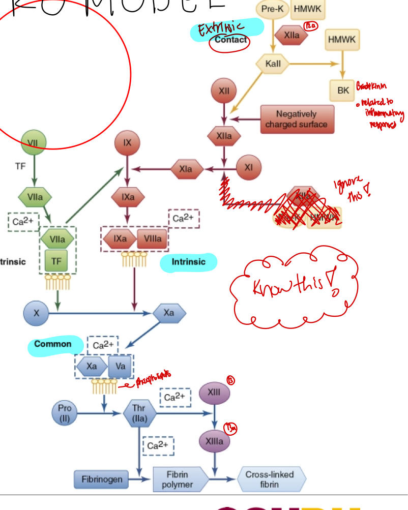
tell me the cascade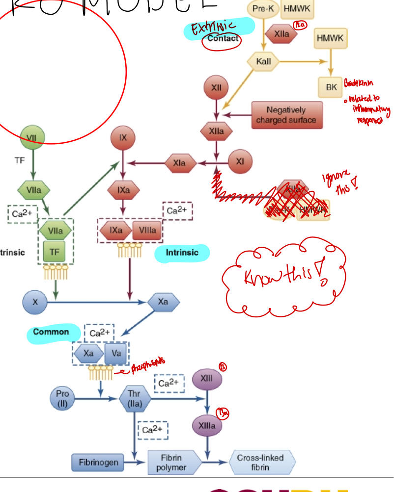
72
The in vivo model includes what ?
Initiation and Propagation
73
During initiation, what is the main initiator of thrombin generation?
extrinsic tenase complex
74
which factor circulates but inerts during initiation and how much % wise?
1-2% FVIIa circulates but inert (requires TF)
75
Once FVIIa meets TF, what do they form? and what does it create?
a complex that can turn on other clotting factors (like Factor X (10) and IX (9)) <-low levels;
This creates a small amount of thrombin which is just enough to start a chain reaction (positive feedback loop) that makes more thrombin but still just a little at this stage (about 3% max)
This creates a small amount of thrombin which is just enough to start a chain reaction (positive feedback loop) that makes more thrombin but still just a little at this stage (about 3% max)
76
During propagation, how much of thrombin is generated? and where do these reactions happen?
>95% thrombin is generated.;
happens on the PLT membrane.
happens on the PLT membrane.
77
When does the intrinsice tenase complex start to work? and what does it activate
When lots of platelets stick togehter (form aggregates), they provide a perfect surface for the Intrinsic tenase complex. ;
this super-activates Factor X (FX) - up to 50 to 100 times more than in the initiation phase.
this super-activates Factor X (FX) - up to 50 to 100 times more than in the initiation phase.
78
Once the intrinsic tenase complex super -activates Factor X (FX), what happens?
this leads to the formation of the prothrombinase complex, which causes a big burst of thrombin - enough to really build a strong clot.
79
What does Serpin stand for?
serine protease inhibitor
80
list 4 regulatory proteins of coagulation ?
Anti-thrombin
Protein C
Heparin Cofactor II
TFPI (tissue factor pathway inhibitor)
Protein C
Heparin Cofactor II
TFPI (tissue factor pathway inhibitor)
81
Q1: What is the function of Tissue Factor Pathway Inhibitor (TFPI)?
A1: With Xa, it binds TF:VIIa.
82
Q2: What is Thrombomodulin?
A2: It is an EC surface receptor for thrombin.
83
Q3: What is the role of Protein C?
A3: Serine protease.
84
Q4: What is the function of Protein S?
A4: Cofactor.
85
Q5: What is Antithrombin?
A5: Serpin.
86
Q6: What is the function of Heparin Cofactor II
A6: Serpin.
87
Q7: What is Protein Z-dependent Protease Inhibitor?
A7: Serpin.
88
What is Fibrinolysis?
Final stage of hemostatic activation (hours after)
89
As fibrin is forming, what happens?
Fibrinolytic proteins
90
What is the main substrate in fibrinolysis?
Main substrate is plasminogen produced by the liver
91
Q: What do endothelial cells secrete to activate plasminogen?
A: TPA (tissue plasminogen activator) and UPA (urokinase plasminogen activator); TPA is more effective.
92
Q: What does TPA do in the fibrinolytic process?
A: It degrades fibrin-bound plasminogen to generate plasmin.
93
Q: What does plasmin do?
A: It degrades fibrin and fibrinogen into smaller fragments: X, Y, D, E, and D-D.
94
Q: What is D-D (D-dimer) and why is it important?
A: D-dimer is fibrin-derived and is used as a diagnostic test for thrombosis and fibrinolysis.
95
Q: What is the main inhibitor of fibrinolysis?
A: Plasminogen activator inhibitor-1 (PAI-1), which inactivates TPA and UPA.
96
Q: What type of molecule is PAI-1 and where is it mostly produced?
A: It’s a serpin (serine protease inhibitor), mostly produced by endothelial cells (ECs) and megakaryocytes (MKs).
97
Q: What does α₂-antiplasmin (α₂-AP) do?
A: It binds to and inactivates free plasmin, not plasmin that's bound to fibrin.
98
Q: Where is α₂-antiplasmin produced?
A: In the liver.
99
Q: What activates thrombin activatable fibrinolysis inhibitor (TAFI)?
A: The thrombin-thrombomodulin complex.
100
Q: What is the function of TAFI in fibrinolysis control?
A: It blocks TPA and plasminogen from binding to fibrin and prevents the formation of plasmin.
101
Acquired bleeding disorders are usually caused by?
Trauma or liver disease
102
When they body goes into shock (aka systemic shock) from major trauma, is can lead to?
- A drop in ADAMTS13 (a protein that controls clotting)
- Activation of clotting factors
- Release of tissue factor
- Hyperfibrinolysis (breaking down clots too much)
- Activation of clotting factors
- Release of tissue factor
- Hyperfibrinolysis (breaking down clots too much)
103
The liver makes most clotting factors, so if the liver is sick, what happens?
bleeding can happen.
104
What happens to clotting factors when a patient has liver disease?
SCZ27910 levels drop in liver disease ( this protein helps with clotting)
- The liver also makes Factor VII (7), which has the shortest half-life, so it drops first.
- Since PT (prothrombin time) is very senstitive to FVII (7) levels, PT becomes PROLONGED early in liver disease -- A USEFULL EARLY WARNING SIGN!
- Vitamin K deficiency also prolongs PT (because it's needed to make FVII (7).
- The liver also makes Factor VII (7), which has the shortest half-life, so it drops first.
- Since PT (prothrombin time) is very senstitive to FVII (7) levels, PT becomes PROLONGED early in liver disease -- A USEFULL EARLY WARNING SIGN!
- Vitamin K deficiency also prolongs PT (because it's needed to make FVII (7).
105
How do you tell the difference between liver disease and vitamin K deficiency?
Doctors look at Factor V (5);
- FV is normal in vitamin K deficiency
- FV is low in liver disease
- FV is normal in vitamin K deficiency
- FV is low in liver disease
106
When the liver isn't working well, how can it affect blood clotting?
1. Low Platelets (Thrombocytopenia)
2. DIC can happen in Liver Disease
3. Acute DIC signs
4. Chronic (compensated) DIC
2. DIC can happen in Liver Disease
3. Acute DIC signs
4. Chronic (compensated) DIC
107
When the liver isn't working well, why are there low platelets (thrombocytopenia)?
- An enlarged spleen (common in liver disease) traps and destroys platelets.
- Platelets that are present don't work properly bc of it-- they don't stick together well (shown by low results in platelet aggregation tests)
- Platelets that are present don't work properly bc of it-- they don't stick together well (shown by low results in platelet aggregation tests)
108
When the liver isn't working well, why can DIC happen?
DIC = Disseminated Intravascular Coagulation= body uses up clotting factors too fast
- Liver makes natural clot blockers like antithrombin or protein C, and protein S.
- If the liver is damaged, it doesn't make enough, leading to unregulated clotting (DIC).
- This can be chronic /(body adjusts) or acute (sudden and dangerous)
- Liver makes natural clot blockers like antithrombin or protein C, and protein S.
- If the liver is damaged, it doesn't make enough, leading to unregulated clotting (DIC).
- This can be chronic /(body adjusts) or acute (sudden and dangerous)
109
What are expected results when the liver isn't working well due to acute DIC,?
Long clotting times: PT, PTT, and TT are all prolonged
Low fibrinogen ( used up from too much clotting)
High D-dimer test (shoes fibrin is being broken down a lot)
Low fibrinogen ( used up from too much clotting)
High D-dimer test (shoes fibrin is being broken down a lot)
110
What is chronic (compensated) DIC? and what tests are increased?
the body adjusts over time ;
ONLY D-dimer is increased , other labs might look normal.
ONLY D-dimer is increased , other labs might look normal.
111
Q: What happens to fibrinogen levels in liver disease?
->400 mg/dL (elevated) in early, mild liver disease
<200 mg/dL in moderate to severe liver disease, which causes dysfibrinogenemia
and hypofibrinogenemia
<200 mg/dL in moderate to severe liver disease, which causes dysfibrinogenemia
and hypofibrinogenemia
112
What is the difference between dysfibrinogenemia and hypofibrinogenemia?
Dysfibrinogenemia: Normal amount of fibrinogen is made, but it doesn’t work properly — it's defective and can’t help form clots.
Hypofibrinogenemia: The body doesn’t make enough fibrinogen, leading to a shortage that affects clot formation.
Hypofibrinogenemia: The body doesn’t make enough fibrinogen, leading to a shortage that affects clot formation.
113
Q: What does a prolonged thrombin time (TT) indicate in liver disease?
It suggests dysfibrinogenemia, hypofibrinogenemia, or elevated fibrin degradation products; can also be affected by heparin.
114
Q: Why would you use reptilase time instead of TT?
It’s not affected by heparin and is prolonged in dysfibrinogenemia or hypofibrinogenemia.
115
Q: Which lab test is prolonged even in mild liver disease and why?
Prothrombin time (PT) — because of reduced production of vitamin K–dependent factors like prothrombin (II), VII, and X.
116
Q: When is PTT prolonged in liver disease?
A: In severe liver disease due to loss of clotting factors or DIC.
Mildly prolonged in severe liver disease because of disseminated
intravascular coagulation (DIC) or des-γ-carboxyl factors II
(prothrombin), IX, and X
Mildly prolonged in severe liver disease because of disseminated
intravascular coagulation (DIC) or des-γ-carboxyl factors II
(prothrombin), IX, and X
117
Q: How can Factor V levels help differentiate liver disease from vitamin K deficiency?
A: Factor V is low in liver disease but normal in vitamin K deficiency.
118
Q: What platelet count is seen in liver disease?
A: Mild thrombocytopenia with a platelet count less than 150,000/μL.
119
Q: What does platelet aggregometry show in liver disease?
Mild suppression of platelet aggregation and secretion in response
to most agonists
to most agonists
120
Q: What D-dimer level indicates increased fibrinolysis in liver disease?
A: >240 ng/mL or >500 ng/mL FEUs (fibrinogen equivalent units)
121
Q: What causes acquired hemophilia?
A: Autoantibodies against Factor VIII (8) that inhibit its function.
122
Q: Who is most likely to develop autoantibodies against Factor VIII?
A: Patients over 65 years old.
123
Q: What symptoms can acquired hemophilia cause?
A: Sudden bleeding into soft tissue or the gastrointestinal (GI) tract.
124
Q: What conditions are associated with the development of acquired hemophilia?
Rheumatoid arthritis (RA), inflammatory bowel disease (IBD), lupus (SLE), and chronic lymphocytic leukemia (CLL).
125
Q: What is the mortality rate for acquired hemophilia, even with treatment?
A: Up to 20%.
126
Q: What lab tests should be ordered for patients with sudden bleeding that resembles acquired hemophilia?
A: PT, PTT, and TT.
127
Q: What is the typical PTT, PT, and TT pattern in acquired Factor VIII inhibitor?
A: Prolonged PTT, normal PT and TT.
128
Q: What FVIII level supports a diagnosis of acquired hemophilia?
A: A FVIII level below 40 units/dL, confirmed by factor assay.
129
Q: What does it mean if a prolonged PTT corrects after mixing with normal plasma but becomes prolonged again after incubation at 37°C?
A: It suggests a time-dependent inhibitor, like anti-FVIII.
130
Q: What does it indicate if prolonged PTT does not correct even after a 2-hour incubation?
A: The patient may have lupus anticoagulants or is on heparin therapy.
131
Q: What does it mean if PTT becomes prolonged immediately after mixing in a study?
A: The patient has a high-avidity inhibitor (present in ~15% of cases).
132
What is vWD?
genetic bleeding disorder caused by a problem with von Willebrand factor (vWF), a protein that helps platelets stick and protects Factor VIII.
133
Q: How is von Willebrand disease (vWD) inherited, and who does it affect?
A: It is autosomal dominant, affects both sexes, but symptoms are more common in females.
134
Q: What causes vWD?
A: Gene mutations that lead to either too little (quantitative) or abnormal (qualitative) vWF.
135
Q: What type of bleeding disorder is vWD, and what does it impair?
A: It’s the most common inherited mucocutaneous bleeding disorder that impairs platelet adhesion and primary hemostasis.
136
Q: What happens in severe cases of vWD?
A: Severe deficiency of vWF leads to Factor VIII deficiency, worsening the bleeding risk.
137
Q: In mild (“low vWF”) cases, what vWF levels are usually seen and how is Factor VIII affected?
A: vWF levels are around 30–50%, which is usually enough to maintain normal Factor VIII levels.
138
Q: What is the most common type of von Willebrand Disease?
A: Type 1 — a quantitative deficiency found in 40–70% of cases; usually mild to moderate.
139
Q: What defines Type 2 von Willebrand Disease?
A: It is a qualitative disorder, meaning vWF is present but doesn’t function properly.
140
Q: What is vWD Type 2A?
A: A subtype of Type 2 where large vWF multimers are missing, leading to poor platelet adhesion.
141
Q: What is vWD Type 2B?
A: A subtype where vWF has increased affinity for platelets, causing them to be cleared from circulation and possibly leading to thrombocytopenia.
142
Q: What is vWD Type 2M?
A: A qualitative defect where vWF has reduced platelet binding, but multimers are normal.
143
Q: What is vWD Type 2N?
A: A subtype with defective binding of vWF to Factor VIII, leading to low FVIII levels, mimicking mild hemophilia A.
144
Q: What is vWD Type 3?
A: A null phenotype — almost no vWF is produced, causing severe bleeding and very low Factor VIII levels.
145
Q: What clinical history is very helpful in diagnosing vWD?
A: History of mucocutaneous bleeding and decreased vWF concentration or activity.
146
Q: What is the purpose of ordering a CBC in suspected vWD?
A: To rule out thrombocytopenia (low platelet count) as a cause of bleeding.
147
Q: Why are PT and PTT included in vWD evaluation?
A: To rule out deficiencies in other clotting factors unrelated to vWF.
148
Q: What does the vWF: RCo test measure?
A: It measures vWF activity — how well vWF helps platelets stick together.
149
Q: What does the vWF: Ag test measure?
A: The amount of von Willebrand factor in the blood.
150
Q: What does the vWF activity to antigen ratio tell us?
A: Whether the vWF is functioning properly — a low ratio suggests dysfunctional vWF.
151
Q: What is the purpose of RIPA(ristocetin-induced platelet agg ) in vWD testing?
A: To help detect vWD Type 2B, which causes abnormal platelet aggregation.
152
Q: Why is Factor VIII activity tested in vWD workup?
A: Because vWF protects Factor VIII, so low vWF may lead to low FVIII activity.
153
Q: What are vWF multimers used for?
A: To evaluate the structure and size of vWF — useful for identifying vWD subtypes.
154
Q: In vWD Type 1, how are vWF:Ag and vWF:RCo affected?
A: Both are low due to a quantitative deficiency of vWF.
155
Q: What is unique about Type 2A in terms of vWF multimers?
A: Large and intermediate multimers are absent, affecting vWF function.
156
Q: Which vWD subtype is associated with a decreased platelet count and increased RIPA?
A: Type 2B — due to increased binding of vWF to platelets, causing platelet clearance.
157
Q: What lab results are normal in vWD Type 2M?
A: vWF:Ag and vWF multimers are normal, but vWF activity is low.
158
Q: Which vWD subtype mimics hemophilia A due to low Factor VIII?
A: Type 2N — vWF can't bind/stabilize FVIII.
159
Q: What are the lab findings in vWD Type 3?
A: Very low/absent vWF and FVIII, prolonged PTT, absent RIPA (ristocetin-induced platelet aggregation), and no multimers.
160
Q: What is typically true of the PTT in vWD Type 1?
A: It is normal to slightly prolonged.
161
Q: What distinguishes Type 2N from other vWD types in terms of lab values?
A: vWF is normal, but Factor VIII is low.
162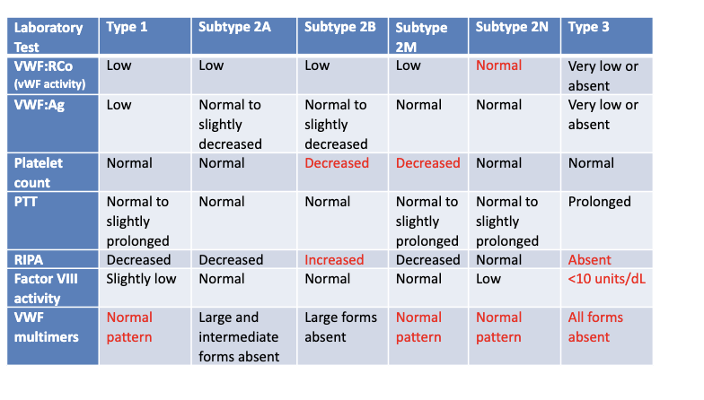
163
What is hemophilia?
a genetic bleeding disorder that runs in families and is present from birth
164
What is the cause of hemophilia?
X-linked recessive: lack of a single clotting factor, which causes bleeding into muscles, joints, or soft tissue (not sure surface bleeding like in vWD.
- Males (who have only one X) usually have the disease.
- Females (who have two X chromosomes) are usually carriers and may not show symptoms.
- Males (who have only one X) usually have the disease.
- Females (who have two X chromosomes) are usually carriers and may not show symptoms.
165
How many types of hemophilia are there?
3:
Hemophilia A, B, C
Hemophilia A, B, C
166
What factor is missing in Hemophilia A?
Missing Factor VIII (most common - 85% of cases)
167
What factor is missing in Hemophilia B?
Missing Factor IX (about 14% of cases)
168
What factor is missing in Hemophilia C?
Missing Factor XI (very rare - about 1% of cases, and not X-linked)
169
Q: What type of deficiency is hemophilia?
A: A congenital single-factor deficiency, meaning a person is born missing one clotting factor.
170
Q: What type of bleeding is common in hemophilia?
A: Anatomic soft tissue bleeding, such as into joints and muscles.
171
Q: What is the inheritance pattern of Hemophilia A and B?
A: X-linked recessive — males are affected, females are typically carriers.
172
Q: What clotting factor is deficient in Hemophilia A and how common is it?
A: Factor VIII (FVIII) — it accounts for 85% of hemophilia cases.
173
Q: What clotting factor is deficient in Hemophilia B and how common is it?
A: Factor IX (FIX) — it causes about 14% of hemophilia cases.
174
Q: What clotting factor is deficient in Hemophilia C and how common is it?
A: Factor XI (FXI) — very rare, found in only 1% of cases.
175
Q: Where is the gene for Factor VIII (FVIII) located?
A: On the X chromosome.
176
Q: What types of genetic mutations can cause hemophilia A?
A: Deletions, stop codons, nonsense, or missense mutations, which reduce FVIII production.
177
Q: How does FVIII deficiency affect coagulation?
A: It slows down thrombin production, which is essential for clotting, leading to increased bleeding.
178
Q: What are the major clinical signs of Hemophilia A?
Anatomic bleeds
Deep muscle and joint hemorrhages
Hematomas (deep bruises)
Wound oozing after injury or surgery
Internal bleeding in the CNS, GI tract, kidneys, and more
Deep muscle and joint hemorrhages
Hematomas (deep bruises)
Wound oozing after injury or surgery
Internal bleeding in the CNS, GI tract, kidneys, and more
179
What reflex tests do you suggest for a patient with normal platelet count and function, normal PT, PTT, TT tests, but with a bleeding history?
You should consider testing Factor XIII, because:
Factor XIII deficiency causes normal PT, PTT, and TT,
But patients still bleed, especially after trauma or surgery.
It's not detected by routine clotting tests — so you need a Factor XIII quantitative assay to diagnose it.
Factor XIII deficiency causes normal PT, PTT, and TT,
But patients still bleed, especially after trauma or surgery.
It's not detected by routine clotting tests — so you need a Factor XIII quantitative assay to diagnose it.
180
Q: What are mixing studies used for?
A: To tell the difference between factor deficiencies and factor inhibitors.
181
Q: What are the 3 main causes mixing studies help differentiate?
Factor deficiencies (single or multiple)
Specific factor antibodies or inhibitors
Non-specific antibodies or inhibitors
Specific factor antibodies or inhibitors
Non-specific antibodies or inhibitors
182
Q: How much of a clotting factor is needed for a normal PT or APTT result?
A: At least 50% of the normal level.
183
Q: Why are mixing studies helpful?
A: They help decide the next steps to find the cause of abnormal PT or APTT.
184
Q: What does it mean if the prolonged PT or APTT corrects after mixing with normal plasma?
A: It suggests a clotting factor deficiency.
185
Q: What does it mean if the prolonged PT or APTT does NOT correct after mixing?
A: It suggests the presence of an inhibitor, like a factor antibody.
186
Q: What does it mean if the PT or APTT corrects initially but becomes prolonged after incubation?
A: It indicates a time-dependent inhibitor, such as an anti-Factor VIII antibody.
187
list the procedure for PT and APTT 50:50 Mixing Studies
Procedure:
1. Repeat PT and/or APTT using patient PPP (platelet poor plasma)
2. Run Normal Pooled Plasma alone
• Ensure normal results or 100% of factor level
3. Run patient PPP mixed 1:1 with NPP
4. Compare results
• Initial testing is compared to mix
• Did mix correct? Did mix not correct?
1. Repeat PT and/or APTT using patient PPP (platelet poor plasma)
2. Run Normal Pooled Plasma alone
• Ensure normal results or 100% of factor level
3. Run patient PPP mixed 1:1 with NPP
4. Compare results
• Initial testing is compared to mix
• Did mix correct? Did mix not correct?
188
Q: What additional test is done if the 1:1 mix does not correct and an inhibitor is suspected?
A: An inhibitor assay, such as DRVVT (dilute Russell viper venom time).
189
Q: What is the next step if the 1:1 mix does correct the result initially?
A: Incubate the mix for 1–2 hours at 37°C to distinguish between a factor deficiency and a factor antibody.
190
Q: After incubation, if the result remains corrected, what is the likely cause?
A: Factor deficiency.
191
Q: After incubation, if the result is no longer corrected, what does it indicate?
A: Presence of a factor-specific antibody.
192
Q: What test is used to confirm the presence of a factor antibody?
A: Factor antibody assays.
193
Q: What does it mean if a 1:1 mix does not correct the prolonged clotting time?
A: It suggests the presence of an inhibitor, such as lupus anticoagulant.
194
Q: What is the purpose of the 5 M urea solubility test?
A: To specifically assess factor XIII (FXIII) deficiency or inhibition.
195
Q: Is FXIII soluble or insoluble in urea?
A: FXIII is insoluble in urea.
196
Q: What happens to a fibrin clot in the presence of urea if FXIII is absent?
A: The fibrin clot will dissolve rapidly.
197
Q: What should be done if the 5 M urea solubility test gives a positive result?
A: The result should be confirmed by an FXIII assay.
198
Q: What type of FXIII assay is used to confirm positive urea solubility results?
A: A quantitative FXIII activity assay.
199
Q1: What does thrombin time (TT) measure?
A1: The time it takes for a fibrin clot to form after thrombin is added to plasma.
200
Q2: What reagent is used in the thrombin time test?
A2: A commercially available bovine thrombin reagent.
201
Q3: At what temperature and for how long are thrombin and plasma pre-warmed before testing?
A3: 37°C for 3 minutes.
202
Q4: How much thrombin is added to the patient’s plasma in the test?
A4: 200 µL.
203
Q5: What is the reference interval (normal range) for thrombin time?
A5: 15–20 seconds.
204
Q6: What conditions can prolong thrombin time?
A6: Hypofibrinogenemia, afibrinogenemia, or dysfibrinogenemia.
205
Q7: What test can be used in place of TT with the same procedure?
A7: The Reptilase time test.
206
Q1: What does the Prothrombin Time (PT) test measure?
A1: It measures how long it takes plasma to clot via the extrinsic and common coagulation pathways.
207
Q2: At what temperature is the PT reagent warmed before testing?
A2: 37°C.
208
Q3: How long should the patient's plasma be warmed before testing?
A3: 3–10 minutes.
209
Q4: Why should the plasma not be incubated for more than 10 minutes?
A4: Coagulation factors may deteriorate or be affected by pH changes and evaporation.
210
Q5: What does the PT reagent contain?
A5: Thromboplastin (tissue factor), phospholipids, and calcium.
211
Q6: What happens after adding PT reagent to plasma?
A6: A timer is started, and time to clot formation is recorded.
212
Q7: How is clot formation detected in the PT test?
A7: Visually or with optical/electromechanical sensors.
213
Q8: What is the normal reference interval (RI) for PT clotting time?
A8: 9–15 seconds.
214
Q1: Why was the International Normalized Ratio (INR) developed?
A1: It was developed to standardize PT reporting across labs due to variations in thromboplastin reagents.
215
Q2: What does the INR do for PT results?
A2: It standardizes PT results and reporting, making it possible to monitor Vitamin K antagonist therapy across different labs.
216
Q3: What is an example of a Vitamin K antagonist that INR helps monitor?
A3: Warfarin or Coumadin.
217
Q4: How is INR reported in relation to PT?
A4: INR is reported alongside the PT result.
218
Q1: What are PT and ISI used for in coagulation testing?
A1: PT (in seconds) and ISI are used to calculate the INR (International Normalized Ratio).
219
Q2: What does ISI stand for?
A2: ISI stands for International Sensitivity Index.
220
Q3: Who provides the ISI value, and when is it provided?
A3: Commercial suppliers provide the ISI value with each thromboplastin reagent lot.
221
Q4: What does the ISI indicate about a reagent?
A4: It indicates how sensitive the reagent is to deficiencies in Vitamin K–dependent factors compared to the WHO reference standard.
222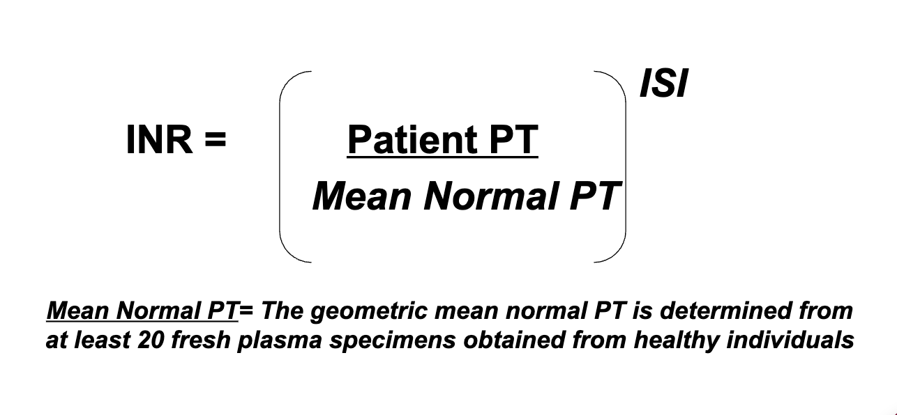
What is the formula for INR?
223
What is the reference interval for manual PT?
10.6 – 14.6 seconds
224
What is the reference interval for automated PT?
9.5 – 13.5 seconds
225
What is the normal INR range?
0.9-1.2
226
Why do PT and INR values vary?
Because they can vary from lab to lab.
227
How often should reference intervals be re-established?
Once a year.
228
How is PT reported in manual systems?
PT is reported in seconds and INR is calculated manually.
229
What do automated analyzers do regarding INR?
They automatically calculate the INR.
230
What does APTT stand for and what type of test is it?
Activated Partial Thromboplastin Time; it is a clot-based routine coagulation test.
231
What is APTT used to assess?
APTT is used to assess deficiencies or inhibitors of the factors in the intrinsic and common pathways.
232
Which clotting factors are involved in the intrinsic and common pathways assessed by APTT?
Factors XII (12), XI (11), IX (9), VIII (8), X (10), V (5), II (prothrombin), and I (fibrinogen).
233
Which clotting factor is NOT assessed by APTT?
Factor VII is not assessed by APTT.
234
What type of anticoagulant therapy is monitored using APTT?
Standard (unfractionated) Heparin anticoagulant therapy.
235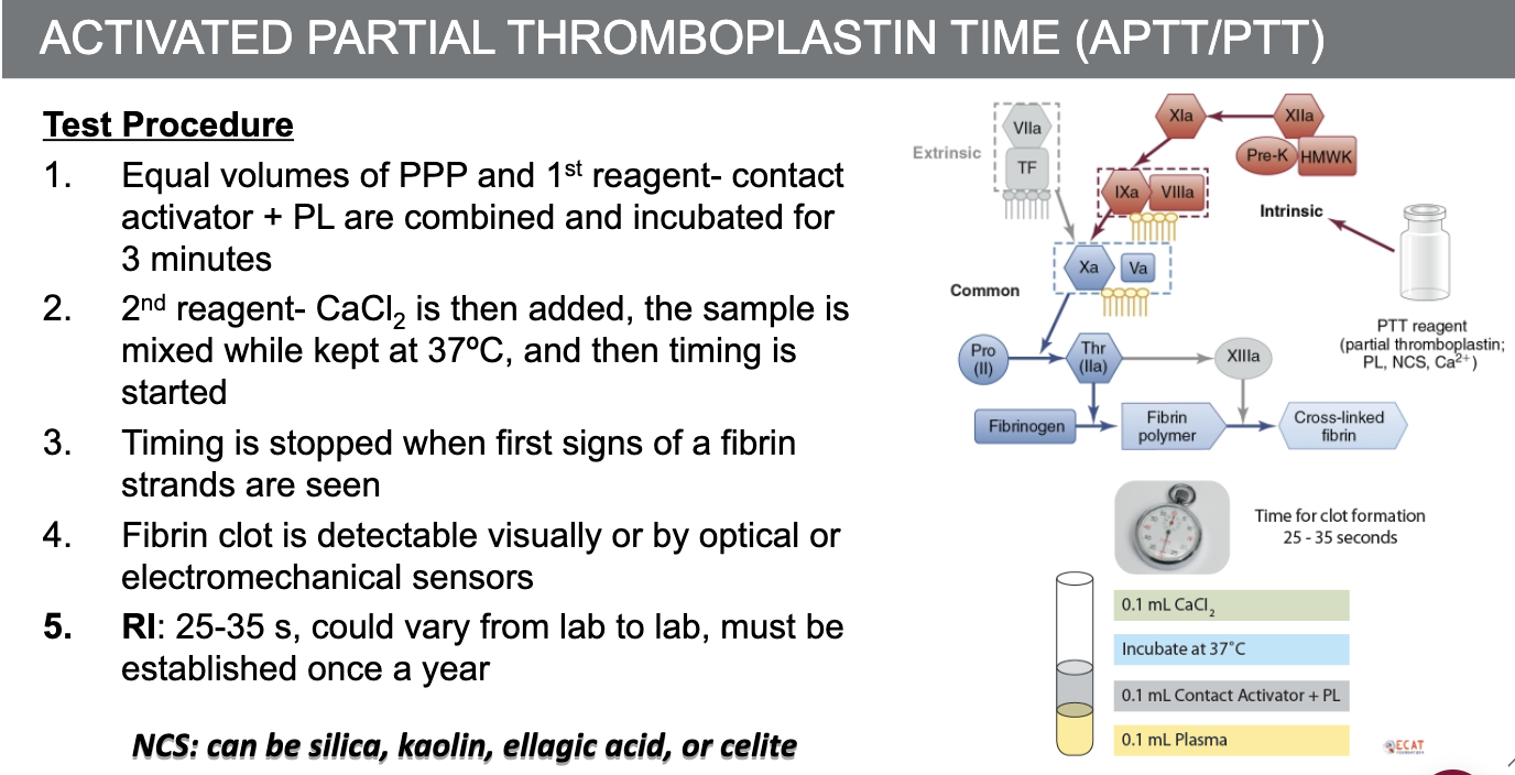
What is the procedure for Activated Partial Thromboplastin Times (aPTT)?
236
What test tube is used for coagulation studies?
3.2% sodium citrate blue top tube;
leaves all clotting factors available except
Calcium, Citrate binds the calcium in the plasma and
therefore prevents coagulation, but not as strongly as EDTA
leaves all clotting factors available except
Calcium, Citrate binds the calcium in the plasma and
therefore prevents coagulation, but not as strongly as EDTA
237
What does plasma contain in the context of coagulation sample collection?
Plasma contains coagulation factors.
238
How is plasma separated from platelets during coagulation sample preparation?
Plasma is separated from platelets by centrifugation. at 3000 RPM for 15 min at room temperature
239
What is Platelet Poor Plasma (PPP)?
Platelet Poor Plasma (PPP) is plasma with a platelet count less than 10x10^9/L.
240
what is the optimal blood to anticoagulant ratio for optimum results
9:1 blood-to-anticoagulant ratio is required for optimum
results (9 parts whole blood to 1 part sodium citrate)
results (9 parts whole blood to 1 part sodium citrate)
241
what affects the ratio? can you fix it?
• High Hematocrit (≥ 55%) affects ratio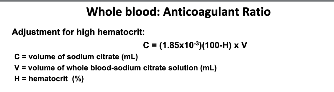
242
243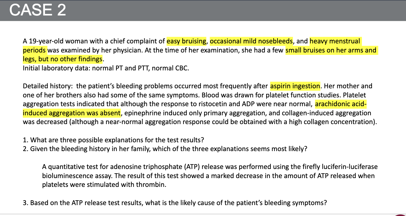
244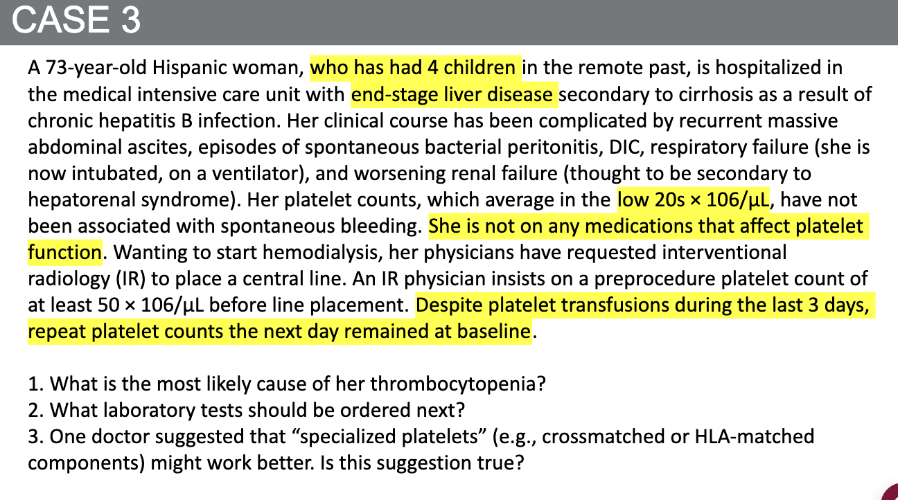
245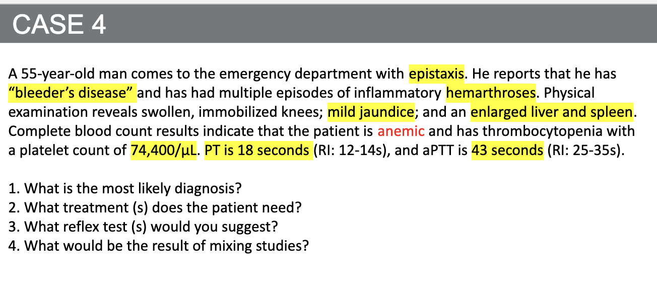
246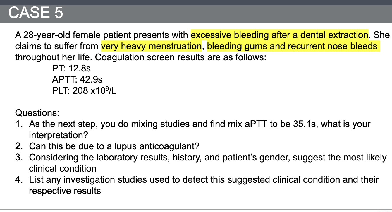
247
248