Lecture 1 - hemostatis part 1
Study these flashcardsFree · spaced repetition · iPhone, Android, Mac & web
All cards
1
What is hemostatis?
the process by which the body stops bleeding after an injury;
2
What does hemo stand for?
BLOOD
3
What does STASIS stand for?
Stop
4
What is coagulation? and when can it happen?
Clotting; it is always ongoing but at low levels, unless a big injury occurs.
5
What is hemorrhage?
bleeding
6
In simple terms, what happens when an injury occurs?
- Produces a clot to stop the bleeding
- Confines the clot to the site of injury
- Dissolves the clot as the wound heals
- Confines the clot to the site of injury
- Dissolves the clot as the wound heals
7
What are the 3 components/properties of hemostasis?
Procoagulant properties
Anticoagulant properties
Fibrinolytic
Anticoagulant properties
Fibrinolytic
8
In simple terms, what do procoagulant properties do?
Initiate clotting if required
9
In simple terms, what do anticoagulant properties do?
Prevent undesirable clotting.
10
In simple terms, what do Fibrinolytic properties do?
Clot breakdown.
11
Breakdown hemostasis, what keeps the balance between bleeding and clotting?
Primary hemostasis: Formation of platelet plug
Secondary Hemostasis: Formation of Fibrin clot
Fibrinolysis: Break down of the Fibrin clot.
Secondary Hemostasis: Formation of Fibrin clot
Fibrinolysis: Break down of the Fibrin clot.
12
what happens when primary hemostasis is activated? and what cellular components are involved?
By desquamation (shedding of the inner lining of blood vessels) and small injuries to blood vessels ; involves vascular intima and platelets
13
How is secondary hemostatis activated? and what cellular components are involved?
By large injuries to blood vessels and surrounding tissues; involves platelets and coagulation system
14
which is rapid, short-lived response and which is delayed, long-term response?
rapid, short-lived response: Primary Hemostasis
delayed, long-term response): Secondary Hemostasis
delayed, long-term response): Secondary Hemostasis
15
What is released when Primary hemostasis occurs?
Procoagulant substanaces exposed or released by damages or activated endothelial cells
16
What is released when Secondary hemostasis occurs?
the activator, tissue factor, is exposed on cell membranes.
17
In general, what are the components involved in hemostasis?
BLOOD VESSELS
PLATELETS
PLASMA COMPONENTS
FIBRINOLYTIC PROTEINS
PLATELETS
PLASMA COMPONENTS
FIBRINOLYTIC PROTEINS
18
What do the blood vessels contain?
Tissue factor and undergo vasoconstriction
19
What do platelets do to help stop the bleeding?
platelet adhesion, aggregation and secretion
20
What are the plasma components consist of?
zymogens, cofactors, control proteins, and fibrinogen
21
What do fibrinolytic proteins consist of?
fibrin clot breakdown.
22
What do blood vessels do? and how many layers does it consist of? what are they
help circulate blood throughout body ;
3 layers:
inner layer - tunica intima
middle layer - tunica media
outer layer - tunica adventitia
3 layers:
inner layer - tunica intima
middle layer - tunica media
outer layer - tunica adventitia
23
What does the inner (tunica intima) layer of the blood vessel composed of? and does it release anything?
vascular endothelium ; yes it releases proteins based on need.
24
What proteins are released from vascular endothelium (inner layer of blood vessel)?
Anticoagulant - normally to prevent spontaneous clotting or when clot is formed
Procoagulant - when there is injury
Fibrinolytic - involved in clot dissociation.
Procoagulant - when there is injury
Fibrinolytic - involved in clot dissociation.
25
What does the middle (tunica media) layer consist of?
muscle depending on BV type and size
smooth muscle cells contract = vasoconstriction
smooth muscle cells contract = vasoconstriction
26
What does the outer (tunica adventitia) layer consist of?
Sub-endothelium
Support a surface protein called Tissue factor
- Procoagulant - important for the initiation of coagulation.
Support a surface protein called Tissue factor
- Procoagulant - important for the initiation of coagulation.
27
List 10 properties that are released from the blood vessels?
ADAMTS13 - A disintegrin and metalloprotease with a thrombospondin type 1 motif, member 13
ECs - endothelial cells
EPCR - endothelial cell protein C receptor
PAI - 1 = plasminogen activator inhibitor-1
PGI2 = prostacyclin or prostaglandin I2
TAF1 = thrmbin activatable fibrinolysis inhibitor
TF = tissue factor
TFP1 = tissue factor pathway inhibitor
TPA = tissue plasminogen activator
VWF = von willebrand factor
ECs - endothelial cells
EPCR - endothelial cell protein C receptor
PAI - 1 = plasminogen activator inhibitor-1
PGI2 = prostacyclin or prostaglandin I2
TAF1 = thrmbin activatable fibrinolysis inhibitor
TF = tissue factor
TFP1 = tissue factor pathway inhibitor
TPA = tissue plasminogen activator
VWF = von willebrand factor
28
what are the 3 blood cells found in the blood?
RBCS
WBC
platelets
WBC
platelets
29
What do RBCs do in the blood?
add bulk and structural integrity to final fibrin clot
30
What do WBCs do in the blood?
help stimulate wound healing; monos and lymphs have tissue factor on surface that can trigger coagulation
31
Are platelets anticoagulant or procoagulant?
procoagulant only -- initiate and control hemostasis; they adhere, aggregate and secrete their granule contents to help form a clot.
32
What are the 8 different zymogens in plasma components?
Prekallikrein
FXII - 12
FXI - 11
FIX - 9
FX - 10
FVII - 7
PROTHROMBIN
FXIII - 13
FXII - 12
FXI - 11
FIX - 9
FX - 10
FVII - 7
PROTHROMBIN
FXIII - 13
33
What are the 7 different control proteins in plasma components?
Antithrombin
Heparin cofactor II
TFPI
Protein C
alpha2 - macroglobulin
alpha1 - antitrypsin
ZPI
Heparin cofactor II
TFPI
Protein C
alpha2 - macroglobulin
alpha1 - antitrypsin
ZPI
34
What are the 7 different co-factors in plasma components?
HMWK
Tissue factor
FVIII - 8
FV - 5
Protein Z
Protein S
Thrombomodulin
Tissue factor
FVIII - 8
FV - 5
Protein Z
Protein S
Thrombomodulin
35
Where are platelets coming from? via which process?
Megakaryocytes; endomitosis
36
Megakaryocytes are derived from which CFU? under the influence of what?
CFU - GEMM ;
IL-3, TPO, AND MEG-CSF
IL-3, TPO, AND MEG-CSF
37
What are the 3 MK progenitors?
BFU- Meg - clones hundreds of daughter cells
CFU - Meg - clones a dozen daughter cells
LD-CFU-Meg - undergoes the first stage of endomitosis
CFU - Meg - clones a dozen daughter cells
LD-CFU-Meg - undergoes the first stage of endomitosis
38
what does endomitosis do?
DNA is duplicated without cell division
39
true/false: MKs become polyploid?
TRUE
40
What are the size of MKs?
30-100 mm; largest normally occurring cells in the marrow; only polyploid cells in body
41
8N OR 16N MK cells can produce how many platelets?
~ 2000-4000 platelets
42
What is thrombopoiesis?
the process of producing platelets (also known as thrombocytes), the cell fragments essential for blood clotting, within the bone marrow
43
What is demarcation system or DMS
a network of internal membranes found within megakaryocytes (MKs), the cells that produce platelets;
biologically identical to MK plasma membrane and form basis for fragmentation into single PLTs;
DMS dilates and tubules called 'proplatelet processes' develop
biologically identical to MK plasma membrane and form basis for fragmentation into single PLTs;
DMS dilates and tubules called 'proplatelet processes' develop
44
After 'proplatelets' develop, what happens?
squeeze through or between endothelial cells and break off or shed into the central vein of BM
45
After platelets are released, what happens in the MK nucleus?
the MK nucleus is reabsorbed by macrophages.
46
What markers are used to ID MKs and platelets?
MPL
CD34
CD41
CD42
PF4
VWF
FIBRINOGEN
CD34
CD41
CD42
PF4
VWF
FIBRINOGEN
47
Where can you specifically find these markers?
MPL
CD34
CD41
CD42
PF4
vWF
Fibringoen
MPL
CD34
CD41
CD42
PF4
vWF
Fibringoen
MPL - expressed in ALL maturation stages
CD34 - only on MK progenitors
CD41 - all positive except BFU - Meg, αIIb portion of αIIbβ3
CD42 - MKI through PLT positive, GPIb portion of vWF receptor (GPIb/IX)
PF4 - MKI through PLT positive
vWF - MKI through PLT positive
Fibrinogen -
CD34 - only on MK progenitors
CD41 - all positive except BFU - Meg, αIIb portion of αIIbβ3
CD42 - MKI through PLT positive, GPIb portion of vWF receptor (GPIb/IX)
PF4 - MKI through PLT positive
vWF - MKI through PLT positive
Fibrinogen -
48
Which hormone helps produce more platelets?and where is it produced/
TPO, thromobopoietin; produced by liver (also by kidney and smooth muscle cells
49
What does TPO attach to?
to circulating platelets via membrane receptor
50
if low platelets are present, what happens?
TPO increases in circulation
51
in general, what are the steps after TPO is present?
Stem cells to differentiate into MK progenitors -> MK progenitors to differentiate into MK -> Proliferation and maturation of MK -> platelet release from MK.
52
What state can platelets be in?
Normal and activated state
53
Platelets have plasma membrane receptors for?
ADP
Serotinin
Collagen (GP VI, GPIa/IIa)
vWF (GP Ib/IX/V)
Fibrinogen (GP IIb/IIIa)
Thrombin
Epinephrine
Serotinin
Collagen (GP VI, GPIa/IIa)
vWF (GP Ib/IX/V)
Fibrinogen (GP IIb/IIIa)
Thrombin
Epinephrine
54
what are platelet granules
membrane-bound compartments within platelets that store and release various molecules crucial for hemostasis (blood clotting)
55
What are the 2 main types of granules in platelets?
alpha granules and dense granules
56
How many alpha granules do platelets have? and what does it contain?
50-80/plts;
PROTEINS:
Fibrinogen
Factor V
beta-thromboglobulin
HMWK
PAI-1
Plasminogen
PROTEINS:
Fibrinogen
Factor V
beta-thromboglobulin
HMWK
PAI-1
Plasminogen
57
How many dense granules do platelets have? and what does it contain?
2-7 per platelets;
ADP
ATP
Serotonin
Ca2+
Mg2+
ADP
ATP
Serotonin
Ca2+
Mg2+
58
At the time of an injury, what do platelets do? ( 3 steps)
Adhesion - adhere to site of injury/disruption
Aggregation - aggregate to with other
Secretion - secrete their granule contents
Aggregation - aggregate to with other
Secretion - secrete their granule contents
59
Specifically, during the adhesion process, what happens?
Platelets bind collagen directly through GPVI and GPIa/IIa or indirectly through vWF (which binds GPIb/IX/V on platelets), ADP TXA2, "inside-out", "outside-in" signaling . 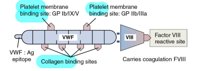
60
What happens in the aggregation step ? (specifically)
Platelets bind to each other through GPIIb/IIIa, shape and membrane platelet flip-flopping. 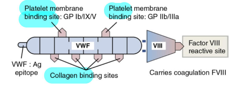
61
What happens in the secretion step? (specifically)
Platelets release granular contents (alpha and dense)
Mostly coagulation proteins
Launching platform for secondary hemostasis
Mostly coagulation proteins
Launching platform for secondary hemostasis
62
Primary Hemostasis Disorders involved which components?
Either platelets or vascular (rare) abnormalities
63
Are primary hemostasis disorders qualitative or quantitative?
can be either qualitative or quantitative.
Qualitative - number of platelets are abnormal; function is normal
Quantitative - platelets are low or high
Qualitative - number of platelets are abnormal; function is normal
Quantitative - platelets are low or high
64
What do primary hemostasis diorders manifest as? and what are common presentations?
Mucocutaneous bleeding
Common presentation:
bruising
petechiae
purpura
ecchymosis
epistaxis
gingival bleeding
Common presentation:
bruising
petechiae
purpura
ecchymosis
epistaxis
gingival bleeding
65
what is thrombocytopenia?
decreased platelets
66
what is thrombocytosis?
increased platelets
67
What are 2 disorders associated with thrombocytosis?
Reactive thrombocytosis - secondary to other conditions
Myeloproliferative disorders
Myeloproliferative disorders
68
List reactive thrombocytosis - secondary to other conditions?
For ex: post-surgery, post-splenectomy, IDA, inflammation or disease, strenuous exercise
Not associated with thrombosis or hemorrhage
Disappears when underlying condition under control
Not associated with thrombosis or hemorrhage
Disappears when underlying condition under control
69
List Myeloproliferative disorders
Essential thrombocythemia, Polycythemia Vera, CML, or primary myelofibrosis.
Can cause either thrombosis or bleeding
Marked and persistent elevations in platelet count are seen.
Can cause either thrombosis or bleeding
Marked and persistent elevations in platelet count are seen.
70
Thrombocytopenia is categorized by how many platelets?
< 150 x 10^9/L
71
Clinically SIGNIFICANT bleeding is categorized by how many platelets?
< 100 x 10^9/L
72
What is the pathophysiologic processes that result in thrombocytopenia?
1. Impaired or decreased platelet production
- Lack of adequate bone marrow megakaryocytes
- Ineffective thrombopoiesis
2. Increased/Accelerated platelet destruction
- Caused by immunologic responses
- Caused by mechanical damage, consumption or sequestration
3. Abnormal platelet distribution or diluition
- Splenic sequestration due to splenomegaly (abnormal sequestration
- Lack of adequate bone marrow megakaryocytes
- Ineffective thrombopoiesis
2. Increased/Accelerated platelet destruction
- Caused by immunologic responses
- Caused by mechanical damage, consumption or sequestration
3. Abnormal platelet distribution or diluition
- Splenic sequestration due to splenomegaly (abnormal sequestration
73
What causes Immune thrombocytopenic purpura?
Destruction of platelets by an antibody-mediated mechanism (antibody against GPIIb/IIIa, GPIb/IX/V, GPIa/IIa)
74
immune thrombocytopenic purpura can be seen in what age range?
2-5 years old, following viral infections or vaccinations (MMR, DTP, polio)
75
What is the key hematologic feature in thrombocytopenia?
3-4% cases: < 10,000uL
76
What might you see in the chart of a child with Acute ITP?
- Severe thrombocytopenia, signs of hemorrhage, normal RBCS and WBC in CBC
- Severe thrombocytopenia, sudden hemorrhage, no family history
- Severe thrombocytopenia, sudden hemorrhage, no family history
77
Recover time for most acute cases of ITP?
3 weeks
78
Can chronic ITP be seen in children?
NO, is in mostly adults ( woman > men)
79
What are 3 qualitative platelet disorders?
Glanzmann's Thrombasthenia
Bernard Soulier Syndrome
May-Hegglin Anomaly
Bernard Soulier Syndrome
May-Hegglin Anomaly
80
What is Glanzmann's Thrombasthenia?
Extremely rare coagulopathy, hemorrhage may be severe and disabling, all types of bleedings: epistaxis, ecchymosis, hemarthrosis, subcutaneous hematoma, menorrhagia, and GI and UT hemorrhage, have been reported.
81
What type of disorder is Glanzmann's Thrombasthenia?
Autosomal recessive disorder, heterozygotes clinically normal
82
when does Glanzmann's Thrombasthenia usually manifest?
In neonatal period or infancy
83
What causes Glanzmann's Thrombasthenia?
Mutation in genes encoding the subunits of GPIIb/IIIa (Fibrinogen & vWF receoptor)
84
What is the lab diagnosis for Glanzmann's Thrombasthenia?
Platelet numbers and morphology are normal
Aggregometry: no aggregation in response to all activating agents (ADP, collagen, thrombin, epinephrin, AA), normal response to ristocetin (which is similar to vWF).
Aggregometry: no aggregation in response to all activating agents (ADP, collagen, thrombin, epinephrin, AA), normal response to ristocetin (which is similar to vWF).
85
What is Bernard Soulier Syndrome? what is the inheritance type?
Rare disorder of platelet adhesion ; autosomal recessive inheritance
86
When is Bernard Soulier Syndrome discovered? what are the presentations?
Usually in early childhood; Ecchymosis, epistaxis, and gingival bleeding
87
What causes Bernard Soulier Syndrome?
Mutation in GPIbalpha of the GP Ib/IX/V receptor complex (vWFR): defective PLT adhesion
88
What is the lab diagnosis of Bernard Soulier syndrome?
Thrombocytopenia (decrease PLT survival/production)
Giant PLTs
Aggregometry: normal response to ADP , collagen, thrombin, epinephrin, AA but lack of aggregation in response to ristocetin.
Giant PLTs
Aggregometry: normal response to ADP , collagen, thrombin, epinephrin, AA but lack of aggregation in response to ristocetin.
89
What is the inheritance pattern for May-Hegglin Anomaly?
Rare, autosomal dominant disorder
Exact frequency unknown
Exact frequency unknown
90
What is seen in May-Hegglin Anomaly? (in general)
Thrombocytopenia (low platelet count)
Characteristic abnormally enlarged or misshapen platelets (giant PLTs seen)
Characteristic abnormally enlarged or misshapen platelets (giant PLTs seen)
91
What is seen in May-Hegglin Anomaly? (on PBS)
- Thrombocytopenia with giant PLTs
- Basophilic inclusions (called Dohle-like bodies) within the granulocytes and occasionally monocytes.
- Basophilic inclusions (called Dohle-like bodies) within the granulocytes and occasionally monocytes.
92
Do platelets function normally in May-Hegglin Anomaly? and what's another name it's classified as
- PLTS function usually normal
- Also classified as Inherited Giant PLT syndrome.
- Also classified as Inherited Giant PLT syndrome.
93
What are the consequences of
Short draw
Specimen clot
Visible hemolysis
Lipemia or icterus
Tourniquet application > 1 minute
Specimen storage at 1-6oC
Specimen storage at > 25oC
Short draw
Specimen clot
Visible hemolysis
Lipemia or icterus
Tourniquet application > 1 minute
Specimen storage at 1-6oC
Specimen storage at > 25oC
94
What is the order of draw?
Yellow - blood culture; sterile
Light blue - 3.2% sodium citrate
Red
Yellow
Green
Lavender - EDTA
Gray
Light blue - 3.2% sodium citrate
Red
Yellow
Green
Lavender - EDTA
Gray
95
What is the anticoagulant of choice for coagulation studies?
sodium citrate - blue top tube
96
In order to test platelet functions, what are some tests you could do?
bleeding time - OG test of platelet function now replaced by:
PFA - 100 Analyzers - automated platelet function testing
Platelet Aggregometry Studies - measure how well PLTs aggregate together to form clots
PFA - 100 Analyzers - automated platelet function testing
Platelet Aggregometry Studies - measure how well PLTs aggregate together to form clots
97
What types of sample does the PFA 100 Platelet Function Analyzer use? and how does it work?
Citrated whole blood - is aspirated at high shear rates through disposable test cartridges
Test cartridges contain 2 membranes: one coated collagen/epinephrin (CEPI) and the other one coated with collagen/ADP (CADP) and an aperture <- AGONISTS
Test cartridges contain 2 membranes: one coated collagen/epinephrin (CEPI) and the other one coated with collagen/ADP (CADP) and an aperture <- AGONISTS
98
How does the PFA 100 Platelet Function analyzer indicate PLT function?
Time required for a PLT plug to occlude aperture (small opening)
99
Why would the PFA 100 platelet function analyzer be used?
Assessment for inherited, acquired, or drug-induced platelet dysfunction
- can be used as an initial screening test for patients with impaired primary hemostasis (Von Willebrand Disease) <- Disease where vW is missing or doesn't work. ... quite successful
- to monitor DDAVP (desmopressin) therapy (DDAVP is a medication that helps release vWF from cells; used in partients before surgery to reduce bleeding)
- to check how aspirin affects platelets (Aspirin blocks platelet activation, so this test can show if the medication is working; also helps identify if aspirin is causing bleeding problems)
- can be used as an initial screening test for patients with impaired primary hemostasis (Von Willebrand Disease) <- Disease where vW is missing or doesn't work. ... quite successful
- to monitor DDAVP (desmopressin) therapy (DDAVP is a medication that helps release vWF from cells; used in partients before surgery to reduce bleeding)
- to check how aspirin affects platelets (Aspirin blocks platelet activation, so this test can show if the medication is working; also helps identify if aspirin is causing bleeding problems)
100
What does platelet aggregometry measure?
how well PLTs aggregate together to form clots
101
Why is sample procurement very important?
MUST NOT ACTIVATED PLATELETS
USE LARGER BORE NEEDLE !!!!
USE LARGER BORE NEEDLE !!!!
102
How is the sample prepared for platelet aggregometry?
Spin low to produce PLATELET RICH PLASMA (PRP)
- allow platelets to regain function - let sample site for ~ 30 minutes
- Test within 4 hrs of collection to avoid spontaneous in vitro platelet activation and loss of normal activity
-chilling destroys PLT activity - samples held at 15-25oC until tested.
- allow platelets to regain function - let sample site for ~ 30 minutes
- Test within 4 hrs of collection to avoid spontaneous in vitro platelet activation and loss of normal activity
-chilling destroys PLT activity - samples held at 15-25oC until tested.
103
`What is the testing procedure for PLT Aggregoemtry - optical aggregometer
PRP in a cuvette with a plasticized magnetic stir-bar:
-keeps platelets in suspension at 800-1200 RPM (gentle speed)
- warm to 37 degrees celsius for 5 minutes.
Photometer directs light through cuvette to photodetector
- OD of PRP is high
- Baseline established = 0% light transmission
-add agonist (PLT activator or aggregating agent)
-keeps platelets in suspension at 800-1200 RPM (gentle speed)
- warm to 37 degrees celsius for 5 minutes.
Photometer directs light through cuvette to photodetector
- OD of PRP is high
- Baseline established = 0% light transmission
-add agonist (PLT activator or aggregating agent)
104
When agonist is added to aggregoemetry what happens?
platelets shape change - increase in %T
platelets start aggregating - increase in %T
platelets form large aggregate - up to 100%T
platelets start aggregating - increase in %T
platelets form large aggregate - up to 100%T
105
What are the steps in this Platelet Aggregometry (optical aggreg.) test?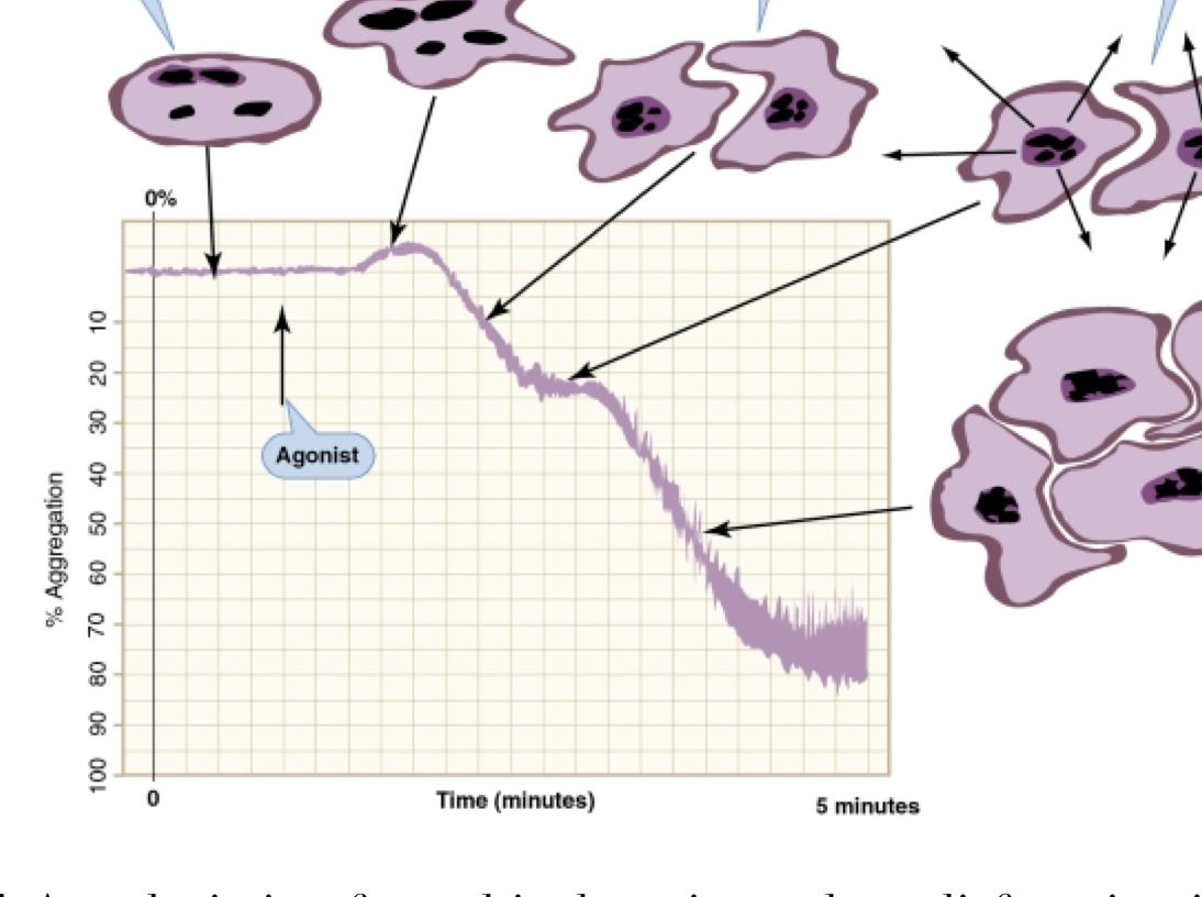
-Resting platelet; stable baseline
- Shape change
- Primary Aggregation
-Secretion
-Secondary aggregation
- Shape change
- Primary Aggregation
-Secretion
-Secondary aggregation
106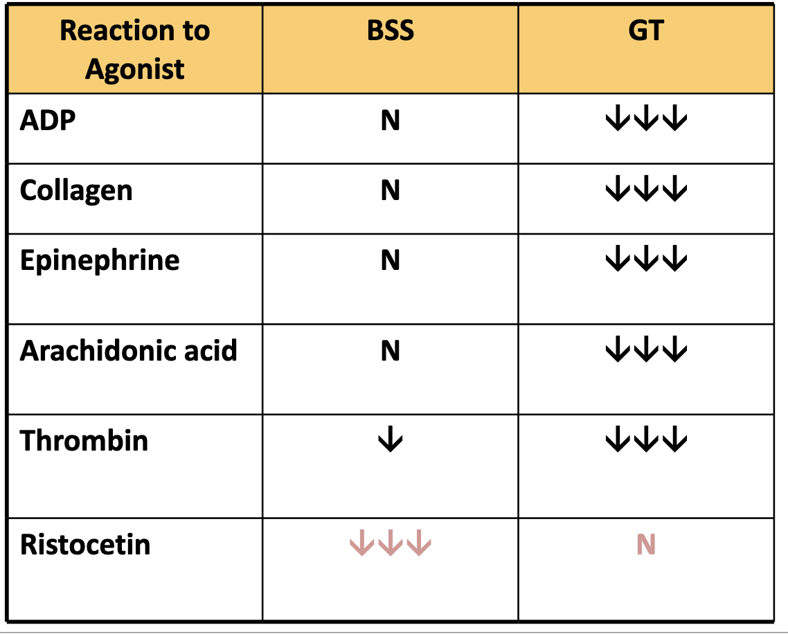
Fill this chart related to platelet aggregation studies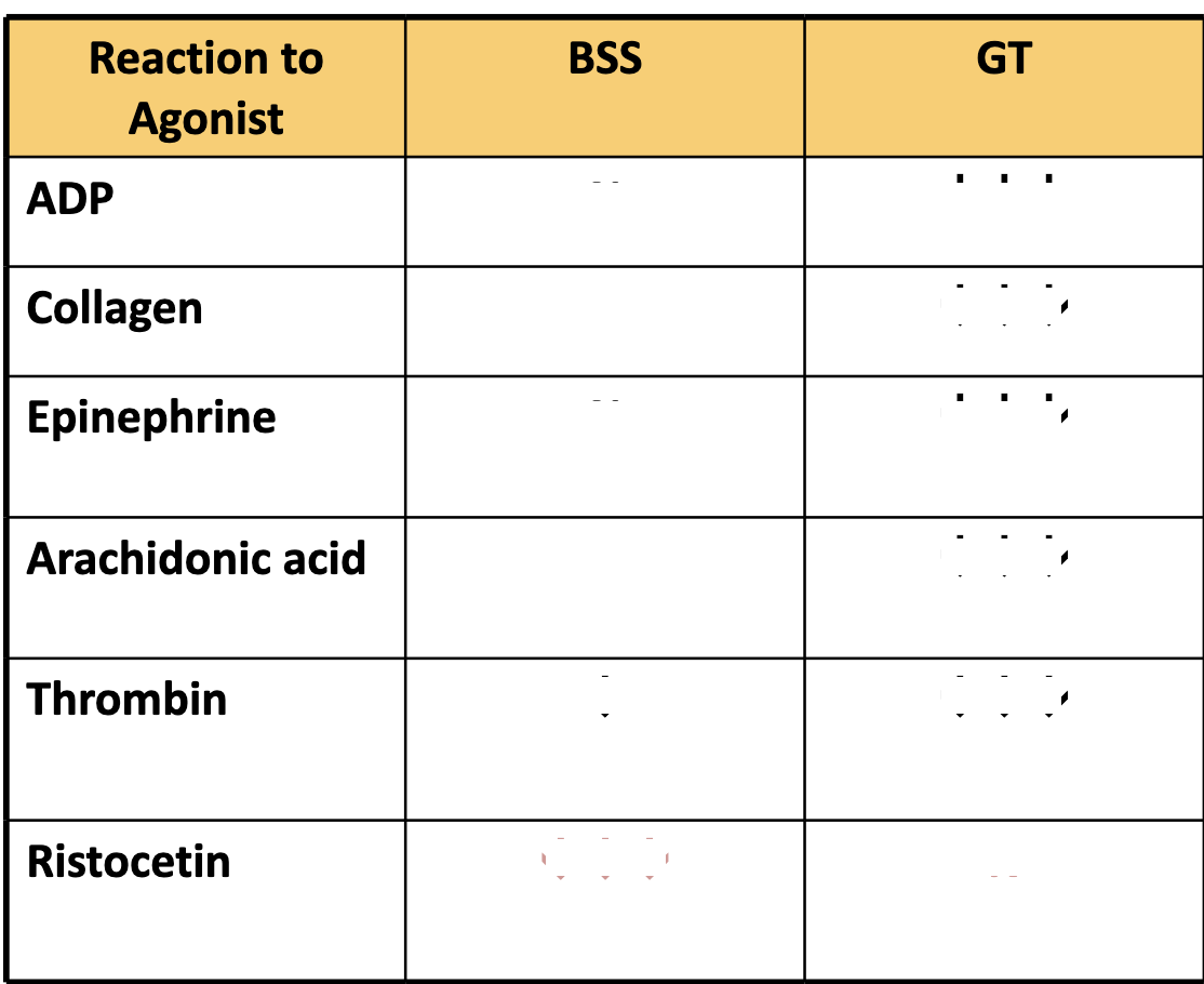
107
What types of disorders do platelet function tests detect?
Platelet secretion/aggregation defects → e.g., Storage pool disease (dense granule deficiency)
Platelet-related acquired disorders → e.g., HIT (Heparin-Induced Thrombocytopenia)
Vascular disorders → e.g., abnormal vessel wall function that affects platelet interaction
Platelet-related acquired disorders → e.g., HIT (Heparin-Induced Thrombocytopenia)
Vascular disorders → e.g., abnormal vessel wall function that affects platelet interaction
108
What test does Storage pool defects - dense granules use? and what do you expect
Bleeding time or PFA-100 → Prolonged/abnormal
Platelet aggregation studies → Decreased secondary wave of aggregation with some agonists (e.g., ADP, epinephrine, collagen)
Normal response with ristocetin
Platelet aggregation studies → Decreased secondary wave of aggregation with some agonists (e.g., ADP, epinephrine, collagen)
Normal response with ristocetin
109
How do you measure HIT (heparin-induced thrombocytopenia)
Early monitoring using 4Ts Scoring system
Enzyme Imunoassay Ex ELISA
Confirmatory PLT Activation Assay (Serotonin Release Assay)
Rapid PLT agglutination immunoassay also available
Enzyme Imunoassay Ex ELISA
Confirmatory PLT Activation Assay (Serotonin Release Assay)
Rapid PLT agglutination immunoassay also available
110
What may be prolonged or abnormal in vascular disorders?
May have prolonged BT or abnormal PLT function testing
111
What do physicians work to rule out in cases of vascular disorders?
Physicians work to rule out other causes for bleeding.
112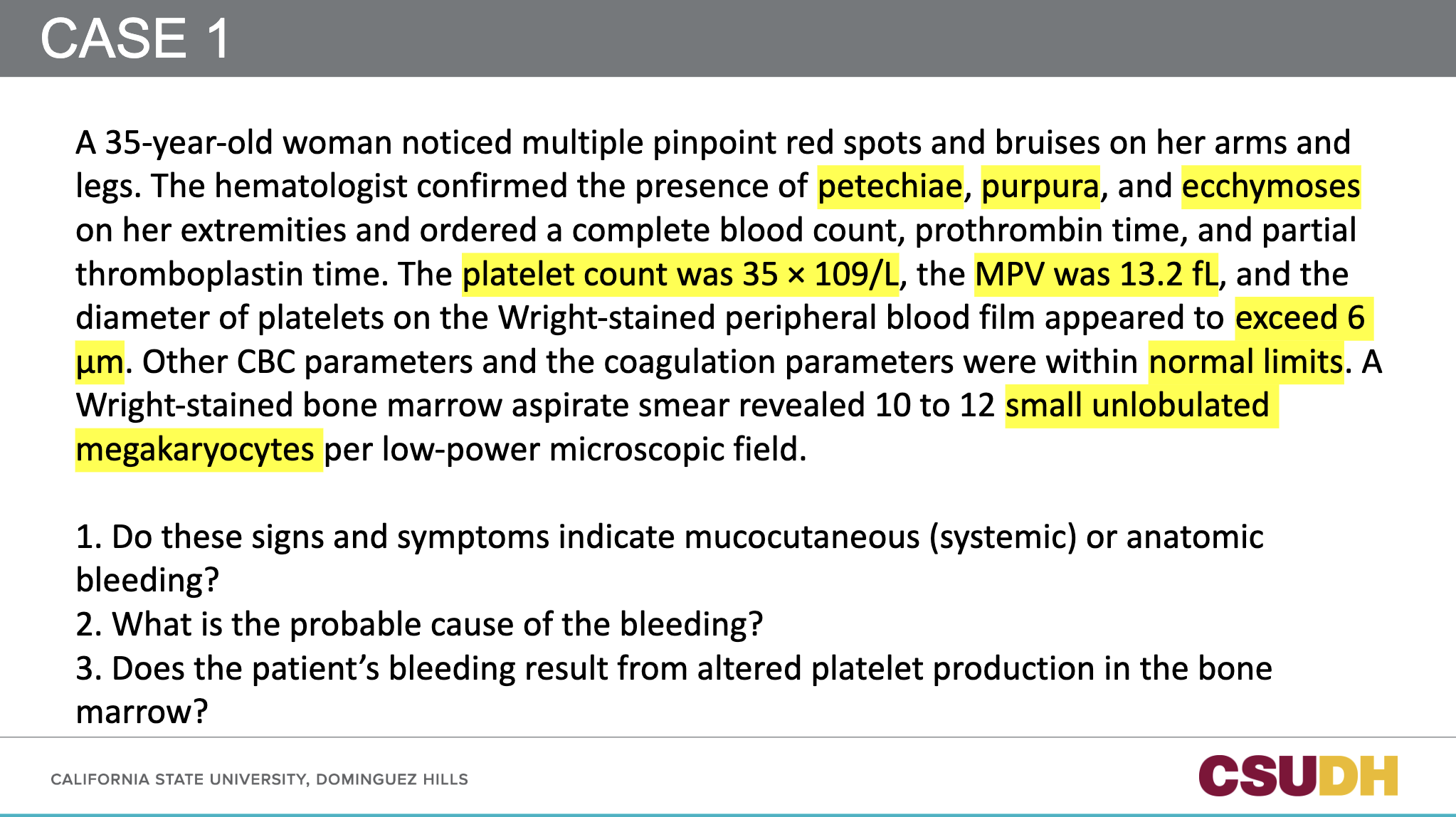
113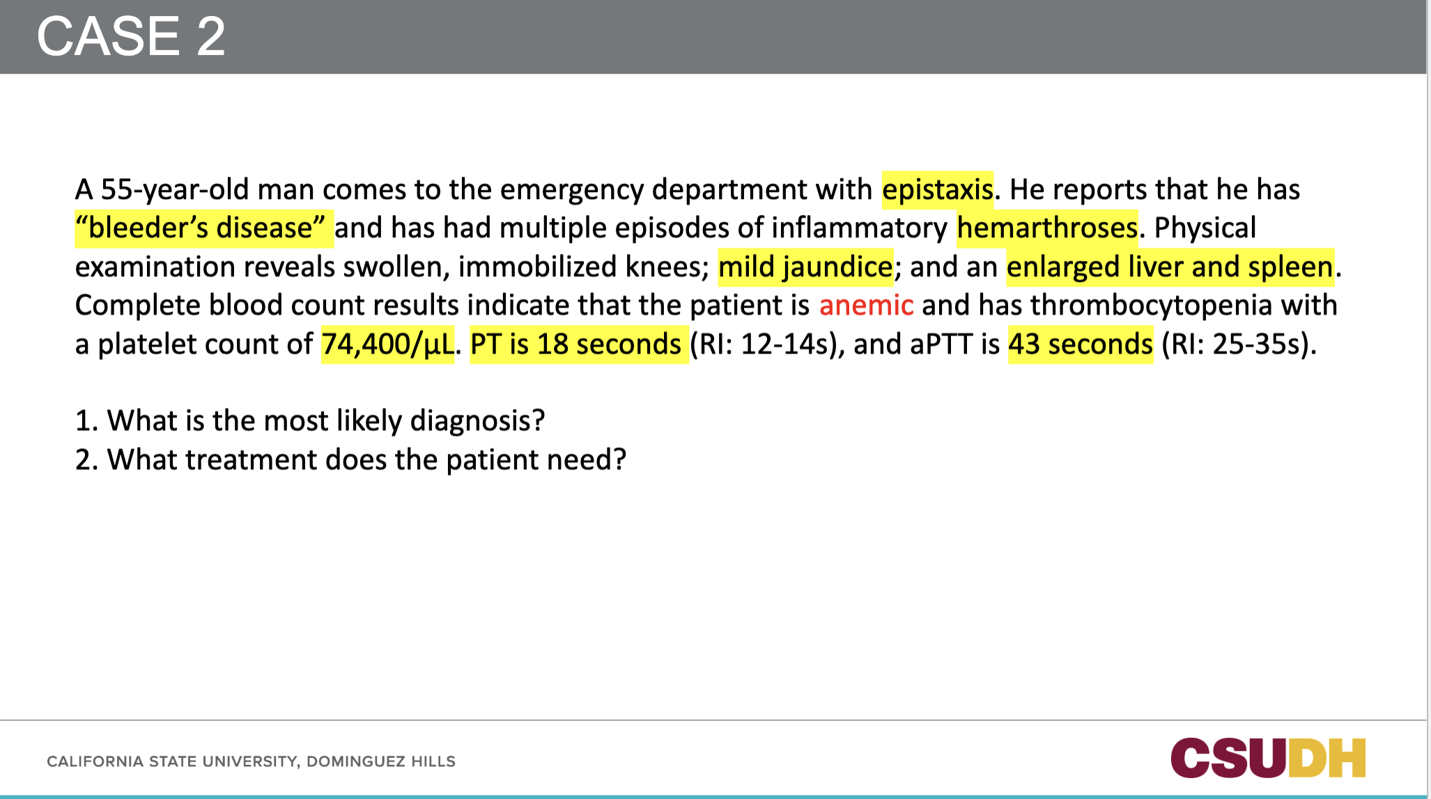
114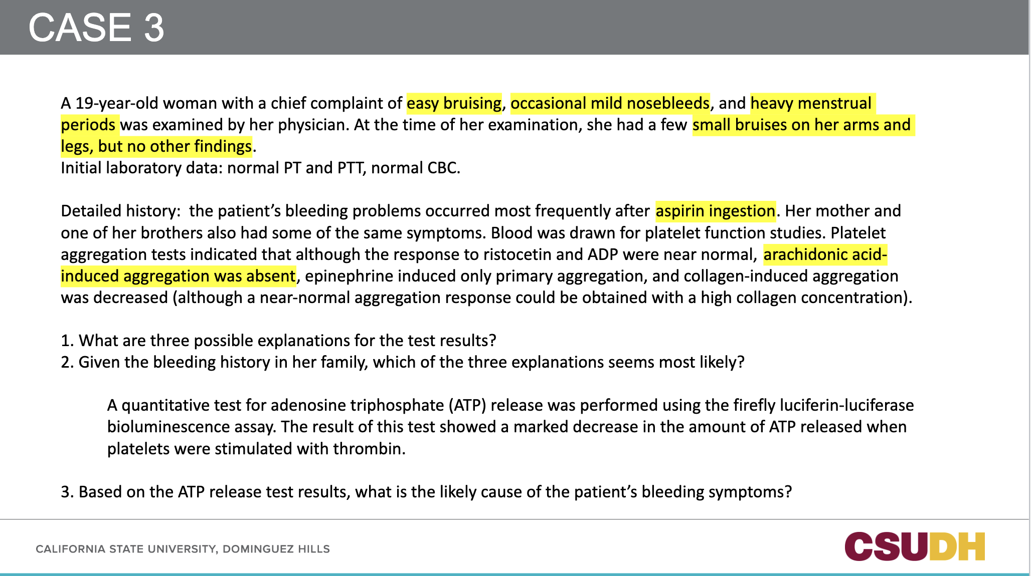
115
116
What are the consequences of short draw?
Leads to excess anticoagulant (e.g., sodium citrate) relative to plasma, which prolong clotting times like PT and aPTT falsely.
117
What are the consequences of a specimen clot?
Clotting has already occurred, so fibrinogen and clotting factors are consumed, making the sample unacceptable for testing. Results will be invalid or falsely shortened
118
What are the consequences of a visible hemolysis?
Hemolysis releases intracellular contents (like ADP and potassium) which can interfere with platelet function tests, activate coagulation, or affect optical readings. Results may be falsley shortened or prolonged, depending on the test.
119
What are the consequences of lipemia or icterus?
Causes optical interference in photo-optic instruments used in coagulation testing. It can lead to inaccurate results due to turbidity or color interference.
120
what are the consequences of Tourniquet application >1 minute?
Prolonged stasis causes hemoconcentration, which elevates proteins like fibrinogen and factors V and VIII, possibly falsely shortening clotting times (e.g., shortened aPTT)
121
What are the consequences of specimen storage at 1-6oC?
Cold temperatures can activate Factor VII (7) and cause platelet activation, leading to inaccurate results, especially in platelet function or factor assays.
122
What are the consequences of specimen storage at >25oC?
Room temperature above 25oC can cause degradation of labile factors (especially factor V and VIII), leading to falsely prolonges PT and aPTT.