Lecture 3 - Thrombophilia
Study these flashcardsFree · spaced repetition · iPhone, Android, Mac & web
All cards
1
What is thrombosis?
blood clots that can block vessels
2
What is thrombophilia?
condition that increases the risk of forming clots
3
what are 4 causes of thrombophilia?
- Chronic or acute inflammation
- Uncontrolled platelet activation
- Blood coagulation control protein deficiencies
- Uncontrolled suppression of fibrinolysis
- Uncontrolled platelet activation
- Blood coagulation control protein deficiencies
- Uncontrolled suppression of fibrinolysis
4
What can thrombophilia lead to?
dangerous conditions like DVT (deep vein thrombosis), PE (pulmonary embolism), or artery blockages
5
What are some risk factors that raise the risk of blood clots?
Age
Diet
Immobilization
Lipid metabolism disorders
Pregnancy
Femoral or tibial fracture
Surgeries (hip, ..)
Smoking
Diet
Immobilization
Lipid metabolism disorders
Pregnancy
Femoral or tibial fracture
Surgeries (hip, ..)
Smoking
6
Q: How does MPN (Myeloproliferative Neoplasms) increase the risk of thrombosis?
A: It activates platelets and increases plasma viscosity, making blood more likely to clot.
7
Q: Why does liver disease increase the risk of thrombosis?
A: It reduces the production of coagulation regulatory proteins, which can lead to abnormal clotting.
8
Q: What type of leukemia is most associated with a high risk of clotting?
A: Acute Promyelocytic Leukemia (APL), which increases the risk of Disseminated Intravascular Coagulation (DIC).
9
Q: How does PNH (Paroxysmal Nocturnal Hemoglobinuria) contribute to thrombosis?
A: It causes platelet activation, which can lead to blood clots.
10
Q: What are some chronic inflammatory conditions that increase clotting risk?
A: Diabetes, cancer, infection, and autoimmune diseases.
11
Q: What is Anti-phospholipid Syndrome (APS)?
A: It’s an autoimmune disorder where antibodies trigger increased clotting, raising the risk of thrombosis.
12
list congential risk factors of thrombophili
- AT deficiency
- Protein C deficiency
- (free) protein S deficiency
- APC resistance [FV Leiden (R506Q): GOF mutation in FV gene], has highest rate of thrombotic events (20-25%)
- Prothrombin G20210A: mutation causes FII elevation
- Hyperfibrinogenemia
- Protein C deficiency
- (free) protein S deficiency
- APC resistance [FV Leiden (R506Q): GOF mutation in FV gene], has highest rate of thrombotic events (20-25%)
- Prothrombin G20210A: mutation causes FII elevation
- Hyperfibrinogenemia
13
Q: What does Antithrombin (AT) deficiency do?
A: It reduces the body's ability to stop clotting, increasing the risk of thrombosis.
14
Q: What is the risk of having a Protein C deficiency?
A: It causes less control over clotting, making blood clots more likely to form.
15
Q: Why is free Protein S important in clotting?
A: It works with Protein C to control clotting, so a deficiency raises clotting risk.
16
Q: What is Factor V Leiden (APC resistance), and why is it important?
A: It’s a mutation in the Factor V gene that makes it hard for the body to stop clotting, leading to the highest rate of clotting events (20–25%).
17
Q: What does the Prothrombin G20210A mutation do?
A: It causes increased production of Factor II (prothrombin), leading to a higher clot risk.
18
Q: What is hyperfibrinogenemia?
A: It’s a condition with high levels of fibrinogen, which makes clotting more likely.
19
Q: If you have a clotting deficiency, does it guarantee you will develop thrombosis?
A: No, clot formation depends on genetics, disease, and lifestyle factors.
20
What falls under THROMBOPHILIA INVESTIGATION TEST PANEL
APC resistance
FV Leiden mutation
Prothrombin mutation G20210A
LAC profile
AT activity
PC activity
PS activity
Fibrinogen
FV Leiden mutation
Prothrombin mutation G20210A
LAC profile
AT activity
PC activity
PS activity
Fibrinogen
21
Q: What does an APC (activated protein C) resistance test check, and what is a normal result?
A: It checks how well your body responds to Activated Protein C; a normal ratio is >1.8.
22
Q: What is the normal result for a Factor V Leiden mutation test?
A: Wild type (meaning no mutation is present).
23
Q: What is the Prothrombin G20210A mutation, and what is a normal test result?
A: It’s a genetic mutation that increases clot risk; normal result is wild type (no mutation).
24
Q: What does the LAC profile test for, and what is the normal result?
A: It checks for Lupus Anticoagulant; normal is negative.
25
Q: What is the reference range for AT (Antithrombin) activity
A: 78%–126%.
26
Q: What is the reference interval (RI) for Protein C activity?
A: 70%–140%.
27
Q: What is the reference interval (RI) for Protein S activity
A: 65%–140%.
28
Q: What is the normal reference range for Fibrinogen levels?
A: 220–498 mg/dL.
29
What does APLAs stand for and what is it?
"these are immune system proteins (antibodies) that accidentally attack your own body, especially the parts involved in blood clotting." - chat
A: APLAs (anti-phospholipid antibodies) are immunoglobulins (IgG or IgM) that bind to protein-phospholipid complexes and act as non-specific inhibitors.
A: APLAs (anti-phospholipid antibodies) are immunoglobulins (IgG or IgM) that bind to protein-phospholipid complexes and act as non-specific inhibitors.
30
Q: What are the three major types of anti-phospholipid antibodies?
A:
- Lupus anticoagulants (LACs),
- anticardiolipins (ACLs),
- anti-β2 glycoprotein I (anti-β2-GPI).
- Lupus anticoagulants (LACs),
- anticardiolipins (ACLs),
- anti-β2 glycoprotein I (anti-β2-GPI).
31
Q: What syndrome is associated with APL antibodies?
A: Antiphospholipid syndrome (APS), which increases the risk of clots and pregnancy complications.
32
Q: What is the clinical significance (% of pop) of APLAs (anti-phospholipid antibodies) in the population?
Clinical correlation:
General population: 1–2%
5–15% of patients with thrombosis, mostly transient following infections (alloimmune)
or arise as autoimmune Abs in , and in 50% of patients with lupus (SLE).
General population: 1–2%
5–15% of patients with thrombosis, mostly transient following infections (alloimmune)
or arise as autoimmune Abs in , and in 50% of patients with lupus (SLE).
33
Q: What is the first step in diagnosing APS (Antiphospholipid Syndrome)when PTT is prolonged?
A: Perform a Thrombin Time (TT) test to rule out heparin or dabigatran interference.
34
Q: What does a normal TT suggest in a prolonged PTT workup?
A: Heparin or thrombin inhibitor is unlikely—proceed with mixing study.
MIXING STUDY: Mix test plasma 1:1 with NP PTT on mixture.
MIXING STUDY: Mix test plasma 1:1 with NP PTT on mixture.
35
Q: What does a long TT suggest in the PTT workup?
A: Possible presence of heparin or dabigatran.
36
Q: What is the next step if TT is prolonged?
A: Add Hepzyme® to neutralize heparin and repeat the PTT.
37
Q: What does a corrected PTT after Hepzyme® indicate?
A: Heparin was the cause; confirm with heparin assay.
38
Q: What should you do if dabigatran is present?
A: Discontinue dabigatran and retest later.
39
Q: What does it mean if the PTT mixing study corrects immediately? (in general)
A: Suggests a factor deficiency—confirm with factor assays.
40
Q: What does it mean if the PTT mixing study does not correct immediately?
A: Suggests the presence of an inhibitor, likely lupus anticoagulant (LAC).
41
Q: What should be done if the PTT mix is uncorrected after incubation at 37°C for 1–2 hours?
A: Suspect a specific factor inhibitor or LAC; perform an assay for each.
42
Q: What does it mean if the PTT corrects after incubation?
A: Suggests a time-dependent factor deficiency.
43
What are common test for LAC (lupus anticoagulant)? and what do they test?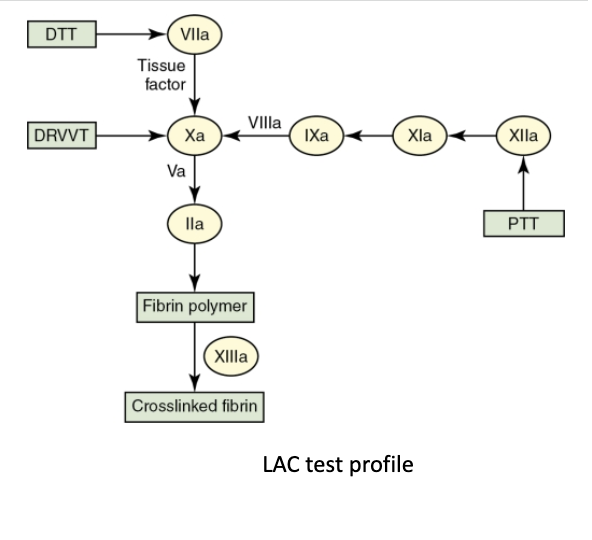
-DTT - Dilute Thromboplastin Time → A sensitive test for LAC (Uses tissue factor and measures activation through Factor VIIa)
-DRVVT - Dilute Russell Viper Venom Test → Directly activates Factor X
- LAC-sensitive partial thromboplastin time (PTT) -> monitors intrinsic and common pathways
-DRVVT - Dilute Russell Viper Venom Test → Directly activates Factor X
- LAC-sensitive partial thromboplastin time (PTT) -> monitors intrinsic and common pathways
44
Q: Which test monitors the intrinsic pathway in the LAC profile?
A: PTT (Partial Thromboplastin Time)
45
Q: What does DRVVT test for, and how does it work?
A: DRVVT tests for lupus anticoagulant by directly activating Factor Xa and measuring the common pathway.
46
Q: What does DTT (Dilute Thromboplastin Time) test for?
A: It evaluates the extrinsic pathway and is sensitive to LAC interference.
47
Q: How does lupus anticoagulant affect clotting tests?
A: It prolongs clotting times by interfering with phospholipid-dependent steps in the coagulation cascade.
48
To diagnose Lupus Anticoagulant (LAC), labs follow how many step procedure?
4 step procedure
49
Q: What is the first step in detecting lupus anticoagulant (LAC) based on the 2009 ISTH guidelines?
A: Perform a phospholipid-dependent clotting test such as a low-phospholipid PTT or DRVVT, known as the PTT or DRVVT screen.
50
Q: What does it mean if the prolonged clotting time does not correct after mixing with normal plasma?
A: It suggests the presence of an inhibitor like LAC rather than a clotting factor deficiency.
51
Q: What result confirms LAC in the confirmatory step of testing?
A: The clotting time shortens or corrects when excess phospholipids are added, using a test like PTT or DRVVT confirm.
52
Q: What is the final step in confirming lupus anticoagulant (LAC)?
A: Rule out other coagulation disorders (coagulopathies).
53
Q: What mutation is responsible for APC resistance?
FV R506Q mutation aka FV Leiden
54
Q: What is the mechanism of action behind Factor V Leiden?
A: Factor V resists hydrolysis (breakdown) by activated protein C (APC).
55
Q: What is the clinical significance of the FV Leiden mutation? homo vs. hetero
A: It causes a 3-fold increase in thrombosis risk, or up to 18-fold in homozygous individuals.
56
Q: What are two ways to detect APC resistance in the lab?
A: A clot-based APC ratio assay and a molecular FV mutation test (to check for zygosity).
57
Q: What is a normal APC ratio, and what does a low ratio indicate?
A: A normal APC ratio is >1.8. A ratio ≤1.8 implies APC resistance and must be confirmed with molecular testing.
58
Q: In the clot-based assay, what result suggests APC resistance?
A: If adding APC doesn’t significantly prolong the clotting time (PTT), it suggests APC resistance.
59
Q: What type of mutation occurs in Prothrombin G20210A?
A: A guanine (G) to adenine (A) mutation at the 3' untranslated region (3'UTR) of the prothrombin gene.
60
Q: How does the Prothrombin G20210A mutation affect prothrombin levels and thrombosis risk?
A: It increases prothrombin levels and raises thrombosis risk by 2–3 times in heterozygotes.
61
Q: What is the worldwide prevalence of the Prothrombin G20210A mutation in the general population and in families with thrombosis?
A: 0.3–2.4% in the general population and 5–18% in familial thrombosis cases.
62
Q: How is the Prothrombin G20210A mutation detected?
A: By using molecular genetic assays.
63
Q: What is the main function of antithrombin (AT)?
A: Antithrombin inhibits thrombin, Xa (10), IXa (9), XIa. (11), and XIIa (12) to prevent excessive clotting.
64
Q: How is AT deficiency defined?
A: AT activity is less than 70% of normal.
65
Q: What are some acquired causes of AT deficiency?
A: Liver disease, nephrotic syndrome, prolonged heparin therapy, contraceptives, and DIC.
66
Q: What characterizes congenital AT deficiency?
A: More than 90% are quantitative, meaning low AT antigen levels.
67
Q: How is Protein C or S deficiency defined?
A: Activity less than 65% of normal.
68
Q: How much does the risk of thrombosis increase in heterozygotes with Protein C or S deficiency?
A: 1.6 to 11.5 times higher risk of DVT and PE.
69
Q: What condition can occur in infants with homozygous Protein C or S deficiency?
A: Neonatal purpura fulminans, which is deadly if untreated.
70
Q: When can PC and PS assays not be used to diagnose congenital deficiency?
When levels are falsely decreased due to:
Within 14 days of acute inflammation
After stopping Coumadin therapy
During pregnancy
In DIC, liver or kidney disease, vitamin K deficiency, or oral contraceptive use.
Within 14 days of acute inflammation
After stopping Coumadin therapy
During pregnancy
In DIC, liver or kidney disease, vitamin K deficiency, or oral contraceptive use.
71
Q: What causes Disseminated Intravascular Coagulation (DIC)?
A: It is triggered by a systemic disease and causes generalized, uncontrolled activation of the hemostasis system.
72
Q: What is the major clinical issue in DIC despite microthrombi formation?
A: Hemorrhage (bleeding) is the major issue.
73
Q: What are other names for DIC?
A: Consumptive coagulopathy or defibrination syndrome.
74
Q: What lab findings are associated with DIC?
A:
Abnormal pattern on PBS and CBC
Thrombocytopenia
Prolonged PT, PTT, and TT
Low fibrinogen
Increased FDPs, especially D-dimer
Abnormal pattern on PBS and CBC
Thrombocytopenia
Prolonged PT, PTT, and TT
Low fibrinogen
Increased FDPs, especially D-dimer
75
Q: Why is acute uncompensated DIC considered a medical emergency?
A: It can be fatal in 50% of cases if not managed quickly and requires immediate testing and intervention.
76
Q: How does tissue factor (TF) release trigger DIC?
A: Endothelium damage (e.g., trauma) releases TF, which activates clotting.
77
Q: How do endotoxins contribute to DIC?
A: Bacterial infections or septicemia release endotoxins that over-activate clotting.
78
Q: How does vasodilation from heat stroke trigger DIC?
A: It exposes the endothelium, which activates clotting.
79
Q: How do circulating immune complexes cause DIC?
A: They damage vessels in conditions like HIT, AHTR, and graft rejection, leading to clotting.
80
Q: How do leukemias like APL and AMML trigger DIC?
A: They release proteolytic enzymes that activate the coagulation system.
81
Q: What role do toxins like snake venom play in DIC?
A: They directly activate clotting pathways.
82
Q: How does acidosis (e.g. from diabetes) lead to DIC?
A: It disrupts normal enzyme function and can trigger abnormal clotting.
83
Q: How does platelet activation from surgery or thrombocythemia lead to DIC?
A: It causes excessive clotting, contributing to DIC.
84
What are conditions that may trigger DIC?
TF release: ENDOTHELIUM DAMAGE, E.G. TRAUMA
Endotoxin: SEPTICEMIA, BACTERIAL INFECTION
Endothelium exposure during vasodilation: HEAT STROKE
Circulating immune complexes: HIT, AHTR, GRAFT REJECTION
Secretion of proteolytic enzymes: APL, AMML
Toxins: SNAKE VENOM
Acidosis: DIABETES MELLITUS
PLT activation: THROMBOCYTHEMIA, VASCULAR SURGERY
Endotoxin: SEPTICEMIA, BACTERIAL INFECTION
Endothelium exposure during vasodilation: HEAT STROKE
Circulating immune complexes: HIT, AHTR, GRAFT REJECTION
Secretion of proteolytic enzymes: APL, AMML
Toxins: SNAKE VENOM
Acidosis: DIABETES MELLITUS
PLT activation: THROMBOCYTHEMIA, VASCULAR SURGERY
85
What is the reference interval of platelet count?
and
what is the typical value in DIC?
and
what is the typical value in DIC?
150,000-450,000/ uL; <150,000/ uL
86
What is the reference internal of PT?
and
what is the typical value in DIC?
and
what is the typical value in DIC?
11-14 sec;
>14 sec
>14 sec
87
What is the reference interval of PTT?
and
what is the typical value in DIC?
and
what is the typical value in DIC?
25-35 sec; >35 sec
88
What is the reference interval of TT, reptilase time?
and
what is the typical value in DIC?
and
what is the typical value in DIC?
Prolonged;
Fibrinogen levels <80 mg/dL, elevated FDPs (Fibrin Degradation Products), and soluble fibrin monomer all prolong thrombin time and reptilase time.
Fibrinogen levels <80 mg/dL, elevated FDPs (Fibrin Degradation Products), and soluble fibrin monomer all prolong thrombin time and reptilase time.
89
What is the reference interval of D-dimer?
and
what is the typical value in DIC?
and
what is the typical value in DIC?
0-240 ng/mL DDU or 0-500 ng/mL FEU;
>240 ng/mL DDU or >500 ng/mL FEU, often 10,000-20,000 ng/mL
>240 ng/mL DDU or >500 ng/mL FEU, often 10,000-20,000 ng/mL
90
What is the reference internal of Fibrinogen? and what is the typical value in DIC?
220-498 mg/ dL;
<220 mg/dL, often higher, because fibrinogen is an acute phase reactant.
<220 mg/dL, often higher, because fibrinogen is an acute phase reactant.
91
List the 7 routine laboratory tests
Platelet count
Peripheral blood film exam
PT
PTT
Thrombin time, reptilase time
D-dimer
Fibrinogen
Peripheral blood film exam
PT
PTT
Thrombin time, reptilase time
D-dimer
Fibrinogen
92
List specialized laboratory tests of DIC
-Protein C, Protein S, and AT activity assays
-Plasminogen, tissue plasminogen activator, plasminogen activator
inhibitor-1 (PAI-1)
-Soluble fibrin monomer
-Factor assays: II (prothrombin, V, VIII, X).
-Plasminogen, tissue plasminogen activator, plasminogen activator
inhibitor-1 (PAI-1)
-Soluble fibrin monomer
-Factor assays: II (prothrombin, V, VIII, X).
93
What value is seen in DIC with -Protein C, Protein S, and AT activity assays? and why is this test used
<50%; use to monitor plasma, AT concentrate
94
What value is seen in -Plasminogen, tissue plasminogen activator, plasminogen activator inhibitor-1 (PAI-1)?and why is this test used?
Decreased; may be useful for analyzing systemic fibrinolysis; specimen management protocol must be strictly observed
95
What value is seen in -Soluble fibrin monomer
and why is this test used?
and why is this test used?
Positive; hemagglutination assay
96
What value is seen in --Factor assays: II (prothrombin, V, VIII, X). and why is this test used?
<30%; Factors V and VIII rise in inflammation; assays may give misleading results
97
Q: What causes heparin-induced thrombocytopenia (HIT)?
A: An immune response against platelets after exposure to heparin (UFH or LMWH), leading to thrombocytopenia
98
Q: What is the major clinical concern in HIT?
A: Risk of thrombosis (blood clots), which occurs in 0.5–5% of patients on UFH (less with LMWH).
99
Q: What are some thrombotic complications of HIT?
A: DVT, PE, MI, stroke, limb ischemia, and skin lesions at injection sites.
100
Q: What is the mortality rate of HIT, and what may happen to survivors?
A: Mortality is 30%, and 20% of survivors may require amputation.
101
Q: Why is diagnosing and treating HIT (heparin-induced thrombocytopenia) difficult?
A: Diagnosis and treatment are complex due to the immune-mediated nature and need for specific testing and management.
102
Q: What triggers the immune reaction in HIT?
A: An IgG antibody targets the PF4-heparin complex (requires heparin to be present).
103
Q: What does the Fc portion of the IgG:PF4:heparin complex bind to on platelets?
A: FcγRIIa (CD32) receptor on platelets.
104
Q: What are the consequences of platelet activation in HIT?
A: Platelet clearance, microparticle release (which promotes clotting), and thrombocytopenia.
105
Q: What happens when the immune complex also binds to endothelial cells (ECs)?
A: It triggers inflammation involving monocytes, PMNs, and macrophages.
106
Q: What three major things occur in HIT?
A: Platelet activation, thrombosis (clotting), and inflammation.
107
Q: How often should platelet counts be monitored in patients receiving heparin?
A: Every 48 hours during and after heparin therapy.
108
Q: What drop in platelet count suggests HIT?
A: A 30–50% decrease in platelet count after heparin treatment compared to before.
109
Q: What is the typical platelet count in HIT and does it usually cause bleeding?
A: Often below 150,000/ uL, but bleeding is rare in HIT patients.
110
Q: When does the platelet count usually fall in primary HIT?
A: 5–10 days after starting heparin.
111
Q: What happens if the patient had heparin within the last 120 days?
A: Platelet count may drop rapidly, within hours.
112
Q: When can delayed HIT occur?
A: Up to 30 days after stopping heparin therapy.
113
Q: What is a strong signal for HIT?
A: Thrombosis in 30% of heparin-treated patients with thrombocytopenia. Even if no clot is seen, monitor for 30 days after platelet drop.
114
Q: Why are anticoagulant drugs given to some patients?
A: To help those at risk of serious cardiovascular events caused by blood clots.
115
Q: How do anticoagulants help reduce morbidity?
A: By preventing the enlargement of existing thrombi and formation of new ones.
116
Q: What do anticoagulants do to the location of a clot?
A: They help keep the thrombus localized.
117
Q: How do anticoagulants assist the body’s natural defenses?
A: They help the body’s thrombolytic system dissolve existing clots.
118
Q: What is one way anticoagulants can work?
A: By causing a deficiency of active clotting factors.
119
Q: What is an indirect inhibitor in anticoagulant therapy?
A: A substance that inhibits clotting factors through an intermediate mechanism.
120
Q: What is a direct inhibitor in anticoagulant therapy?
A: A substance that directly blocks clotting factors.
121
What are the 3 mode of action of anticoagulants?
Can cause deficiency of active clotting factors
• Can be indirect inhibitors of factors
• Can be direct inhibitors of factors
• Can be indirect inhibitors of factors
• Can be direct inhibitors of factors
122
Q: What is the most serious and common side effect of anticoagulant therapy?
A: Uncontrolled bleeding.
123
Q: What are common minor bleeding side effects of anticoagulants?
A: Nosebleeds, bruising, and bleeding after minor injuries.
124
Q: What is the risk of fatal or life-threatening bleeding with anticoagulants?
A: Between 1% and 3%.
125
Q: What factors influence the risk of bleeding from anticoagulants?
A: Dose, patient condition, and age.
126
Q: What should all anticoagulants have due to bleeding risks?
A: A reversal mechanism.
127
List the 3 anticoagulants used in therapy
Coumadin/Warfarin
Heparin
Direct Oral Anticoagulants or DOACs
Heparin
Direct Oral Anticoagulants or DOACs
128
Q: What is the mechanism of action of Coumadin/Warfarin?
A: It blocks vitamin K, which is needed to make clotting factors.
129
Q: How does STANDARD unfractionated heparin (UFH) work?
A: It indirectly inhibits clotting by activating antithrombin (AT), which blocks thrombin, IXa (9), Xa (10), XIa (11), and XIIa (12).
130
Q: What factor does fondaparinux (heparin) specifically inhibit?
Xa inhibitor
131
Q: What does Dabigatran (oral) directly inhibit?
A: Thrombin (it is a Direct Thrombin Inhibitor or DTI).
132
Q: What factor does Apixaban (oral) directly inhibit?
A: Factor Xa.
133
Where is heparin primarily used in hospital?
in ER
during surgery
post-surgery
'bridging' to Coumadin or DOACs
Effect is immediate
during surgery
post-surgery
'bridging' to Coumadin or DOACs
Effect is immediate
134
Is coumadin and DOAC used in short or long term?
long term use; choice depends on physician, comorbidities and condition
135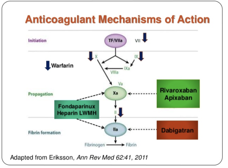
136
Q: Which two anticoagulants require regular lab monitoring?
A: Heparin and Coumadin (Warfarin).
137
Q: What lab tests are used to monitor Heparin and Coumadin?
A: PT/INR, PTT, and FXa assay.
138
Q: Why don’t DOACs require routine monitoring?
A: Standard doses are effective and have little variability between patients.
139
Q: Do DOACs affect lab tests?
A: Yes, they can interfere with lab tests even though they don't require routine monitoring.
140
Q: When might testing be done for DOACs?
A: To check if they are present in the body, such as before surgery or during emergencies.
141
Which anticoagulants have well-established reversal protocols?
Heparin and Coumadin (covered individually)
142
Which anticoaglant contain reversals that are just emerging?
DOAC
143
Generic name of Coumadin? what are the stats for this drug
Warfarin - generic name but coumadin is mostly used; one of the 20 most prescribed drugs in North America
144
How does Coumadin work?
it blocks the activity of vit K epoxide reductase required for gamma-carboxylation of vita K-dependent coag factors (SCZ27910)
145
What is coumadin used to prevent?
prevent stroke and heart attacks, post-surgery/trauma VTE and recurrence of DVTs & PE
146
How was coumadin discovered? how is it used besides antithrombotic therapy
-from moldy sweet clover- discovered when cows died after eating moldy clover in fields.
-Also used as rat poison in very different dose and form
-Also used as rat poison in very different dose and form
147
When does coumadin take into full effect?
around 5 days (bridging with UFH, LMWH or Fondaparinux)
148
How is coumadin therapy monitored? what is the therapeutic range by physician and by cardiac valves?
with INR ;
INR of 2-3
Cardiac valves 2.5-3.5
INR of 2-3
Cardiac valves 2.5-3.5
149
WHen is the patient done after starting therapy? and how often are they checked?
24 hrs ;
-checked every 2-4 days until INR consistent (2-3 times a week)
-checked very 4 weeks when stabilized until discontinued
some patients on long-term therapy
-checked every 2-4 days until INR consistent (2-3 times a week)
-checked very 4 weeks when stabilized until discontinued
some patients on long-term therapy
150
what vitamin reduces coumadin drug effects?
diet high in Vitamin K!
151
What are the 2 categories of reversal of coumadin?
non-urgent and urgent
152
What constitues non-urgent reversal of coumadin?
This is when there's no bleeding or surgery, just a high INR (meaning too much anticoagulation).
153
What to do ? (non-urgent reversal of coumadin)
Stop taking Coumadin
Take oral or injection of Vitamin K
Take oral or injection of Vitamin K
154
Q: What three things are done in an urgent reversal of Coumadin?
A: Stop Coumadin, give IV Vitamin K, and give a blood product that provides clotting factors.
155
Q: What is the preferred blood product used to reverse Coumadin in an emergency?
A: Prothrombin Complex Concentrate (PCC).
156
Q: What blood product can be used if PCC is not available?
plasma
157
Q: What conditions is heparin commonly used to treat or prevent?
A: DVT, PE, and myocardial infarction.
158
Q: What are the two main forms of heparin?
A: Unfractionated heparin (UFH) and low molecular weight heparin (LMWH).
159
Q: How does UFH work?
A: It activates antithrombin to block clotting enzymes (serine proteases), except Factor VIIa. (7)
160
Q: What is a key difference between LMWH and UFH in terms of function?
A: LMWH cannot inhibit thrombin but efficiently inhibits Factor Xa. (10)
161
Q: Why is LMWH preferred for some patients?
A: It can be given in fixed doses, works as well as UFH, is easier to use, and usually doesn’t require lab monitoring.