Clinicals: RBC morphology presenation
Study these flashcardsFree · spaced repetition · iPhone, Android, Mac & web
All cards
1
Name the different RBC Maturation Series 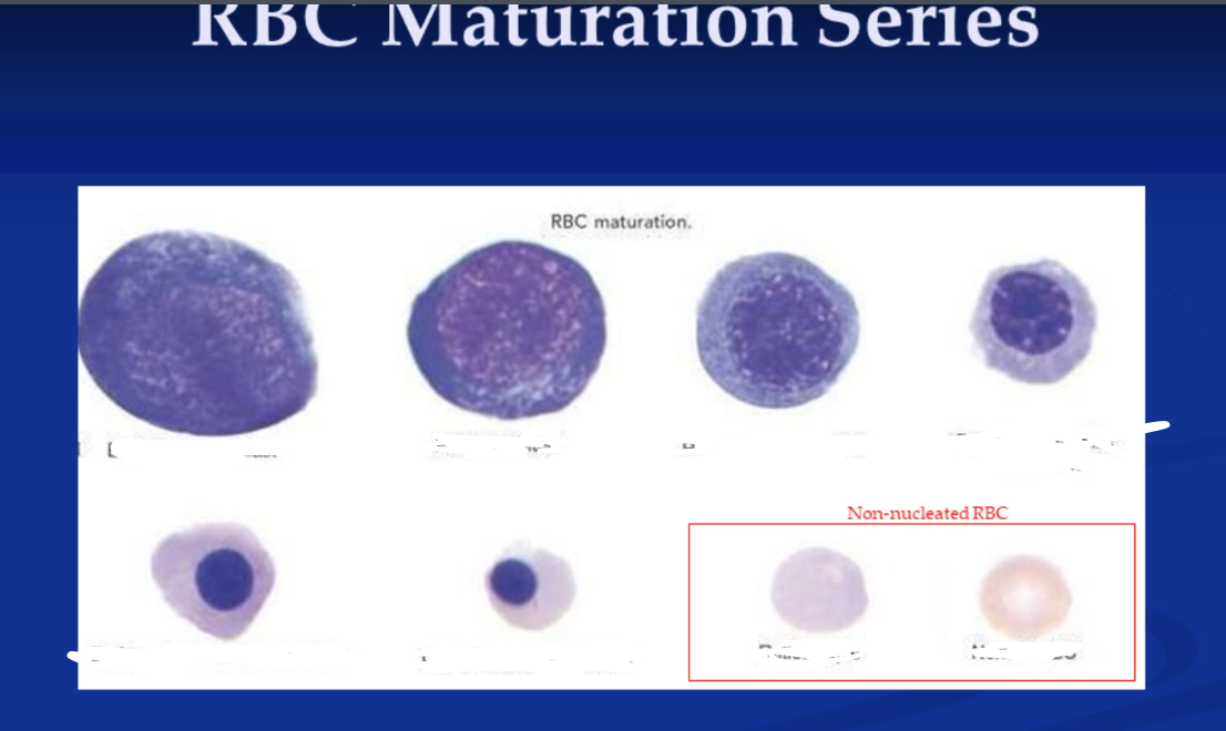
(left to right)
Early Pronormoblast
Pronormoblast
Basophilic normoblast
Polychromatophilic normoblast
Early othrochromic normoblast
Orthochromic normoblast
Reticulocyte
Normal RBC
Early Pronormoblast
Pronormoblast
Basophilic normoblast
Polychromatophilic normoblast
Early othrochromic normoblast
Orthochromic normoblast
Reticulocyte
Normal RBC
2
What are normal RBCs called?
normocytic, normochromic
3
Q: What is the typical shape and nuclear status of a normal red blood cell?WhWha
A: Non-nucleated biconcave red cell
4
Q: What is the normal diameter, MCV, MCHC, and RDW range for a red blood cell?
A: Diameter 6.7–7.8 μm;
MCV 80–99 fL;
MCHC 32–36%;
RDW 12–15
MCV 80–99 fL;
MCHC 32–36%;
RDW 12–15
5
Q: What color does a normal red blood cell stain?
A: Pink-red
6
Q: What is the typical shape of a normal red blood cell?
A: Round in shape
7
Q: How much of a normal red blood cell's area is central pallor?
A: About 1/3 of the area
8
Approximately how long do they circulate in the blood?
120 days
9
What is anisocytosis?
generic term used to indicate variation in size or erythrocytes
10
What is poikilocytosis?
variation in shape of RBCs (e.g., oval, pear-shaped, teardrop-shaped, saddle-shaped, helmet-shaped, sickle-shaped, and irregularly shaped).
11
what is the size of a microcyte?
smaller than normal RBC (MCV <80 fL)
12
What conditions are microcytes seen?
Iron deficiency anemia, thalassemias, sideroblastic anemia and lead poisoning
13
What is the size of macrocytes? (diameter and MCV)
Larger than normal RBC ( >8um in diameter)
MCV >100 fL
MCV >100 fL
14
What is the usual shape of a macrocytes?
Round or oval shape
15
Macrocytes are usually seen in what conditions? and what type of patients is it commonly seen in?
liver disease, reticulocytosis, hypothyroidism and postsplenectomy states;
seen in babiessss
seen in babiessss
16
Oval macrocytes may be seen in what type of conditions?
folate or B12 deficiency
17
How is hypochromasia characterized?
large area of central pallor
18
What is hypochromasia caused by? What is the MCHC?
decreased concentration of hemoglobin;
MCHC < 32.0%
MCHC < 32.0%
19
What conditions is hypochromasia seen in?
Characteristically seen in iron deficiency anemia but present in other forms of anemia
20
What do spherocytes lack?
central pallor
21
What conditions are spherocytes usually seen in?
hemolytic anemia, hereditary spherocytosis, ABO hemolytic disease of the newborn, burns, cold agglutinin disease, drug-induced hemolytic anemia
22
What causes spherocytes?
abnormality or deficiency of spectrin, ankyrin and protein 4.1
23
Q: What shape do ovalocytes (elliptocytes) have?
A: RBCs shaped like a pencil or thin cigar, with blunt ends and parallel sides.
24
Q: Where is hemoglobin often concentrated in ovalocytes?
A: At the ends of the ovalocyte.
25
Q: Can small numbers of ovalocytes be found in healthy individuals?
A: Yes, small numbers can be seen in normal individuals.
26
Q: What percentage of ovalocytes may be seen in patients with hereditary elliptocytosis?
A: Greater than 10–25%.
27
Q: What causes hereditary elliptocytosis?
A: Abnormalities of erythrocyte skeletal membrane proteins.
28
Q: In which other conditions are ovalocytes commonly increased?
A: Iron deficiency anemia and other types of anemia.
29
What specifically causes Elliptocytosis membrane abnoamlity?
Spectrin abnormality
Deficiency of protein 4.1
Hereditary elliptocytosis worldwide prevalence of at least 1 in 2000 persons
Deficiency of protein 4.1
Hereditary elliptocytosis worldwide prevalence of at least 1 in 2000 persons
30
What's another name for target cell?
Codocyte
31
How do target cells (codocytes) look like?
central hemoglobinized area
32
What conditions can target cells be seen in?
Hemoglobinopathies, liver disease, thalassmeias, sideroblastic anemia, obstructive jaundice
33
Describe a stomatocyte
RBCs with a rectangular area of pallor
34
What conditions are stomatocytes seen?
May be seen in hereditary stomatocytosis, liver disease, hydroxyurea therapy, electrolyte imbalance and myelodysplastic syndrome
35
What is another name for sickle cell?
Drepanocyte
36
Describe a sickle cell
RBCs in shape of crescent with 2 pointed ends
37
What type of conditions are seen with sickle cell?
sickle cell anemia, hemoglobin SC disease, SD disease and S-betea thalassemia
38
What causes sickle cell? specificaly
caused by amino acid substitution valine for glutamic acid, in the 6th position of the beta chain
39
What's another name for TEARDROP CELL?
Dacrocyte
40
Describe a teardrop cell.
RBCs appearing in the shape of a teardrop or pear with a single, short, or long often blunted or rounded end.
41
what conditions are associated with tear drop cell?
bone marrow fibrosis, pernicious anemia, anemia of renal disease, hemolytic anemias, and other forms of severe anemia
42
Q: What are schistocytes?
A: RBCs that have been physically cut (mechanical damage).
43
Q: Name some conditions where schistocytes may be seen.
A: Severe burns, DIC (disseminated intravascular coagulation), TTP (thrombotic thrombocytopenia purpura), HUS (hemolytic uremic syndrome), and other microangiopathic hemolytic anemias.
44
Q: What is a helmet cell (keratocyte)?
A: A type of schistocyte found in G6PD deficiency and pulmonary emboli
45
Q: What is the shape of schistocytes?
A: Triangularly-shaped cells.
46
Q: What is a blister cell also known as?
A: Pre-keratocyte.
47
Q: What does a blister cell look like?
A: Vacuolated, non-nucleated red blood cell.
48
Q: With which condition are blister cells commonly associated?
A: MAHA (microangiopathic hemolytic anemia).
49
Q: How are blister cells formed?
A: RBCs containing inclusions are “pitted” or removed by macrophages in the spleen, or when RBCs are impaled by fibrin strands, the membrane reforms and produces a vacuole.
50
What cells are associated with disorders with Heinz body formation?
bite cells
51
Q: What is another name for a burr cell?
A: Echinocyte.
52
Q: What do burr cells look like?
A: RBCs with evenly spaced pointed projections.
53
Q: Name some conditions where burr cells may be seen.
A: Uremia, pyruvate kinase deficiency, cancer of the stomach, acute blood loss, severe burns, liver disease, and renal disease.
54
Q: What does the term “echino-” in echinocyte mean?
A: It comes from Greek meaning “prickly” or “spiny,” like a sea urchin.
55
Q: What is the cause of crenated red blood cells?
A: They are an artifactual change.
56
Q: How do crenated RBCs appear under the microscope?
A: RBCs with blunt projections evenly distributed over the surface.
57
Q: What are common causes of RBC crenation?
A: Faulty drying of the blood smear, change in pH, or excess EDTA causing shrinkage of the RBC.
58
Q: What is another name for an acanthocyte?
A: Spur cell.
59
Q: How do acanthocytes appear under the microscope?
A: RBCs with irregularly spaced, thorn-like projections.
60
Q: Name some conditions where acanthocytes may be seen.
A: Abetalipoproteinemia, liver disease, alcohol intoxication, anorexia nervosa, post-splenectomy, and hepatorenal failure.
61
Q: What is polychromasia?
A: A condition where red blood cells (RBCs) appear bluish-gray or pale purple on Wright-Giemsa stain due to retained RNA, indicating young RBCs (reticulocytes).
62
Q: Are polychromatophilic RBCs nucleated?
A: No, they are non-nucleated.
63
Q: What is the shape of polychromatophilic RBCs?
A: Round or ovoid.
64
Q: What does polychromatophilic RBC represent in RBC maturation?
A: The final stage of RBC maturation after exiting the bone marrow.
65
Q: How do polychromatophilic RBCs compare in size to mature RBCs?
A: They are larger than mature RBCs.
66
Q: Do polychromatophilic RBCs have central pallor?
A: They usually lack central pallor.
67
Q: What cellular component makes polychromatophilic RBCs stain differently?
A: They contain RNA.
68
Q: What stain color is typical for polychromatophilic RBCs with Wright-Giemsa?
A: Pink-gray to pale purple.
69
Q: In what conditions are polychromatophilic RBCs typically seen?
A: Various anemias.
70
Q: How do polychromatophilic RBCs stain with supravital stains (e.g., new methylene blue)?
A: They show deep blue granular and/or filamentous structures (reticulin).
71
Q: How can basophilic stippling appear under the microscope?
A: It may be fine or coarse.
72
Q: What are the granules in basophilic stippling composed of?
A: Unstable RNA.
73
Q: In which conditions can basophilic stippling be found?
A:
Lead poisoning
Alcoholism
Megaloblastic anemias
Dyserythropoiesis
Anemias with impaired hemoglobin synthesis (e.g., thalassemia)
Lead poisoning
Alcoholism
Megaloblastic anemias
Dyserythropoiesis
Anemias with impaired hemoglobin synthesis (e.g., thalassemia)
74
Q: What are Pappenheimer bodies?
A: Small, dark, angular inclusions (<1 µm) in RBCs that contain iron.
75
Q: How do Pappenheimer bodies compare in size to Howell-Jolly bodies?
A: They are smaller than Howell-Jolly bodies.
76
Q: Where in the cell are Pappenheimer bodies typically located?
A: Near the periphery of the cell, usually in clusters.
77
Q: On what type of stain are Pappenheimer bodies seen?
A: On Wright-stained smear.
78
Q: What are siderocytes?
A: RBCs that contain iron granules visible with iron stains (confirmed Pappenheimer bodies).
79
Q: Which stain is used to confirm siderocytes?
A: Prussian blue (iron stain).
80
Q: How are siderocytes related to Pappenheimer bodies?
A: Pappenheimer bodies that turn positive on iron stain are called siderocytes.
81
Q: In which anemia are siderocytes classically present?
A: Sideroblastic anemia.
82
Q: What other anemia type may show siderocytes?
A: Megaloblastic anemia.
83
Q: What substance use is associated with siderocytes?
A: Alcoholism.
84
Q: After which surgery might siderocytes appear?
A: Splenectomy (postsplenectomy).
85
Q: Besides anemias, what other disorder group can show siderocytes?
A: Some hemoglobinopathies.
86
Q: What are Howell-Jolly bodies?
A: Small, round RBC inclusions ~1 µm in diameter.
87
Q: What are Howell-Jolly bodies composed of?
A: DNA (nuclear remnants).
88
Q: On which stain are Howell-Jolly bodies typically seen?
A: Wright/Wright-Giemsa as dark, round inclusions.
89
Q: In which conditions are Howell-Jolly bodies commonly seen?
A: Megaloblastic anemia and myelodysplastic syndromes (MDS).
90
Q: What organ normally removes Howell-Jolly bodies from circulating RBCs?
A: The spleen (splenic pitting).
91
Q: When do Howell-Jolly bodies persist in circulation?
A: With asplenia, hyposplenia, or postsplenectomy.
92
Q: How do Howell-Jolly bodies compare in size to Pappenheimer bodies?
A: They are typically larger and appear singly; Pappenheimer bodies are smaller and clustered.
93
Q: Do Howell-Jolly bodies contain iron?
A: No—iron stains (e.g., Prussian blue) are negative; they are DNA, not iron.
94
Q: What does the term nRBC indicate in peripheral blood?
A: Presence of normoblasts (nucleated RBC precursors) in circulation, regardless of maturation stage.
95
Q: At what maturation stage are circulating nRBCs typically seen?
A: Orthochromic normoblast stage.
96
Q: When can nRBCs be released prematurely into blood?
A: Under prolonged/severe hemolytic stress or with extramedullary hematopoiesis.
97
Q: Are nRBCs normal in any population?
A: They may be seen in newborns (physiologic).
98
Q: What general pathologies can cause nRBCs in adults?
A: Severe hemolysis, hypoxia/stress erythropoiesis, marrow infiltration, or extramedullary hematopoiesis.
99
Q: What do nRBCs in blood suggest about erythropoiesis?
A: Stress or disordered erythroid maturation in the marrow (or production outside marrow).
100
Q: What cytologic changes may be seen in circulating nRBCs?
A: Megaloblastic and dysplastic features can appear.
101
Q: Why should we be cautious calling a circulating nRBC “dysplastic”?
A: Abnormal nuclear shapes (lobated/fragmented) can occur during egress from marrow and may not reflect true marrow dysplasia.
102
Q: What organ process normally prevents nRBCs from circulating?
A: Proper marrow maturation with enucleation before release into blood.
103
Q: Name two conditions specifically mentioned that can lead to premature nRBC release.
A: Prolonged/severe hemolytic stress and extramedullary hematopoiesis.
104
Q: In a smear report, how are nRBCs usually expressed?
A: As number of nRBCs per 100 WBCs (and automated analyzers correct WBC count accordingly).
105
Q: What are Cabot rings?
A: Purple-staining, threadlike filaments forming a ring or loop inside an RBC.
106
Q: What are Cabot rings thought to represent?
A: Nuclear remnants or denatured proteins (mitotic spindle remnants).
107
Q: On what stain are Cabot rings typically seen?
A: Wright/Wright–Giemsa as thin purple rings/figure-8 shapes.
108
Q: In which conditions are Cabot rings (rarely) seen?
A: Pernicious anemia and lead poisoning.
109
Q: How do Cabot rings differ from Howell-Jolly bodies?
A: Cabot rings are filamentous ring/figure-8 structures; Howell-Jolly bodies are single round DNA dots.
110
Q: How do Cabot rings differ from Pappenheimer bodies?
A: Cabot rings are non-iron filaments; Pappenheimer bodies are small clustered iron granules (Prussian blue positive).
111
Q: What are Heinz bodies?
A: Aggregates of denatured hemoglobin attached to the RBC membrane.
112
Q: What is the typical size and number of Heinz bodies in a cell?
A: Large (≈1–3 µm) inclusions; may be single or multiple.
113
Q: How can Heinz bodies be made visible on a smear?
A: They require supravital stains.
114
Q: Which supravital stains are used to detect Heinz bodies?
A: New methylene blue, crystal violet, Nile blue, or methyl violet.
115
Q: Are Heinz bodies visible on a routine Wright/Wright-Giemsa stain?
A: Generally no; they’re best seen only with supravital staining.
116
Q: What are Heinz bodies composed of?
A: Precipitated hemoglobin within RBCs.
117
Q: In G6PD deficiency, what kind of hemoglobin forms Heinz bodies?
A: Precipitated normal hemoglobin (oxidant stress denatures it).
118
Q: In unstable hemoglobin disorders, what kind of hemoglobin forms Heinz bodies?
A: Structurally defective (unstable) hemoglobin that precipitates.
119
Q: What is the unifying mechanism behind Heinz body formation across diseases?
A: Hemoglobin denaturation → precipitation → membrane-bound inclusions.
120
Q: What do Hemoglobin H inclusions represent?
A: Precipitated excess beta globin chains (β4).
121
Q: When are HbH inclusions visible on a smear?
A: Only after supravital staining.
122
Q: Which stains can reveal HbH inclusions?
A: Supravital stains such as brilliant cresyl blue or new methylene blue.
123
Q: What is the characteristic appearance of HbH inclusions?
A: Small, evenly dispersed deposits giving a “golf-ball” pattern.
124
Q: In which condition are HbH inclusions classically found?
A: Hemoglobin H disease (α-thalassemia with three α-gene deletions).
125
Q: Mechanistically, why do HbH inclusions form?
A: Lack of α chains → excess β chains tetramerize (β4) → precipitate within RBCs.
126
Q: Are HbH inclusions typically seen on routine Wright-Giemsa stain?
A: No—they require supravital staining to be seen.
127
Q: What are HbC crystals?
A: Dense hemoglobin C deposits forming crystals inside (or outside) RBCs.
128
Q: What shapes do HbC crystals typically have?
A: Rhomboid, tetragonal, or rod-shaped.
129
Q: How do HbC crystals affect the RBC?
A: They often distort the cell and can project beyond the cell rim.
130
Q: Where can HbC crystals be located relative to the cell?
A: Intra-erythrocytic or extracellular.
131
Q: In which patients are HbC crystals commonly seen post-splenectomy?
A: Patients with Hb CC disease or Hb SC disease.
132
Q: On routine Wright–Giemsa, how do HbC crystals appear?
A: Very dense, pink-red, angular crystals within/distorting RBCs.
133
Q: Which five Plasmodium species cause human malaria?
A: P. falciparum, P. vivax, P. ovale, P. malariae, and P. knowlesi.
134
Q: What feature helps distinguish Plasmodium species on smear?
A: The distinctive shapes/appearances of developmental stages (ring, trophozoite, schizont, gametocyte).
135
Q: Which infection can look similar to malaria ring forms on blood smear?
A: Babesiosis.
136
Q: Which Plasmodium species is associated with crescent (banana-shaped) gametocytes?
A: P. falciparum.
137
Q: What stain is standard for thick and thin malaria smears?
A: Giemsa (Romanowsky).
138
Q: Which species classically shows oval RBCs with fimbriated edges?
A: P. ovale.
139
Q: Which species has a quartan (72-hour) fever periodicity?
A: P. malariae.
140
Q: Which species is known for high parasitemia and severe disease?
A: P. falciparum.
141
Q: What common smear artifact can mimic an RBC inclusion?
A: A platelet sitting on top of an RBC.
142
Q: Should a platelet on top of an RBC be reported as an RBC inclusion?
A: No—it should not be confused with an inclusion.
143
Q: What visual clue helps you recognize a platelet lying on an RBC rather than an inclusion?
A: A non-staining halo around the platelet where it sits on the RBC surface.
144
Q: Why does a halo appear around a platelet on top of an RBC?
A: The platelet sits on the cell surface and displaces stain/light at the contact area, creating a clear rim.
145
Q: What is rouleaux?
A: Linear stacking of ≥4 RBCs that looks like a “stack of coins.”
146
Q: What causes rouleaux formation?
A: Increased plasma proteins that reduce RBC zeta potential.
147
Q: Which proteins are most implicated in rouleaux?
A: Acute-phase proteins, especially fibrinogen and globulins.
148
Q: In which disorders is rouleaux commonly seen?
A: Multiple myeloma, Waldenström macroglobulinemia, and autoimmune/inflammatory conditions.
149
Q: How does rouleaux affect ESR?
A: ESR is elevated (faster settling due to RBC aggregation).
150
Q: What causes agglutination of RBCs?
A: Agglutinins (antibodies) that cross-link RBCs, most often IgM cold agglutinins.
151
Q: How does agglutination look compared with rouleaux?
A: Irregular, round grape-like clumps rather than the linear “stack of coins” of rouleaux.
152
Q: Key difference in cause vs. rouleaux?
A: Agglutination = antibodies cross-link RBCs; Rouleaux = increased plasma proteins reduce zeta potential.
153
Q: In what infections can RBC agglutination be found?
A: Viral or Mycoplasma infections (e.g., Mycoplasma pneumoniae; some viral illnesses).
154
Q: In what hematologic disorders can agglutination be seen?
A: Plasma-cell or lymphoid neoplasms (e.g., Waldenström macroglobulinemia, CLL/lymphoma).