Lecture 4: Protozoa (Lumen Dwelling): Coccidia and Ciliate
Study these flashcardsFree · spaced repetition · iPhone, Android, Mac & web
All cards
1
What is this?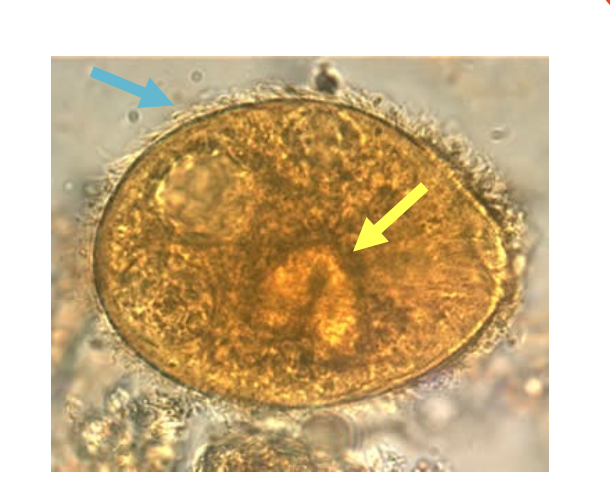
Ciliates
2
What do ciliates usually contain in at least one stage of their life cycle?
cilia
3
What are the 2 different types of nuclei for Ciliates?
1 macronucleus
1 micronucleus
1 micronucleus
4
Which ciliates is the only ciliate that infects humans?
Balantidium coli
5
What are the blue and yellow arrows?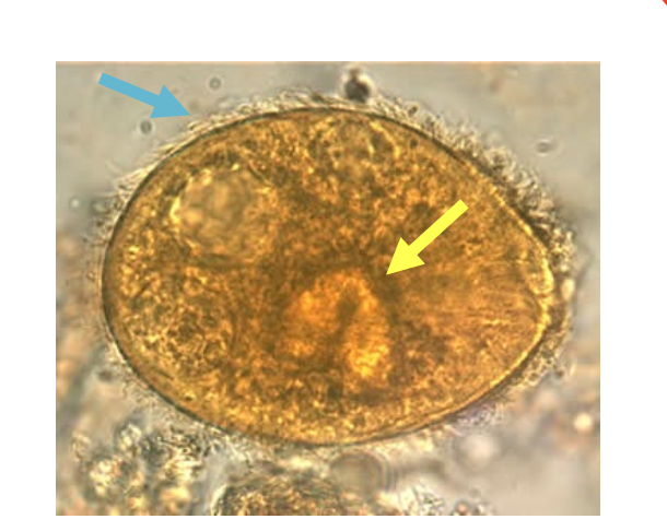
Blue: cilia
Yellow: Macronucleus
Yellow: Macronucleus
6
Are balantidium coli big or small?
HUGE
7
What is the bottom arrow?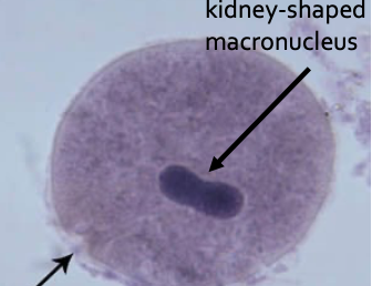
Cytostome "cell mouth"
8
What is the peanut shape organelle?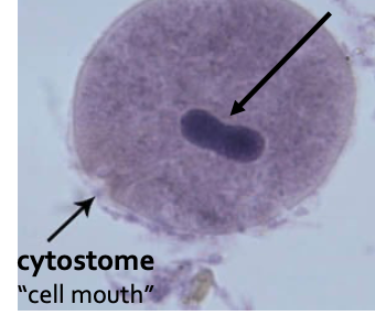
Kidney-shaped macronucleus
9
Which is the trophozoite and which is the cyst ?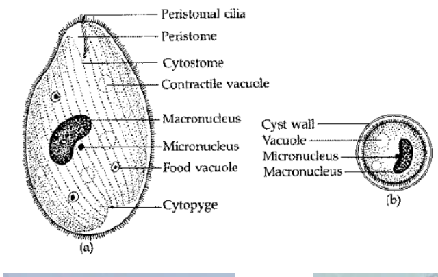
Left: Trophozoites
Right: Cysts
Right: Cysts
10
Which is the trophozoite and which is the cyst ?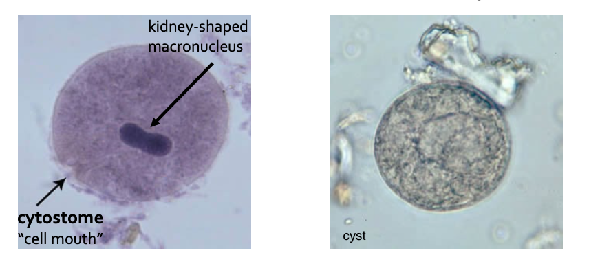
Left: Trophozoites
Right: Cysts
Right: Cysts
11
Are these trophozoites or cysts of B. coli?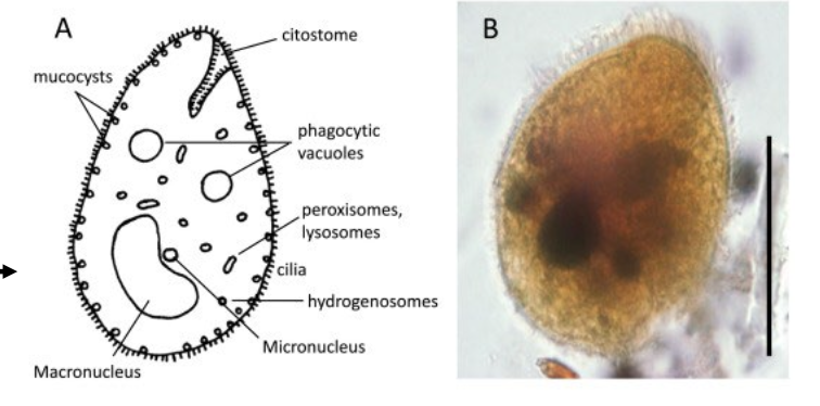
Trophozoites
12
Are these trophozoites or cysts of B. coli?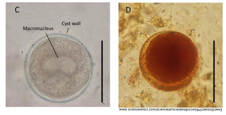
Cysts
13
What is Balantidium coli usually associated with? (animal)
PIGS
14
What disease does B. coli cause?
Balantidiasis
15
Is B. coli asymptomatic or symptomatic?
Mainly asymptomatic, but can lead to serious disease
16
What clinical symptoms are associated with serious B. coli infection?
Persistent diarrhea
Abdominal pain
Perforated colon <- remember very dangerous. can become septic
Abdominal pain
Perforated colon <- remember very dangerous. can become septic
17
Where in the world is B coli found?
Worldwide; rare in US (temperate and wamr climated)
18
In which areas are non-human primates infected with B. coli?
Tropical areas
19
In which areas are pigs infected with B. coli?
Other areas
20
How is one exposed to B. coli?
Inadvertently ingesting contaminated human/animal fecal matter (wash your hands); drinking and washing food with contaminated water
21
What is the treatment for B. coli?
Tetracycline, Metronidazole, Iodoquinol
22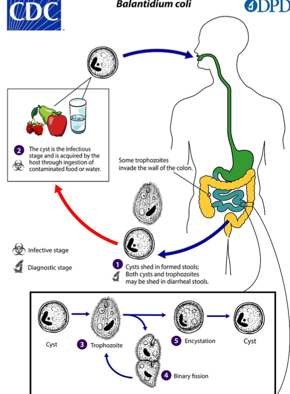
What is the Life Cycle of Balantidium coli?
23
What is the mode of transmission for Balantidium coli?
Fecal-oral
24
Which stage is B. coli infectious?
Cyst stage - responsible for transmission
25
Where does excystation occur for trophozoites of B coli?
small intestine
26
What invades the large intestine in B. coli?
Trophozoites
27
Where do trophozoites of B. coli colonize and invade?
they colonize the large intestine (lumen) and applendix. Some invade the wall of the colon, causing ulcerative pathology.
28
What type of cysts gets excreted in the feces for B. coli?
Mature cysts
29
What are the 4 types of Coccidia?
(Cysto)isospora
Microsporidia
Cryptosporidium
Cyclospora
Microsporidia
Cryptosporidium
Cyclospora
30
What is the clinical presentation of (Cysto)isospora belli?
Acute, non-bloody diarrhea
Crampy abdominal pain, which can last for weeks and results in malabsoption and weight loss
Eosinophilia may be present.
Crampy abdominal pain, which can last for weeks and results in malabsoption and weight loss
Eosinophilia may be present.
31
Who is at risk for (Cysto)isospora belli?
Immunocompromised and infants and children at increased risk.
32
What type of situations is (Cysto)isospora belli seen?
Outbreaks in institutionalized groups/food products
33
Give examples of the types of foods that (Cysto)isospora belli can be found?
Basil, cilantro, mesclun lettuce, raspberries, snow peas, bagged salad
34
Where in the world and which areas is (Cysto)isospora belli found?
Worldwide (tropical/subtropical)
35
What is the treatment for (Cysto)isospora belli?
Trimethoprim-sulfamethoxazole
Pyrimethamine (if can't take TMP-SMX)
Ciprofloxacin (second line)
Pyrimethamine (if can't take TMP-SMX)
Ciprofloxacin (second line)
36
What is this called and what is the stained used?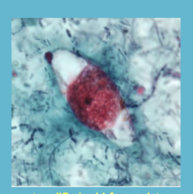
(Crysto)isospora belli; modified acid-fast stain
37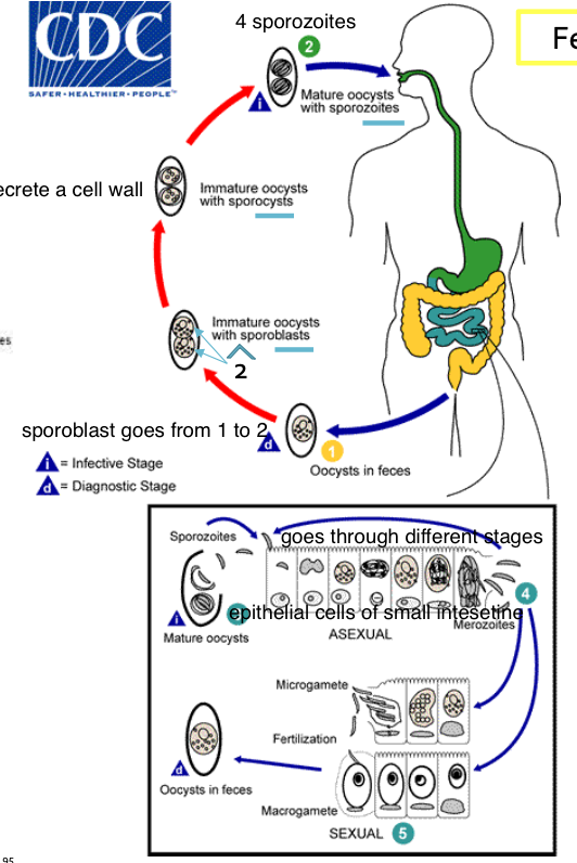
What is the life cycle of (Cysto)isospora belli?
38
Starting at excretion, what is released? c. belli
Immature oocysts with one sporoblast (more rarely two)
39
What happens after excretion of the immature oocyst of C. belli?
The sporoblast divides in 2 (the oocysts now contains 2 sporoblasts)
40
What happens after the sporoblasts divide into 2 for C. belli?
The sporoblasts secrete a cyst wall, this becoming sporocysts.
41
What happens after sporocysts are formed in C. belli?
The sporocytes divide twice to produce 4 sporozoites each - mature
42
What happens after sporozoites are mature? (C. belli)
The mature oozytes excysts in the small intestine and release their sporozoites, which invade the epithelial cells and initiate schizogony.
43
What is shizogony?
a form of asexual reproduction where a single cell divides multiple times to produce many daughter cells.
44
What happens after schizogony occurs in C. belli?
The merozoites are released, invade new epithelial cells, and continue the cycle of asexual multiplication.
45
How long does it take for C. belli gametocytes to start the sexual stage?
Minimum one week.
46
How are gametocytes of C. belli formed?
the development of male and femal gametocytes
47
What is considered fertilization of oocysts for C. belli?
when Oocysts are exreted in the stool.
48
What is this?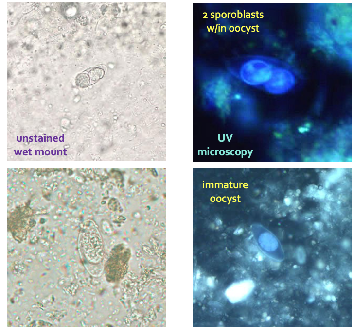
(Cysto)isospora belli oocysts in feces
49
What type of microscopy is this and for which organism?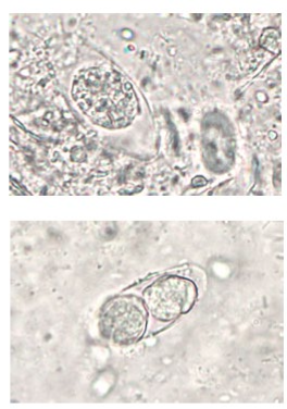
Wet mount; (Cysto)isospora oocysts
50
What type of microscopy is this and for which organism?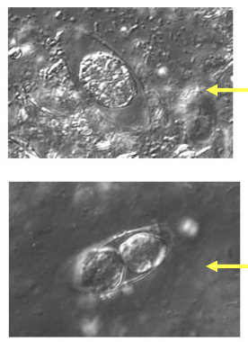
DIC (differential interference contrast); Cystoisospora Oocysts
51
What type of microscopy is this and for which organism?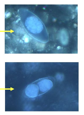
UV; (Cysto)isospora Oocysts
52
What do Oocysts from Cystoisospora look like under the wet mount?
Oocysts are large (25 to 30 by 10 to 19 um) and
have a typical ellipsodial shape.
have a typical ellipsodial shape.
53
Which microscopy is required for (Cysto)isospora Oocysts?
Fluorescent microscope.
54
What parasite is this?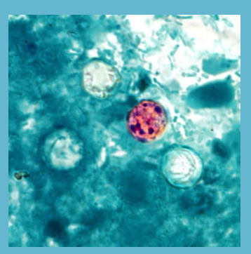
Cyclospora cayetanensis
55
What stain is used for this parasite?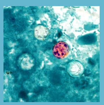
acid-fast stain
56
What disease is associated with Cyclospora cayetanensis?
Cyclosporiasis
57
Is Cyclosporiasis asymptomatic or severe?
can vary from asymptomatic -> severe
58
How long does untreated cyclosporiasis last?
10-12 weeks and may follow a relasping course
59
What are symptoms of cyclosporiasis?
Watery diarrhea
Anorexia, weight loss
Abdominal pain
Nausea and vomiting
Myalgias, low-grade fever, and fatigue.
Anorexia, weight loss
Abdominal pain
Nausea and vomiting
Myalgias, low-grade fever, and fatigue.
60
Is Cyclospora cayetanensis fecal-oral transmitted?
NOT directly
person-person transmission unlikely
person-person transmission unlikely
61
When is the oocyst in Cyclospora cayetanensis infective and how long does it take?
Days to weeks in the environment
62
What is the usual source for Cyclospora cayetanensis?
Fresh produce/water; associated with raspberries and basil
63
Whom is the major host for Cyclospora cayetanensis?
Humans
64
Where is Cyclospora cayetanensis primarily found? (location)
Nepal, Peru, Haiti, Guatemala (also US)
65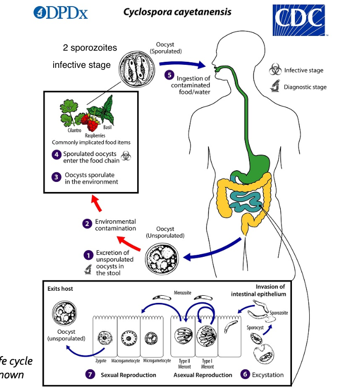
What is the life cycle of Cyclospora cayetensis?
66
What is the 1st step of Cyclospora cayetensis life cycle?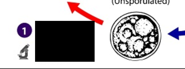
Excretion of unsporulated oocyts in the stool 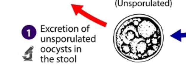
67
What is the 2nd step of Cyclospora cayetensis life cycle?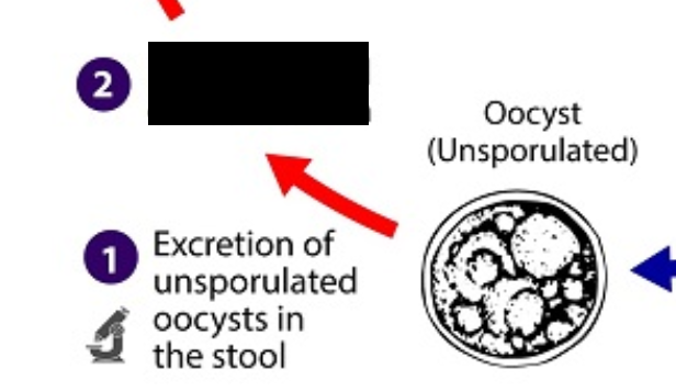
Environmental contamination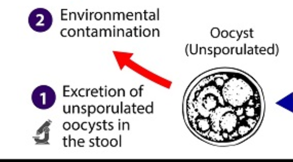
68
What is the 3rd step of Cyclospora cayetensis life cycle?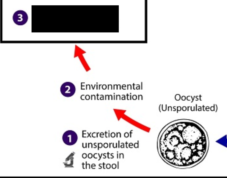
Oocysts sporulate in the environment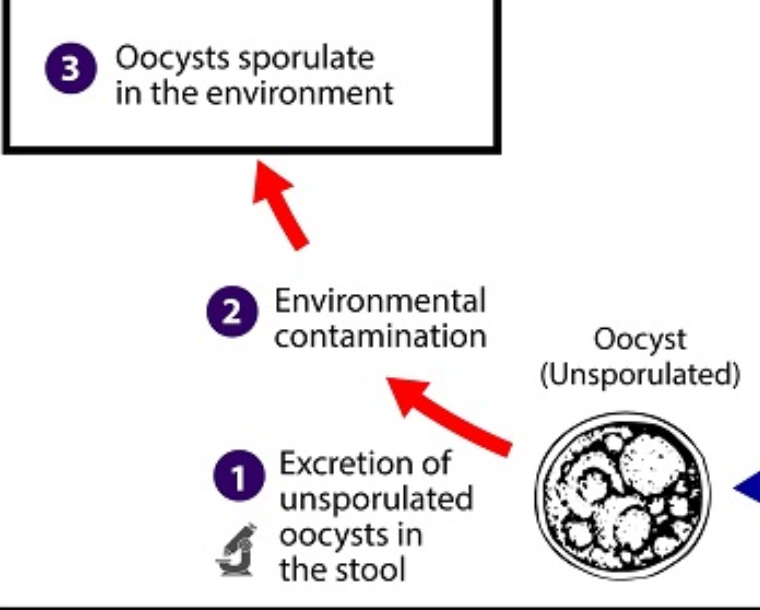
69
What is the 4th step of Cyclospora cayetensis life cycle?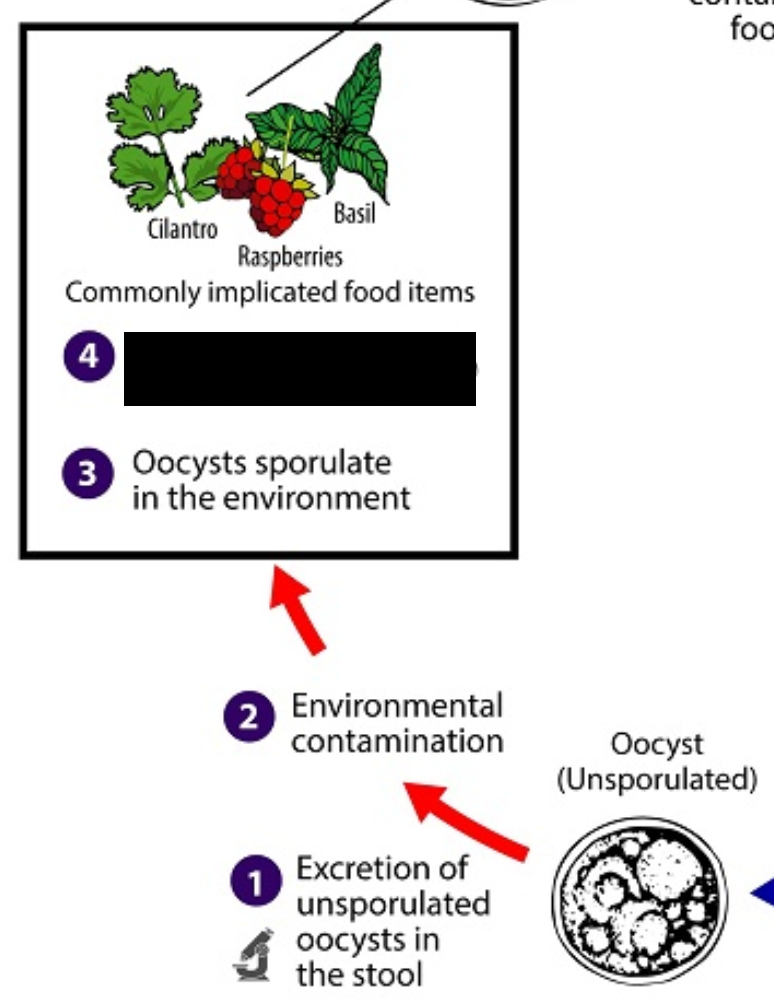
Sporulated Oocysts enter the food chain 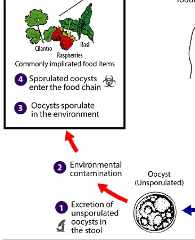
70
What is the 5th step of Cyclospora cayetensis life cycle?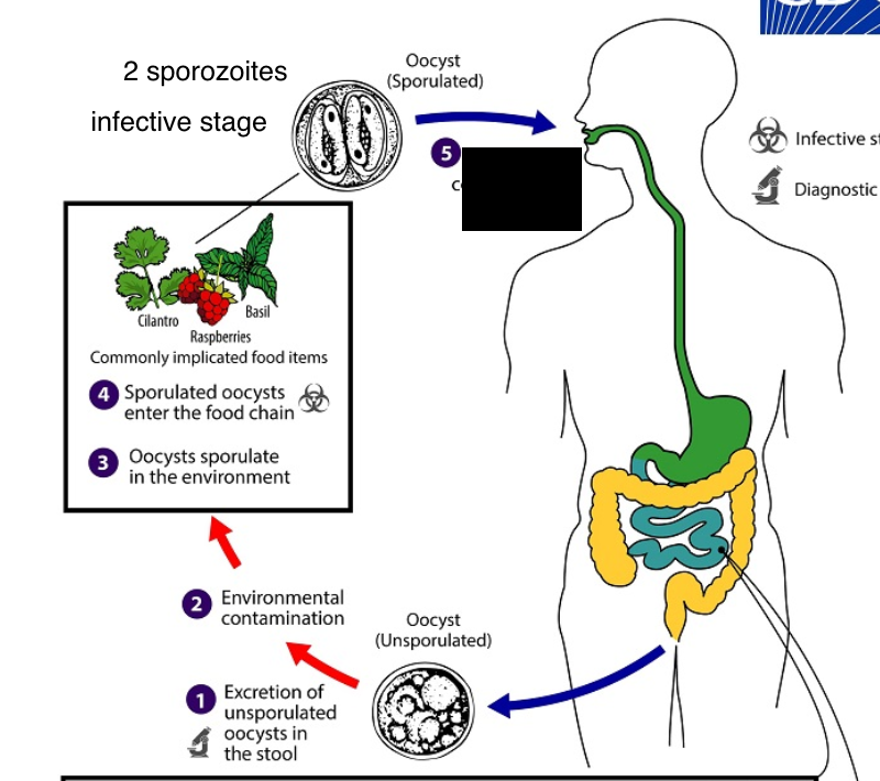
Ingestion of contaminated food/water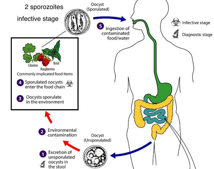
71
When Oocysts are sporulated, how many sporozoites are there?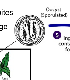
2 sporozoites
72
What is the 6th step of Cyclospora cayetensis life cycle?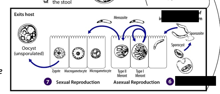
Excystation; invasion of intestinal epithelium 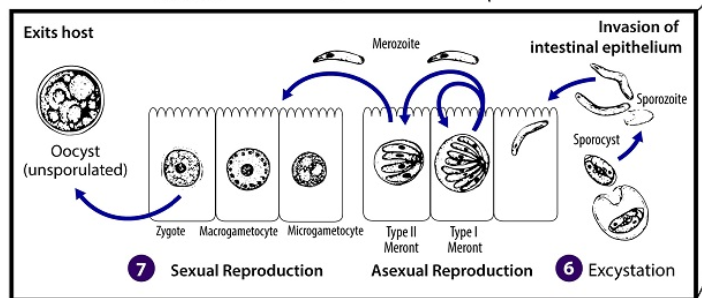
73
What is the 7th step of Cyclospora cayetensis life cycle?t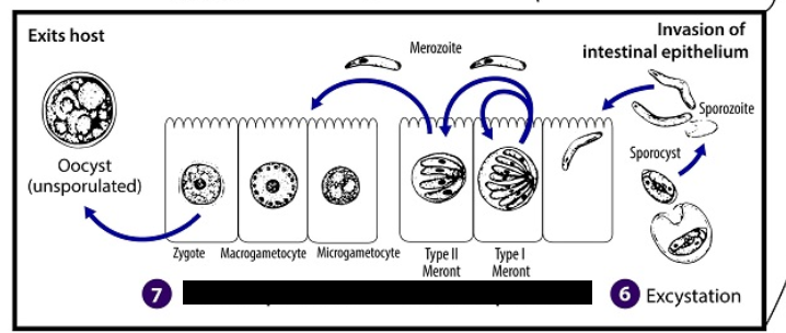
asexual and sexual reproduction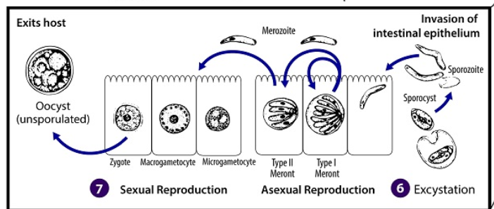
74
During excystation of Cyclospora cayetensis life cycle, what exits out of it?n
2 Sporozoites within the sporocysts enter the intestinal epithelium 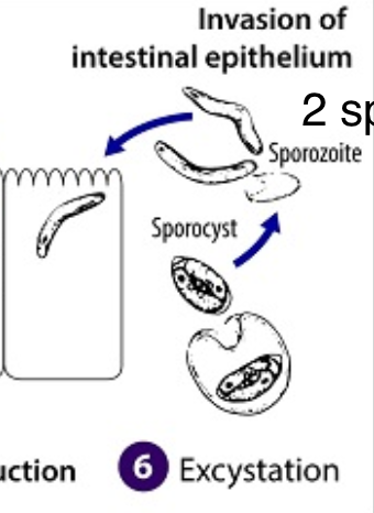
75
What differentiates Cyclospora cayetensis from Cryptosporidium?
The oocyst is not infective (direct fecal-oral transmission cannot occur) in Cyclospora cayetensis
76
At what temperature does sporulation occur and when for Cyclospora cayetensis?
Days or weeks at 22oC to 32oC.
77
What happens during sporulation for Cyclospora cayentensis?
Division of the sporont into 2 sporocysts, each containing 2 elongated sporozoites
78
At what step does sporulation happen? (Cyclospora cayetanensis)
3rd step.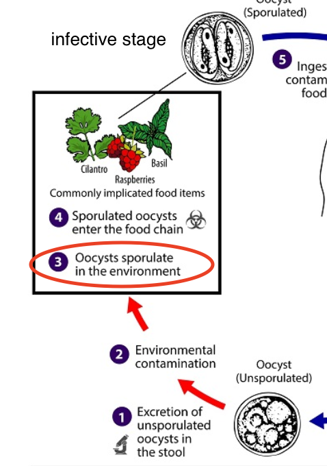
79
Where do the oocysts excyst for Cyclospora cayentensis?
Gastrointestinal tract
80
Specifially, what cells and where do the sporozoites of oocysts invade for Cyclospora cayentensis?
Epithelial cells of the small intestine
81
What type of reproduction do the sporozoites undergo for Cyclospora cayentensis when they enter the epithelial cells of the small intestine?
Asexual multiplication
82
What is produced during asexual multiplication of cyclospora caytenensis?
Type I & type II meronts
83
Which meront undergoes sexual development into macrogametocytes and microgametocytes?
Type II meronts
84
The macrogametocyte and microgametocyte combine together to form what? (Cyclospora cayentensis)
Zygote
85
How do you ID Oocysts?
Look for unsporulated oocysts in feces or small bowel aspirate
86
What do spherical oocysts measure?
7-10 um
87
What do spherical oocysts look like? Cyclospora cayetenensis
Contain a cluster of refractile globules surrounded by a thick membrane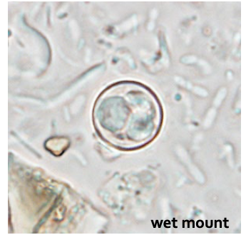
88
What type of stains and microscopy are used to ID Oocysts?
Trichrome, iodine, and modified acid-fast stain,
UV micrscopy, PCR.
UV micrscopy, PCR.
89
What colors are seen in modified acid-fast stains?
variable: pink-red-deep purple
90
What is this?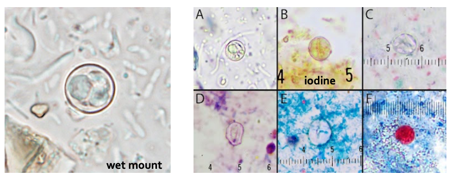
Oocysts
91
What type of staining is this?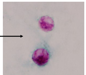
Modified acid-fast stains
92
What type of microscopy is this?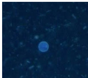
UV microscopy
93
What type of staining is this?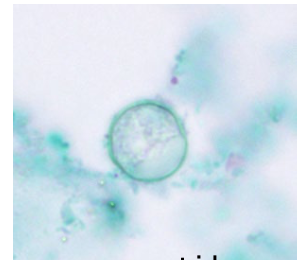
Trichrome
94
What intestinal parasite is this?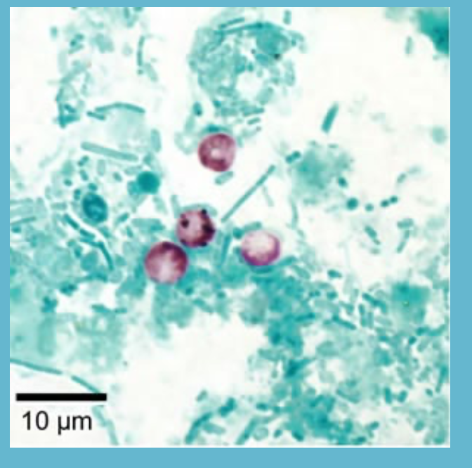
Cyptosporidium parvum and cryptosporidium hominis
95
What is the major cause of protracted (prolonged) diarrhea in immunocompromised patients?
Cyptosporidium parvum and cryptosporidium hominis
96
What can Cyptosporidium parvum and cryptosporidium hominis lead to in immunocompromised patients?
Can lead to life-threatening malabsorption and wasting
97
What does Cyptosporidium parvum and cryptosporidium hominis cause in immunocompetent patients?
Diarrhea lasting for 2-3 weeks (self-limiting)
98
What is the mode of transmission for Cyptosporidium parvum and cryptosporidium hominis?
Mainly thorugh ingestion of fecally contaminated water (e.g. drinking or recreational water) or food (e.g., raw milk) or following direct contact with infect animals or people.
99
Where, specifically, are there number outbreaks for Cyptosporidium parvum and cryptosporidium hominis?
swimming pools
water playgrounds/spalsh pads
contact with animals (C. parvum association with cattle, particularly calves)
childcare settings
food-borne: unpasteruized cider, juice, and milk
water playgrounds/spalsh pads
contact with animals (C. parvum association with cattle, particularly calves)
childcare settings
food-borne: unpasteruized cider, juice, and milk
100
Where can Cyptosporidium parvum and cryptosporidium hominis be seen on earth?
Worldwide
101
Who can Cyptosporidium parvum and cryptosporidium hominis infect?
Wide range of vertebrates (humans and animals)
102
What is the treatment for Cyptosporidium parvum and cryptosporidium hominis?
Replace fluid/electrolyte loss
Nitazoxanide to treat diarrhea in immunocompentent
Antivirals (HIV+) to control underlying immunosuppression
Nitazoxanide to treat diarrhea in immunocompentent
Antivirals (HIV+) to control underlying immunosuppression
103
Out of 105 outbreaks with a laboratory-confrimed etiology, how many were caused by parasites?
64.8%
104
What is the etiologic agent of 60 (44.8% of 134 outbreaks associated with treated recreational water?
Crytosporidium
105
What is the 1st step for Cryptosporidium spp?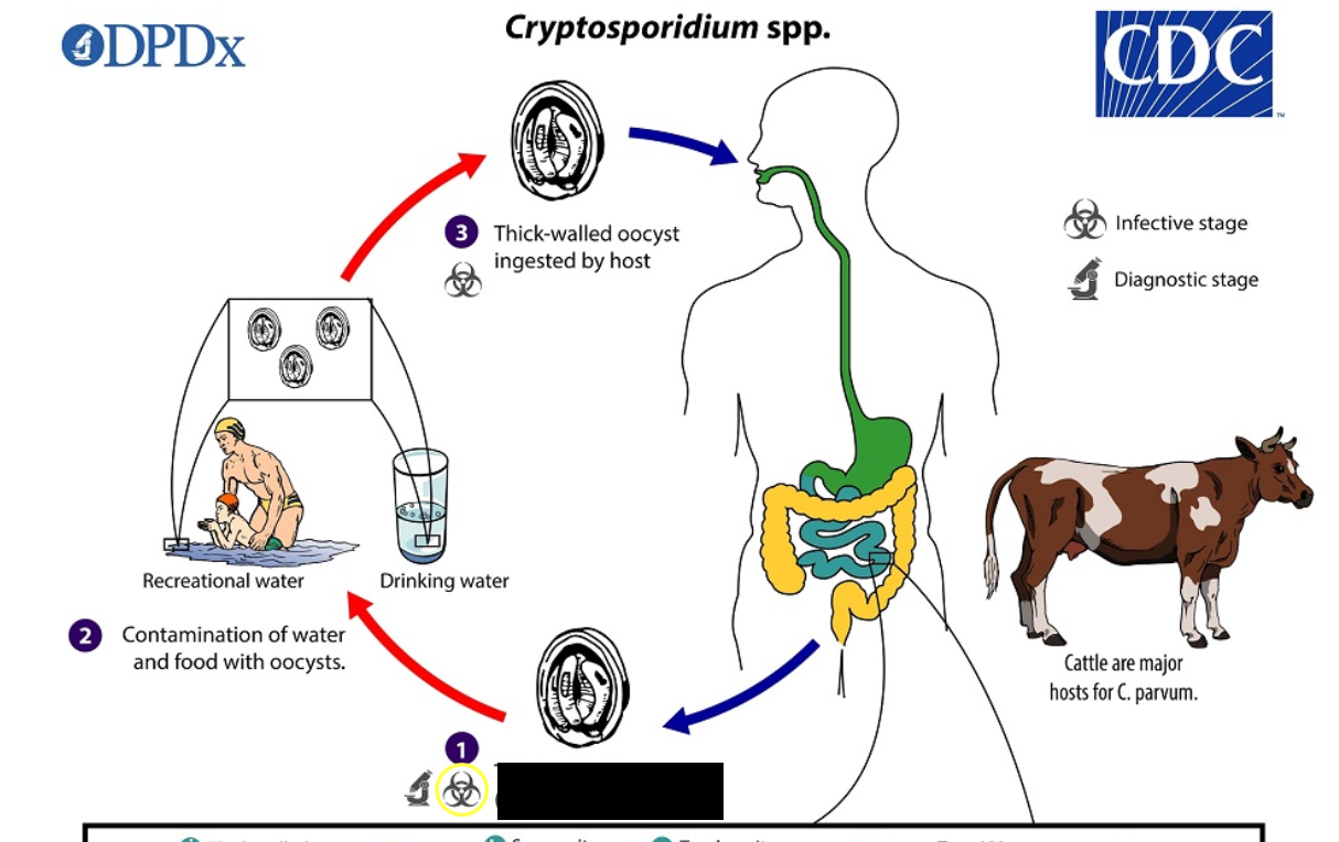
Thick-walled oocysts (sporulated) exists host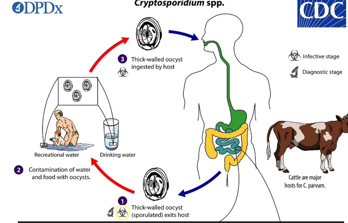
106
What is the 2nd step for Cryptosporidium spp?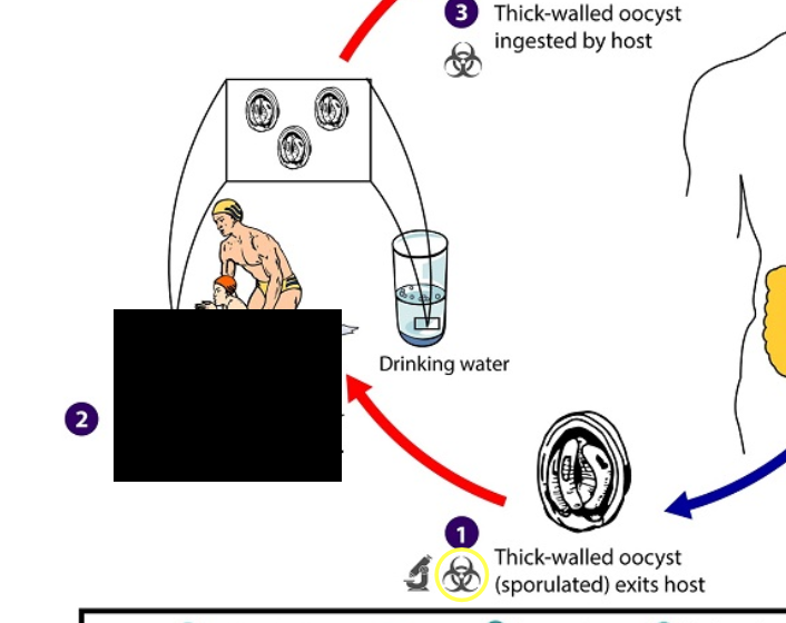
Contamination of water and food with oocysts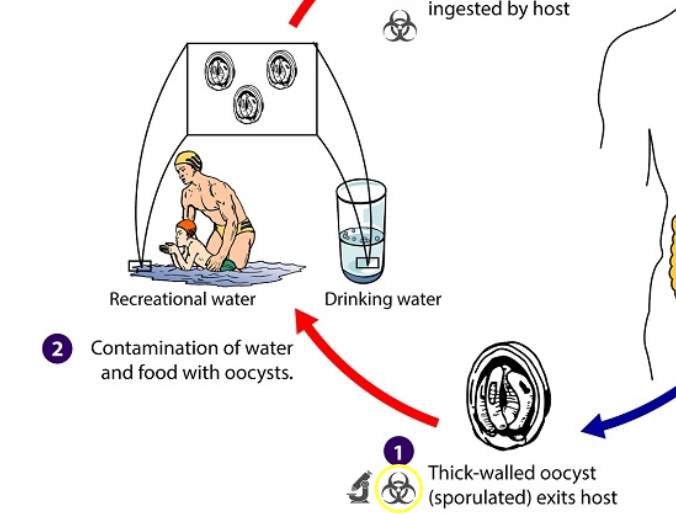
107
What is the 3rd step for Cryptosporidium spp?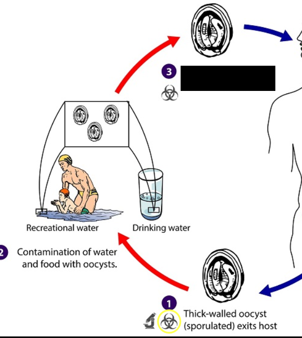
Thick-walled oocysts ingested by host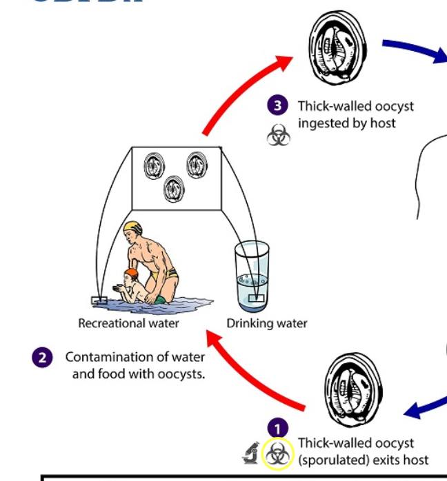
108
Which is the diagnostic and infective stage of Cryptosporidium spp?
1st 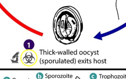
109
Which is the infective stage of Cryptosporidium spp?
3rd and first. seen in feces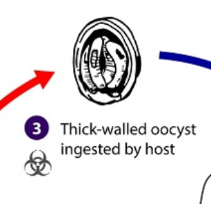
110
How many sporozoites do the sporulated oocysts have that are excreted from the host? cryptosporidium spp.
4 sporozoites
111
After zygotes are formed in Cryptosporidium spp, what types of oocysts are produced and how many?
2 different types:
Thick-walled & thin-walled
Thick-walled & thin-walled
112
Where do thick-walled oocysts from Cryptosporidium spp life cycle go?
excreted from the host to the environment and considered infective!
113
Where do thin-walled oocysts from Cryptosporidium spp life cycle go?
not recovered from stool, but involved in the internal autoinfective cycle.
114
What is the mode of transmission for Cryptosporidium spp?
115
What are the methods of detection for Cryptosporidium oocysts?
PCR
Immunoflourescence
Direct microscopy
Immunoflourescence
Direct microscopy
116
What method of detection is commonly included on GI panels?
PCR
117
How is immunoflouresence used for parasites?
Kits contain antibodies against other intestinal parasites
118
What stain is used for direct microscopy of Cryptosporidium oocysts?
Modified acid-fast staining
119
What do the oocysts of Cryptosporidium look like under direct micrscopy and modified acid-fast stain?
Bright red with granules that stain black.
120
What stain is not used because Cyprtosporidium is too small/easy to miss?
Trichrome
121
Cryptosporidium oocysts measure about?
4.2 um-5.4 um in diameter
122
What can cryptosporidium oocysts be confused with?
Yeasts, fat globules, fungal and bacterial spores
123
Which is Cryptosporidia and which is Giardia?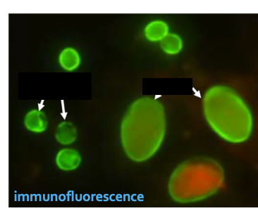
Left: crytosporidia
right: giardia
right: giardia
124
Which one is a yeast and which one is an oocysts?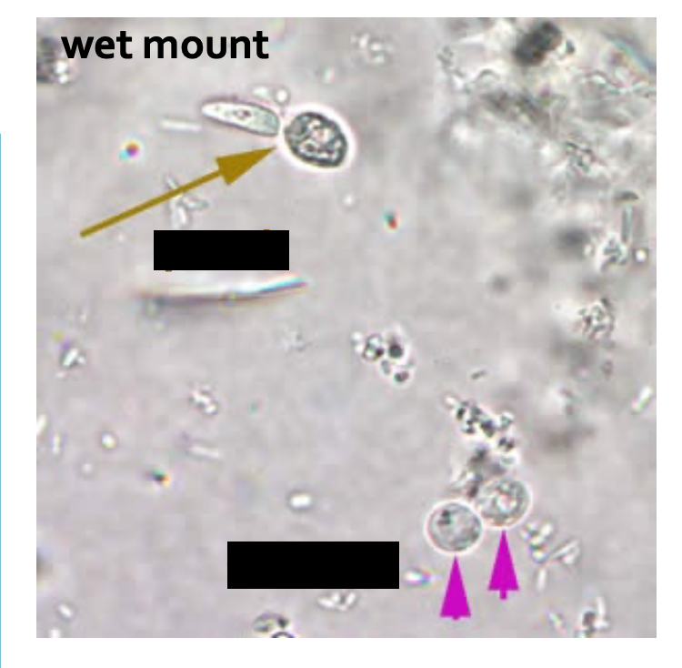
yellow: yeast
purple: oocysts
purple: oocysts
125
Which one is a yeast and which one is an oocysts?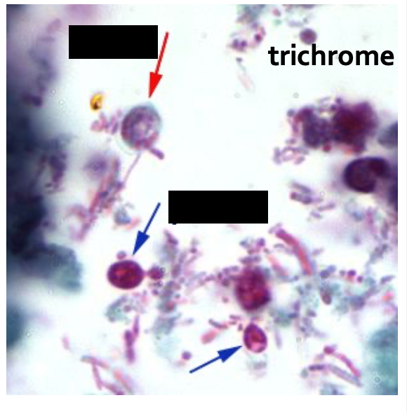
red: oocyst
blue: yeasts
blue: yeasts
126
What stain is used? & label the parasites (oocyts).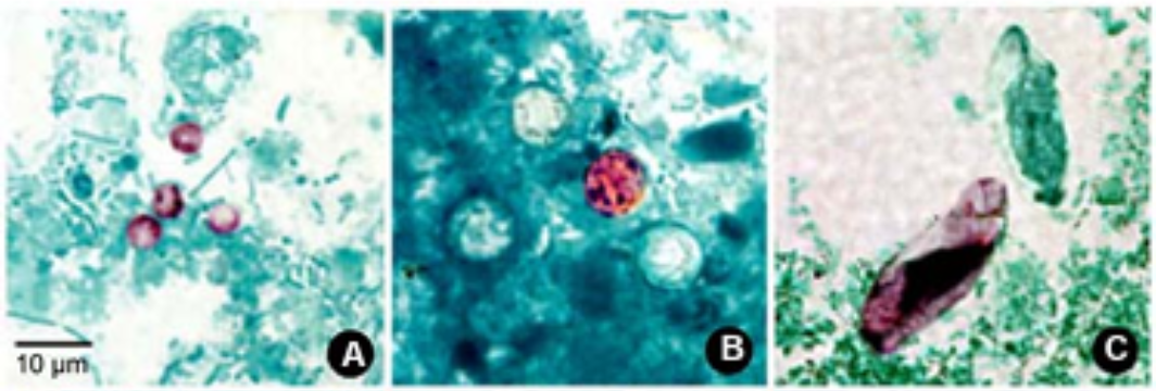
modified acid-fast stain
A. Cryptosporidium sp.
B. Cyclospora cayetanensis
C. Cystoisospora belli
A. Cryptosporidium sp.
B. Cyclospora cayetanensis
C. Cystoisospora belli
127
What stain is used? & label the parasites (oocysts) 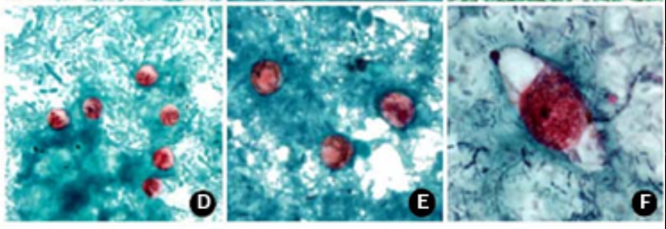
safranin stain
D. Cryptosporidium sp.
E. Cyclospora cayetanensis
F. Cystoiospora belli
D. Cryptosporidium sp.
E. Cyclospora cayetanensis
F. Cystoiospora belli
128
What stain is this? and label the parasites.
Wet mount preparations
G. Cyclospora cayetanensis
H. Cystoisospora belli (UV fluorescence microscopy)
I Cyclospora cayetanensis (UV fluoresence microscopy)
G. Cyclospora cayetanensis
H. Cystoisospora belli (UV fluorescence microscopy)
I Cyclospora cayetanensis (UV fluoresence microscopy)
129
How many microsporidian species have been identified as human pathogens?
15
130
What are microsporidia?
are a group of spore-forming unicellular & intracellular organisms.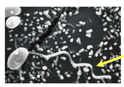
131
Are microsporidia prokaryotic or eukaryotic?
Eukaryotic with nucleus and organelles
132
What is a major microsporidia species?
Enterocytozoon bieneusi
133
what is a unique organelle for microsporidia?
Polar tubules or polar filament
134
What is the polar tubule used for in microsporidia?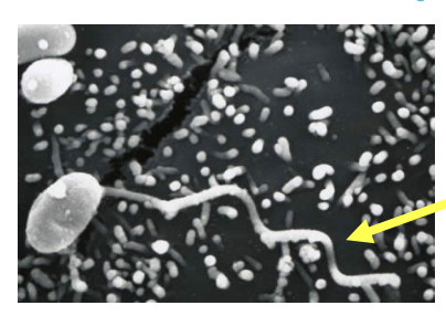
It is used during infection: allows organism to infect host cells at a distance.
135
What does the spore release via polar tubule during infection?
Infective sporoplasms
136
What were microconidia considered as and what are they considered as now?
before: protozoa
now: obligate intracellular fungi
now: obligate intracellular fungi
137
Who can microsporidia infect?
infect vertebrates and invertebrates worldwide
138
What are other species that fall under microsporidia?
Encephalitozoon species:
E. cuniculi
E.hellem
E. intestinalis = Septate intestinalis
E. cuniculi
E.hellem
E. intestinalis = Septate intestinalis
139
What is the known site of localization for Entercytozoon bieneusi?
Small intestine, biliary tree
140
Microsporidia is associated with this type of important disease?
opportunistic -> associated with immunocompromiseds (ex: AIDS patients), but can also be found in immunocompetent
141
Microsporidia can get exposed to which organs in the body?
GI tract
142
Specifically, Enterocytozoon bieneusi causes this most common symptom?
Diarrhea
143
Clinical presentation for microsporidia infections depend on what?
Species and route of infection.
144
which organs can be infected with microsporidia?
ALMOST every organ system: encephalitis, ocular infection, sinusitis, myositis, etc.
145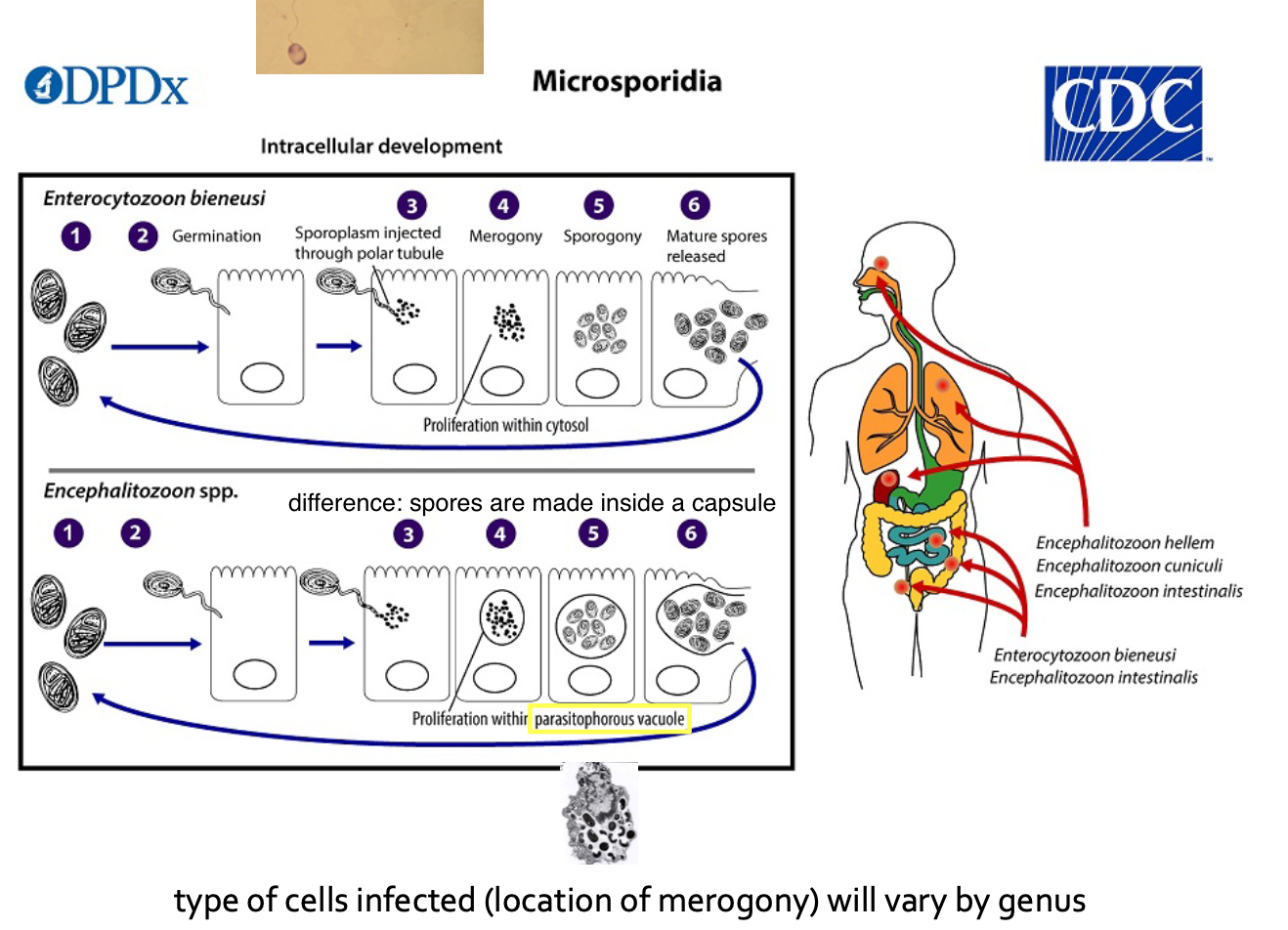
What is the life cycle of microsporidia?
146
What is the 1st step of the Microsporidia life cycle?
Infective form of microsporidia (resistant spore) is in the environment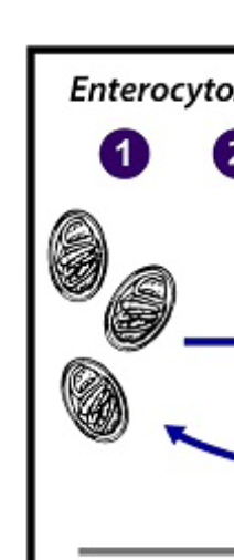
147
What is the 2nd step of the Microsporidia life cycle?
Germination 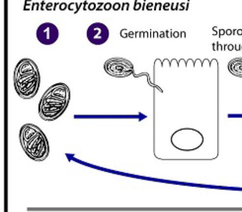
148
What is the 3rd step of the Microsporidia life cycle?
Sporoplasm injected through polar tubule;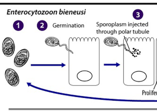
149
What is the 4th step of the Microsporidia life cycle?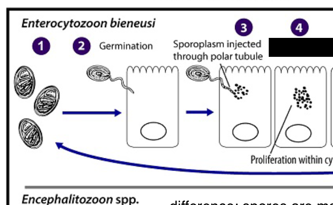
Merogony. Inside the cell, the sporoplasm enters the proliferative phase. (inside the cytosol)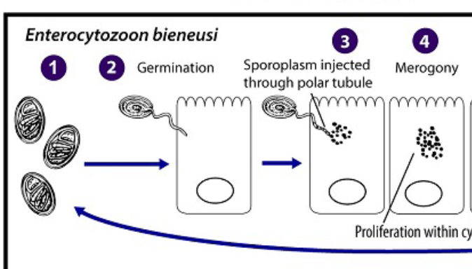
150
What is Mergony?
Extensive multiplication (binary fission or multiple fission), creating
151
What is the 5th step of the Microsporidia life cycle?
Meronts undergo sporogony
152
What is sporogony?
Thick spore wall and invasion pparatus develop, creating sporonts and eventually mature spores.
153
What is the 6th step of the Microsporidia life cycle?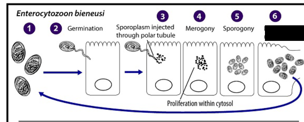
Mature spores are released due to cell membrane disruption. 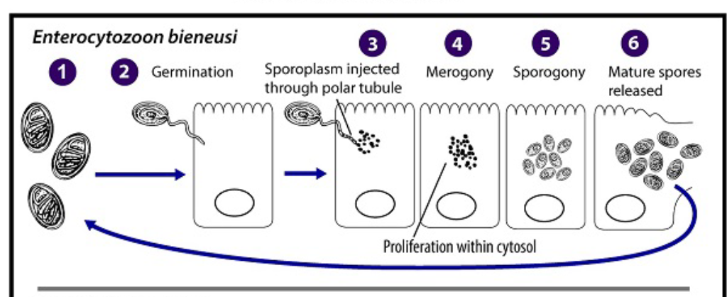
154
Which cells are infected with microsporidia?
depends on the genus due to the location of merogony.
155
What are the issues in microsporidium diagnosis?
Small size of spores (0.8-4 um)
Failure of spores to stain well.
Failure of spores to stain well.
156
What is usually needed to help with the diagnosis of microsporidium?
Ultra-thing histologic specimens of infected tissues.
157
What do the arrows indicate?i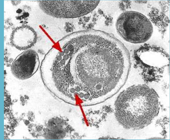
the double rows of polar tubule coils in cross section which characterize a mature E. bieneusi spore.
158
What are the 3 different types of diagnostic tests for microsporidium?
Microscopy
Immunofluorescent assays
PCR (molecular)
Immunofluorescent assays
PCR (molecular)
159
Which test is hard/ tricky for ID of microsporidium?
Microscopy
160
What is used in microscopy for ID of microsporidium? (stain)
Chromotrope 2R method and variations
Calcofluor white (chemofluorescent agent) which helps see spores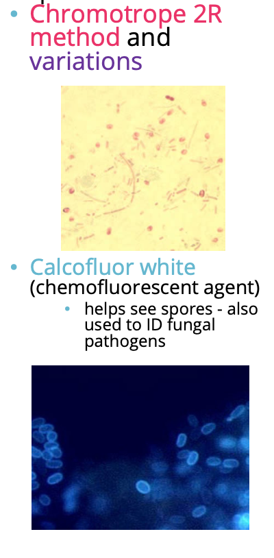
Calcofluor white (chemofluorescent agent) which helps see spores
161
What tools are used for immunofluorescent assays?
Monoclonal and polyclonal antibodies (kits)
162
Who tests for microsporidium for PCR?
CDC and ARUP