Lecture 1_Immunodeficiency_pt1: disease & lab diagnosis
Study these flashcardsFree · spaced repetition · iPhone, Android, Mac & web
All cards
1
What is immunodeficiency?
A part of the immune system is missing or dysfunctional.
2
What does immunodeficiency increase the risk of?
Recurrent infections or some types of cancer.
3
What is primary immunodeficiency?
It is inherited.
4
What is secondary immunodeficiency?
it is acquired
5
What are examples of secondary (acquired) immunodeficiency causes?
AIDS, malignancies, immunosuppressive drugs.
6
How many primary immunodeficiency diseases (PIDs) have been identified, and what are they like?
More than 350 different PIDs have been identified; most are rare but clinically significant.
7
What causes primary immune deficiency diseases (PID)?
PID results from genetic defects.
8
What types of defects can cause PID? (4 things)
Defects in lymphoid cells, phagocytic cells, regulatory molecules, and complement proteins.
9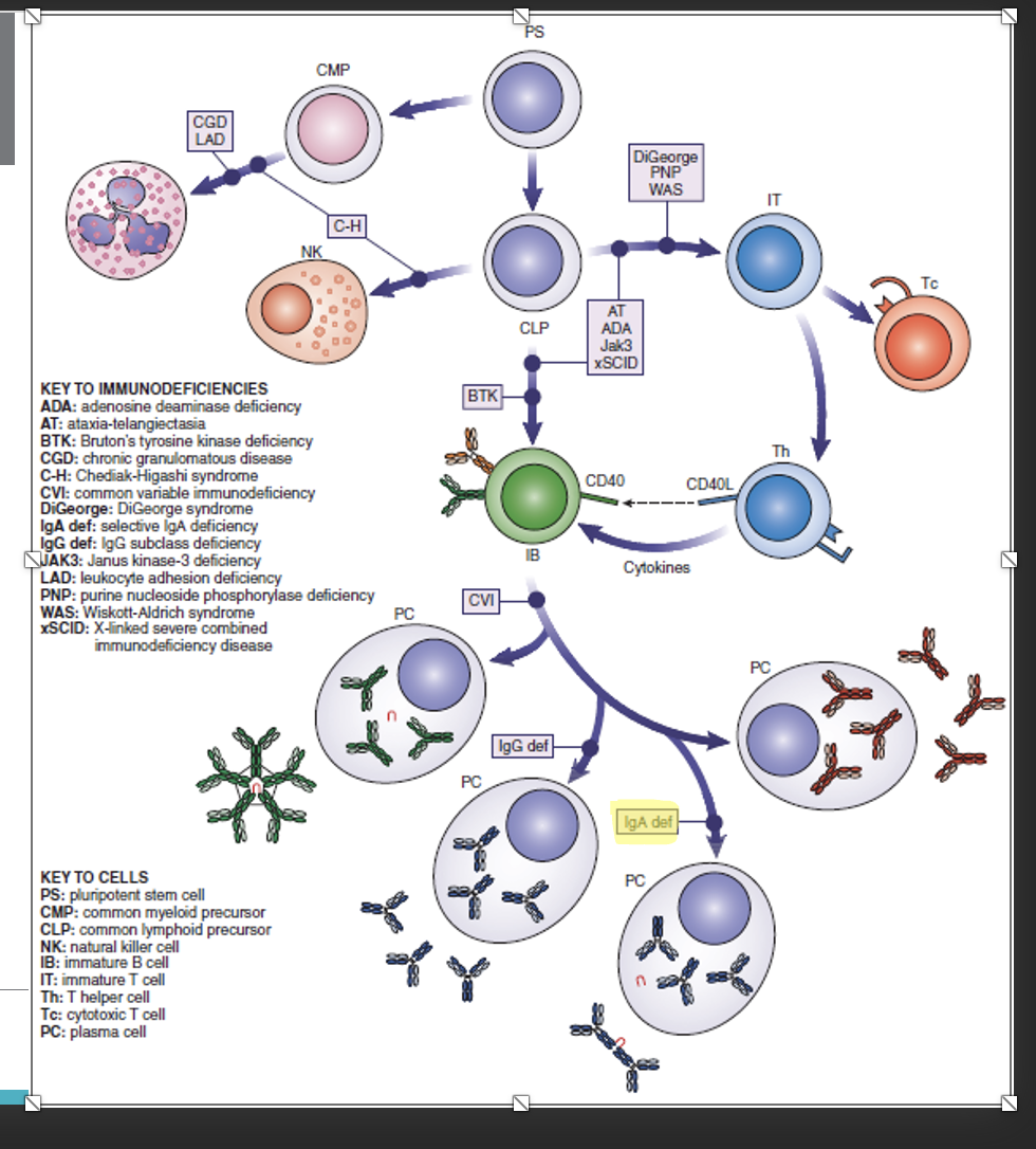
10
What causes primary immunodeficiencies?
They are genetic; the child is born with a defect.
11
In what age group are most primary immunodeficiency cases diagnosed?
At least 58% of cases are diagnosed in children under 15 years old.
12
What is the gender and inheritance pattern seen in most primary immunodeficiencies?
The vast majority (83%) are males with X-linked recessive, autosomal recessive, autosomal dominant, and sporadic inheritance patterns.
13
What causes secondary immunodeficiencies?
They are acquired and may be transient or permanent due to diseases, protein-caloric malnutrition, chemotherapy, irradiation, malignancies, or immunosuppression.
14
What can be absent or malfunctioning in immunodeficiency?
Bone marrow precursor stem cells.
15
What types of blood cells can be affected in immunodeficiency?
Lymphocytes, neutrophils, and monocytes.
16
What soluble molecules can be affected in immunodeficiency?
Antibody, cytokines, and complement components.
17
What are the four major host defense mechanisms that can be affected in primary immunodeficiencies?
1. B-cell immunity
2. T-cell immunity
3. Phagocytosis
4. Complement system
2. T-cell immunity
3. Phagocytosis
4. Complement system
18
Which type of immunity is affected most often in primary immunodeficiencies?
B-cell (humoral) immunity — 50% of cases.
19
What percentage of primary immunodeficiencies affect T-cell (cellular) immunity?
10%.
20
What percentage of primary immunodeficiencies affect phagocytic function?
18%.
21
What percentage of primary immunodeficiencies affect the complement system?
2%.
22
What percentage of primary immunodeficiencies involve combined B- and T-cell defects?
20%.
23
What are common clinical findings in immunodeficiency?
Very mild or subclinical to severe recurrent infections or failure to thrive.
24
What do defects in humoral immunity result in?
Pyogenic bacterial infections (Staphylococcus aureus, Haemophilus influenzae, pneumococcus).
25
Which areas of the body are particularly affected by humoral immunity defects? as a result in pyogenic bacterial infections
Upper and lower respiratory tract, GI tract (malabsorption), and slow growth.
26
What recurrent infections are seen with humoral immunity defects? as a result in pyogenic bactieral infections
Otitis media, sinusitis, and pneumonia.
27
Front: What do defects in T-cell-mediated immunity result in?
Back: Recurrent infections with intracellular pathogens such as viruses, fungi (commonly Candida), and intracellular bacteria.
28
Front: What types of viral infections are patients with T-cell defects prone to?
Back: Disseminated viral infections, especially with latent viruses such as herpes simplex, varicella zoster, and CMV.
29
Front: What conditions are patients with T-cell defects more susceptible to?
Back: Developing certain types of cancer.
30
Front: What do neutrophils do in phagocytic immunity?
Back: They adhere to vascular endothelial lining cells, migrate through the capillary wall to a site of infection, and ingest and kill microbes.
31
Front: What happens if any step in neutrophil function is defective?
Back: It can lead to increased susceptibility to pyogenic infections.
32
Front: What is the normal function of the complement system?
Back: To produce inflammatory molecules and cause lysis of organisms.
33
Front: What do defects in the complement system result in?
Back: Pyogenic infections.
a. Recurrent Neisseria infections
b. Rheumatic and connective tissue diseases
a. Recurrent Neisseria infections
b. Rheumatic and connective tissue diseases
34
Phagocytic and complement system is part of which immunity? (innate or adaptive)
INNATE!!!!
35
B-cell and T-cell is part of which immunity (innate or adaptive) ?
ADAPTIVE!!!
36
Front: What subgroup of immunodeficiency involves B-cell differentiation and antibody production?
Back: Disorders of humoral immunity (B-cell differentiation and antibody production).
37
Front: When do disorders of humoral immunity typically present?
Back: After 6 months of age; can also be present in adulthood.
38
Front: What are other features of humoral immunity disorders?
Recurrent infections: sinus infections, otitis media, bronchiectasis;
Chronic gastrointestinal tract problems including malabsorption;
Autoimmunity
Chronic gastrointestinal tract problems including malabsorption;
Autoimmunity
39
Front: What subgroup involves combined B-cell and T-cell defects?
Back: T-cell and mixed disorders (combined B-cell and T-cell defects).
40
Front: When do T-cell and mixed disorders usually appear?
Back: Before 6 months of age.
41
Front: What are other features of T-cell and mixed disorders?
Failure to thrive
Oral thrush
GVHD from maternal lymphocytes
Excess diarrhea
Oral thrush
GVHD from maternal lymphocytes
Excess diarrhea
42
Front: When do phagocytic disorders usually present?
Back: Infancy or childhood.
43
Front: What are common clinical features of phagocytic disorders?
Unusually severe infections by common pathogens
Granuloma formation
Poor wound healing
Abscesses and skin infections
Oral cavity infections
Granuloma formation
Poor wound healing
Abscesses and skin infections
Oral cavity infections
44
Front: When can complement disorders occur?
Back: At any age.
45
Front: What infections are associated with complement disorders?
Back: Neisseria infections, including meningococcal and gonococcal infections.
46
Front: What other conditions are associated with complement disorders?
Rheumatoid disorders
Lupus-like syndrome
Scleroderma
Lupus-like syndrome
Scleroderma
47
Front: What is Category 3 of PIDs?
Back: Predominantly Antibody Deficiencies.
48
Front: What is Bruton’s agammaglobulinemia and who does it primarily affect?
Back: Bruton’s agammaglobulinemia, first described in 1952, is X-linked and affects males almost exclusively.
49
Front: What do patients with X-linked agammaglobulinemia lack?
Back: They lack circulating mature CD19 positive B cells and have no plasma cells.
50
Front: How can Bruton’s agammaglobulinemia be differentiated from transient hypogammaglobulinemia of infancy?
Back: By the absence of CD19 positive B cells in the peripheral blood.
51
Front: What type of deficiency do patients with Bruton’s agammaglobulinemia exhibit?
Back: A deficiency or lack of all immunoglobulins of all classes.
52
Front: What do patients with Bruton’s agammaglobulinemia lack in their lymphoid tissues, and what do they have in their bone marrow?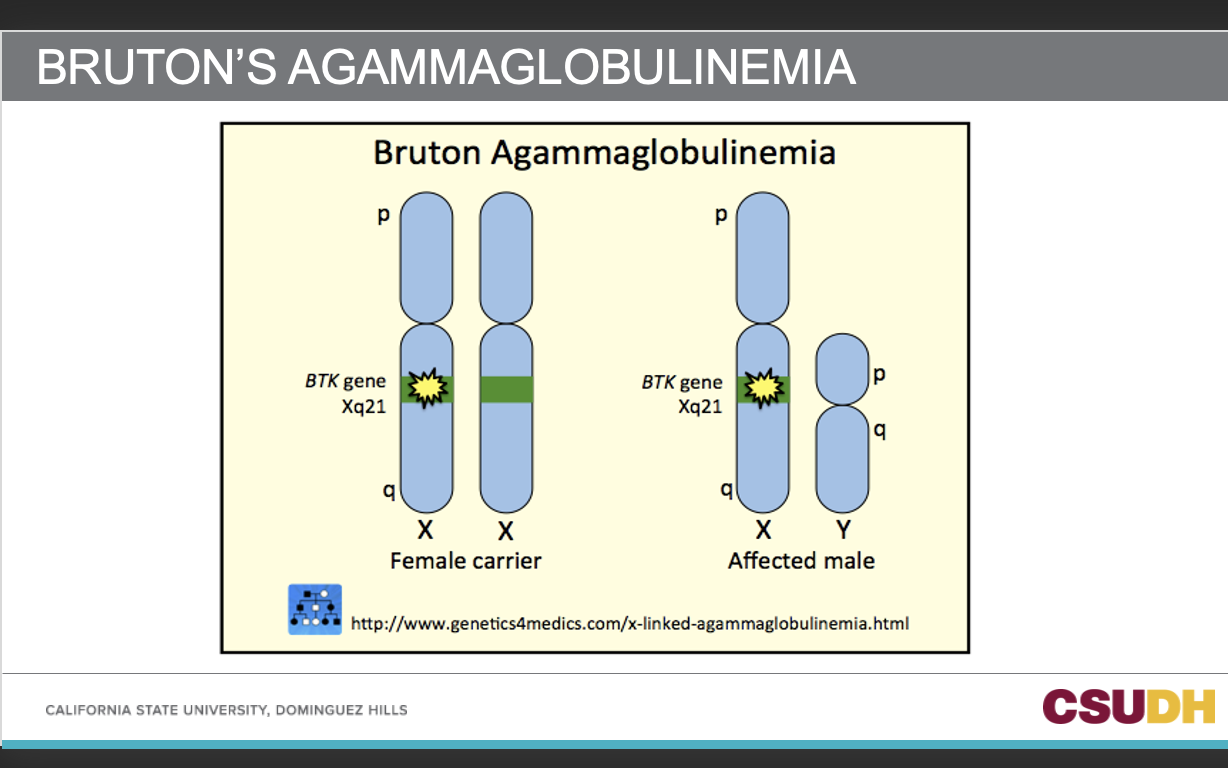
Back: They have no plasma cells in their lymphoid tissues but do have pre-B cells in their bone marrow.
53
Front: What types of infections are common in Bruton’s agammaglobulinemia?
Back: Recurrent sinopulmonary infections by encapsulated organisms (Streptococcus pneumoniae, Haemophilus influenzae) and bacterial otitis media.
54
Front: How are T cells affected in Bruton’s agammaglobulinemia?
Back: T cells are normal in number and function.
55
Front: When do recurrent bacterial infections typically begin in Bruton’s agammaglobulinemia?
Back: In infancy, as maternal antibody is cleared.
56
Front: What enzyme is lacking in Bruton’s agammaglobulinemia, and what does it cause?
Back: Lack of the Btk enzyme causes failure of Vh gene rearrangement.
57
Front: What is the treatment for Bruton’s agammaglobulinemia?
Back: Intramuscular (IM) or intravenous (IV) immunoglobulin (IG).
58
Front: What percentage of patients with IgA deficiency are asymptomatic?
Back: 70% of patients with IgA deficiency are asymptomatic.
59
Front: What type of symptoms do patients with IgA deficiency have?
Sino-pulmonary infections
GI infections
Allergies
An increased tendency to autoimmune diseases (SLE, RA, immune thrombocytopenia)
GI infections
Allergies
An increased tendency to autoimmune diseases (SLE, RA, immune thrombocytopenia)
60
What symptoms are more common in patients with selective IgA deficiency?
Allergic disorders and malignancy are also more common among these patients
61
Front: What is the underlying cause of IgA deficiency?
Back: Impaired differentiation of B cells to become IgA-producing plasma cells.
62
Front: What may be found in patients with very severe IgA deficiency?
Back: IgE antibodies to IgA in very severe IgA deficiency
63
Front: Why must patients with IgA deficiency receive plasma products only from other IgA-deficient patients?
Back: To prevent reactions caused by anti-IgA antibodies.
64
Front: Why is intramuscular (IM) or intravenous (IV) IgA therapy not adequate for IgA deficiency?
Back: IgA has a very short half-life and cannot be transported to secretory sites such as mucosal surfaces.
65
Front: What other treatments can be used for IgA deficiency?
Back: Antibiotics and possible gene therapy.
66
Front: What type of disorder is Common Variable Immunodeficiency (CVID)?
Back: A heterogeneous group of disorders, sometimes difficult to diagnose.
67
Front: What makes CVID clinically significant?
Back: It is the most common PID with a severe clinical syndrome that requires medical attention.
68
Front: What happens to immunoglobulin levels in CVID?
Back: There is a slow decline in more than one class of immunoglobulins.
69
Front: What childhood condition is often part of the history in CVID?
Back: Recurrent otitis media.
70
Front: When do sinopulmonary infections usually begin in CVID, and what are they typically caused by?
Back: They usually begin in adolescence and are bacterial in origin.
71
Front: What are common complications of CVID?
Back: Increased episodes of pneumonia and an increased risk of autoimmune diseases.
72
Front: What is the number of mature B cells in Common Variable Immunodeficiency (CVID)?
Back: Normal numbers of mature B cells.
73
Front: What is the issue with B cells in CVID?
Inability to become memory B cells (CD27+) and plasma cells
Decreased activation of B cells in germinal centers
Decreased activation of B cells in germinal centers
74
Front: What immunoglobulin is primarily deficient in CVID?
Back: IgA; patients also show a marked decrease in IgG levels due to certain subclasses.
75
Front: What does hypogammaglobulinemia cause in CVID?
Back: Recurrent bacterial infections, particularly sinusitis and pneumonia.
76
Front: What is the most common genetic susceptibility locus for CVID?
Back: MHC.
77
Front: When is CVID diagnosed?
Back: When patients with recurrent bacterial infections demonstrate a low serum immunoglobulin level.
78
Front: What happens to blood group isohemagglutinins in CVID?
Back: They are absent or decreased.
79
What are the complications of "“Lack or decreased levels of blood group isohemagglutinins”?
discrepancies in ABO typing
80
Front: What defines an isolated IgG subclass deficiency?
Back: The levels of one or more of the four IgG subclasses are lower than the reference interval (RI).
81
Front: What is the normal distribution of total IgG subclasses?
IgG1: 66%
IgG2: 23%
IgG3: 7%
IgG4: 4%
IgG2: 23%
IgG3: 7%
IgG4: 4%
82
Front: When should IgG subclass levels be measured?
Back: In patients with recurrent infections, even if the total IgG level is normal but the clinical picture suggests immunoglobulin deficiency.
83
Front: What outcomes of IgG1 or IgG3 deficiency is mentioned for IgG subclass deficiencies?
when the immune system is responding to protein antigens (i.e. RH blood group, Duffy, Lutheran, or structure on a pathogen ) these antibodies are involved
84
Front: What outcomes of IgG2 deficiency are mentioned for IgG subclass deficiencies?
polysaccharides ???why
85
Front: What are combined immunodeficiencies?
Back: Defects in both humoral (B cell) and cell-mediated (T cell) immunity.
86
Front: What can cause combined immunodeficiencies?
Back: Mutations in any of several genes.
87
Front: What are two possible types of defects that lead to combined immunodeficiencies?
a. A defect that affects development of both types of lymphocytes.
b. A defective interaction between the two limbs of the immune system.
b. A defective interaction between the two limbs of the immune system.
88
Front: How are combined immunodeficiencies abbreviated?
Back: Using a shorthand of T⁺/⁻ B⁺/⁻ NK⁺/⁻, with the superscript +/− showing whether each cell type is deficient.
89
Front: What is the most serious congenital immune deficiency?
Back: Severe Combined Immunodeficiency (SCID).
90
Front: What does SCID represent?
Back: A group of related diseases that all affect T- and B-cell function but have differing etiologies.
91
Front: What is the most common form of SCID?
Back: X-linked SCID.
92
Front: What is the most frequent mutation causing SCID?
Back: Mutation in the IL2RG gene located on the X chromosome — the root cause of most SCID cases.
93
Front: What does the IL2RG gene encode?
Back: The common gamma chain, a cytokine receptor subunit common to receptor complexes for at least six different interleukin receptors.
94
Front: Which interleukin receptors use the common gamma chain encoded by IL2RG?
Back: IL-2, IL-4, IL-7, IL-9, IL-15, and IL-21 receptors.
95
Front: What is the outcome of a defective IL2RG gene?
Back: Halting lymphocyte maturation due to defective cytokine signaling.
96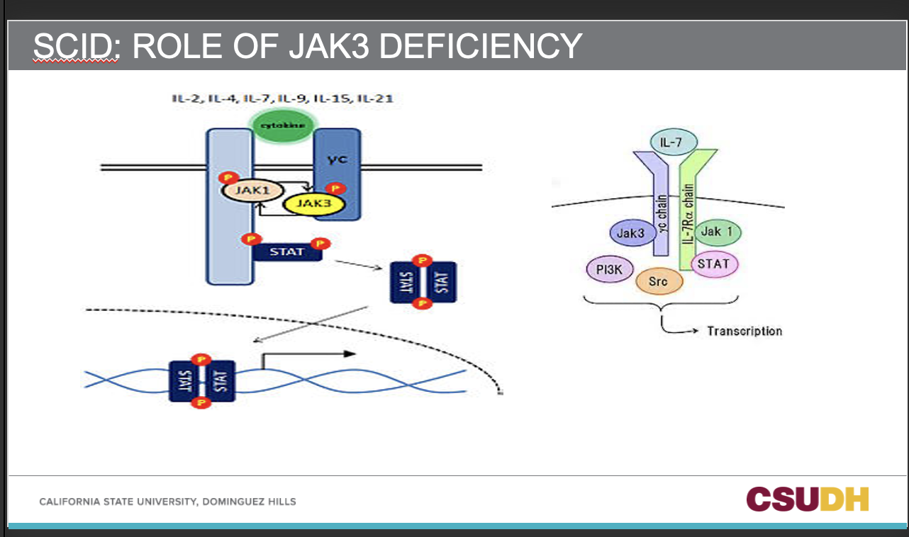
97
Front: Where is JAK3 most commonly expressed?
Back: In T cells and NK cells.
98
Front: What is the function of JAK3?
Back: It processes interleukin-binding signals from the cell membrane to the nucleus.
99
Front: What receptors involve JAK3 in signal transduction?
Back: Receptors that employ the common gamma chain (γc).
100
Front: What type of immunodeficiency can JAK3 cause and what is the shorthand annotation for this
Back: An autosomal SCID (Severe Combined Immunodeficiency) with the T⁻B⁺NK⁻ phenotype.
101
Front: What percentage of SCID patients have adenosine deaminase (ADA) deficiency, and what phenotype does it lead to?
Back: About 15–20% of SCID patients have ADA deficiency, leading to a T⁻B⁻NK⁻ phenotype.
102
Front: Where is the ADA gene located?
Back: On chromosome 1, region q21.
103
Front: What does ADA deficiency affect?
Back: An enzyme involved in the metabolism of purines, similar to PNP deficiency.
104
Front: What happens in ADA deficiency?
Back: Toxic metabolites of purines accumulate in lymphoid cells, impairing proliferation of both B and T cells.
105
Front: What occurs in both ADA and PNP deficiencies?
Back: There is a progressive decrease in lymphocyte numbers.
106
Front: What cells are lacking and which are functioning in RAG-1 or RAG-2 gene deficiency?
Back: Lack of T cells and B cells, but NK cells remain functioning.
107
Front: What does RAG-1 or RAG-2 deficiency cause?
Lack of both T cells and B cells but with functioning NK cells .
Profound lymphocytopenia (elimination of B and T cell precursors during development)
Decreased MHC-II molecule expression
Profound lymphocytopenia (elimination of B and T cell precursors during development)
Decreased MHC-II molecule expression
108
What decreases in RAG-1 OR RAG-2 (recombinase-activatin) gene deficiency? and what is the consequence of it?
SCID: OTHER MOLECULAR DEFECTS
SCID: OTHER MOLECULAR DEFECTS
Back: Decreased MHC-II expression (leads to immune dysfunction/ aka impaired activation of CD4+ T helper cells).
109
Front: What is the function of the CD45 antigen?
Back: It is a transmembrane phosphatase that regulates signal transduction of T- and B-cell receptors.
110
Q: What deficiencies cause T–, B–, NK– SCID?
A: Reticular dysgenesis (Adenylate kinase 2 deficiency — no myeloid cells)
ADA deficiency (enzyme involved in purine metabolism).
ADA deficiency (enzyme involved in purine metabolism).
111
Q: What causes T–, B–, NK+ SCID?
A: Rag 1 and 2 deficiency/ recombination deficiency.
112
Q: What causes T–, B cells immature, NK– SCID?
γc-chain deficiency (IL-2 receptor; NK low or defective)
JAK3 deficiency (interleukin-binding signal; NK near absent)
JAK3 deficiency (interleukin-binding signal; NK near absent)
113
Q: What causes T–, B cells immature, NK+ SCID?
A: IL-7Rα deficiency.
114
Q: What immunodeficiency presents with T cells positive (clonal), B cells low, NK cells +?
A: Omenn Syndrome.