Lecture 3 - Protozoa Lumen Dwelling Flagellates
Study these flashcardsFree · spaced repetition · iPhone, Android, Mac & web
All cards
1
List 3 nonpathogenic flagellates?
Chilmastix mesnili
Dientamoeba fragilis
Pentatrichomonas hominis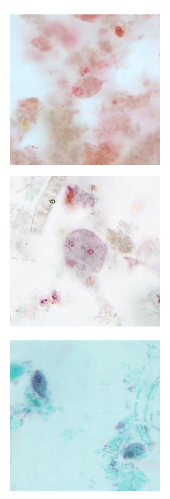
Dientamoeba fragilis
Pentatrichomonas hominis
2
What are pathogenic flagellates? List 2.
Giardia lamblia/G. intestinalis/ G. duodenalis
Trichomonas vaginalis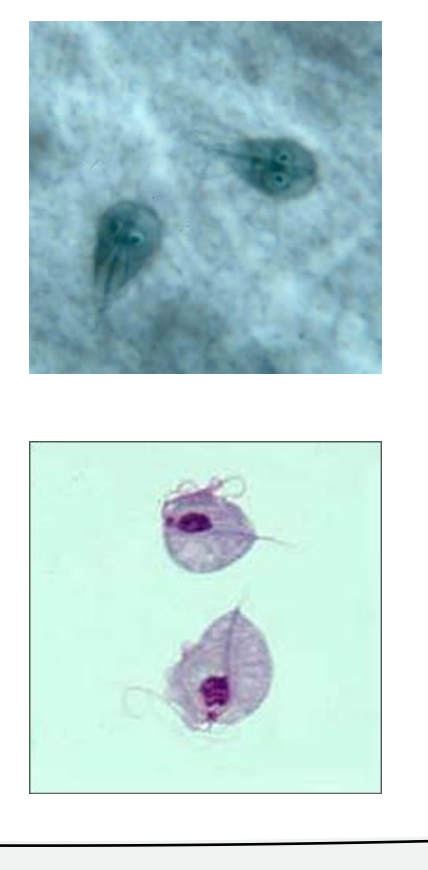
Trichomonas vaginalis
3
What is this?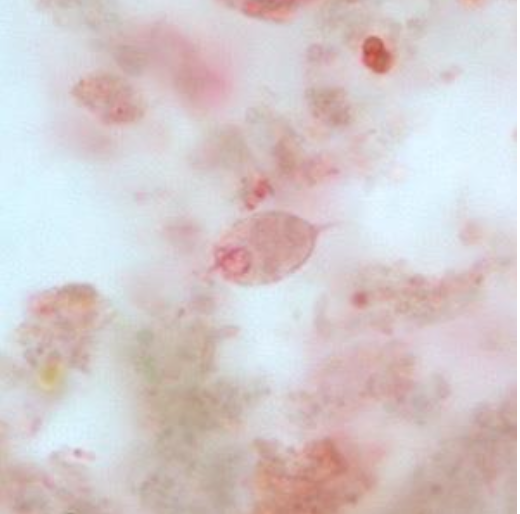
Chilomastix mesnili
4
What is this ? 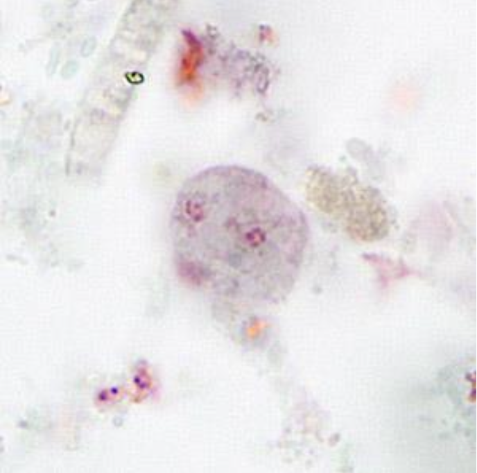
Dientamoeba fragilis
5
What is this?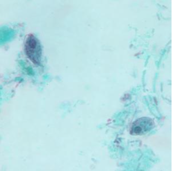
Pentatrichomonas hominis
6
What is this? 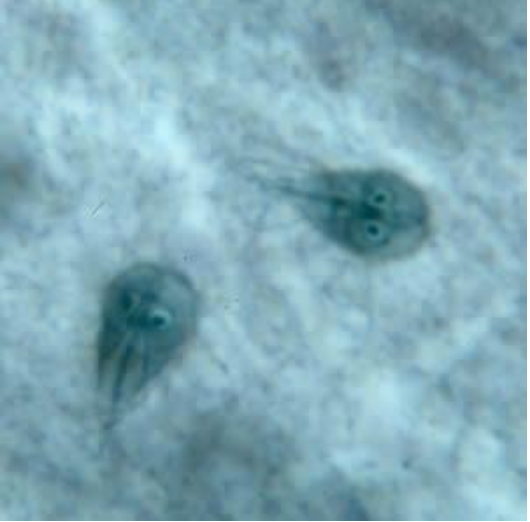
Giardia lamblia/ G. intestinalis/G. duedenalis
7
What is this? 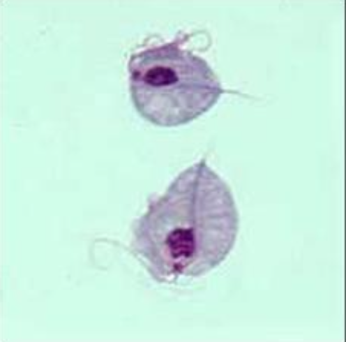
Trichomonas vaginalis
8
What is trichomonas vaginalis?
sexually transmitted infection (STI) caused by a parasite.
9
What % of cases are trichomonas vaginalis are pathogenic?
30% of cases
10
What is trichomonas vaginalis called when its a female sample?
Vaginal trichomoniasis
11
What is trichomonas vaginalis called when its a male sample?
Prostate and epididymis infection
12
What are symptoms for vaginal trichomoniasis?
Itching, burning, redness, or soreness of the genitals.
Discomfort when peeing
A clear, white, yellowish, or greenish vaginal discharge with a fishy smell.
Discomfort when peeing
A clear, white, yellowish, or greenish vaginal discharge with a fishy smell.
13
What are symptoms for prostate and epididymis infections?
Itching or irritation inside the penis.
Burning after peeing or ejaculating.
Discharge from the penis.
Burning after peeing or ejaculating.
Discharge from the penis.
14
What is the sample being collected when testing trichomonas vaginalis?
Vaginal/ genital fluid specimen of choise; can also see in urine.
15
Can trichomonas vaginalis occure through fomites?
YES!
16
What are fomites?
objects or materials which are likely to carry infection, such as clothes, utensils, and furniture
17
What is the morphology of trichomonas vaginalis?
Pear-shaped.
Large nuclear at anterior end
undulating membrane extends halfway down organim
many granules in cytoplasm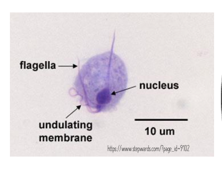
Large nuclear at anterior end
undulating membrane extends halfway down organim
many granules in cytoplasm
18
what is this?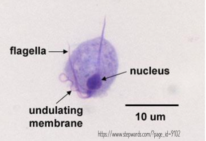
Trichomonas vaginalis
19
What does the nucleus in trichomonas vaginalis look like?
large nucleus at anterior end.
20
What is one feature of trichomonas vaginalis that can differentiate it from other parasites?
undulating membrane extends halfway down organism
21
Which is the undulating membrane?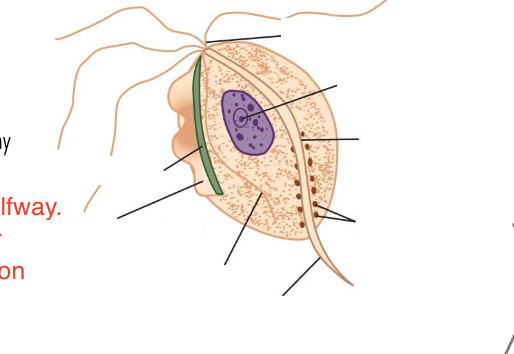
second line from the left
22
what does the cytoplasm look like in trichomonas vaginalis?
Many granules!
23
What are the cases/year in the US for trichomonas vaginalis?
2 mil cases/year
24
What type of slide is prepared for trichomonas vaginalis?
wet prep.
25
What is the left arrow called?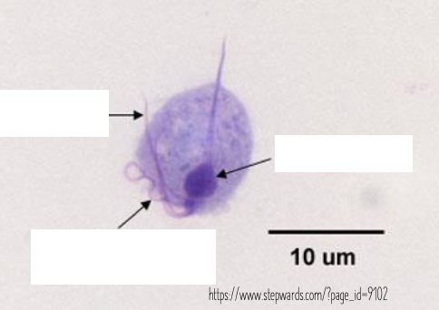
Flagella
26
What is the right arrow called?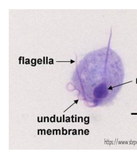
Nucleus
27
What is the middle arrow called?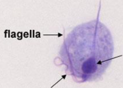
Undulating membrane
28
Does trichomonas vaginalis have a cysts stage?
no
29
How does trichomonas vaginalis multiply?
Longitudinal binary fission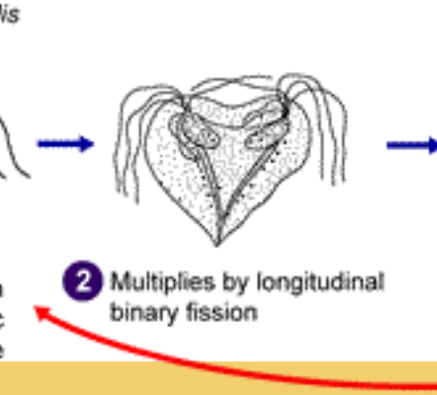
30
How does trichomonas vaginalis move?
Twitchy/ jerky movements
31
What is this?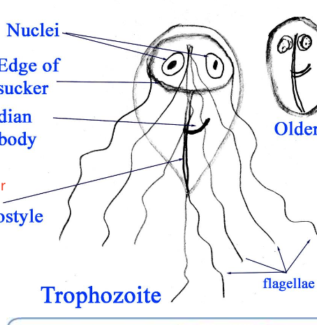
Giardia Lamblia
32
What is Giardia lamblia?
Most frequently diagnosed intestinal parasite in the U.S.
33
What type of outbreaks are common for Giardia Lamblia?
Waterborne outbreaks
34
What does Giardia Lamblia cause?
Giardiasas.
35
What are the clinical symptoms for Giardia Lamblia?
CHRONIC diarrhea, gas, cramps, greasy/floaty stools, nausea and vomiting.
36
What deficiency can Giardia Lamblia cause?
Vitamin A and B12 deficiencies
37
How often are samples collected for testing Giardia Lamblia?
Collect 3 stool specimens on separate days.
38
How are slides prepared for Giardia Lamblia?
Concentrated wet mount (iodine)
Trichrome staining.
Fluorescent testing kits (immunofluorescent anitbodies)
PCR: used to subtype.
Trichrome staining.
Fluorescent testing kits (immunofluorescent anitbodies)
PCR: used to subtype.
39
What is the treatment for Giardia Lamblia?
Metronidazole (Flagyl), tinidazole, and nitazoxanide
40
Which one is the older cysts (for Giardia Lamblia)?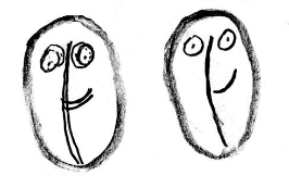
Left
41
Which one is the younger cysts (for Giardia Lamblia)?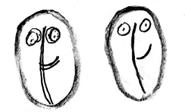
Right
42
Name the structures. (from left to right)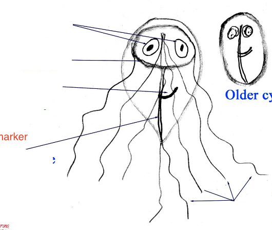
Nuclei
Edge of sucker
Median body
Axostyle
Trophozoite
Flagellae
Edge of sucker
Median body
Axostyle
Trophozoite
Flagellae
43
What is this organism?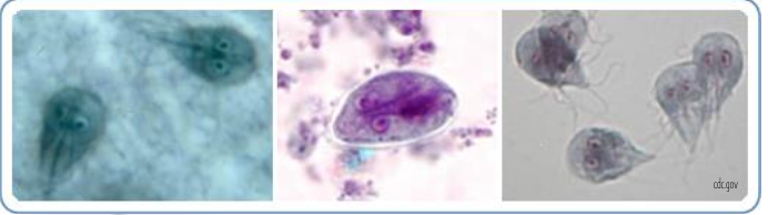
Giardia Lamblia aka Giardia duodenalis aka Giardia intestinalis
44
Are trophozoites or cysts attaching to the intestinal mucosa?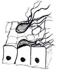
Trophozoites
45
What is this?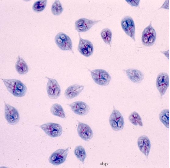
Trophozoite - Giardia Spp.
46
What is this?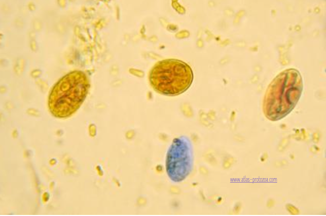
Cyst - Giardia Spp.
47
How is Giardia transmitted?
Fecal-Oral. Contaminated water, food, hands, fomites. Recently associated with anal sex (person-person). Associated with drinking contaminated water with nature.
48
How long can Giardia cysts survive?
several months in cold water and chlorine disinfection.
49
Where does excystation occur during the giardia life cycle?
in the small intestine.
50
What happens during excystation for Giardia?
Trophozoites are released (each cyst produces 2 trophozoites). 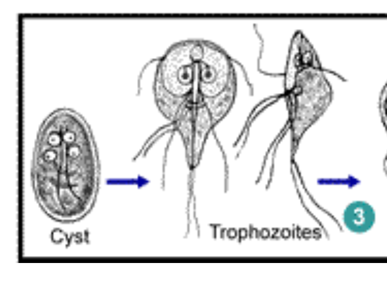
51
How are Giardia trophozoites multiply?
Longitudinal binary fission.
52
What happens when the giardia - trophozoites - reach the small bowel?
Remain in the lumen of the small bowel where they can be free or attached to the mucosa by a central sucking disk.
53
When does encystation occure during the Giardia life cycle?
As the parasites transit toward the colon.
54
What does encystation mean?
the process of forming a cyst or capsule around an organism to protect it from unfavorable conditions
55
What is Giardiasis associated with?
Beaver fever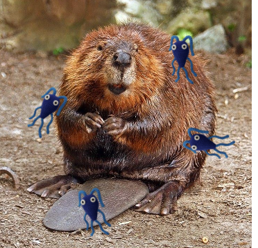
56
What is Chilomastix mesnili?
A non-pathogenic parasite. (BUT presence could indicate exposed to contaminated food-water, may have other parasites lurking).
57
Where is Chilomastix mesnili found?
Worldwide.
58
Where are Chilomastix mesnili usually found?
Large (mostly) and small intestine
59
What do Chilomastix mesnili (trophozoites) look like?
pear-shaped. 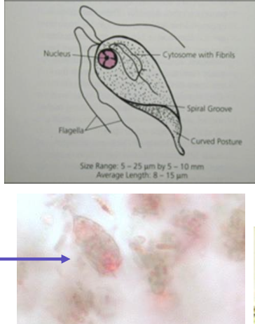
60
What do Chilomastix mesnili (cysts) look like?
Lemon-shaped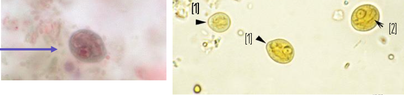
61
What is this?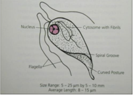
Chilomastix mesnili
62
Which is the trophozoite and which is the cyst? (Chilomastix mesnili) 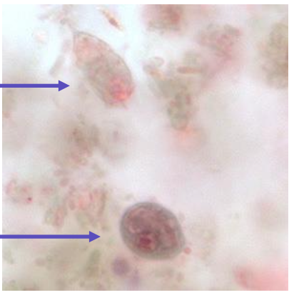
Top: trophozoite
Bottom: Cyst
Bottom: Cyst
63
How do trophozoites move? (Chilomastix mesnili)
rotary movement; 3 anterior flagella
64
What is a prominent cytosome in Chilomastix mesnili?
Extends 1/3-1/2 length of body.
"Shepherd's crook" fibril curves around cytosome
"Shepherd's crook" fibril curves around cytosome
65
What are Chilomastix mesnili cysts shaped like?
Lemon-shaped. Anterior hyaline knob/nipple-like protuberance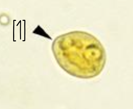
66
What does Chilomastix Mesnili cysts nucleus look like?
Large & may have large karyosome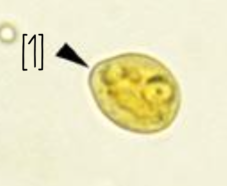
67
What do the fibrils in the cysts resemble in Chilomastix mesnili?
"safety pins"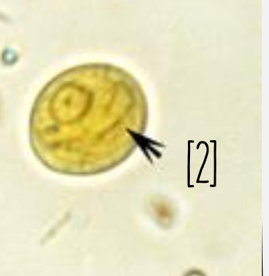
68
Label 1 and 2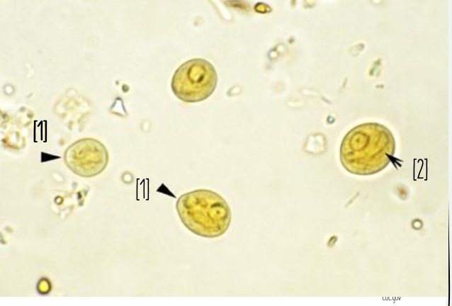
1) Anterior hyaline knob/nipple-like protuberance
2) safety pin
2) safety pin
69
what is this?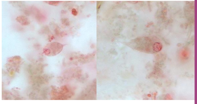
trophozoites - Chilomastix mesnili
70
what is this?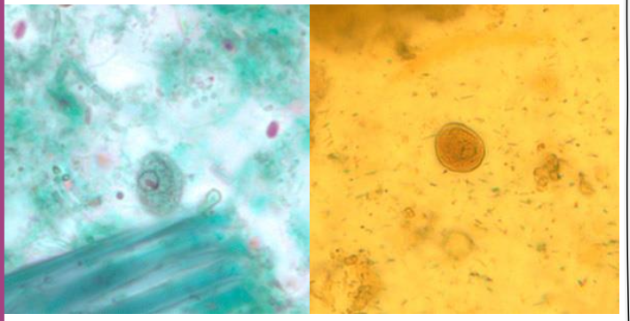
cysts - Chilomastix mesnili
71
How is Chilomastix mesnili transmitted?
Fecal-oral
72
What happens during the cyst stage of Chilomastix mesnili?
Infectious stage
Resistant to environemtnal pressure
Resistant to environemtnal pressure
73
Where are cysts from Chilomastix mesnilli usually found?
Feces
74
Where are the trophs released when infected with Chilomastix mesnili?
Large (possible small) instestine
75
Where does the trophozoite stage reside for Chilomastix mesnili?
Resides in the cecum and/or colon
76
What is Dientamoeba fragilis?
Protozoan flagellate (NOT AMOEBA)
77
Where is Dientamoeba fragilis found in the world?
Worldwide
78
What is the morphology for Triphozoites - Dientamoeba fragilis?
No evident flagella
Variable size (even in same specimen)
Most have 2 nuclei, 30-40% have only 1.
Nuclei show a mass of karysomal material, usually in a cluster of 4-8 granules.
Cytoplasm may be vaculoated.
Variable size (even in same specimen)
Most have 2 nuclei, 30-40% have only 1.
Nuclei show a mass of karysomal material, usually in a cluster of 4-8 granules.
Cytoplasm may be vaculoated.
79
Do Dientamoeba fragilis have cysts?
No definitively proven cyst stage.
80
What is this?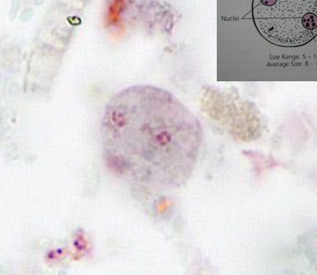
Dientamoeba fragilis with distinctive granules in nuclei.
81
How is dientamoeba fragilis transmitted?
Fecal-oral
82
Which flagelate life cycle is not yet determined?
Dientamoeba fragilis
83
Where are trophozoites (Deintamoeba fragilis) usually found?
Large intestine
84
How do trophozoites (Dientamoeba fragilis) multiply?
binary fission
85
Which stage of D. gragilis is detected in clinical specimens?
Only trophozoite stage
86
What is Pentatrichomonas hominis aka Trichomonas hominis?
Non-pathogenic / asymptomatic colonization of the large intestine. Possibly opportunistic (linked to diarrhea in immunocompromised)
87
Which host is Pentatrichomonas hominis usually found?
animals (mostly), but can be found in humans as well.
88
How many flagella does P. hominis have?
5
4 directed anteriorly and 5th directed posteriorly, that forms the outer edge of the undulating membrane and projects beyond the posterior as a free flagellum.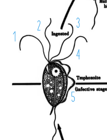
4 directed anteriorly and 5th directed posteriorly, that forms the outer edge of the undulating membrane and projects beyond the posterior as a free flagellum.
89
Does P. hominis have a cyst stage?
No
90
Name these parasites. Left to right. 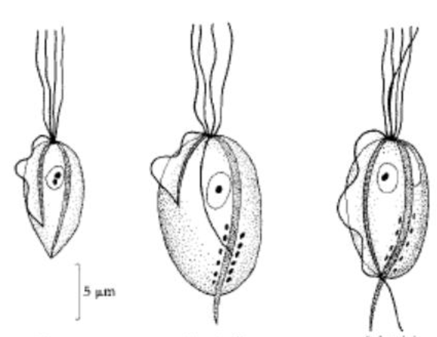
Trichomonas tenax
Trichomonas vaginalis
Pentatrichomonas hominis
Trichomonas vaginalis
Pentatrichomonas hominis