Lecture 12 - Serous Fluid
Study these flashcardsFree · spaced repetition · iPhone, Android, Mac & web
All cards
1
What does serous membrane cover?
lungs, heart, and abdominal organs
2
what does visceral membrane cover?
abdominal organs
3
what does parietal membrane cover?
internal surfaces of body cavity wall.
4
What is in between parietal and visceral membranes?
fluid-filled space or cavity btw these membranes
5
Pleural refers to what?
lungs
6
Pericardial refers to what?
heart
7
Peritoneal refers to what?
abdominal organs
8
What type of process is the formation and absorption of fluid called?
dynamic
9
Fluid formation is controlled by how many factors and what are they?
FOUR FACTORS:
permeability of capillaries in parietal membrane.
Hydrostatic pressure in these capillaries.
Oncotic pressure (colloid osmotic pressure) produced by plasma proteins in capillaries.
Absorption of fluid by lymphatic system.
permeability of capillaries in parietal membrane.
Hydrostatic pressure in these capillaries.
Oncotic pressure (colloid osmotic pressure) produced by plasma proteins in capillaries.
Absorption of fluid by lymphatic system.
10
What pressure forces plasma ultrafiltrate to form in cavity?
hydrostatic pressure (blood pressure)
11
What pressure opposes hydrostatic pressure?
oncotic (osmotic) pressure by plasma proteins in capillaries
12
What is effusion? and what does it indicate?
accumulation of fluid in a body cavity indicating an abnormal or pathologic process.
13
What happens to hydrstatic and oncotic pressure when fluid seeps into the membrane?
Hydrostatic is HIGH
oncotic pressure is low.
oncotic pressure is low.
14
How is serous fluid collected? (what is the process called?)
Paracentesis
15
What is paracentesis?
puncture of a body cavity for aspiration of fluid.
16
What is thoracentesis?
to obtain pleural fluid
17
What fluid is collected in pericardiocentesis?
for pericardial fluid
18
What fluid is collected in peritoneocentesis (or abdominal paracentesis)?
for peritoneal fluid
19
What is ascites?
an effusion in peritoneal cavity
20
What is ascitic fluid?
same as peritoneal fluid
21
Paracentesis is usually performed what which provider?
physician
22
What type of tube is used to collect serous fluid?
sterile tubes coated with anticoagulant
23
Sterile anticoagulant tubes with serous fluid is used for what studies?
microscopic exam or microbiological studies.
24
Why is a sterile tube with anticoagulant used to collect serous fluid?
to prevent clotting of abnormal specimens containing fibrinogen or blood
25
What type of tube is used for chemistry studies? what color?
nonabticoagulant (red-top)
26
How are tubes with serous fluid transported? at what temp?
room temperature and immediately sent to lab
27
What else is collected besides serous fluid and why?
collect a blood sample for comparison purposes.
28
What else should be recorded during collection?
color and clarity
29
How is pH maintained during collection? (what syringe should be used)
heparinized syringes
30
How are serous fluids classified?
Transudate vs exudate
31
What does transudate result from?
systemic disease that causes an increase in hydrostatic pressure OR
a decrease in plasma oncotic pressure (Congestive heart failure)
a decrease in plasma oncotic pressure (Congestive heart failure)
32
Are
Congestive heart failure, hepatic cirrhosis, and
nephrotic syndrome noninflammatory or inflammatory conditions?
Congestive heart failure, hepatic cirrhosis, and
nephrotic syndrome noninflammatory or inflammatory conditions?
noninflammatory
33
What does exudate result from?
inflammatory processes that increase permeability of capillary endothelium OR
a decrease in absorption by lymphatic system
OR
Infections, neoplasms, trauma, inflammatory conditions
a decrease in absorption by lymphatic system
OR
Infections, neoplasms, trauma, inflammatory conditions
34
Does transudate or exudate clot?
Exudate clots
35
What color may exudate appear?
pink, orange, and yellow
36
What color is transudates?
clear, pale yellow
37
How is the viscosity like in transudates?
similar to serum
38
Do transudates clot and why?
do not clot bc they have no fibrinogen
39
Describe exudate.
usually cloudy
various colors
may have shimmer or sheen
can form clots
various colors
may have shimmer or sheen
can form clots
40
Describe cloudy paracentesis fluid.
Large numbers of WBCs, other cells, chyle, and/or lipids; milky after centrifugation indicates chyle
41
What is chylous effusions caused by? describe it.
obstruction of or damage to lymphatic system.
sample is milky
tryglycerides are high (>110 mg/dL)
lymphs are present
happens bc of trauma.
sample is milky
tryglycerides are high (>110 mg/dL)
lymphs are present
happens bc of trauma.
42
What are Pseudochylous effusions?
chronic effusions, can be differentiated from chylous effusions by their lipid content.
43
What is chylous?
high triglycerides and chylomicrons (lipoproteins) present
44
What is pseudochylous?
low triglycerides and no chylomicrons
45
What does a microscopic exam for serous fluid include?
Total cell count
Red and white blood cell counts
Differential cell count
ID of crystals
Gram stain
Cytologic exam for malignant cells
Red and white blood cell counts
Differential cell count
ID of crystals
Gram stain
Cytologic exam for malignant cells
46
What is the white blood cell (WBC) count typically found in transudates?
A) Greater than 1000 cells/μL
B) Less than 1000 cells/μL
C) Equal to 1000 cells/μL
D) Not measurable
A) Greater than 1000 cells/μL
B) Less than 1000 cells/μL
C) Equal to 1000 cells/μL
D) Not measurable
B) Less than 1000 cells/μL
47
What is the WBC count typically found in exudates?
A) Greater than 1000 cells/μL
B) Less than 1000 cells/μL
C) Equal to 1000 cells/μL
D) Not measurable
A) Greater than 1000 cells/μL
B) Less than 1000 cells/μL
C) Equal to 1000 cells/μL
D) Not measurable
Answer: A) Greater than 1000 cells/μL
48
What can cause the presence of red blood cells (RBCs) in fluid?
A) Traumatic taps
B) Disease processes
C) Both A and B
D) Neither A nor B
A) Traumatic taps
B) Disease processes
C) Both A and B
D) Neither A nor B
Answer: C) Both A and B
49
What characteristic is associated with traumatic taps during fluid collection?
A) RBCs increase as the collection progresses
B) RBCs decrease later in the collection
C) RBCs remain constant throughout the collection
D) No RBCs are present
A) RBCs increase as the collection progresses
B) RBCs decrease later in the collection
C) RBCs remain constant throughout the collection
D) No RBCs are present
B) RBCs decrease later in the collection
50
What is a key characteristic of hemorrhagic effusion?
A) Blood is heterogeneously distributed with clots
B) Blood is homogeneously distributed with no clotting
C) Blood is only found at the start of the collection
D) No blood is present
A) Blood is heterogeneously distributed with clots
B) Blood is homogeneously distributed with no clotting
C) Blood is only found at the start of the collection
D) No blood is present
Answer: B) Blood is homogeneously distributed with no clotting
51
How is serous fluid prepared in a differential cell count?
cytocentrifuge-prepared slide
52
Which WBCs predominate in most exudates of pleural/ peritoneal fluid?
Neutrophils
53
Which WBCs predominate in pleural effusions and peritoneal transudates and exudates? and in what conditions?
lymphocytes; tuberculosis (TB), neoplasms, and systemic diseases
54
Peritoneal transudates and exudates are caused by what?
decreased lymphatic absorption
55
What are other cells that could be seen in differential cell count?
monocytes, macrophages, eosinophils, plasma cells, lupus erythematosus (LE) cells, malignant cells, and mesothelial cells.
56
what type of cell is this?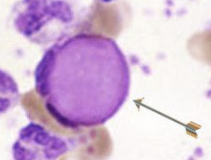
LE cell aka lupus erythematosus
57
What is the fluid/serum protein ratio for a transudate and exudate?
< 0.5 transudate
> 0.5 exudate
> 0.5 exudate
58
What is the fluid/serum lactate dehydrogenase (LD) ratio for a transudate and exudate?
< 0.6 transudate
> 0.6 exudate
> 0.6 exudate
59
What happens when protein increases in serous fluid, glucose goes?
down.
60
What does a decrease in glucose associated with?
variety of disease processes
61
What is amylase?
enzyme produced in the pancreas
62
What is a high pleural or peritoneal fluid amylase associated with?
Pancreatitis
Edophageal rupture
Gastric duodenal perforation
Metastatic disease
Edophageal rupture
Gastric duodenal perforation
Metastatic disease
63
What is lipids in a chemistry exam used to identify?
chyloud effusions
64
What does pH in a chemistry exam used to identify?
abnormally low pH identifies pleural exudates caused by pneumonia or lung abscess
65
What happens when the pH is < 7.30 while on antibiotics?
requires more aggressive treatment
66
What does CEA stand for? and what is it?
Carcinoembryonic antigen; it is a tumor marker
67
If CEA is low, how big is the tumor?
small
68
If CEA is high, what is the size of the tumor?
big
69
CEA is an indication of what?
sign of colon cancer, rectal cancer
70
What type of stains may be used for microbiological exam?
gram stain
acid-fast stain
other stains
bacterial cultures
acid-fast stain
other stains
bacterial cultures