Lecture 10: Chapter 9 Cerebrospinal Fluid Analysis
Study these flashcardsFree · spaced repetition · iPhone, Android, Mac & web
All cards
1
Why is CFS a stat specimen?
because cell count decreases as time goes on.
2
what color is normal CFS?
colorless
3
what does CFS with WBCs and/or RBCs and/or hemorrhage mean?
sample is turbid
4
When is pressure recorded during CSF extraction?
before and after
5
At what pressure is 20mL of CFS collected?
50-180 mmHg
6
At what pressure is 1-2 mL of CSF collected?
if it is outside the range: 50-180 mmHg
7
Where is the puncture site for adults?
3 & 4 lumbar
8
Where is the puncture site for children?
4 & 5 lumbar
9
from inner to outer, what are the 3 different layers of meninges?
dura mater
arachnoid mater
pia mater
arachnoid mater
pia mater
10
Where can CSF be found?
in the subarachnoid space
11
What are the 3 protective coverings of the brain?
bone, meninges, and fluid meninges
12
Absorption and production of CSF are both at the same rate. true/false
TRUE
13
How many mL of CSF is seen in the subarachnoid space?
80-150 mL
14
Where is the subarachnoid space located?
between pia and arachnoid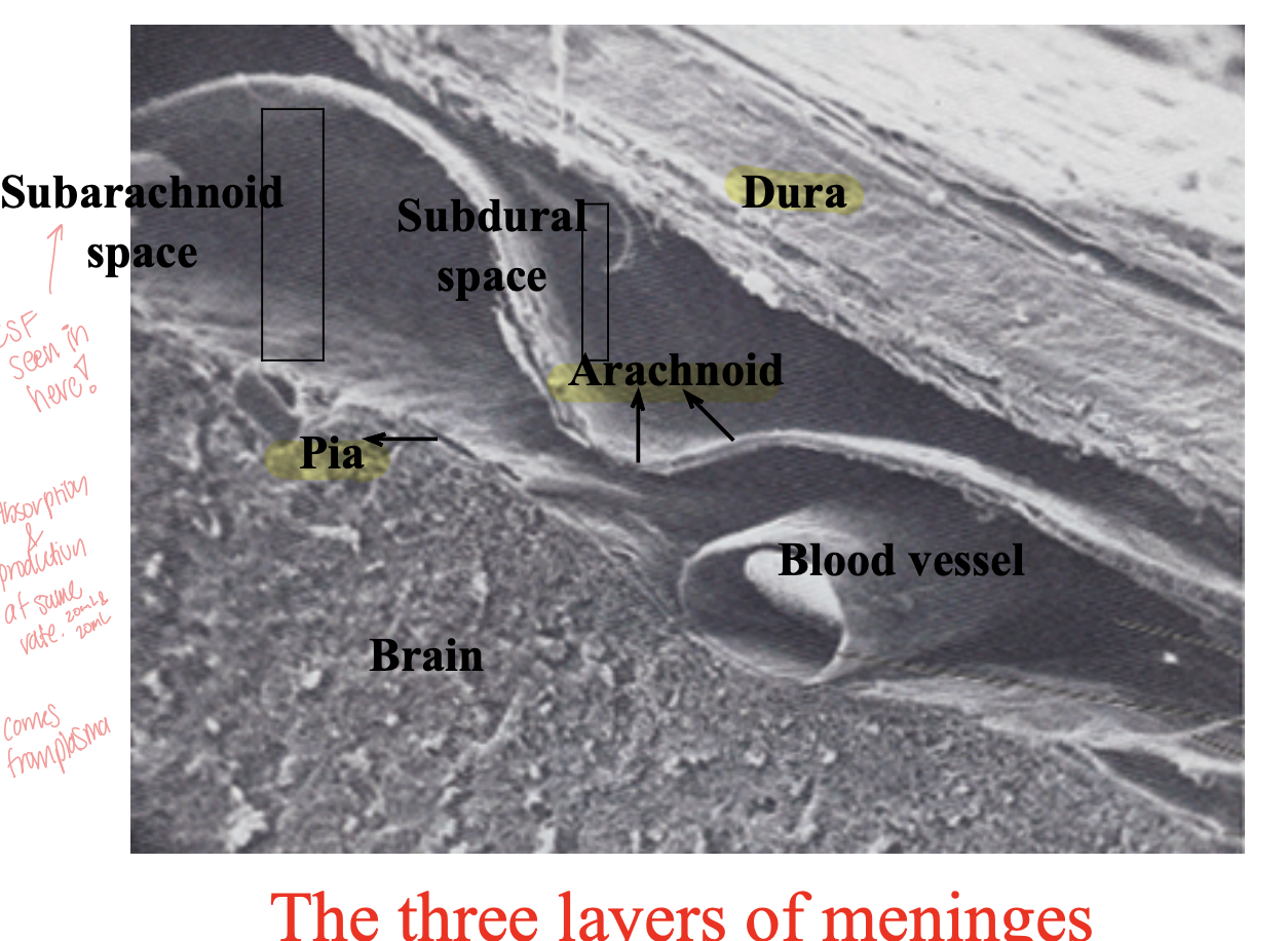
15
what does CSF consist of?
glucose, proteins, and ions
16
What are the 3 functions of CSF?
Mechanical protection
Chemical protection
Circulation
Chemical protection
Circulation
17
How does cerebrospinal fluid (CSF) provide mechanical protection to the brain?
Cerebrospinal fluid (CSF) acts as a cushion that "floats" the brain, reducing its effective weight and softening the impact against the hard skull. This helps protect the brain from injury by absorbing shocks and reducing the force from sudden movements or impacts.
18
How does cerebrospinal fluid (CSF) provide chemical protection to the brain?
optimal ionic concentrations for action potentials
19
How does cerebrospinal fluid (CSF) provide circulation to the brain?
circulates nutrients and waste products to and from bloodstream
20
Where is CSF produced?
from arterial blood by the choroid plexuses
21
What is the structure of the choroid plexus, and how does it relate to its function?
The choroid plexus is made up of clusters of capillaries with thin, fenestrated (small opening) endothelial cells. These capillaries are covered by specialized ependymal cells that have tiny hair-like projections called microvilli. This structure allows the choroid plexus to produce cerebrospinal fluid (CSF) efficiently, filtering substances from the blood while maintaining a controlled environment around the brain.
22
where is CSF reabsorbed?
through arachnoid villi; which are grapelike clusters of arachnoid layer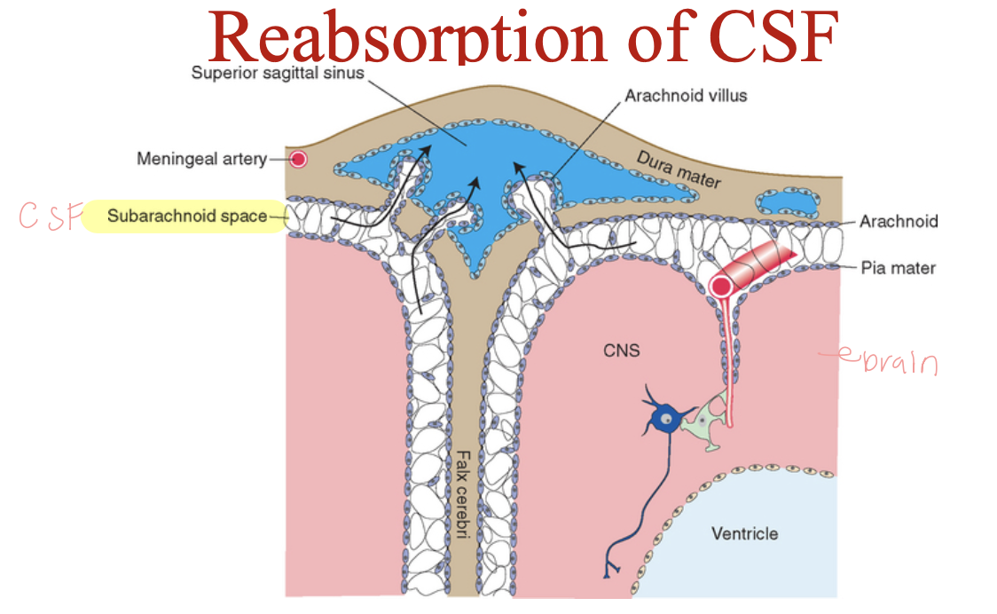
23
What is the reabsorption rate of CSF and what is the production rate? (mL/hour)
both reabsorption rate and production rate are the same; so 20 mL/hour
24
What is hydrocephalus?
a condition where there is an abnormal buildup of cerebrospinal fluid (CSF) in the brain’s ventricles, which are fluid-filled spaces.
25
what does the choroid plexus consist of?
Lateral ventricle (right and left)
Third ventricle,
Fourth ventricle
Third ventricle,
Fourth ventricle
26
What are some causes of hydrocephalus?
tumor, inflammation, developmental malformation, meningitis, hemorrhage or injury
27
What happens when a patient has hydrocephalus?
continued production of CSF causing an increase in pressure!
28
what is the treatment for this condition: hydrocephalus?
a drain shunting the CSF to the veins of the neck or the abdomen.
29
What type of anestesia is used to collect CSF sample?
local anesthesia
30
How is pressure taken prior and after collection?
with manometer attached to the spinal needle
31
What is the normal recumbent pressure of CSF?
50 to 180 mmHg
32
What should the closing pressure taken after collection of sample should be?
10-20 mm less
33
what should always be recorded when collecting CSF sample?
pressure values and amount collected!
34
How many tubes of CSF are collected and which departments are they sent to?
3 tubes:
1. chemistry
2. microbiology
3. cell counts
1. chemistry
2. microbiology
3. cell counts
35
Why is CSF sent to microbiology department?
to check for infections in the central nervous system, such as meningitis or encephalitis.
36
what does normal CSF look like?
clear and colorless with viscosity like water
37
What does clarity or turbidity relate to?
number of cells
38
What is pleocytosis?
increased number of cells in CSF
39
Cloudy CSF can be associated with what type of cells?
WBCs (>200 cells/mL)
or
RBCs (>400 cells/mL)
OR
microorganisms
OR
increased protein
or
RBCs (>400 cells/mL)
OR
microorganisms
OR
increased protein
40
What does not cause turbidity?
glucose
41
What is xanthochromia?
abnormal color of CSF like orange, red/pink, yellow
42
What color is CSF when there is cerebral hemmorhage?
pink bc blood is in sample.
43
What does a traumatic tap mean?
occurs when blood unintentionally enters the cerebrospinal fluid (CSF) sample during a lumbar puncture (spinal tap) procedure. This usually happens when a small blood vessel is accidentally punctured as the needle is inserted into the spinal canal.
44
How do you know when a CSF sample is a traumatic tap and NOT hemorrhage?
theres a lot of blood in tube1, least in tube 3
AND
After centrifugation, supernatant is colorless
AND
After centrifugation, supernatant is colorless
45
How do you know when a CSF sample is a hemorrahge and NOT a traumatic tape?
There's consistent amount of blood in all 3 tubes; meaning there's internal bleeding
AND
After centrifugation, xanthochromic supernatant
AND
Macrophages containing phagocytosed RBCs within
AND
After centrifugation, xanthochromic supernatant
AND
Macrophages containing phagocytosed RBCs within
46
What is the normal cell count for WBCs per microliter?
0 to 5 WBCs/ uL
47
Are RBCs normally present in CSF?
Noooooooo; could be due to traumatic tape or hemorrhage
48
Why is CSF samples stat?
because WBCs lyse pretty quickly, so we want to prevent that from happening
49
What should be done to slow down the lysing of WBCs in a CSF sample?
Store in fridge at 4oC
50
How are cell counts performed?
with hemacytometer on undiluted specimens counting all nine squares on both sides
51
If the CSF contains 90% of neutrophils, what is the condition?
bacterial meningitis
52
What other conditions is associated with an increase of neutrophils?
early viral, fungal, TB, or parasitic infections
and
some non infectious conditions
and
some non infectious conditions
53
An increase of lymphocytes is associated with what condition?
Viral meningitis, TB, fungal or syphilitic meningitis particularly in later stages
54
An increase in plasma cells (antibodiy producing) indicate what?
multiple sclerosis and acute viral and chronic inflammatory conditions.
55
what happens to glucose and proteins when 90% of WBCs in CSF are neutrophils?
decrease in glucose; because bacteria uses it up!
increase in proteins; because of inflammatory proteins!
increase in proteins; because of inflammatory proteins!
56
Increase in eosinophil production is associated with what?
10% or greater with parasitic, fungal, or allergic reactions
57
An increase in monocytes is associated with what?
fungal meningitis, TB, chronic bacterial meningitis, or rupture off cerebral abscess
58
What is the normal range of total protein seen in CSF?
15 - 45 mg/dL; higher in infants and older adults
59
An increase in proteins can be due to what?
contamination with blood during traumatic tap
change in blood-brain barrier
decreased reabsorption into venous blood
increased synthesis in central nervous system (CNS)
change in blood-brain barrier
decreased reabsorption into venous blood
increased synthesis in central nervous system (CNS)
60
A decrease in proteins can be due to what?
the body is rapidly producing spinal fluid &
loss of fluid
loss of fluid
61
Which disorders is increased protein usually seen in?
hemorrhage
bactieral, viral ,and other forms of meningitis
cerebral infarction
MS
obstruction of CSF flow
trauma
bactieral, viral ,and other forms of meningitis
cerebral infarction
MS
obstruction of CSF flow
trauma
62
What is Cerebrospinal fluid (CSF)/Serum albumin index used for?
to assess permeability of blood-brain barrier
63
What is the CSF/Serum albumin index for a normal CSF?
< 9 is normal
64
What is the CSF/Serum albumin index for a minimal impairment CSF?
9 to 14
65
What is the CSF/Serum albumin index for a moderate to severe CSF?
15 to 100
66
What is the CSF/Serum albumin index for a complete breakdown of barrier CSF?
exceeding 100
67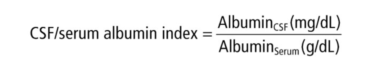
How do you calculate CSF/serum albumin index?
68
What are other tests done?
glucose and lactate
69
Where is glucose measured from? and when?
plasma value is measured 30 to 90 minutes before CSF collection.
70
A decrease glucose is associated with what?
meningitis
71
What usually increases when there's impairing blood suply or transport of oxygen to the CNS?
lactate
72
A patient containing viral meningitis has increase/decrease/normal levels of lactate?
normal; other conditions are elevated.
73
an increase in lymphocytes is assocaited with?
autoimmune
74
What is a myelin sheath? and what does it do?
a protective, fatty layer that surrounds nerve fibers (axons) in the nervous system; to insulate the nerve cells and to speed up the transmission of electrical signals (nerve impulses) along the axon
75
What are schwann cells?
a type of glial cell in the peripheral nervous system (PNS) responsible for producing and maintaining the myelin sheath around peripheral nerve fibers.
76
If immune system destroys the myelin sheath, what happens to the nerves?
cells can't communicate with other cells.
77
what is released with mylein sheath is damaged? and what happens?
folded MBP (myelin basic proteins) becomes unfolded MBP which can not be phaocytized by a phagocytic cell.
78
What conditions are myelin basic proteins seen in?
multiple sclerosis and other demyelinating diseases
79
What stain is used to detect bacteria and fungi?
gram stain
80
What stain is used to detect Cryptococcus neoformans?
India ink
81
What stain is used to detect TB meningitis?
Acid fast stain
82
What stain is used to detect Naegleria fowleri (amoeba)?
Wright stain
83
What test to done to detect bacterial cause?
culture!