Lecture 3 (PT1) Anemias: hypoproliferative (role of vit b12 and folate)
Study these flashcardsFree · spaced repetition · iPhone, Android, Mac & web
All cards
1
What is the difference between megaloblastic and non-megaloblastic anemia?
Megaloblastic → DNA synthesis problem (B₁₂/folate deficiency), oval macrocytes, hypersegmented neutrophils.
Non-megaloblastic → Other causes (alcohol, liver disease, etc.), round macrocytes, no hypersegmented neutrophils.
Non-megaloblastic → Other causes (alcohol, liver disease, etc.), round macrocytes, no hypersegmented neutrophils.
2
Q: What does an MCV > 100 fL indicate?
A: Possible macrocytic anemia.
3
What are some megaloblastic characteristics?
oval macrocytes, hypersegmented neutrophils, and pancytopenia
4
if megaloblastic characteristics are checked out, what test would you do?
Serum folate and vitamin B 12 levels
5
Q: What are some non-megaloblastic causes of MCV > 100 fL?
A: Normal newborns, reticulocytosis, liver disease, chronic alcoholism, bone marrow failure.
6
Q: What are the megaloblastic characteristics of macrocytic anemia?
A: Oval macrocytes, hypersegmented neutrophils, pancytopenia.
7
Q: What are the causes of Vitamin B₁₂ deficiency?
A:
Inadequate intake
Increased need
Impaired absorption:
Inability to split B₁₂ from food
Inability to split B₁₂ from haptocorrin
Lack of intrinsic factor
Pernicious anemia
H. pylori infection
Gastrectomy
Hereditary intrinsic factor deficiency
General malabsorption
Inherited errors in transport/absorption
Competition for vitamin B₁₂ (D. latum infection, blind loop syndrome)
Inadequate intake
Increased need
Impaired absorption:
Inability to split B₁₂ from food
Inability to split B₁₂ from haptocorrin
Lack of intrinsic factor
Pernicious anemia
H. pylori infection
Gastrectomy
Hereditary intrinsic factor deficiency
General malabsorption
Inherited errors in transport/absorption
Competition for vitamin B₁₂ (D. latum infection, blind loop syndrome)
8
Q: What are the causes of folate deficiency?
A:
Inadequate intake
Increased need
Impaired absorption
Impaired use
Excessive loss (renal dialysis)
Inadequate intake
Increased need
Impaired absorption
Impaired use
Excessive loss (renal dialysis)
9
Q: If no folate or vitamin B₁₂ deficiency is found, what are other causes of macrocytosis?
A: Myelodysplastic syndrome, acute erythroid leukemia, congenital dyserythropoietic anemia, reverse transcriptase inhibitors.
10
Q: What is another name for Vitamin B12?
A: Cobalamin.
11
Q: What is the structure of Vitamin B12?
A: It has a tetrapyrrole ring with cobalt in the middle, attached to a ribonucleotide (5,6-dimethylbenzimidazolyl).
12
Q: What are the different forms of Vitamin B12?
A: Hydroxy-cobalamin, cyano-cobalamin, methyl-cobalamin, and 5′-deoxyadenosylcobalamin.
13
Q: Which forms of Vitamin B12 are found in foods?
A: Hydroxy-cobalamin and cyano-cobalamin.
14
Q: Which forms of Vitamin B12 act as co-enzymes?
A: Methyl-cobalamin and 5′-deoxyadenosylcobalamin.
15
Q: What is the first step where Vitamin B12 absorption can fail?
A: P separation from CBL (failure to release B12 from proteins in food).
16
Q: What is the second step where Vitamin B12 absorption can fail?
A: R separation from CBL (failure to release B12 from R-binder/haptocorrin).
17
Q: What happens if there is impaired IF-CBL binding?
A: Lack of intrinsic factor prevents Vitamin B12 binding, leading to pernicious anemia.
18
Q: How can malabsorption cause Vitamin B12 deficiency?
A: Intestinal conditions (like celiac disease or Crohn’s) prevent absorption in the ileum.
19
Q: How does competition for Vitamin B12 affect absorption?
A: Organisms like Diphyllobothrium latum (fish tapeworm) or bacterial overgrowth use available Vitamin B12, reducing absorption.
20
Q: What type of symptoms are more prominent in Vitamin B12 deficiency?
A: Neurologic symptoms.
21
Q: What neurologic symptoms occur in Vitamin B12 deficiency?
A: Memory loss, numbness and tingling in fingers, and loss of vibratory sense.
22
Q: Are CNS symptoms common in folate deficiency?
A: No, CNS involvement is not common.
23
Q: What risk is associated with folate deficiency?
A: Increased risk of cardiovascular disease due to high serum homocysteine.
24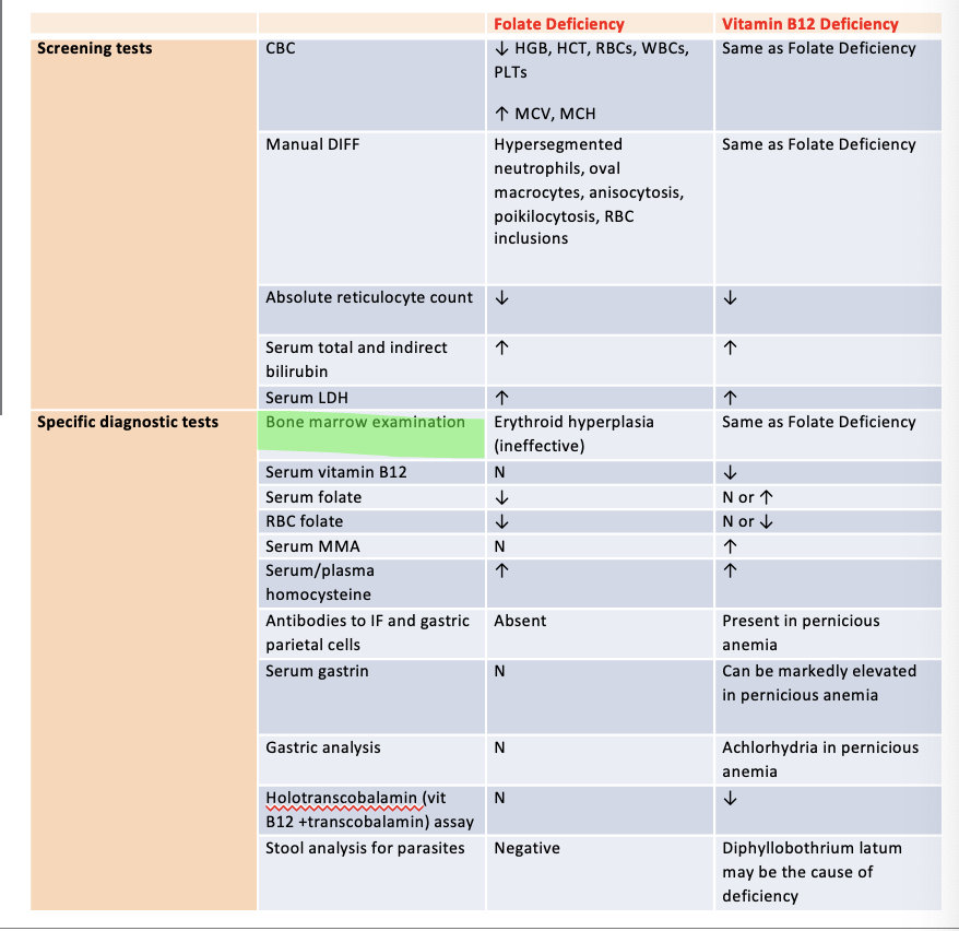
25
What are screening tests for megaloblastic anemia?
CBC, MANUAL DIFF, ABSOLUTE RETICULOCYTE COUNT, SERUM TOTAL AND INDIRECT BILIRUBIN, AND SERUM LDH
26
What are specific diagnositic tests for megaloblastic anemia?
bone marrow examination, serum vitamin B12, serum folate , rbc folate, serum MMA, serum/plasma homocysteine, antibdoesi to IF and gastric parietal cells, serum gastrin, gastric analysis, holotranscobalamin (vit b12 + transcobalamin) assay, stoll analysis for parasties.
27
Q: What happens to HGB, HCT, RBCs, WBCs, and PLTs in folate deficiency?
A: Decreased.
28
Q: What happens to MCV and MCH in folate deficiency?
A: Increased.
29
Q: What are the CBC findings in Vitamin B12 deficiency?
A: Same as folate deficiency (↓ HGB, HCT, RBCs, WBCs, PLTs; ↑ MCV, MCH).
30
Q: What does the manual differential show in folate deficiency?
A: Hypersegmented neutrophils, oval macrocytes, anisocytosis, poikilocytosis, RBC inclusions.
31
Q: What does the manual differential show in Vitamin B12 deficiency?
A: Same as folate deficiency.
32
Q: What happens to the absolute reticulocyte count in folate deficiency?
A: Decreased.
33
Q: What happens to the absolute reticulocyte count in Vitamin B12 deficiency?
A: Decreased.
34
Q: What happens to serum total and indirect bilirubin in folate deficiency?
A: Increased.
35
Q: What happens to serum total and indirect bilirubin in Vitamin B12 deficiency?
A: Increased.
36
Q: What happens to serum LDH in folate deficiency?
A: Increased.
37
Q: What happens to serum LDH in Vitamin B12 deficiency?
A: Increased.
38
Q: What does bone marrow show in folate deficiency?
A: Erythroid hyperplasia (ineffective).
39
Q: What does bone marrow show in Vitamin B12 deficiency?
A: Same as folate deficiency.
40
Q: What happens to serum vitamin B12 in folate deficiency?
A: Normal.
41
Q: What happens to serum vitamin B12 in Vitamin B12 deficiency?
A: Decreased.
42
Q: What happens to serum folate in folate deficiency?
A: Decreased.
43
Q: What happens to serum folate in Vitamin B12 deficiency?
A: Normal or increased.
44
Q: What happens to RBC folate in folate deficiency?
A: Decreased.
45
Q: What happens to RBC folate in Vitamin B12 deficiency?
A: Normal or decreased.
46
Q: What happens to serum methylmalonic acid (MMA) in folate deficiency?
A: Normal.
47
Q: What happens to serum methylmalonic acid (MMA) in Vitamin B12 deficiency?
A: Increased.
48
Q: What happens to serum/plasma homocysteine in folate deficiency?
A: Increased.
49
Q: What happens to serum/plasma homocysteine in Vitamin B12 deficiency?
A: Increased.
50
Q: Are antibodies to intrinsic factor (IF) and gastric parietal cells present in folate deficiency?
A: Absent.
51
Q: Are antibodies to intrinsic factor (IF) and gastric parietal cells present in Vitamin B12 deficiency?
A: Present in pernicious anemia.
52
Q: What happens to serum gastrin in folate deficiency?
A: Normal.
53
Q: What happens to serum gastrin in Vitamin B12 deficiency?
A: Markedly elevated in pernicious anemia.
54
Q: What does gastric analysis show in folate deficiency?
A: Normal.
55
Q: What does gastric analysis show in Vitamin B12 deficiency?
A: Achlorhydria in pernicious anemia.
56
Q: What happens to holotranscobalamin (Vitamin B12 + transcobalamin) assay in folate deficiency?
A: Normal.
57
Q: What happens to holotranscobalamin (Vitamin B12 + transcobalamin) assay in Vitamin B12 deficiency?
A: Decreased.
58
Q: What does stool analysis for parasites show in folate deficiency?
A: Negative.
59
Q: What does stool analysis for parasites show in Vitamin B12 deficiency?
A: Diphyllobothrium latum may be the cause of deficiency.
60
Q: What is pernicious anemia?
A: An autoimmune disease in which autoantibodies are raised against intrinsic factor (IF) or gastric parietal cells.
61
Q: How does pernicious anemia develop through immune response?
A: T CD4-mediated response destroys parietal cells over time → decreased IF secretion → impaired B12 absorption.
62
Q: Besides IF loss, what else do T cells destroy in pernicious anemia?
A: The H⁺/K⁺-ATPase pump on parietal cell membranes, leading to reduced HCl production (achlorhydria).
63
Q: What test can detect absence of intrinsic factor in pernicious anemia?
A: Schilling test.
64
Q: What happens to serum gastrin levels in pernicious anemia?
A: Highly increased (especially in achlorhydria).
65
Q: Which antibodies are detected in pernicious anemia?
A:
Anti-IF antibodies (blocking antibodies against B12 binding sites on IF, found in 70–90% of patients).
Anti-parietal cell antibodies (found in 50–90% of patients).
Anti-IF antibodies (blocking antibodies against B12 binding sites on IF, found in 70–90% of patients).
Anti-parietal cell antibodies (found in 50–90% of patients).
66
Q: What infection may also lead to pernicious anemia?
A: Helicobacter pylori (causes parietal cell destruction).
67
Q: How does megaloblastic anemia typically develop?
A: Slowly, but usually very severe (Hb < 7).
68
Q: What is the earliest sign of megaloblastic anemia?
A: Slight macrocytosis.
69
Q: What is the typical MCV range in megaloblastic anemia?
A: 100–150 fL, mostly >120 fL.
70
Q: What hematologic finding often accompanies megaloblastic anemia?
A: Pancytopenia.
71
Q: How can coexisting conditions (IDA, chronic inflammation, or beta-thal minor) affect MCV in megaloblastic anemia?
A: They can normalize MCV.
72
Q: What happens to MCH and MCHC in megaloblastic anemia?
A: MCH is high, but MCHC is normal.
73
Q: What happens to the reticulocyte count in megaloblastic anemia?
A: Low retic count (no polychromasia).
74
Q: What RBC morphology is a very important clue for megaloblastic anemia?
A: Oval macrocytes.
75
Q: What abnormal RBC morphologies may be present in megaloblastic anemia (besides oval macrocytes)?
A: Dacrocytes, RBC fragments, micro-spherocytes (with high RDW).
76
Q: What rare RBC inclusions may be seen in megaloblastic anemia?
A: nRBCs, Howell-Jolly bodies, basophilic stippling, Cabot rings.
77
Q: What neutrophil finding is a very important clue for megaloblastic anemia?
A: Neutrophil hypersegmentation.
78
Q: When is neutrophil hypersegmentation reported as significant in megaloblastic anemia?
A: When at least 5 five-lobed neutrophils per 100 WBCs, or at least 1 neutrophil with 6 lobes, is seen.
79
Q: How long does neutrophil hypersegmentation persist after treatment in megaloblastic anemia?
A: It appears early and can stay for 2 weeks after treatment.
80
What do you expect to see on the PBS? (describe RBC and WBC morphology)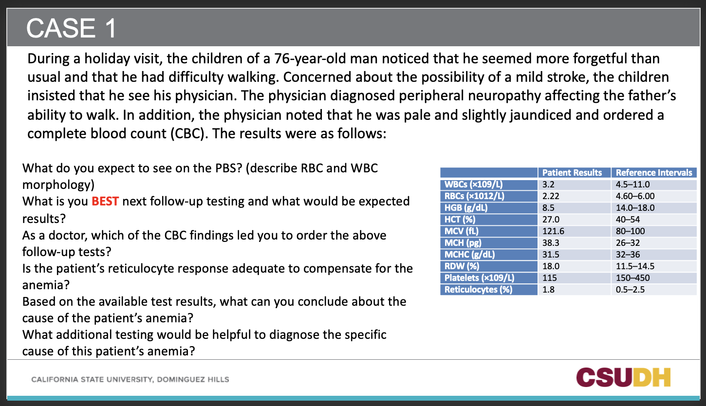
RBC: Oval macrocytes, anisocytosis, poikilocytosis, possible Howell-Jolly bodies, RBC fragments, dacryocytes.
WBC: Hypersegmented neutrophils (≥5 lobes).
Platelets: Decreased.
WBC: Hypersegmented neutrophils (≥5 lobes).
Platelets: Decreased.
81
What is you BEST next follow-up testing and what would be expected results?
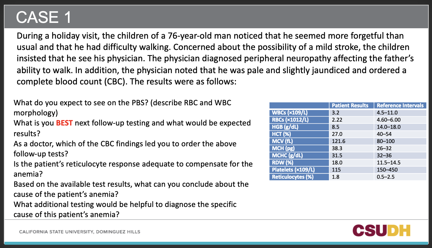
Serum Vitamin B12 and Folate levels
Expect low Vitamin B12 (given neurologic findings).
Could also check MMA (↑ in B12 deficiency) and homocysteine (↑ in both B12 & folate deficiency).
Expect low Vitamin B12 (given neurologic findings).
Could also check MMA (↑ in B12 deficiency) and homocysteine (↑ in both B12 & folate deficiency).
82
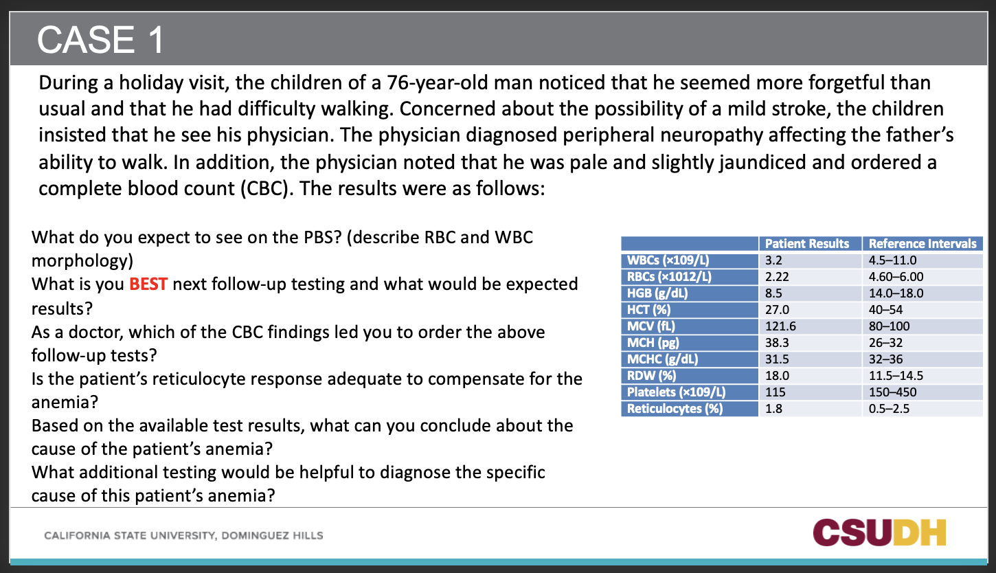
Q3: As a doctor, which CBC findings led you to order the above follow-up tests?
A:
High MCV (121.6 fL) → macrocytosis.
Pancytopenia (low WBCs, RBCs, platelets).
High RDW (18.0).
These findings suggest a megaloblastic process.
High MCV (121.6 fL) → macrocytosis.
Pancytopenia (low WBCs, RBCs, platelets).
High RDW (18.0).
These findings suggest a megaloblastic process.
83
Q4: Is the patient’s reticulocyte response adequate to compensate for the anemia?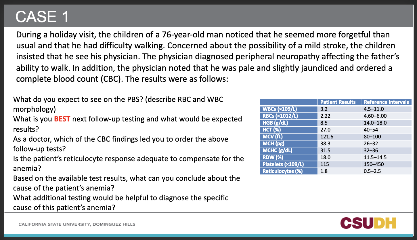
A: No.
Reticulocytes are 1.8%, which is within normal range, but with severe anemia (Hb 8.5, RBC 2.22), you would expect a strong compensatory increase.
Therefore, inadequate reticulocyte response → ineffective erythropoiesis.
Reticulocytes are 1.8%, which is within normal range, but with severe anemia (Hb 8.5, RBC 2.22), you would expect a strong compensatory increase.
Therefore, inadequate reticulocyte response → ineffective erythropoiesis.
84
Q5: Based on the available test results, what can you conclude about the cause of the patient’s anemia?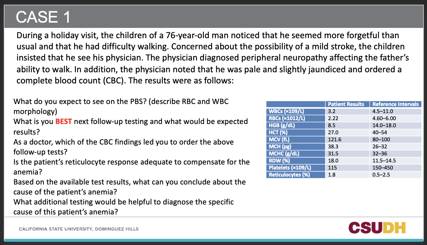
A:
Likely megaloblastic anemia due to Vitamin B12 deficiency.
Neurologic symptoms (neuropathy, forgetfulness, gait disturbance) point more toward B12 deficiency rather than folate deficiency
Likely megaloblastic anemia due to Vitamin B12 deficiency.
Neurologic symptoms (neuropathy, forgetfulness, gait disturbance) point more toward B12 deficiency rather than folate deficiency
85
Q6: What additional testing would be helpful to diagnose the specific cause of this patient’s anemia?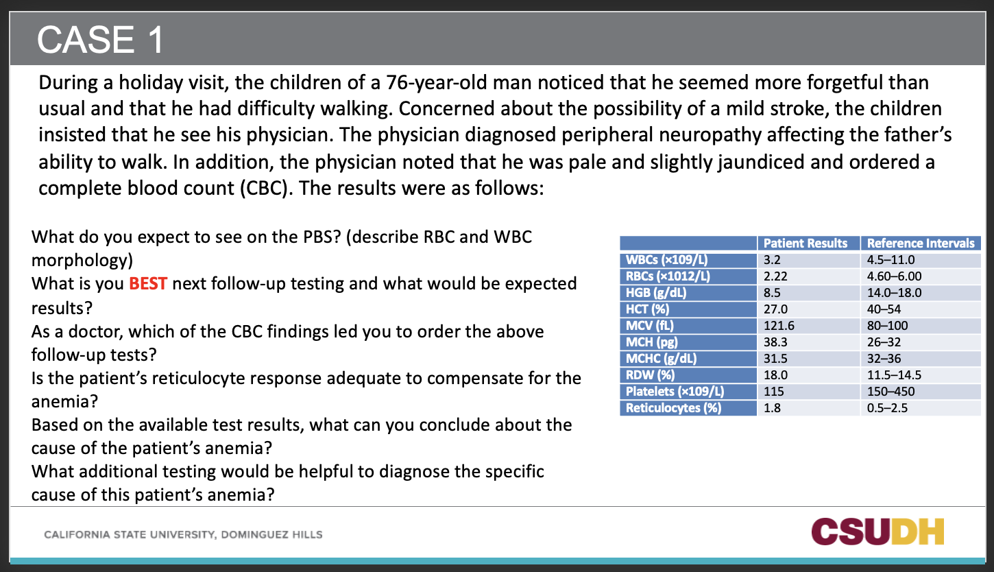
A:
Anti-intrinsic factor antibodies (pernicious anemia).
Anti-parietal cell antibodies.
Serum MMA and homocysteine.
Serum gastrin (↑ in pernicious anemia with achlorhydria).
Schilling test (historical, rarely used now).
Stool exam for Diphyllobothrium latum (if dietary risk)
Anti-intrinsic factor antibodies (pernicious anemia).
Anti-parietal cell antibodies.
Serum MMA and homocysteine.
Serum gastrin (↑ in pernicious anemia with achlorhydria).
Schilling test (historical, rarely used now).
Stool exam for Diphyllobothrium latum (if dietary risk)
86
Q: What is the purpose of the Schilling test?
A: To distinguish cobalamin (Vitamin B12) deficiency due to malabsorption, dietary deficiency, or absence of intrinsic factor (IF).
87
Q: What does the Schilling test measure?
A: The amount of radioactively labeled crystalline Vitamin B12 absorbed in the gut and excreted in the urine.
88
Q: What is the “flushing dose” in the Schilling test?
A: An intramuscular injection of unlabeled Vitamin B12 given to saturate tissue B12 receptors so excess labeled B12 is excreted in urine.
89
Q: What does it mean if >7.5% of the oral dose of labeled B12 is excreted in urine?
A: Normal absorption.
90
Q: What does it mean if <7.5% of the oral dose of labeled B12 is excreted in urine (Part I)?
A: Abnormal absorption → could be pernicious anemia (PA) or another malabsorption syndrome.
91
Q: What is the purpose of Part II of the Schilling test?
A: To distinguish pernicious anemia from other malabsorption causes.
92
Q: In Part II, what is given along with the oral labeled B12?
A: Intrinsic factor.
93
Q: If Part II shows >7.5% excretion, what is the diagnosis?
A: Pernicious anemia (due to lack of intrinsic factor).
94
Q: If Part II still shows <7.5% excretion, what does it suggest?
A: Another malabsorption defect (e.g., sprue, ileal disease).
95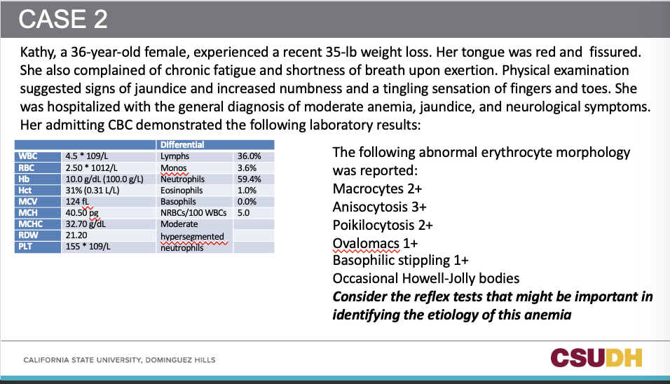
Case 2: Summary
36-year-old woman, weight loss, glossitis (red fissured tongue), fatigue, SOB, jaundice, neurological symptoms.
CBC: Anemia (Hb 10, Hct 31), macrocytic (MCV 124), ↑MCH, ↑RDW, PLT slightly low-normal, WBC normal-low.
PBS: Macrocytes, anisocytosis, poikilocytosis, ovalocytes, basophilic stippling, Howell-Jolly bodies, hypersegmented neutrophils.
Reflex test: serum b12 or folate levels
Follow-up tests: Low B12 (50 pg/mL), folate normal, ↑bilirubin (2.5), bone marrow = erythroblastic hyperplasia with megaloblasts.
Special tests: Schilling test abnormal, anti-IF blocking antibodies strongly positive.
36-year-old woman, weight loss, glossitis (red fissured tongue), fatigue, SOB, jaundice, neurological symptoms.
CBC: Anemia (Hb 10, Hct 31), macrocytic (MCV 124), ↑MCH, ↑RDW, PLT slightly low-normal, WBC normal-low.
PBS: Macrocytes, anisocytosis, poikilocytosis, ovalocytes, basophilic stippling, Howell-Jolly bodies, hypersegmented neutrophils.
Reflex test: serum b12 or folate levels
Follow-up tests: Low B12 (50 pg/mL), folate normal, ↑bilirubin (2.5), bone marrow = erythroblastic hyperplasia with megaloblasts.
Special tests: Schilling test abnormal, anti-IF blocking antibodies strongly positive.
96
1. What is the morphologic classification of the patient’s anemia?
2. Based on the information obtained so far, what is the most likely defect?
3. What is the significance of the AST/ALT results?
4. What further testing can be done to obtain a definitive diagnosis?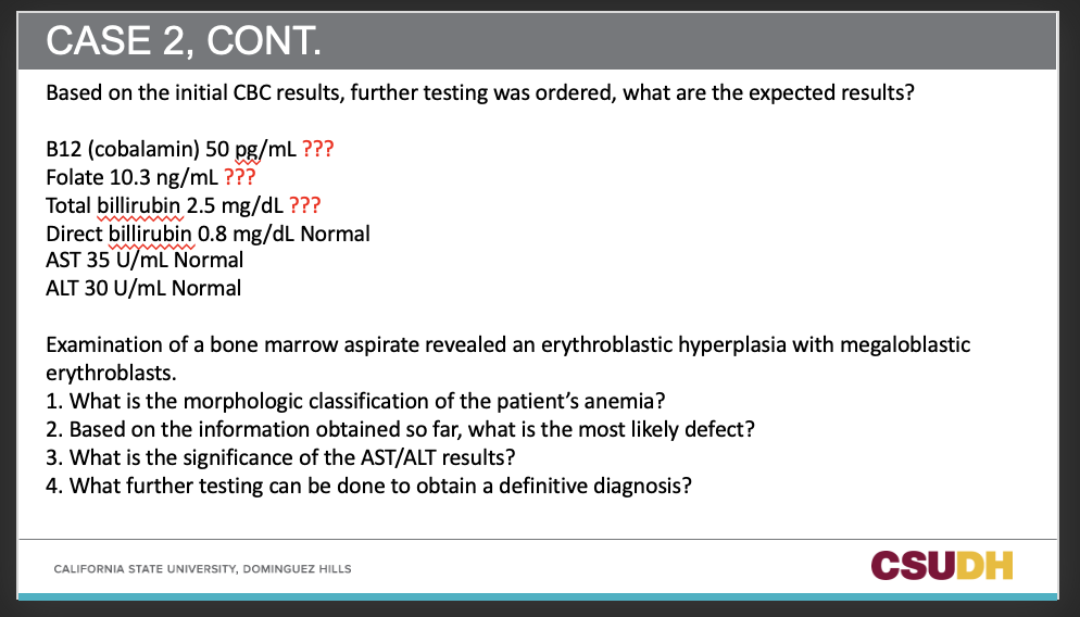
2. Based on the information obtained so far, what is the most likely defect?
3. What is the significance of the AST/ALT results?
4. What further testing can be done to obtain a definitive diagnosis?
Answers
Q1: What is the morphologic classification of the patient’s anemia? A: Macrocytic, megaloblastic anemia.
Q2: Based on the information obtained so far, what is the most likely defect? A: Vitamin B12 deficiency (not folate, since folate is normal).
Q3: What is the significance of the AST/ALT results? A: They are normal → excludes liver disease as the cause of macrocytosis.; prehepatic case
Q4: What further testing can be done to obtain a definitive diagnosis? A: MMA (to confirm vitamin b12 deficiency) , antibody assay, Schilling test.
Q1: What is the morphologic classification of the patient’s anemia? A: Macrocytic, megaloblastic anemia.
Q2: Based on the information obtained so far, what is the most likely defect? A: Vitamin B12 deficiency (not folate, since folate is normal).
Q3: What is the significance of the AST/ALT results? A: They are normal → excludes liver disease as the cause of macrocytosis.; prehepatic case
Q4: What further testing can be done to obtain a definitive diagnosis? A: MMA (to confirm vitamin b12 deficiency) , antibody assay, Schilling test.
97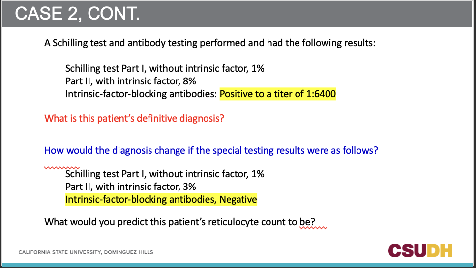
Q5: What is this patient’s definitive diagnosis?
A: Pernicious anemia (Vitamin B12 deficiency due to intrinsic factor-blocking antibodies). Titer is high!!
Q6: How would the diagnosis change if special testing showed:
Schilling test with IF improves absorption (3%),
Anti-IF antibodies negative?** A: Suggests malabsorption cause (e.g., ileal disease, bacterial overgrowth, or Diphyllobothrium latum) rather than pernicious anemia.
Q7: What would you predict this patient’s reticulocyte count to be? A: Low to inappropriately normal, because marrow is ineffective (megaloblastic anemia has poor retic response).
Q6: How would the diagnosis change if special testing showed:
Schilling test with IF improves absorption (3%),
Anti-IF antibodies negative?** A: Suggests malabsorption cause (e.g., ileal disease, bacterial overgrowth, or Diphyllobothrium latum) rather than pernicious anemia.
Q7: What would you predict this patient’s reticulocyte count to be? A: Low to inappropriately normal, because marrow is ineffective (megaloblastic anemia has poor retic response).
98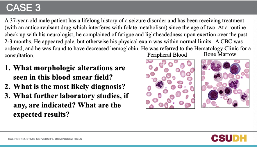
1. The blood smear likely shows macro-ovalocytes, hypersegmented neutrophils, and RBC anisocytosis, possibly with macrocytosis. Basophilic stippling is not typical for folate deficiency; it’s more associated with lead poisoning or thalassemia.
2. The diagnosis: Megaloblastic anemia from folate deficiency, likely due to anticonvulsant therapy (phenytoin).
3. Further labs: CBC would show increased MCV (>100 fL), elevated MCH, RDW raised, and a low or normal reticulocyte count.
2. The diagnosis: Megaloblastic anemia from folate deficiency, likely due to anticonvulsant therapy (phenytoin).
3. Further labs: CBC would show increased MCV (>100 fL), elevated MCH, RDW raised, and a low or normal reticulocyte count.
99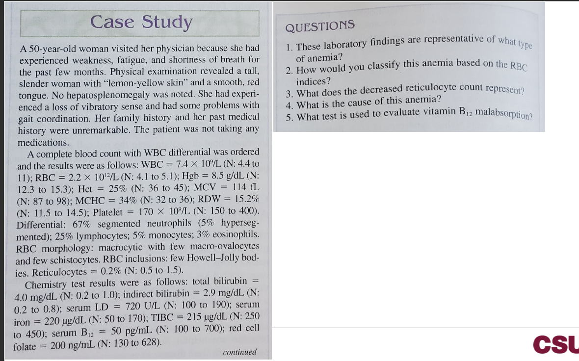
1. Type of anemia:
Megaloblastic anemia (vitamin B₁₂ deficiency picture) with ineffective erythropoiesis.
2. Classification by RBC indices:
Macrocytic, normochromic anemia (MCV 114 fL; MCHC normal; RDW ↑).
3. Meaning of the low reticulocyte count (0.2%):
Poor marrow response / decreased RBC production due to ineffective erythropoiesis (intramedullary destruction typical of megaloblastic states).
4. Cause of this anemia:
Vitamin B₁₂ deficiency, most likely pernicious anemia (autoimmune intrinsic-factor deficiency causing malabsorption), supported by neurologic signs (loss of vibration sense, gait issues), glossitis, ↑ LDH and indirect bilirubin.
5. Test to evaluate vitamin B₁₂ malabsorption:
Schilling test (historical “gold standard”: radiolabeled B₁₂ with/without intrinsic factor).
Modern practice also uses intrinsic-factor antibody testing ± parietal cell antibodies, and MMA/homocysteine levels.
Megaloblastic anemia (vitamin B₁₂ deficiency picture) with ineffective erythropoiesis.
2. Classification by RBC indices:
Macrocytic, normochromic anemia (MCV 114 fL; MCHC normal; RDW ↑).
3. Meaning of the low reticulocyte count (0.2%):
Poor marrow response / decreased RBC production due to ineffective erythropoiesis (intramedullary destruction typical of megaloblastic states).
4. Cause of this anemia:
Vitamin B₁₂ deficiency, most likely pernicious anemia (autoimmune intrinsic-factor deficiency causing malabsorption), supported by neurologic signs (loss of vibration sense, gait issues), glossitis, ↑ LDH and indirect bilirubin.
5. Test to evaluate vitamin B₁₂ malabsorption:
Schilling test (historical “gold standard”: radiolabeled B₁₂ with/without intrinsic factor).
Modern practice also uses intrinsic-factor antibody testing ± parietal cell antibodies, and MMA/homocysteine levels.