Wbc Lecture 5 Chronic Lymphoproliferative/Lymphoma Multiple Myeloma/ Plasma Cell Disorders
Study these flashcardsFree · spaced repetition · iPhone, Android, Mac & web
All cards
1
What are the classifications under Mature B cell neoplasms according to the WHO Classification of Mature Lymphoid Neoplasms?
- Chronic lymphocytic leukemia
- B cell prolymphocytic leukemia (B cell PLL)
- Hairy cell leukemia (HCL)
- Waldenström’s macroglobulinemia (WM)
- Monoclonal gammopathy of undetermined significance (MGUS)
- Plasma cell myeloma (aka MM)
- Follicular lymphoma
- Mantle cell lymphoma
- Burkitt lymphoma
- B cell prolymphocytic leukemia (B cell PLL)
- Hairy cell leukemia (HCL)
- Waldenström’s macroglobulinemia (WM)
- Monoclonal gammopathy of undetermined significance (MGUS)
- Plasma cell myeloma (aka MM)
- Follicular lymphoma
- Mantle cell lymphoma
- Burkitt lymphoma
2
What are the classifications under Mature T and Natural Killer (NK) Neoplasms according to the WHO Classification of Mature Lymphoid Neoplasms?
- Adult T cell leukemia/lymphoma
- Mycosis fungoides
- Sézary syndrome
- Mycosis fungoides
- Sézary syndrome
3
What is the final category listed in the WHO Classification of Mature Lymphoid Neoplasms?
Hodgkin Lymphoma
4
What does CBC reveal in lab investigation of lymphoid neoplasms?
Normal lymphocyte count or automated diff, WHICH does not exclude lymphoid neoplasm
5
What method is used for Immunophenotyping in lab investigation of lymphoid neoplasms?
Flow cytometry
6
What studies are included under .Cytogenetics in lab investigation of lymphoid neoplasms?
(karyotyping) and fluorescent in situ hybridization (FISH)
7
What biochemical analyses are performed in lab investigation of lymphoid neoplasms?
Serum Cr,
uric acid,
SPE,
retic count, DAT and haptoglobin in CLL presenting with anemia, LDH and β2-MG as indicators of tumor load,
ESR in Hodgkin lymphoma (high ESR predictive of early relapse and decreased survival)
uric acid,
SPE,
retic count, DAT and haptoglobin in CLL presenting with anemia, LDH and β2-MG as indicators of tumor load,
ESR in Hodgkin lymphoma (high ESR predictive of early relapse and decreased survival)
8
Pros and cons of periphereal blood investigation.
Peripheral blood is easy
but less informative, biopsy from lymph nodes more informative
but less informative, biopsy from lymph nodes more informative
9
What is considered the gold standard for lymphoma diagnosis?
Excisional biopsy (gold standard),
but fine needle aspiration (FNA) has become popular due to the ease with which specimens can be obtained
but fine needle aspiration (FNA) has become popular due to the ease with which specimens can be obtained
10
What imaging studies may be needed in lab investigation of lymphoid neoplasms?
Imaging studies may be needed (CT scan)
11
What genetic abnormality is associated with Chronic lymphocytic leukemia (CLL)?
Chronic lymphocytic leukemia (CLL): 13q14 del
12
What genetic abnormality is associated with Prolymphocytic leukemia (PLL)?
Prolymphocytic leukemia (PLL): t(8;14), MYC
13
What mutation is associated with Hairy cell leukemia (HCL)?
Hairy cell leukemia (HCL): BRAF V(600)E
14
What are the four types of leukemias classified under Mature lymphoid neoplasms?
Chronic lymphocytic leukemia (CLL),
Prolymphocytic leukemia (PLL),
Hairy cell leukemia (HCL),
Large granular lymphocytic (LGL) leukemia
Prolymphocytic leukemia (PLL),
Hairy cell leukemia (HCL),
Large granular lymphocytic (LGL) leukemia
15
What is the most common leukemia in adults?
Chronic lymphocytic leukemia (CLL)
16
What type of disorder is CLL?
B cell disorder
17
What is the median age of patients with CLL?
72
18
What percentage of lymphocytes appear small and mature in typical CLL?
More than 85%
19
Describe the appearance of lymphocytes in typical CLL.
In typical CLL: >85% of the lymphocytes appear as small and mature with scant cytoplasm and a dense nucleus with a condensed chromatin pattern without a defined nucleolus (“cobblestone” or soccer balll appearance)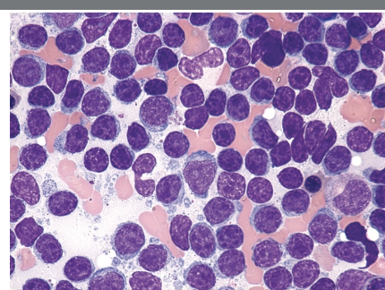
20
What percentage of CLL cases are typical versus atypical?
80% cases are typical CLL, and 20% atypical CLL
21
What are smudge cells and why are they important in CLL?
Smudge cells are very important cells that must be counted as lymphocytes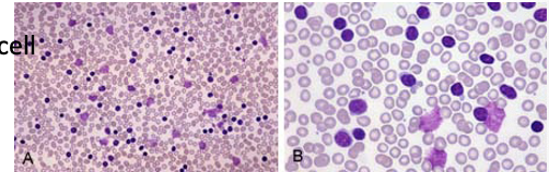
22
What are the immunophenotyping markers for CLL?
CD19, CD20, CD23, and T cell marker CD5 (aberrant)
CD 38 AND ZAP70
CD 38 AND ZAP70
23
What is the Catovsky-Matutes Scoring System used for?
Diagnosis of Chronic Lymphocytic Leukemia (CLL)
24
How many points are assigned for each feature in the Catovsky-Matutes scoring system and what are they?
Score 1 point for each of following:
- Weak expression of surface immunoglobulin
- Expression of CD5
- Expression of CD23
- No expression of FMC7
- Absent or weak expression of CD79b or CD22
- Weak expression of surface immunoglobulin
- Expression of CD5
- Expression of CD23
- No expression of FMC7
- Absent or weak expression of CD79b or CD22
25
What total score is consistent with a diagnosis of CLL?
Total score greater than or equal to 4 is consistent with CLL.
26
What do low scores of 1 or 2 suggest in the Catovsky-Matutes scoring system?
Low scores of 1 or 2 suggest Non-Hodgkin lymphoma (NHL)
27
What does an intermediate score of 3 indicate in the Catovsky-Matutes scoring system?
An intermediate score of 3 remains problematic,
and
these cases may require biopsy of a node or molecular data to establish the diagnosis
and
these cases may require biopsy of a node or molecular data to establish the diagnosis
28
Which abnormalities are considered the worst prognostic indicators, drug resistance in CLL?
Del(17p) and TP53 are the worst prognostic indicators
29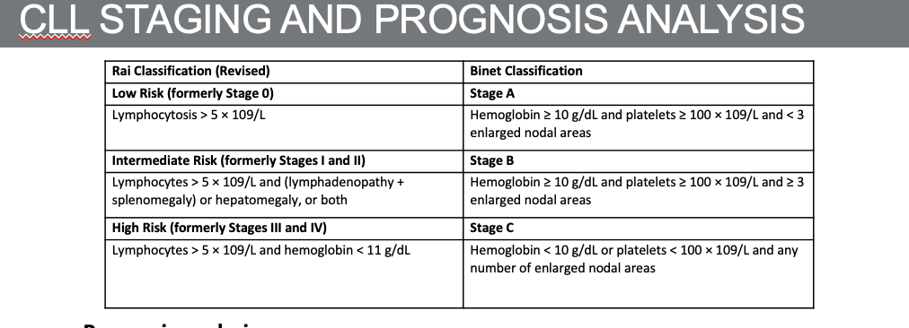
30
What is the prognostic significance of IGHV mutational status in CLL?
Mutational status of the heavy chain immunoglobulin gene (IGHV) is a strong prognostic factor.
No mutation is associated with pre-germinal center type of CLL and more aggressive clinical course.
Mutated IGHV indicates better prognosis and better response to drug treatment.
No mutation is associated with pre-germinal center type of CLL and more aggressive clinical course.
Mutated IGHV indicates better prognosis and better response to drug treatment.
31
Which additional markers are tested for correlation with IGHV mutational status?
Testing for ZAP70 and CD38: correlated with IGHV mutational status.
32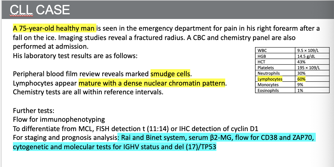
33
What is Prolymphocytic Leukemia (PLL)?
A rare and very aggressive leukemia with a unique clinical course
34
What is the median age for Prolymphocytic Leukemia (PLL)?
70
35
What percentage of Prolymphocytic Leukemia cases are B cell type?
More than 75%
36
What are key clinical features of Prolymphocytic Leukemia (PLL)?
Very aggressive, massive splenomegaly, marked absolute lymphocytosis (>100 × 10⁹/L)
37
What laboratory clue may indicate a diagnosis other than CLL in PLL?
A rapidly rising white blood cell (WBC) count may be a clue that the diagnosis is not CLL!!!
38
Describe the typical cell morphology seen in PLL.
Medium to large lymphocytes with ample cytoplasm, a more open chromatin pattern, and a prominent nucleolus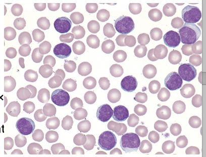
39
What are the immunophenotypic findings in PLL?
CD7 is positive, and inv(14) is present in approximately 70% of patients (using FISH)
40
What findings can be observed on the hematology analyzer in PLL?
Increased WBC, increased lymphocytes, but blast or atypical/variant lymph flag, PBS exam is a must!
41
What type of disease is Hairy Cell Leukemia (HCL)?
An indolent disease of B cell lineage most commonly found in middle age (median age, 50 years), mostly in males
42
Where is the major focus of disease in Hairy Cell Leukemia (HCL)?
In the spleen, blood, and bone marrow with splenomegaly and cytopenias
43
Describe the characteristic appearance of hairy cells in HCL.
Characteristic hairy cells have round to ovoid nuclei, lack nucleoli, and have relatively abundant cytoplasm with ragged projections that extend circumferentially around the entire cell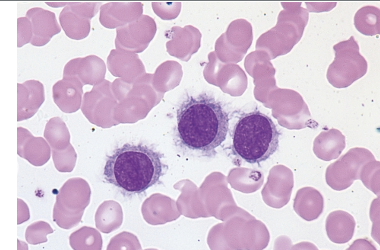
44
What causes a “dry tap” in Hairy Cell Leukemia?
Fibrotic bone marrow due to fibrosis, requiring a biopsy
45
What diagnostic staining tests are specific for Hairy Cell Leukemia (HCL)?
Monoclonal anti-CD20 and Annexin A (specific to HCL) using Immunohistochemistry
46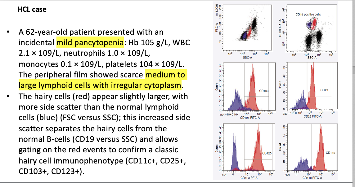
47
What is Large Granular Lymphocytic (LGL) Leukemia?
A rare disease characterized by an increase in large granular lymphocytes (>2 × 10⁹/L)
48
What age group is most commonly affected by LGL Leukemia?
Older adults with a median age of 60 years
49
What hematology analyzer finding may indicate LGL Leukemia?
Variant or atypical lymphocyte flag or blast flag on the hematology analyzer
50
What markers are expressed in T cell LGL Leukemia?
CD3, CD8, and CD57 (clonal expansion of cytotoxic T cells)
51
What markers are expressed in NK cell LGL Leukemia?
CD3 negative but CD56 positive
52
What virus is Adult T Cell Leukemia/Lymphoma (ATLL) associated with?
Associated with retroviral infection by the human T lymphotropic virus type 1 (HTLV-1)
53
Where is HTLV-1 endemic?
HTLV-1 is endemic in Japan, the Caribbean islands, Africa, South America, the Middle East, and northern Oceania
54
How is HTLV-1 transmitted?
The virus is transmitted via placental circulation, breastfeeding, blood transfusion, or sex.
55
What is the estimated risk of developing ATLL among HTLV-1 carriers?
Approximately 2.5% (women) to 5% (men) over a latency period of 55 to 70 years
56
What hematologic finding is characteristic of the leukemic phase of ATLL?
Marked leukocytosis in the leukemic phase of ATLL
57
Describe the morphology of ATLL cells.
ATLL cells are medium to large in size and have accentuated, convoluted nuclei, coarsely clumped chromatin, and deeply basophilic cytoplasm (aka “flower cell”)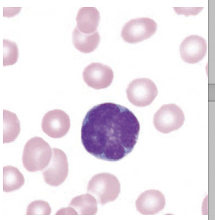
58
What is the typical immunophenotype of ATLL?
Generally consistent with T helper cells;
CD3 and CD4 are expressed,
whereas CD7 and CD8 are absent
CD3 and CD4 are expressed,
whereas CD7 and CD8 are absent
59
Which additional markers are highly expressed in ATLL?
CD25 and CCR4
60
What cytokine has prognostic significance in ATLL and can be used as a tumor marker?
The soluble form of interleukin 2 (IL-2R) has prognostic significance and can be used as a tumor marker for assessing disease status.
61
What type of cancer is Burkitt Lymphoma/Leukemia (BL)?
An aggressive cancer of mature B cells associated with a fulminant clinical presentation
62
How many subtypes of Burkitt Lymphoma exist and what are they?
Three subtypes — endemic form, sporadic form, and HIV form
63
Where is the endemic form of Burkitt Lymphoma primarily found?
The endemic form is found primarily in childhood in equatorial Africa.
64
What are the typical sites of extranodal involvement in the endemic form of Burkitt Lymphoma?
Extranodal involvement is common, with the orbits and mandible as typical
65
What viral genome is found in the neoplastic cells of endemic Burkitt Lymphoma?
the EBV genome has been found to be present in the neoplastic cells.
66
How does sporadic Burkitt Lymphoma typically present?
sporadic BL presents mostly as an abdominal disease, extranodal involvement is not uncommon.
67
What characterizes the HIV form of Burkitt Lymphoma?
Profound immunosuppression with blood and bone marrow as primary sites of disease
68
Describe the morphology of Burkitt Lymphoma cells.
Medium to large in size, finely clumped chromatin, deeply basophilic cytoplasm with distinct vacuoles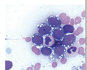
69
What is the classic biopsy appearance of Burkitt Lymphoma?
Both bone marrow and lymph node biopsies may show a classic “starry sky” appearance, which results from the interspersed tingible body macrophages suspended in a “sea” of malignant cells.
70
Which markers are Burkitt Lymphoma cells positive and negative for?
BL cells are positive for sIgG, CD10, and negative for CD34 and Tdt
71
What type of disorder is Follicular Lymphoma?
A neoplastic disorder of germinal B cells accounting for approximately 12% of NHL cases
72
What age group is most commonly affected by Follicular Lymphoma?
Generally, a disease of middle to older age groups.
73
How is the clinical course of Follicular Lymphoma typically described?
Indolent, but most often incurable with current therapies
74
What are common symptoms of Follicular Lymphoma?
Most patients are asymptomatic, sometimes present with painless lymphadenopathy
75
What percentage of Follicular Lymphoma cases show peripheral blood involvement?
10%
76
Describe the morphology of circulating FL cells.
circulating FL cells have a condensed chromatin pattern and distinct nuclear clefts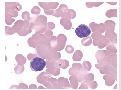
77
Can bone marrow be involved in Follicular Lymphoma?
Yes, bone marrow biopsy may reveal FL cells
78
What percentage of NHL cases does Mantle Cell Lymphoma (MCL) constitute?
Approximately 3% to 6% of NHL cases
79
What is the median age and predominant gender affected by Mantle Cell Lymphoma?
Median age 68, with male predominance
80
How is Mantle Cell Lymphoma clinically characterized?
Clinically aggressive with extensive lymphadenopathy
81
Why is diagnosis of MCL based on morphology challenging?
Diagnosis based on morphology is challenging:
- The small cell form of MCL can easily be mistaken for CLL
- A blastoid variant of MCL can be confused with ALL.
- In some cases, circulating mantle cells exhibit morphologic changes that can be confused with hairy cells, or prolymphocytes.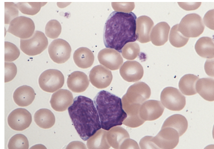
- The small cell form of MCL can easily be mistaken for CLL
- A blastoid variant of MCL can be confused with ALL.
- In some cases, circulating mantle cells exhibit morphologic changes that can be confused with hairy cells, or prolymphocytes.
82
What is required for an accurate diagnosis of Mantle Cell Lymphoma?
Demonstration of either t(11;14) via FISH or overexpression of cyclin D1 by immunohistochemistry
83
What is the most common form of cutaneous T cell lymphoma?
Mycosis Fungoides (MF) and Sézary Syndrome (SS)
84
Which population is most affected by MF/SS?
Elderly individuals, with male predominance
85
Which is more common, Mycosis Fungoides or Sézary Syndrome?
Mycosis Fungoides, accounting for 70% of cases and confined to the skin
86
Describe the appearance of malignant T cells in MF and SS.
Malignant T cells in MF and SS have an abnormal appearance with scant cytoplasm and a cerebriform, folded nucleus, variably condensed chromatin, and inconspicuous nucleoli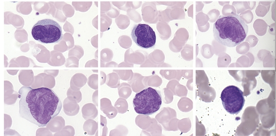
87
What type of T cells are involved in MF and SS?
CD4+ T helper cells
88
What are the characteristic features of Mycosis Fungoides?
Psoriatic-like skin lesions requiring a skin biopsy
89
What are the characteristic features of Sézary Syndrome?
Erythroderma, generalized lymphadenopathy, and presence of clonal T cells in skin, lymph nodes, peripheral blood and severe pruritus
90
What type of cells do plasma cell neoplasms originate from?
Neoplasms of terminally differentiated B cells
91
What is the prototype of plasma cell neoplasms?
Multiple myeloma (plasma cell myeloma), median age 69
92
What is Monoclonal Gammopathy of Undetermined Significance (MGUS)?
The precursor state of multiple myeloma; M spike in SPE helps predict the risk for MM transformation
93
Where is multiple myeloma (MM) primarily confined to, and what is rare in this condition?
Confined to the bone marrow; plasma cell leukemia (PCL) is rare (>20% plasma cells in peripheral blood)
94
What causes RBC rouleaux formation in plasma cell neoplasms?
Paraprotein in plasma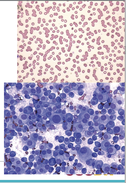
95
How can MM cells be differentiated from reactive plasma cells?
By immunohistochemistry (IHC) with CD138 or kappa and lambda clonality
96
Which markers do MM cells strongly express?
CD38 and CD138, and CD56 (aberrant)
97
What type of lymphoma is Waldenström’s Macroglobulinemia (WM)?
A low-grade lymphoplasmacytic lymphoma associated with aberrant secretion of IgM
98
What complication can high levels of IgM cause in Waldenström’s Macroglobulinemia?
Hyperviscosity syndrome requiring emergent plasmapheresis to alleviate symptoms
99
What sites can show extramedullary involvement in Waldenström’s Macroglobulinemia?
Extramedullary involvement includes nodal disease and hepatosplenomegaly.
100
What gene mutation is commonly found in Waldenström’s Macroglobulinemia?
Somatic mutation in myeloid differentiation factor 88 (MYD88) gene
101
What percentage of patients with Waldenström’s Macroglobulinemia have a MYD88 mutation?
Over 90% of patients
102
What type of lymphoma is Hodgkin Lymphoma (HL)?
A nodal-based lymphoma
103
What are the two types of Hodgkin Lymphoma?
Classic and lymphocyte-predominant
104
How is the diagnosis of Hodgkin Lymphoma made?
Always based on node biopsy
105
What percentage of HL cases are Classic HL and what group is commonly affected?
More than 85% of cases, usually in young adults
106
How does Classic HL typically respond to treatment?
Responds well to chemotherapy
107
What is the characteristic cell type seen in Classic Hodgkin Lymphoma?
Characterized by Reed-Sternberg (RS) cells (binucleated or multinucleated with abundant cytoplasm and distinct nuclei)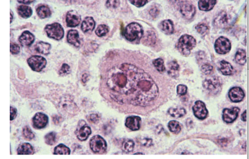
108
What is the origin and immunophenotype of RS cells in Classic HL?
Of B cell origin (but do not express most B cell markers); CD30 and CD15 positive; never seen in peripheral blood
109
What is the malignant cell in Lymphocyte-predominant Hodgkin Lymphoma?
No RS cells; the malignant cell is a lymphocytic histiocytic (L&H) cell, also known as “popcorn” cells because of their distinct morphology.
110
What is the immunophenotype of popcorn cells in Lymphocyte-predominant HL?
Negative for CD15 and CD30 expression