Traumatic Brain Injury (TBI)
Study these flashcardsFree · spaced repetition · iPhone, Android, Mac & web
All cards
1
Traumatic brain injury (TBI)
is a nondegenerative, noncongenital insult to the brain from an external mechanical
force, possibly leading to permanent or temporary impairment of cognitive, physical, and psychosocial functions, with an associated diminished or altered state of consciousness.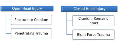
force, possibly leading to permanent or temporary impairment of cognitive, physical, and psychosocial functions, with an associated diminished or altered state of consciousness.
2
What is the most common cause of TBI in all age groups?
Fall
3
TBI Epidemiology
• US Annual Incidence: 180-200 cases/100,000
• Approximately 600,000 new cases each year
• Nearly 550,000 patients hospitalized
• Approximately 75-80% cases are mild
• Up to 10% of cases are fatal
• High risk of permanent disability:
• Nearly 100% with severe TBI
• Up to 2/3 with moderate TBI
• Annual direct cost of care: \$25 billion
• Approximately 600,000 new cases each year
• Nearly 550,000 patients hospitalized
• Approximately 75-80% cases are mild
• Up to 10% of cases are fatal
• High risk of permanent disability:
• Nearly 100% with severe TBI
• Up to 2/3 with moderate TBI
• Annual direct cost of care: \$25 billion
4
Consequences of TBI
Cognitive:
• Lack of concentration
• Limited attention span
• Short term memory loss
• Trouble with planning, writing, reading, making judgments
Physical:
• Speech difficulties
• Loss of vision, hearing
• Loss of balance, coordination
Psychosocial:
• Depression
• Anxiety
• Mood swings
• Lack of concentration
• Limited attention span
• Short term memory loss
• Trouble with planning, writing, reading, making judgments
Physical:
• Speech difficulties
• Loss of vision, hearing
• Loss of balance, coordination
Psychosocial:
• Depression
• Anxiety
• Mood swings
5
Cerebrum
Memory, problem solving, thinking, feeling, movement
6
Cerebellum
Coordination, balance
7
Brain Stem
Autonomic functions (breathing, digestion, HR, BP)
8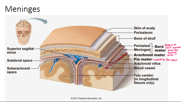
Image occlusion: name the hidden labels
9
Image occlusion: name the hidden labels
10
Image occlusion: name the hidden labels
11
Image occlusion: name the hidden labels
12
Image occlusion: name the hidden labels
13
Image occlusion: name the hidden labels
14
Image occlusion: name the hidden labels
15
Dural Folds
The dural folds are created by the meningeal layer of the dura mater and divide the cranial cavity into freely communicating spaces. The function of these folds is to limit the rotational displacement of the brain. Some examples of dural folds include the falx cerebri, tentorium cerebelli, falx cerebelli, superior sagittal sinus, and the inferior sagittal sinus.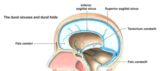
16
Brain Herniation Patterns
Brain herniation occurs when brain tissue is displaced due to increased pressure within the skull. There are several types of brain herniation patterns, including subfalcine herniation where the brain tissue moves underneath a membrane known as the falx cerebri in the middle of the brain. Another type is transtentorial herniation which can be further broken down into two types: descending transtentorial or uncal. Cerebellar tonsillar is another type of brain herniation.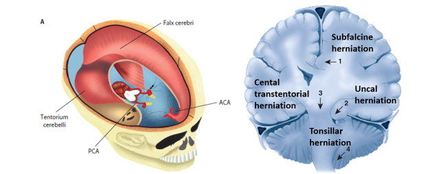
17
Monro-Kellie Doctrine
• The cranial vault is a closed, fixed-space (“bony box”)
• Intracranial volume (constant) = brain tissue (~85-90%) + blood
(~10%) + CSF (<3%)
• Fluid components are nearly non-compressible
• Only a small amount of compensation can occur
• Intracranial Compliance = Change in volume / change in pressure
• Once the cranial vault reaches capacity, ICP rises dramatically
• Brain tissue may be compressed à further injury or death
• Increased ICP can then lead to interruption of cerebral blood flow (CBF) by reducing the CPP
• CPP = Mean Arterial Pressure (MAP) – ICP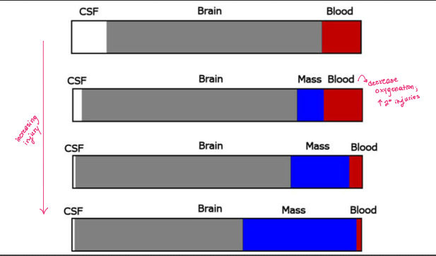
• Intracranial volume (constant) = brain tissue (~85-90%) + blood
(~10%) + CSF (<3%)
• Fluid components are nearly non-compressible
• Only a small amount of compensation can occur
• Intracranial Compliance = Change in volume / change in pressure
• Once the cranial vault reaches capacity, ICP rises dramatically
• Brain tissue may be compressed à further injury or death
• Increased ICP can then lead to interruption of cerebral blood flow (CBF) by reducing the CPP
• CPP = Mean Arterial Pressure (MAP) – ICP
18
Critical Volume
The critical volume refers to the maximum volume that can be added to the intracranial vault before it causes an increase in intracranial pressure. Since the skull is a rigid compartment, any addition of volume to the intracranial vault can cause an increase in ICP. Hence all ICP therapies are directed toward reducing intracranial volume.
19
Primary Brain Injury
• Instantaneous effects caused by external transfer of kinetic energy to various structural components of the brain:
• Neurons
• Nerve synapses
• Glial cells
• Axons
• Cerebral blood vessels
• Biochemical forces --> primary brain injury:
• Contact (e.g., blunt-object blow, penetrating-missile injuries)
• Acceleration/Deceleration (e.g., instantaneous brain movements following motor vehicle accidents)
• Focal versus Diffuse
• Neurons
• Nerve synapses
• Glial cells
• Axons
• Cerebral blood vessels
• Biochemical forces --> primary brain injury:
• Contact (e.g., blunt-object blow, penetrating-missile injuries)
• Acceleration/Deceleration (e.g., instantaneous brain movements following motor vehicle accidents)
• Focal versus Diffuse
20
Secondary Brain Injury
Key pathophysiologic trigger = cerebral ischemia
May occur minutes, hours, or days after initial injury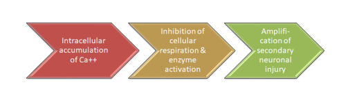
May occur minutes, hours, or days after initial injury
21
Glasgow Coma Scale (GCS)
• Most widely used system to grade arousal and functional capacity of
the cerebral cortex
• Evaluates consciousness according to:
• Eye Opening
• Motor Response
• Verbal Response
• Maximum score: 15
Consider effects of ethanol/drug intoxication, hypotension,
hypoxia, hypothermia, and post-ictal state.
the cerebral cortex
• Evaluates consciousness according to:
• Eye Opening
• Motor Response
• Verbal Response
• Maximum score: 15
Consider effects of ethanol/drug intoxication, hypotension,
hypoxia, hypothermia, and post-ictal state.
22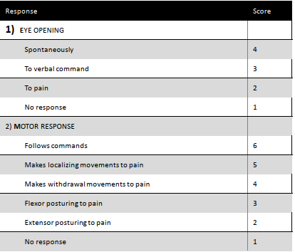
Image occlusion: name the hidden labels
23
Image occlusion: name the hidden labels
24
Image occlusion: name the hidden labels
25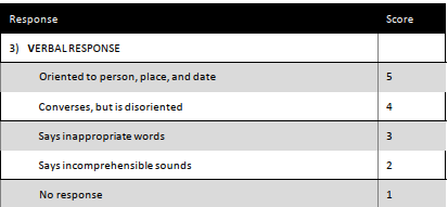
Image occlusion: name the hidden labels
26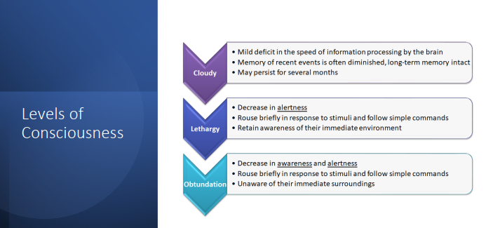
Image occlusion: name the hidden labels
27
Image occlusion: name the hidden labels
28
Image occlusion: name the hidden labels
29
Image occlusion: name the hidden labels
30
Image occlusion: name the hidden labels
31
Image occlusion: name the hidden labels
32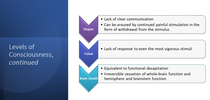
Image occlusion: name the hidden labels
33
Image occlusion: name the hidden labels
34
Image occlusion: name the hidden labels
35
Image occlusion: name the hidden labels
36
Image occlusion: name the hidden labels
37
Image occlusion: name the hidden labels
38
Goals of Acute Management
• Overall Goals:
• Reduce morbidity & mortality
• Optimize long-term functional outcomes
• Short-Term Goals:
• Establishment of an adequate airway and maintenance of ventilation/circulation
• Maintenance of balance between cerebral oxygen delivery (CDo2) and cerebral metabolic rate (CMRo2)
• Prevention/attenuation of secondary neuronal injury
• Prevention and/or treatment of associated medical complications
• Reduce morbidity & mortality
• Optimize long-term functional outcomes
• Short-Term Goals:
• Establishment of an adequate airway and maintenance of ventilation/circulation
• Maintenance of balance between cerebral oxygen delivery (CDo2) and cerebral metabolic rate (CMRo2)
• Prevention/attenuation of secondary neuronal injury
• Prevention and/or treatment of associated medical complications
39
Threshold Values
• Blood Pressure: correct & prevent hypotension (SBP < 90 mm Hg)
• General target: 120-140 mmHg
• 15 to 49 years old: Maintain SBP at ≥110 mm Hg
• 50 to 69 years old: Maintain SBP at ≥100 mm Hg
• ICP: maintain < 22 mm Hg
• NOTE: Normal ICP = 5 to 15 mm Hg
• CPP:
• Adults: maintain between 50-70 mm Hg
• Pediatrics: maintain > 40 mm Hg
• Perfusion: correct & prevent hypoxemia (PaO2 < 60 mm Hg)
• Arterial oxygen saturation: maintain > 90%
• General target: 120-140 mmHg
• 15 to 49 years old: Maintain SBP at ≥110 mm Hg
• 50 to 69 years old: Maintain SBP at ≥100 mm Hg
• ICP: maintain < 22 mm Hg
• NOTE: Normal ICP = 5 to 15 mm Hg
• CPP:
• Adults: maintain between 50-70 mm Hg
• Pediatrics: maintain > 40 mm Hg
• Perfusion: correct & prevent hypoxemia (PaO2 < 60 mm Hg)
• Arterial oxygen saturation: maintain > 90%
40
Multimodal Monitoring
• Detect early worsening before irreversible brain damage occurs
• Individualize patient care decisions
• Guide patient management
• Monitor response to treatment
• Improve understanding of complex disorders
• Develop new treatments
• Individualize patient care decisions
• Guide patient management
• Monitor response to treatment
• Improve understanding of complex disorders
• Develop new treatments
41
Types of Multimodal Monitoring
• Clinical Evaluation
• Intracranial Pressure (ICP)
• Cerebral Perfusion Pressure (CPP)
• Hemodynamic Monitoring
• Cerebral Autoregulation
• Cerebral Blood Flow
• Electrophysiology
• Cerebral Metabolism
• Intracranial Pressure (ICP)
• Cerebral Perfusion Pressure (CPP)
• Hemodynamic Monitoring
• Cerebral Autoregulation
• Cerebral Blood Flow
• Electrophysiology
• Cerebral Metabolism
42
Steps of Treatment
1. Initial Resuscitation (focus on SBP)
• Airway establishment
• Restoration and maintenance of systolic blood pressure (SBP)
2. Postresuscitative Care (focus on ICP)
• Diagnostic evaluation of intracranial and extracranial injuries
• Emergent surgical intervention as needed.
• Airway establishment
• Restoration and maintenance of systolic blood pressure (SBP)
2. Postresuscitative Care (focus on ICP)
• Diagnostic evaluation of intracranial and extracranial injuries
• Emergent surgical intervention as needed.
43
Initial Resuscitation
• Establish airway:
• Immobilize spine
• Intubate
• Oxygenate
• Elevate head of bed (30 degrees)
• Start fluid resuscitation as needed - initial resuscitation fluids of choice:
• Isotonic saline (0.9% normal saline)
• Lactated Ringer’s solution
• Maintain MAP:
• Vasopressors (e.g., norepinephrine, phenylephrine, dopamine)
• Inotropic agents
• Immobilize spine
• Intubate
• Oxygenate
• Elevate head of bed (30 degrees)
• Start fluid resuscitation as needed - initial resuscitation fluids of choice:
• Isotonic saline (0.9% normal saline)
• Lactated Ringer’s solution
• Maintain MAP:
• Vasopressors (e.g., norepinephrine, phenylephrine, dopamine)
• Inotropic agents
44
Postresuscitative Care
• Evaluate for and start surgery (as indicated)
• Insert ICP monitor or extraventricular drain
• Monitor and manage threshold values:
• Mechanism ventilation
• Maintain CPP 60-70 mmHg (fluids ± vasopressors)
• Treat and prevent ICP > 22 mmHg
• Prevent complications:
• Seizure treatment and prophylaxis
• Stress ulcer and venous thromboembolism (VTE) prophylaxis
• Insert ICP monitor or extraventricular drain
• Monitor and manage threshold values:
• Mechanism ventilation
• Maintain CPP 60-70 mmHg (fluids ± vasopressors)
• Treat and prevent ICP > 22 mmHg
• Prevent complications:
• Seizure treatment and prophylaxis
• Stress ulcer and venous thromboembolism (VTE) prophylaxis
45
ICP Monitoring
• Normal ICP = 5 to 15 mm Hg
• Indications for ICP Monitoring (2007 guidelines)
• Severe TBI (GCS ≤ 8) with abnormal CT scan
• Severe TBI (GCS ≤ 8) with normal CT scan and at least 2 of the following:
• Age > 40 years old
• Unilateral or bilateral motor posturing
• SBP < 90 mm Hg
• ICP Monitoring Tools
• ICP Bolt
• External Ventricular Catheter (EVD)
• Indications for ICP Monitoring (2007 guidelines)
• Severe TBI (GCS ≤ 8) with abnormal CT scan
• Severe TBI (GCS ≤ 8) with normal CT scan and at least 2 of the following:
• Age > 40 years old
• Unilateral or bilateral motor posturing
• SBP < 90 mm Hg
• ICP Monitoring Tools
• ICP Bolt
• External Ventricular Catheter (EVD)
46
Pharmacological Treatment of Elevated ICP
Hyperosmolar (osmotic) therapy
• Mannitol
• Hypertonic Saline
Sedation / Pain management
• Fentanyl
• Propofol
Paralytics
• Cisatracurium
• Atracurium
Barbiturate Coma
• Pentobarbital
• Mannitol
• Hypertonic Saline
Sedation / Pain management
• Fentanyl
• Propofol
Paralytics
• Cisatracurium
• Atracurium
Barbiturate Coma
• Pentobarbital
47
Hyperosmolar Therapy
• Basic Principle: draw fluid from the cranial space -> decrease brain
volume -> decreased ICP
• Serum osmolarity = [(2 x Na) + (BUN/2.8) + (glucose/18)]
• Normal serum osmolality = 275-290 mOsm/L
• Target serum osmolality = 300-320 mOsm/L
• Options:
• Mannitol
• Hypertonic Saline (HTS)
volume -> decreased ICP
• Serum osmolarity = [(2 x Na) + (BUN/2.8) + (glucose/18)]
• Normal serum osmolality = 275-290 mOsm/L
• Target serum osmolality = 300-320 mOsm/L
• Options:
• Mannitol
• Hypertonic Saline (HTS)
48
Mannitol
• Dehydrated sugar alcohol
• Provides a large osmotic load causing diuretic effects
• Recommended dose: 0.25-1 g/kg given over 20 minutes every 2-4 hours
• Effects may only last for a short period of time
• May be repeated as needed to reduce ICP
• Monitor serum osmolality every 4-6 hours
• Hold if serum osmolality > 320 mOsm/L
• Hold if serum sodium > 145 mEq/L
• Possible dehydration and renal failure
• Provides a large osmotic load causing diuretic effects
• Recommended dose: 0.25-1 g/kg given over 20 minutes every 2-4 hours
• Effects may only last for a short period of time
• May be repeated as needed to reduce ICP
• Monitor serum osmolality every 4-6 hours
• Hold if serum osmolality > 320 mOsm/L
• Hold if serum sodium > 145 mEq/L
• Possible dehydration and renal failure
49
HTS
• Varied concentrations used in practice (2% - 23.4%)
• Concentrations of 3% or greater require central venous catheter for administration
• Similar MOA to mannitol, except it uses serum sodium as a driver of osmolarity
• Establishes a gradient across an intact blood brain barrier
• Goal Serum Sodium = 145-155 mmol/L
• May be administered as bolus or continuous infusion
• Should not be abruptly discontinued
• High alert medication in hospital settings
• Concentrations of 3% or greater require central venous catheter for administration
• Similar MOA to mannitol, except it uses serum sodium as a driver of osmolarity
• Establishes a gradient across an intact blood brain barrier
• Goal Serum Sodium = 145-155 mmol/L
• May be administered as bolus or continuous infusion
• Should not be abruptly discontinued
• High alert medication in hospital settings
50
Mannitol v. HTS
• Guidelines do not currently recommend one option over the other
• Clinical considerations:
• Does the patient have a central line?
• Is the patient hemodynamically unstable?
• Does the patient have renal impairment?
• Is the patient volume overloaded or dry?
• What is the most recent serum sodium and/or serum osmolarity?
• Clinical considerations:
• Does the patient have a central line?
• Is the patient hemodynamically unstable?
• Does the patient have renal impairment?
• Is the patient volume overloaded or dry?
• What is the most recent serum sodium and/or serum osmolarity?
51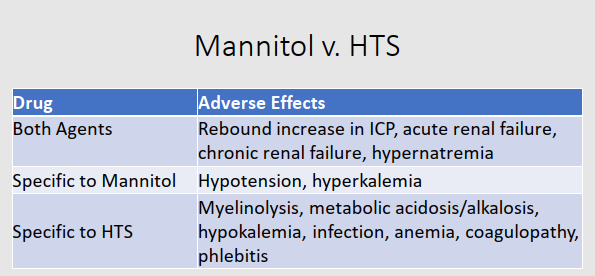
Image occlusion: name the hidden labels
52
Image occlusion: name the hidden labels
53
Image occlusion: name the hidden labels
54
Image occlusion: name the hidden labels
55
Image occlusion: name the hidden labels
56
Image occlusion: name the hidden labels
57
Sedatives & Analgesics
• Agitation & Pain cause Increased ICP
• Sedatives and pain medications may lower metabolic demands
• Short-acting agents are preferred
• Fentanyl
• Propofol (only if intubated)
• Maximum recommended dose of propofol is 50 mcg/kg/min
• Propofol also lowers ICP independent of agitation
• Risks:
• Hypotension (variable effect on CPP)
• Propofol-related infusion syndrome (PRIS) with high-dose prolonged infusion
• Sedatives and pain medications may lower metabolic demands
• Short-acting agents are preferred
• Fentanyl
• Propofol (only if intubated)
• Maximum recommended dose of propofol is 50 mcg/kg/min
• Propofol also lowers ICP independent of agitation
• Risks:
• Hypotension (variable effect on CPP)
• Propofol-related infusion syndrome (PRIS) with high-dose prolonged infusion
58
Paralytics
• Only considered after initial treatment options fail
• Patients MUST be intubated for airway protection with adequate
pain/sedative medications
• Intermittent boluses or continuous infusion
• Preferred agents in critically ill patients
• Cisatracurium
• Atracurium
• Side effects: hypotension, bradycardia
• Patients MUST be intubated for airway protection with adequate
pain/sedative medications
• Intermittent boluses or continuous infusion
• Preferred agents in critically ill patients
• Cisatracurium
• Atracurium
• Side effects: hypotension, bradycardia
59
Barbiturate Coma
• Used in management of super-refractory cases
• Suppresses cerebral metabolism and alters vascular tone → decreasing both oxygen demand and cerebral blood flow
• Pentobarbital Dosing:
• STEP 1: 10 mg/kg over 30 min
• STEP 2: 5 mg/kg every hour x 3 doses
• STEP 3: 1-2 mg/kg/hour infusion
• Titrated to burst suppression on EEG
• Pentobarbital steady state target: 30 to 40 mcg/mL
• Side effects: hypotension, myocardial depression, immune suppression
ileus with subsequent feeding intolerance
• Suppresses cerebral metabolism and alters vascular tone → decreasing both oxygen demand and cerebral blood flow
• Pentobarbital Dosing:
• STEP 1: 10 mg/kg over 30 min
• STEP 2: 5 mg/kg every hour x 3 doses
• STEP 3: 1-2 mg/kg/hour infusion
• Titrated to burst suppression on EEG
• Pentobarbital steady state target: 30 to 40 mcg/mL
• Side effects: hypotension, myocardial depression, immune suppression
ileus with subsequent feeding intolerance
60
Posttraumatic Seizures
• Phenytoin
• Fosphenytoin
• Carbamazepine
• Levetiracetam
• Fosphenytoin
• Carbamazepine
• Levetiracetam
61
Venous Thromboembolism (VTE)
• Mechanical Prophylaxis
• Heparin
• Enoxaparn
• Heparin
• Enoxaparn
62
Treatment of Active Posttraumatic Seizures
• Goals:
• Avoid increases in CMRo2
• Prevent status epilepticus
• Treatment of Active Seizures: Diazepam or Lorazepam
• Diazepam
• Adults: 5-40 mg IV
• Infants & Children: 0.1-0.5 mg/kg IV
• Lorazepam
• Adults: 2-8 mg IV
• Infants & Children: 0.03-0.1 mg/kg IV
• Prevention of Additional Seizures: Phenytoin or Fosphenytoin
• Loading Dose
• Adults: 15-20 mg/kg IV
• Children: 10-15 mg/kg IV
• Maintenance Dose: 4-6 mg/kg/day IV divided Q8H or Q12H
• Avoid increases in CMRo2
• Prevent status epilepticus
• Treatment of Active Seizures: Diazepam or Lorazepam
• Diazepam
• Adults: 5-40 mg IV
• Infants & Children: 0.1-0.5 mg/kg IV
• Lorazepam
• Adults: 2-8 mg IV
• Infants & Children: 0.03-0.1 mg/kg IV
• Prevention of Additional Seizures: Phenytoin or Fosphenytoin
• Loading Dose
• Adults: 15-20 mg/kg IV
• Children: 10-15 mg/kg IV
• Maintenance Dose: 4-6 mg/kg/day IV divided Q8H or Q12H
63
Posttraumatic Seizures Prophylaxis
• Risk factors for early posttraumatic seizures (less than 7 days after injury):
• GCS < 10
• Cortical contusion
• Depressed skull fracture
• Subdural hematoma
• Epidural hematoma
• Intracerebral hematoma
• Penetrating head wound
• Seizure within the first 24 hours of injury
• GCS < 10
• Cortical contusion
• Depressed skull fracture
• Subdural hematoma
• Epidural hematoma
• Intracerebral hematoma
• Penetrating head wound
• Seizure within the first 24 hours of injury
64
VTE Prophylaxis
• Pulmonary embolism is the 3rd leading cause of death in trauma patients who survive first 24 hours
• Severe TBI is an independent risk factor for deep vein thrombosis in poly-trauma patients:
• Primary brain injury
• Prolonged periods of immobilization
• Focal motor deficits
• Severe TBI is an independent risk factor for deep vein thrombosis in poly-trauma patients:
• Primary brain injury
• Prolonged periods of immobilization
• Focal motor deficits
65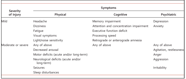
Image occlusion: name the hidden labels
66
Image occlusion: name the hidden labels
67
Image occlusion: name the hidden labels
68
Image occlusion: name the hidden labels
69
Image occlusion: name the hidden labels
70
Image occlusion: name the hidden labels
71
Rates of Psychiatric Disorders in Patients with TBI
MDD most common post-TBI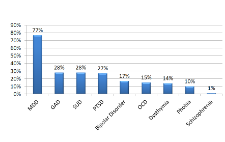
72
General Principles of Sequelae Management
Environmental and behavioral interventions are first-line
in the management of post-TBI behavioral disturbances
When using medications, first, do no harm
• Avoid medications that can:
• Interfere with neurologic recovery or engagement in care
• Lower the seizure threshold
• Cause confusion
• Avoid benzodiazepines, tricyclic antidepressants (TCAs), and monoamine oxidase inhibitors (MAOIs)
• Use antipsychotics, bupropion, and lithium sparingly
Give full therapeutic trials at maximum tolerated doses
Monitor closely for toxicity
Limit quantities of medications in patients at high risk of
suicide
in the management of post-TBI behavioral disturbances
When using medications, first, do no harm
• Avoid medications that can:
• Interfere with neurologic recovery or engagement in care
• Lower the seizure threshold
• Cause confusion
• Avoid benzodiazepines, tricyclic antidepressants (TCAs), and monoamine oxidase inhibitors (MAOIs)
• Use antipsychotics, bupropion, and lithium sparingly
Give full therapeutic trials at maximum tolerated doses
Monitor closely for toxicity
Limit quantities of medications in patients at high risk of
suicide
73
TBI and Agitation, Aggression, and Behavioral Disturbances
• Post-TBI agitation and aggression develop in up to 70% of patients
• First-line options:
• Beta-blockers: propranolol, pindolol
• Mood stabilizers: valproate (VPA), carbamazepine (CBZ), lithium
• Antipsychotics:
• First-generation (FGA): haloperidol
• Second-generation (SGA): clozapine, olanzapine, quetiapine, ziprasidone
• Alternative options:
• Antidepressants: tricyclic antidepressants (TCAs), selective serotonin reuptake inhibitors (SSRIs)
• Dopamine agonist: amantadine
• Serotonin agonist: buspirone
• Stimulant: methylphenidate
• First-line options:
• Beta-blockers: propranolol, pindolol
• Mood stabilizers: valproate (VPA), carbamazepine (CBZ), lithium
• Antipsychotics:
• First-generation (FGA): haloperidol
• Second-generation (SGA): clozapine, olanzapine, quetiapine, ziprasidone
• Alternative options:
• Antidepressants: tricyclic antidepressants (TCAs), selective serotonin reuptake inhibitors (SSRIs)
• Dopamine agonist: amantadine
• Serotonin agonist: buspirone
• Stimulant: methylphenidate
74
Beta-Blockers
• Target symptoms: agitation, restlessness, aggression
• Randomized controlled trial suggest Improvement in agitation and reduced need for additional medications and restraints
• Propranolol and pindolol used due to high relative lipophilicity
• Proposed mechanism is via dampening of exaggerated adrenergic responses (plus 5-HT1a partial agonism in the case of pindolol)
• Rarely associated with hypotension and bradycardia
• Randomized controlled trial suggest Improvement in agitation and reduced need for additional medications and restraints
• Propranolol and pindolol used due to high relative lipophilicity
• Proposed mechanism is via dampening of exaggerated adrenergic responses (plus 5-HT1a partial agonism in the case of pindolol)
• Rarely associated with hypotension and bradycardia
75
Mood Stabilizers
• Target symptoms: agitation, irritability, impulsivity, disinhibition,
aggression
• VPA has the most evidence (uncontrolled) in TBI populations supporting rapid onset of action and overall good tolerability
• Proposed mechanism is via increased GABA-ergic-mediated inhibitory control, neuronal stabilization, and anti-kindling effects in limbic areas
• Good choice for patients with concurrent bipolar disorder
aggression
• VPA has the most evidence (uncontrolled) in TBI populations supporting rapid onset of action and overall good tolerability
• Proposed mechanism is via increased GABA-ergic-mediated inhibitory control, neuronal stabilization, and anti-kindling effects in limbic areas
• Good choice for patients with concurrent bipolar disorder
76
Antipsychotics
• Target symptoms: psychosis, agitation, aggression, irritability
• Use is best reserved as last alternative for the short-term management of acute or severe aggression
• Delay motor and cognitive function recovery
• Relevant adverse reactions: extrapyramidal symptoms, sedation, seizures, and neuroleptic malignant syndrome
• Guidance on use of antipsychotics post-TBI:
• Use SGAs at conservative doses
• Restrict use to treatment of psychosis or as short-term alterative for acute or severe aggression
• Use is best reserved as last alternative for the short-term management of acute or severe aggression
• Delay motor and cognitive function recovery
• Relevant adverse reactions: extrapyramidal symptoms, sedation, seizures, and neuroleptic malignant syndrome
• Guidance on use of antipsychotics post-TBI:
• Use SGAs at conservative doses
• Restrict use to treatment of psychosis or as short-term alterative for acute or severe aggression
77
TBI and Depression
• Most cases develop within the year following TBI
• Non-pharmacologic treatment:
• Psychotherapy adapted to the cognitive limitations of the patient
• Family and social support therapy for caregivers
• Electroconvulsive therapy (ECT)
• Pharmacologic treatment:
• There is limited evidence supporting the efficacy of antidepressants
• Utilize cautious dose escalations, close monitoring, and ongoing counseling
• Selective serotonin reuptake inhibitors (SSRIs) are considered first-line
• Low-dose methylphenidate may improve depression, cognitive deficits and daytime sedation
• TCAs, MOAIs and bupropion are generally avoided
• Non-pharmacologic treatment:
• Psychotherapy adapted to the cognitive limitations of the patient
• Family and social support therapy for caregivers
• Electroconvulsive therapy (ECT)
• Pharmacologic treatment:
• There is limited evidence supporting the efficacy of antidepressants
• Utilize cautious dose escalations, close monitoring, and ongoing counseling
• Selective serotonin reuptake inhibitors (SSRIs) are considered first-line
• Low-dose methylphenidate may improve depression, cognitive deficits and daytime sedation
• TCAs, MOAIs and bupropion are generally avoided
78
TBI and Bipolar Disorder
• Lifetime risk for bipolar disorder is similar between post-TBI patients and
the general population
• Accurate diagnosis of bipolar disorder may be challenging
• Pharmacologic treatment:
• There is very limited data to inform treatment decisions
• Greater caution regarding tolerability and safety
• Medication selection follows bipolar treatment guidelines
• General treatment options: Lithium, SGAs, VPA, CBZ
• Choose agents based on predominant symptoms and those that can treat comorbid
conditions
• Epilepsy/high risk of seizure/alcohol use disorder: anticonvulsants (e.g., VPA, CBZ)
• Headache: VPA
• Psychosis: SGA
the general population
• Accurate diagnosis of bipolar disorder may be challenging
• Pharmacologic treatment:
• There is very limited data to inform treatment decisions
• Greater caution regarding tolerability and safety
• Medication selection follows bipolar treatment guidelines
• General treatment options: Lithium, SGAs, VPA, CBZ
• Choose agents based on predominant symptoms and those that can treat comorbid
conditions
• Epilepsy/high risk of seizure/alcohol use disorder: anticonvulsants (e.g., VPA, CBZ)
• Headache: VPA
• Psychosis: SGA