Immunopharmacology
Study these flashcardsFree · spaced repetition · iPhone, Android, Mac & web
All cards
1
Types of Drugs
- Immunosuppressants
- Immunostimulants/Immunomodulators
– Tolerogens
– Cytokines
– Hematopoietic growth factors
– Antibodies targeting cell receptors/ligands
– Antibody-mediated drug delivery
- Immunostimulants/Immunomodulators
– Tolerogens
– Cytokines
– Hematopoietic growth factors
– Antibodies targeting cell receptors/ligands
– Antibody-mediated drug delivery
2
Innate Immunity
don’t require very specific receptors for activity (broad)
macrophages turn on T cells which then turn on B cells
- macrophages have to process and then present antigens to T cells in order to turn on adaptive immunity
macrophages turn on T cells which then turn on B cells
- macrophages have to process and then present antigens to T cells in order to turn on adaptive immunity
3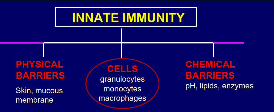
Image occlusion: name the hidden labels
4
Image occlusion: name the hidden labels
5
Image occlusion: name the hidden labels
6
Image occlusion: name the hidden labels
7
Image occlusion: name the hidden labels
8
Image occlusion: name the hidden labels
9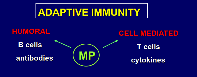
Image occlusion: name the hidden labels
10
Image occlusion: name the hidden labels
11
Image occlusion: name the hidden labels
12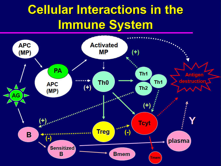
Image occlusion: name the hidden labels
13
Image occlusion: name the hidden labels
14
Image occlusion: name the hidden labels
15
Image occlusion: name the hidden labels
16
Image occlusion: name the hidden labels
17
Image occlusion: name the hidden labels
18
Image occlusion: name the hidden labels
19
Image occlusion: name the hidden labels
20
Image occlusion: name the hidden labels
21
Image occlusion: name the hidden labels
22
Image occlusion: name the hidden labels
23
Image occlusion: name the hidden labels
24
Lymphocytes
Potential targets for intervention
-Antigen recognition and binding (receptors)
-Clonal Proliferation (easiest step to block)
-Maturation and Differentiation
Potential side effects: HIGH
-Increased infections
-Increased cancer
Clinical Uses
-Organ Transplant
-Autoimmune Disease (SLE, RA, MS)
-Graft vs Host Disease
-Cancer
-Antigen recognition and binding (receptors)
-Clonal Proliferation (easiest step to block)
-Maturation and Differentiation
Potential side effects: HIGH
-Increased infections
-Increased cancer
Clinical Uses
-Organ Transplant
-Autoimmune Disease (SLE, RA, MS)
-Graft vs Host Disease
-Cancer
25
Transplantation
Success depends on donor and host “histocompatibility”
Determined genetically by MHC class I proteins (“transplantation antigens”)
Some MHC I antigens stronger than others (ABO and Rh
antigens on blood); elicit stronger immune response; others less important; elicit weak response
Tissue typing: based on analysis of MHC I proteins
- Present on peripheral blood leukocytes
- **Mixed lymphocyte reaction**: mix donor and recipient
lymphocytes; measure proliferation; histocompatibility inversely related to proliferation
- Greater histocompatibility, decreased need for immunosuppressive therapy
Determined genetically by MHC class I proteins (“transplantation antigens”)
Some MHC I antigens stronger than others (ABO and Rh
antigens on blood); elicit stronger immune response; others less important; elicit weak response
Tissue typing: based on analysis of MHC I proteins
- Present on peripheral blood leukocytes
- **Mixed lymphocyte reaction**: mix donor and recipient
lymphocytes; measure proliferation; histocompatibility inversely related to proliferation
- Greater histocompatibility, decreased need for immunosuppressive therapy
26
Autoimmune Disease/Chronic Inflammatory Disease
Disturbance in immunologic tolerance of self (MHC I) markers (become antigenic)
Immune cells react against self antigens; destroy host cells and tissues; chronic inflammation
Antibodies produced against self antigens called “autoantibodies” (ex, anti-nuclear antibodies, ANA)
Examples (etiology unknown): systemic lupus erythrmatosis (↑Th); idiopathic (immunologic) anemia; rheumatoid arthritis, multiple sclerosis, Hashimoto’s
thyroiditis, Grave’s disease; Crohn’s disease
Drug-induced: hydralazine, procainamide ( ↓Ts), troglitazone, tienilic acid
Immune cells react against self antigens; destroy host cells and tissues; chronic inflammation
Antibodies produced against self antigens called “autoantibodies” (ex, anti-nuclear antibodies, ANA)
Examples (etiology unknown): systemic lupus erythrmatosis (↑Th); idiopathic (immunologic) anemia; rheumatoid arthritis, multiple sclerosis, Hashimoto’s
thyroiditis, Grave’s disease; Crohn’s disease
Drug-induced: hydralazine, procainamide ( ↓Ts), troglitazone, tienilic acid
27
Graft Versus Host Disease
⚫ Common after bone marrow (BM) transplant in immunosuppressed patients
⚫ Transplant BM into histo-incompatible host
⚫ Donor cells attack recipient cells
⚫ Rapid proliferation of donor T-helper cells followed by generation of specific cytotoxic T cells
⚫ Difficult to attenuate once established- fatal
Treatment: prophylactic cyclosporine or tacrolimus
⚫ Transplant BM into histo-incompatible host
⚫ Donor cells attack recipient cells
⚫ Rapid proliferation of donor T-helper cells followed by generation of specific cytotoxic T cells
⚫ Difficult to attenuate once established- fatal
Treatment: prophylactic cyclosporine or tacrolimus
28
Cancer
Leukemias and lymphomas
Solid tumors
Treatment:
Cytotoxic agents
Monoclonal antibodies
Checkpoint inhibitors
Autologous T cells (CAR-T cells)
Solid tumors
Treatment:
Cytotoxic agents
Monoclonal antibodies
Checkpoint inhibitors
Autologous T cells (CAR-T cells)
29
Corticosteroids
– Anti-inflammatory activity (block macrophages, neutrophils and mediators)
– Lympholytic
– One of most widely used immunosuppressive agents; especially in autoimmune disorders; used frequently in organ transplants, especially
during rejection crisis
– Low bone marrow toxicity
– Prednisone and dexamethasone: most commonly used
**Lympholytic activity**
-T cells more sensitive than B cells
-May be due to specific lysis Th and Treg cells
-Lysis of precursor B cells
-Reduce size and content lymph nodes and spleen
-Continued use: decreased antibody production
**Anti-inflammatory activity**
-Inhibit macrophage and neutrophil activity
-Inhibits synthesis of eicosanoids (LT, PG), cytokines (TNFa)
-Interfere with transcription factor (NF-kB/AP-1) binding to target genes controlling production of inflammatory mediators
-GR cross talk with TLR resulting in altered expression of NFkB, AP-1, STAT, IRF regulated inflammatory genes
– Lympholytic
– One of most widely used immunosuppressive agents; especially in autoimmune disorders; used frequently in organ transplants, especially
during rejection crisis
– Low bone marrow toxicity
– Prednisone and dexamethasone: most commonly used
**Lympholytic activity**
-T cells more sensitive than B cells
-May be due to specific lysis Th and Treg cells
-Lysis of precursor B cells
-Reduce size and content lymph nodes and spleen
-Continued use: decreased antibody production
**Anti-inflammatory activity**
-Inhibit macrophage and neutrophil activity
-Inhibits synthesis of eicosanoids (LT, PG), cytokines (TNFa)
-Interfere with transcription factor (NF-kB/AP-1) binding to target genes controlling production of inflammatory mediators
-GR cross talk with TLR resulting in altered expression of NFkB, AP-1, STAT, IRF regulated inflammatory genes
30
Anti-Inflammatory Mechanism of Glucocorticoids
The GRE works for anti-inflammatory activity by allowing the GR to regulate the expression of genes that are involved in inflammation. For example, the GR can bind to GREs in the promoters of genes that encode anti-inflammatory mediators. The GR can also bind to GREs that are located near other transcription factors, such as NF-κB and AP-1, that activate inflammatory genes, and interfere with their function. By modulating the expression of these genes, the GR can reduce inflammation and suppress the immune system.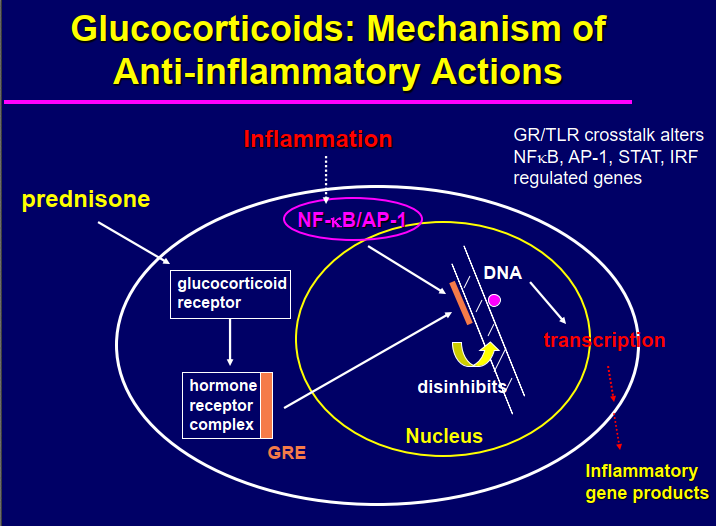
31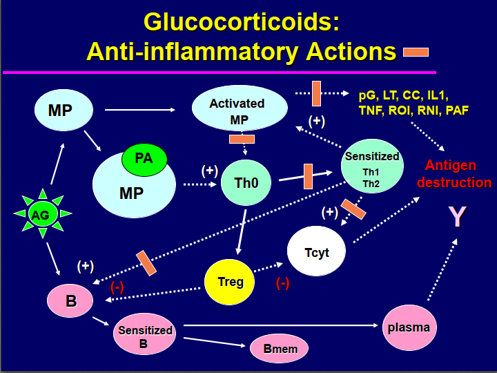
Image occlusion: name the hidden labels
32
Image occlusion: name the hidden labels
33
Image occlusion: name the hidden labels
34
Image occlusion: name the hidden labels
35
Image occlusion: name the hidden labels
36
Image occlusion: name the hidden labels
37
Image occlusion: name the hidden labels
38
Image occlusion: name the hidden labels
39
Image occlusion: name the hidden labels
40
Image occlusion: name the hidden labels
41
Image occlusion: name the hidden labels
42
Image occlusion: name the hidden labels
43
Image occlusion: name the hidden labels
44
Image occlusion: name the hidden labels
45
Image occlusion: name the hidden labels
46
Image occlusion: name the hidden labels
47
Image occlusion: name the hidden labels
48
Image occlusion: name the hidden labels
49
Image occlusion: name the hidden labels
50
Image occlusion: name the hidden labels
51
Image occlusion: name the hidden labels
52
Image occlusion: name the hidden labels
53
Image occlusion: name the hidden labels
54
Image occlusion: name the hidden labels
55
Image occlusion: name the hidden labels
56
Image occlusion: name the hidden labels
57
Image occlusion: name the hidden labels
58
Image occlusion: name the hidden labels
59
Image occlusion: name the hidden labels
60
Image occlusion: name the hidden labels
61
Image occlusion: name the hidden labels
62
Image occlusion: name the hidden labels
63
Image occlusion: name the hidden labels
64
Image occlusion: name the hidden labels
65
Image occlusion: name the hidden labels
66
Image occlusion: name the hidden labels
67
Image occlusion: name the hidden labels
68
Toxicity of Glucocorticoids
Major side effects are common due to high doses necessary for suppression
– Cushings syndrome
– glucose intolerance
– infections
– bone dissolution
– muscle wasting
– Cushings syndrome
– glucose intolerance
– infections
– bone dissolution
– muscle wasting
69
Cytotoxic Agents (nonspecific)
Block DNA synthesis; destroy antigen stimulated proliferation of lymphocytes
Most cytotoxic drugs act nonspecifically; inhibit entire immune response (B and T cells)
Easier to block/attenuate primary immune response than to suppress established response
Classical cytotoxic agents are antiproliferative; overlap with cancer chemotherapeutic drugs
Lymphoid cells most sensitive to cytotoxic agents; macrophages relatively resistant
Most cytotoxic drugs act nonspecifically; inhibit entire immune response (B and T cells)
Easier to block/attenuate primary immune response than to suppress established response
Classical cytotoxic agents are antiproliferative; overlap with cancer chemotherapeutic drugs
Lymphoid cells most sensitive to cytotoxic agents; macrophages relatively resistant
70
Major Cytotoxic Immunosuppressants
⚫ Cyclophosphamide (alkylating agent)
⚫ Azathioprine (purine analog)
⚫ Mycophenolate (MMF; purine analog)
⚫ Leflunomide (pyrimidine analog)
⚫ Methotrexate (antifolate)
Overlap with cancer chemotherapeutics
⚫ Azathioprine (purine analog)
⚫ Mycophenolate (MMF; purine analog)
⚫ Leflunomide (pyrimidine analog)
⚫ Methotrexate (antifolate)
Overlap with cancer chemotherapeutics
71
Immune Cells vs. Cancer Cells
Proliferation
– Immune cells: proliferate in response to specific antigen; synchronized; antigen causes burst of proliferation in all cells at once; all cells enter S phase
– Cancer cells: unstimulated proliferation; random, unsynchronized; only some cells in S phase
Selective Toxicity
– Immune cells: cytotoxic drug given immediately after antigen (transplant); kill high % small number antigen specific cells
– Cancer cells: no selective toxicity; only kill S phase cells
Clinical Use
– Immune cells: drugs administered in low doses on a daily schedule for life time in order to continuously block immune cell proliferation
– Cancer cells: drugs administered in high dose pulse therapy (1-3 days) every 2-4 weeks for up to 6 months to permit immune cell rebound between treatments
– Immune cells: proliferate in response to specific antigen; synchronized; antigen causes burst of proliferation in all cells at once; all cells enter S phase
– Cancer cells: unstimulated proliferation; random, unsynchronized; only some cells in S phase
Selective Toxicity
– Immune cells: cytotoxic drug given immediately after antigen (transplant); kill high % small number antigen specific cells
– Cancer cells: no selective toxicity; only kill S phase cells
Clinical Use
– Immune cells: drugs administered in low doses on a daily schedule for life time in order to continuously block immune cell proliferation
– Cancer cells: drugs administered in high dose pulse therapy (1-3 days) every 2-4 weeks for up to 6 months to permit immune cell rebound between treatments
72
Cyclophosphamide
most commonly used immunosuppressive agent; cell cycle
nonspecific; but toxicity is proliferation dependent
**Mechanism of cytotoxicity**: bifunctional alkylating agent; highly reactive alkyl group forms covalent bonds with nucleophilic groups (amino acids, carboxyl, sulfhydryl and imidazole groups) in DNA (N-7 guanine); cross-links DNA
Cell cycle nonspecific
Can interact with DNA of resting (G0) and proliferating cells; more toxic to growing cells; dormant cells (G0) can repair alkylation damage
Proliferation dependent
Amount of damage dependent on growth fraction of tumor
Drug resistance
Decreased drug uptake
Increased repair of drug defect
Cross resistance between drugs
nonspecific; but toxicity is proliferation dependent
**Mechanism of cytotoxicity**: bifunctional alkylating agent; highly reactive alkyl group forms covalent bonds with nucleophilic groups (amino acids, carboxyl, sulfhydryl and imidazole groups) in DNA (N-7 guanine); cross-links DNA
Cell cycle nonspecific
Can interact with DNA of resting (G0) and proliferating cells; more toxic to growing cells; dormant cells (G0) can repair alkylation damage
Proliferation dependent
Amount of damage dependent on growth fraction of tumor
Drug resistance
Decreased drug uptake
Increased repair of drug defect
Cross resistance between drugs
73
Antimetabolites
Purine Synthesis Inhibitors
– Azathioprine, mycophenolate
Pyrimidine Synthesis Inhibitors
– Leflunomide, teriflunomide
Anti-folates
– methotrexate
– Azathioprine, mycophenolate
Pyrimidine Synthesis Inhibitors
– Leflunomide, teriflunomide
Anti-folates
– methotrexate
74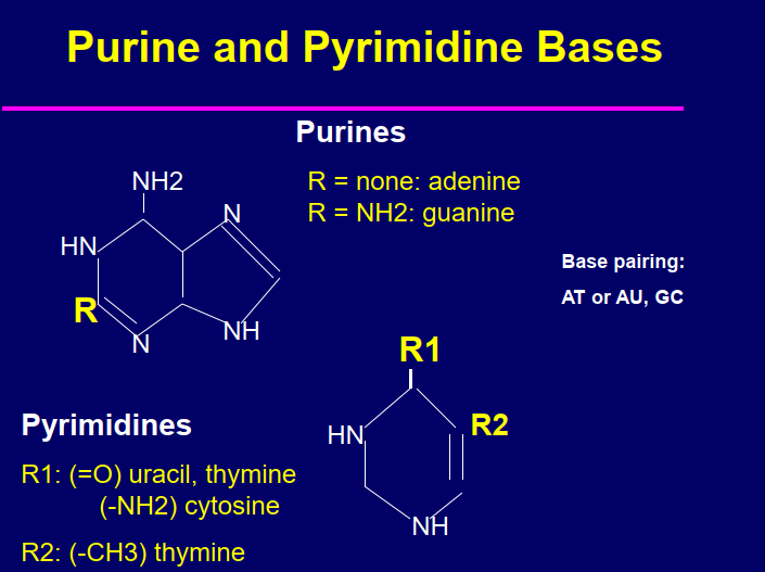
Image occlusion: name the hidden labels
75
Image occlusion: name the hidden labels
76
Image occlusion: name the hidden labels
77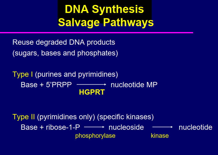
Image occlusion: name the hidden labels
78
Image occlusion: name the hidden labels
79
Image occlusion: name the hidden labels
80
Image occlusion: name the hidden labels
81
Image occlusion: name the hidden labels
82
Image occlusion: name the hidden labels
83
Purine Analogs
Azathioprine
Mycophenolate (MMF)
Imidizole derivatives of 6-mercaptopurine (6-MP) which may be active form of drugs in vivo
**S-phase specific**
**Cytotoxic Actions**: “lethal synthesis” to active form of drug (nucleotide: 6-MPRP) by salvage DNA enzyme (HGPRT); inhibits de novo nucleic acid biosynthesis and proliferation of antigen stimulated lymphocytes; blocks CMI and humoral immunity
Mechanisms of Cytotoxicity: Inhibition of de novo purine biosynthesis;
6-MPRP structural analog inosine monophosphate (IMP), covalently binds to and inhibits IMP dehydrogenase
Inhibition of first step de novo purine synthesis; negative feedback mechanism; “pseudo –feedback inhibitor”
Incorporation of active 6-MPRP into DNA-mutations/strand breaks
Mycophenolate (MMF)
Imidizole derivatives of 6-mercaptopurine (6-MP) which may be active form of drugs in vivo
**S-phase specific**
**Cytotoxic Actions**: “lethal synthesis” to active form of drug (nucleotide: 6-MPRP) by salvage DNA enzyme (HGPRT); inhibits de novo nucleic acid biosynthesis and proliferation of antigen stimulated lymphocytes; blocks CMI and humoral immunity
Mechanisms of Cytotoxicity: Inhibition of de novo purine biosynthesis;
6-MPRP structural analog inosine monophosphate (IMP), covalently binds to and inhibits IMP dehydrogenase
Inhibition of first step de novo purine synthesis; negative feedback mechanism; “pseudo –feedback inhibitor”
Incorporation of active 6-MPRP into DNA-mutations/strand breaks
84
Azathioprine
Used with other drugs to prevent rejection of kidney transplants
Used to treat severe rheumatoid arthritis when other drugs fail
Ulcerative colitis
High toxicity
Used to treat severe rheumatoid arthritis when other drugs fail
Ulcerative colitis
High toxicity
85
Mycophenolate (MMP)
Fungal metabolite
Gradually replacing azathioprine
Typically used in combination with other immunosuppressants (prednisone and calcineurin inhibitor)
Used to prevent transplant rejection and to treat autoimmune diseases
(i.e., Crohn’s disease, psoriasis, lupus)
Better efficacy and less toxicity than cyclophosphamide
Gradually replacing azathioprine
Typically used in combination with other immunosuppressants (prednisone and calcineurin inhibitor)
Used to prevent transplant rejection and to treat autoimmune diseases
(i.e., Crohn’s disease, psoriasis, lupus)
Better efficacy and less toxicity than cyclophosphamide
86
Pyrimidine Analogs
Leflunomide
Teriflunamide (active metabolite)
–Used to treat moderate to severe rheumatoid arthritis and psoriatic arthritis
–S-phase specific
–Inhibits dihydroorotate dehydrogenase, key step in de novo pyrimidine biosynthesis
Teriflunamide (active metabolite)
–Used to treat moderate to severe rheumatoid arthritis and psoriatic arthritis
–S-phase specific
–Inhibits dihydroorotate dehydrogenase, key step in de novo pyrimidine biosynthesis
87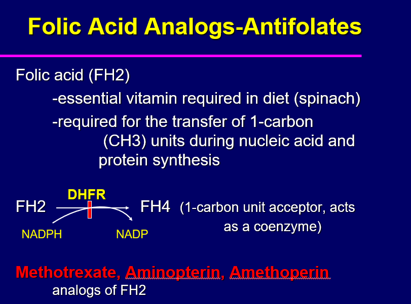
Image occlusion: name the hidden labels
88
Image occlusion: name the hidden labels
89
Image occlusion: name the hidden labels
90
Image occlusion: name the hidden labels
91
Image occlusion: name the hidden labels
92
Methotrexate
Competitive inhibitors of dihydrofolic acid reductase (DHFR)
-Bind tightly to DHFR “pseudo-irreversible”
-Inhibit all one carbon transfer reactions; involved in the synthesis of thymidylate
(dTMP), purines, glycine and methionine; results in inhibition of DNA, RNA and protein synthesis
-S phase specific
-Bind tightly to DHFR “pseudo-irreversible”
-Inhibit all one carbon transfer reactions; involved in the synthesis of thymidylate
(dTMP), purines, glycine and methionine; results in inhibition of DNA, RNA and protein synthesis
-S phase specific
93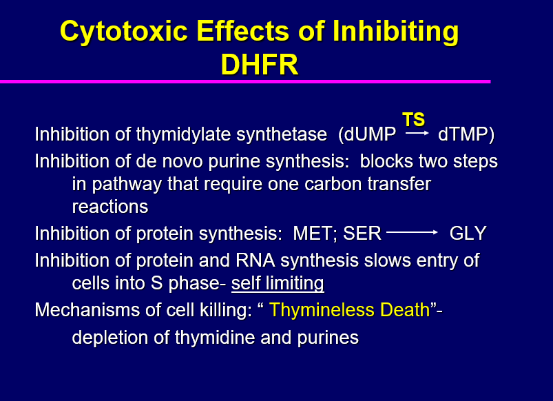
Image occlusion: name the hidden labels
94
Image occlusion: name the hidden labels
95
Image occlusion: name the hidden labels
96
Image occlusion: name the hidden labels
97
Image occlusion: name the hidden labels
98
Selective Immunosuppressants
Block T-helper cell proliferation and function
- Calcineurin Inhibitors
- Mammalian Target-of-Rapamycin (mTOR) Inhibitors
- Calcineurin Inhibitors
- Mammalian Target-of-Rapamycin (mTOR) Inhibitors
99
Targeting T Cells: Major Effectors of Transplant Rejection and Tolerance
**Transplant Rejection**
T-helper cells (CD4+)
Cytotoxic T cells (CD8+)
**Approaches to inhibiting cells**
-Block IL-2 production (CSA; tacrolimus)
-Block T cell proliferation (rapamycin, everolimus)
**Induction of Tolerance**
T-regulatory cells (CD4+/CD25+/FoxP3+)
T-helper cells (CD4+)
Cytotoxic T cells (CD8+)
**Approaches to inhibiting cells**
-Block IL-2 production (CSA; tacrolimus)
-Block T cell proliferation (rapamycin, everolimus)
**Induction of Tolerance**
T-regulatory cells (CD4+/CD25+/FoxP3+)
100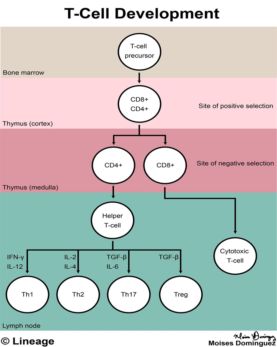
Image occlusion: name the hidden labels
101
Image occlusion: name the hidden labels
102
Image occlusion: name the hidden labels
103
Image occlusion: name the hidden labels
104
Image occlusion: name the hidden labels
105
Image occlusion: name the hidden labels
106
Image occlusion: name the hidden labels
107
Image occlusion: name the hidden labels
108
Image occlusion: name the hidden labels
109
T Cell Subsets
Develop in response to cytokines in tissue microenvironment
Work together to modulate immune response
Actions mediated by cytokines (lymphokines)
**T-helper cells (Th): CD4+**
- Th1 cells: promote CMI and inflammation (IFNg, IL-2, TNFa)
- Th2 cells: promote B cell activity; activate eosinophils; suppress macrophages; asthma, allergy (IL-4, IL-5, IL-10, IL-13)
- Th17 cells: proinflammatory; target extracellular bacteria (skin, intestinal lining) and fungi; autoimmunity (IL-17, IL-21, IL-22);
**T-regulatory (suppressor) cells (Treg)**: surface CD4+/CD25+/intracellular FoxP3+; immune tolerance, lymphocyte homeostasis (TGFb, IL-35, IL-10)
**Cytotoxic (killer) T cells (Tcyt)**: CD8+ (bone marrow)
Work together to modulate immune response
Actions mediated by cytokines (lymphokines)
**T-helper cells (Th): CD4+**
- Th1 cells: promote CMI and inflammation (IFNg, IL-2, TNFa)
- Th2 cells: promote B cell activity; activate eosinophils; suppress macrophages; asthma, allergy (IL-4, IL-5, IL-10, IL-13)
- Th17 cells: proinflammatory; target extracellular bacteria (skin, intestinal lining) and fungi; autoimmunity (IL-17, IL-21, IL-22);
**T-regulatory (suppressor) cells (Treg)**: surface CD4+/CD25+/intracellular FoxP3+; immune tolerance, lymphocyte homeostasis (TGFb, IL-35, IL-10)
**Cytotoxic (killer) T cells (Tcyt)**: CD8+ (bone marrow)
110
Selective Immunosuppressants Prototype: Cyclosporine (CSA)
Discovered by Borel (1976); Sandoz
Extracted from soil fungus
Small molecular weight peptide; first developed as antifungal agent; limited action; potent immunosuppressant
Revolutionized transplantation
Highly selective inhibitor T-helper cells; primary cells involved in graft rejection
Timing of administration critical for efficacy (14 d pre- 4 d post); once immune response started, not effective; suggests actions at early stage
**Pretreatment alone not effective;** suggests CSA acts after exposure to antigen has occurred
In general: **continued life time therapy required**
Selective action on antigen stimulated T-helper cells; inhibits IL-2 (T), IL-3 (B), IFNg (MP) production
Effects reversible
**Long term therapy**: nephrotoxicity; drug interactions due to effects on cyp P450
Extracted from soil fungus
Small molecular weight peptide; first developed as antifungal agent; limited action; potent immunosuppressant
Revolutionized transplantation
Highly selective inhibitor T-helper cells; primary cells involved in graft rejection
Timing of administration critical for efficacy (14 d pre- 4 d post); once immune response started, not effective; suggests actions at early stage
**Pretreatment alone not effective;** suggests CSA acts after exposure to antigen has occurred
In general: **continued life time therapy required**
Selective action on antigen stimulated T-helper cells; inhibits IL-2 (T), IL-3 (B), IFNg (MP) production
Effects reversible
**Long term therapy**: nephrotoxicity; drug interactions due to effects on cyp P450
111
Tacrolimus
Isolated from streptomyces; macrolide antibiotic
Blocks T-helper cell activation and IL-2 production
10-100 times more potent than CSA
Blocks T-helper cell activation and IL-2 production
10-100 times more potent than CSA
112
Calcineurin Inhibitors
**Cyclosporine and Tacrolimus**
Block T lymphocyte transcription of IL-2
Inhibit T cell dependent B cell responses (no direct effect
on B cells) (antibody production)
**Calcineurin**: cytoplasmic phosphatase; required for activation of NF-AT; T cell transcription factor that regulates IL-2 production
Calcineurin inhibitors: bind to specific cytoplasmic isomerases (binding proteins) in T-helper cells; form complexes that inhibits calcineurin
Cyclosporine binds: cyclophilin
Tacrolimus binds: FK-binding protein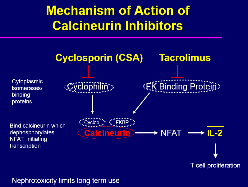
Block T lymphocyte transcription of IL-2
Inhibit T cell dependent B cell responses (no direct effect
on B cells) (antibody production)
**Calcineurin**: cytoplasmic phosphatase; required for activation of NF-AT; T cell transcription factor that regulates IL-2 production
Calcineurin inhibitors: bind to specific cytoplasmic isomerases (binding proteins) in T-helper cells; form complexes that inhibits calcineurin
Cyclosporine binds: cyclophilin
Tacrolimus binds: FK-binding protein
113
Untoward Effects of Calcineurin Inhibitors
Nephrotoxicity
Serum levels of these drugs do not correlate well with the extent of renal damage
Chronic toxicity- allograft damage\/rejection, hypertension, hyperlipidemia, glucose intolerance
Combination therapy: used to achieve adequate immunosuppression and to lower the risk of drug-related toxicity
Most frequently administered regimens include a corticosteroid plus either cyclosporin or tacrolimus plus a lymphocyte-proliferation inhibitor (e.g. mycophenolate)
Serum levels of these drugs do not correlate well with the extent of renal damage
Chronic toxicity- allograft damage\/rejection, hypertension, hyperlipidemia, glucose intolerance
Combination therapy: used to achieve adequate immunosuppression and to lower the risk of drug-related toxicity
Most frequently administered regimens include a corticosteroid plus either cyclosporin or tacrolimus plus a lymphocyte-proliferation inhibitor (e.g. mycophenolate)
114
Mammalian Target-of-Rapamycin (mTOR) Inhibitors
**mTOR**
-serine/threonine protein kinase (Cdk) that regulates cell growth, motility, survival, protein synthesis and transcription
-activated by growth factors, mitogens, cellular stress, nutrients
**Sirolimus (rapamycin) and Everolimus**
-macrolide antibotics
-associate with intracellular receptor (FKBP12); form complex which binds to and inhibits mTOR
-serine/threonine protein kinase (Cdk) that regulates cell growth, motility, survival, protein synthesis and transcription
-activated by growth factors, mitogens, cellular stress, nutrients
**Sirolimus (rapamycin) and Everolimus**
-macrolide antibotics
-associate with intracellular receptor (FKBP12); form complex which binds to and inhibits mTOR
115
Cell Cycle Control
**Cyclins** (intracellular levels rise and fall with stages of cell cycle)
G1 cyclins (D cyclins)
S-phase cyclins (cyclins E and A)
mitotic cyclins (B cyclins)
**Cyclin-dependent kinases (Cdks)**- bind and activate CYCLINS
G1 Cdk (Cdk4)
S-phase Cdk (Cdk2)
M-phase Cdk (Cdk1)
**Anaphase-promoting complex (APC)**. Important in sister chromatids to separate
degrades mitotic (B) cyclins
G1 cyclins (D cyclins)
S-phase cyclins (cyclins E and A)
mitotic cyclins (B cyclins)
**Cyclin-dependent kinases (Cdks)**- bind and activate CYCLINS
G1 Cdk (Cdk4)
S-phase Cdk (Cdk2)
M-phase Cdk (Cdk1)
**Anaphase-promoting complex (APC)**. Important in sister chromatids to separate
degrades mitotic (B) cyclins
116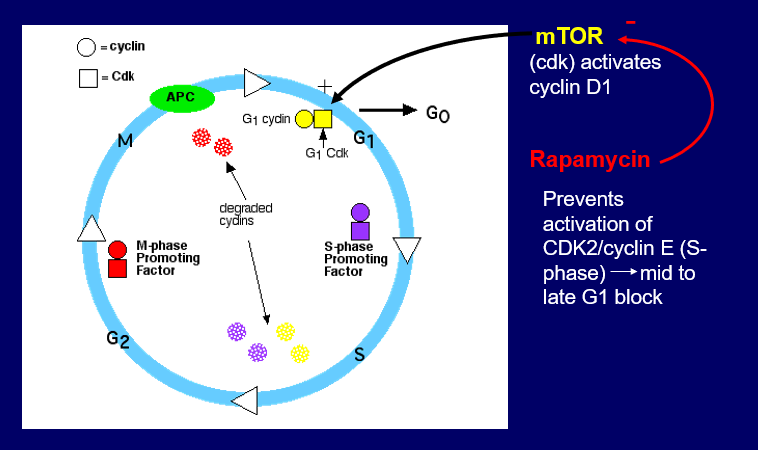
Image occlusion: name the hidden labels
117
Image occlusion: name the hidden labels
118
Image occlusion: name the hidden labels
119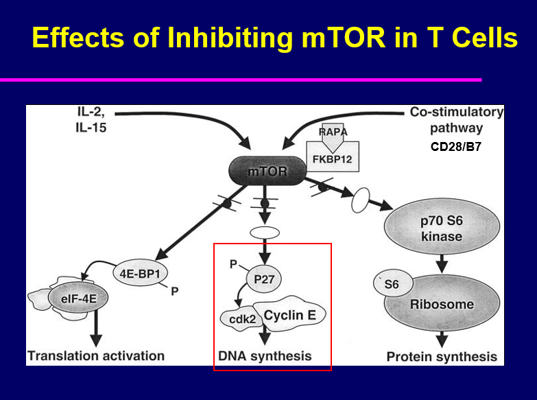
Image occlusion: name the hidden labels
120
Selective Targeting of Specific Immune Cells and Inflammatory Mediators
**Approaches**
Antisera: human and rodent
Monoclonal antibodies: chimeric, fully humanized
Fusion proteins
Recombinant proteins
**Targets**
Cell surface proteins (CD)
Cell surface receptors
Adhesion molecules
Co-stimulatory molecules
Intracellular signaling molecules
Soluble cytokines, chemokines or their receptors
Human immunoglobulins
Non-human polyclonal antibodies (antisera)
Mouse monoclonal antibodies
Chimeric (humanized) antibodies
Chimeric fusion proteins
polyclonal and monoclonal
– Target cell, protein or receptor specific
Antisera: human and rodent
Monoclonal antibodies: chimeric, fully humanized
Fusion proteins
Recombinant proteins
**Targets**
Cell surface proteins (CD)
Cell surface receptors
Adhesion molecules
Co-stimulatory molecules
Intracellular signaling molecules
Soluble cytokines, chemokines or their receptors
Human immunoglobulins
Non-human polyclonal antibodies (antisera)
Mouse monoclonal antibodies
Chimeric (humanized) antibodies
Chimeric fusion proteins
polyclonal and monoclonal
– Target cell, protein or receptor specific
121
Human Antibodies (Immunoglobulins)
**Immune globulin intravenous (IGIV)**
Pooled polyclonal human immunoglobulin prepared from >1000 healthy donors; no antigen
Used in patients with primary immune deficiency; unable to produce IgG; replaces normal IgG
Promotes pathogen destruction
**Hyperimmune immunoglobulin**
IGIV preparations made from pools of “selected donors” with high titer antibodies against particular agents (i.e., viruses); Passive transfer of antibodies
Examples: hepatitis B immune globulin; tetanus immune globulin; rabies immune globulin
Pooled polyclonal human immunoglobulin prepared from >1000 healthy donors; no antigen
Used in patients with primary immune deficiency; unable to produce IgG; replaces normal IgG
Promotes pathogen destruction
**Hyperimmune immunoglobulin**
IGIV preparations made from pools of “selected donors” with high titer antibodies against particular agents (i.e., viruses); Passive transfer of antibodies
Examples: hepatitis B immune globulin; tetanus immune globulin; rabies immune globulin
122
Humanizing Biologics Recombinant Fusion Proteins (Chimeric Proteins)
Proteins created by joining two genes which code for separate proteins
Translation of fusion gene by host cell results in a single chimeric protein with functional properties derived from each of the original proteins
Protein can be engineered to include the full sequence of both original proteins, or only a portion of either
Chimeric antibodies
Fc chimeric fusion proteins
Translation of fusion gene by host cell results in a single chimeric protein with functional properties derived from each of the original proteins
Protein can be engineered to include the full sequence of both original proteins, or only a portion of either
Chimeric antibodies
Fc chimeric fusion proteins
123
Chimeric Antibodies
Humanizing mouse monoclonal antibodies
Use techniques in genetic engineering to replace segments of the mouse antibody molecule (constant regions) most different from human antibody.
Example:
Infliximab (chimeric anti-TNFa antibody). Human constant domains introduced, eliminating the most immunogenic portions of the mouse antibody without altering its antigen specificity.
Use techniques in genetic engineering to replace segments of the mouse antibody molecule (constant regions) most different from human antibody.
Example:
Infliximab (chimeric anti-TNFa antibody). Human constant domains introduced, eliminating the most immunogenic portions of the mouse antibody without altering its antigen specificity.
124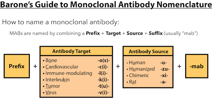
Image occlusion: name the hidden labels
125
Fc Chimeric Fusion Proteins
Examples of proteins fused to the Fc domain (constant regions) of IgG1; adds stability and deliverability to the drug
**Abatacept**
Extracellular domain of human CTLA-4 (co-inhibitory molecule)
Etanercept (TNF inhibitor)
Extracellular domain of human p75 TNF receptor
**Abatacept**
Extracellular domain of human CTLA-4 (co-inhibitory molecule)
Etanercept (TNF inhibitor)
Extracellular domain of human p75 TNF receptor
126
Targets for Therapeutic Biologics
Cell surface targets
T cells
B cells
Cell adhesion molecules
Cell surface receptor antagonists
Intracellular targets
Signaling molecules
Checkpoint inhibitors
Serum targets/cytokines
Complement
Cytokines
Soluble cytokine receptors
T cells
B cells
Cell adhesion molecules
Cell surface receptor antagonists
Intracellular targets
Signaling molecules
Checkpoint inhibitors
Serum targets/cytokines
Complement
Cytokines
Soluble cytokine receptors
127
Examples of Chimeric Fusion proteins
T Cell Inhibitors
Abatracept
Ipilimumab
B Cell Inhibitors
Rituximab
Ocrelizuman
Ofatumumumab
Cell Adhesion Molecules
Vedolizumab
Natalizumab
Abatracept
Ipilimumab
B Cell Inhibitors
Rituximab
Ocrelizuman
Ofatumumumab
Cell Adhesion Molecules
Vedolizumab
Natalizumab
128
TNFα Inhibitors
TNFa: master regulator of inflammatory response; key in autoimmune disease; most widely used biologics
Infliximab (RemicadeR): chimeric monoclonal anti-TNFa antibody
Adalimumab (HumiraR): fully humanized monoclonal anti-TNFa antibody
Golimumab (SimponiR): fully humanized monoclonal anti-TNFa antibody
Certolizumab pegol (CimziaR): fragment of monoclonal antibody specific to TNFa
Etanercept (EmbrelR) decoy receptor
Receptor fusion protein; mimics actions of sTNFR which inactivates TNF and suppresses inflammation; does not neutralize receptor bound TNFa; does not lyse inflammatory cells; longer half life than anti-TNF antibodies
Differences may underlie efficacy and side effects
Infliximab (RemicadeR): chimeric monoclonal anti-TNFa antibody
Adalimumab (HumiraR): fully humanized monoclonal anti-TNFa antibody
Golimumab (SimponiR): fully humanized monoclonal anti-TNFa antibody
Certolizumab pegol (CimziaR): fragment of monoclonal antibody specific to TNFa
Etanercept (EmbrelR) decoy receptor
Receptor fusion protein; mimics actions of sTNFR which inactivates TNF and suppresses inflammation; does not neutralize receptor bound TNFa; does not lyse inflammatory cells; longer half life than anti-TNF antibodies
Differences may underlie efficacy and side effects
129
Chimeric Fusion Proteins/Antibodies Interleukin Inhibitors
IL-1 inhibitors
Rilonacept (ArcalystR): IL-1 trap; dimeric fusion protein consists of extracellular
domain of IL-1 receptor and Fc domain of human IgG1; binds and neutralizes IL-1
Anakinra (KineretR); IL-1 receptor antagonist; genetically engineered E-coli; differs from native human IL-1RA by single methionine residue on amino terminus; inflammation and rheumatoid arthritis
IL-2 inhibitors
Basiliximab (SimulectR): chimeric antibody to CD25, a chain of T cell receptor
IL-4/IL-13 inhibitors
Dupilumab (DupixentR): humanized monoclonal anti-IL4/IL-13 receptor; asthma; dermatitis
IL-5 inhibitors
Mepolizumab (NucalaR): chimeric anti-IL-5 antibody; eosinophilic asthma
Benralizumab (FasenraR): chimeric antibody against a chain IL-5 receptor, asthma
IL-6 inhibitors
Siltuximab (SylvantR): chimeric anti IL-6 antibody
Toclizumab/Atlizumab (ActemraR): chimeric anti-IL-6 receptor monoclonal antibody; cytokine release syndrome
Rilonacept (ArcalystR): IL-1 trap; dimeric fusion protein consists of extracellular
domain of IL-1 receptor and Fc domain of human IgG1; binds and neutralizes IL-1
Anakinra (KineretR); IL-1 receptor antagonist; genetically engineered E-coli; differs from native human IL-1RA by single methionine residue on amino terminus; inflammation and rheumatoid arthritis
IL-2 inhibitors
Basiliximab (SimulectR): chimeric antibody to CD25, a chain of T cell receptor
IL-4/IL-13 inhibitors
Dupilumab (DupixentR): humanized monoclonal anti-IL4/IL-13 receptor; asthma; dermatitis
IL-5 inhibitors
Mepolizumab (NucalaR): chimeric anti-IL-5 antibody; eosinophilic asthma
Benralizumab (FasenraR): chimeric antibody against a chain IL-5 receptor, asthma
IL-6 inhibitors
Siltuximab (SylvantR): chimeric anti IL-6 antibody
Toclizumab/Atlizumab (ActemraR): chimeric anti-IL-6 receptor monoclonal antibody; cytokine release syndrome
130
Targeting Th17 Cytokines with Humanized Monoclonal Antibodies
IL-12 and IL-23
Ustekinumab (StellaraR)- human monoclonal antibody; reduces differentiation of Th0 into Tcyt; plaque psoriasis; psoriatic arthritis
IL-23
Guselkumab (TremfyaR), Tildrakizumab- human monoclonal antibodies selectively binds IL-23; inhibits interaction with IL-23R; blocks proinflammatory/ chemokine release; moderate to severe plaque psoriasis
IL-17A
Secukinumab (CosentyxR)- human monoclonal antibody; plaque psoriasis; ankylosing spondylitis; psoriatic arthritis
Ixekizumab (TaltzR)- human monoclonal antibody; moderate/severe plaque psoriasis; prevents IL17A from binding IL-17R
Ustekinumab (StellaraR)- human monoclonal antibody; reduces differentiation of Th0 into Tcyt; plaque psoriasis; psoriatic arthritis
IL-23
Guselkumab (TremfyaR), Tildrakizumab- human monoclonal antibodies selectively binds IL-23; inhibits interaction with IL-23R; blocks proinflammatory/ chemokine release; moderate to severe plaque psoriasis
IL-17A
Secukinumab (CosentyxR)- human monoclonal antibody; plaque psoriasis; ankylosing spondylitis; psoriatic arthritis
Ixekizumab (TaltzR)- human monoclonal antibody; moderate/severe plaque psoriasis; prevents IL17A from binding IL-17R
131
Immune Checkpoints
Molecules that either stimulate or inhibit T cells
Critical for maintaining self-tolerance and modulating the duration and amplitude of immune responses in order to minimize collateral damage
Antigen Recognition by T Cells
Specific T cell receptor
T cells are **MHC restricted;** only recognize antigen and MHC protein
T-helper cells recognize processed antigen and MHC II (self)
Cytotoxic T cells recognize processed antigen and MHC I (non-self or altered self)
Successful immune response also requires costimulatory and/or coinhibitory molecules: Immune Checkpoints
Critical for maintaining self-tolerance and modulating the duration and amplitude of immune responses in order to minimize collateral damage
Antigen Recognition by T Cells
Specific T cell receptor
T cells are **MHC restricted;** only recognize antigen and MHC protein
T-helper cells recognize processed antigen and MHC II (self)
Cytotoxic T cells recognize processed antigen and MHC I (non-self or altered self)
Successful immune response also requires costimulatory and/or coinhibitory molecules: Immune Checkpoints
132
Immune Checkpoint Inhibitors
Molecules that inhibit T cell activation
Critical for maintaining self-tolerance and modulating the duration and amplitude of immune responses in order to minimize collateral damage
Examples:
CTLA-4
PD-1
PD-L1
LAG-3
Immune checkpoints: function to limit CD8+ cytotoxic T cell (Tcyt) activation
Usurped by tumor cells to prevent immune attack
Checkpoint inhibitors: prevent tumor cells from turning off activated Tcyt cells
Anti-PD-1: Pembrolizumab; Nivolumab; Relatlimab; Cemiplimab
Anti-PD-L1: Atezolizumab; Avelumab; Durvalumab
Anti-CTLA-4: Ipilimumab
Anti-LAG-3: Relatlimab
Critical for maintaining self-tolerance and modulating the duration and amplitude of immune responses in order to minimize collateral damage
Examples:
CTLA-4
PD-1
PD-L1
LAG-3
Immune checkpoints: function to limit CD8+ cytotoxic T cell (Tcyt) activation
Usurped by tumor cells to prevent immune attack
Checkpoint inhibitors: prevent tumor cells from turning off activated Tcyt cells
Anti-PD-1: Pembrolizumab; Nivolumab; Relatlimab; Cemiplimab
Anti-PD-L1: Atezolizumab; Avelumab; Durvalumab
Anti-CTLA-4: Ipilimumab
Anti-LAG-3: Relatlimab
133
Non-human Polyclonal Antibodies Antisera
Serum IgG from animals (donkey, horse, rabbit) immunized with human antigen; not standardized- variability between batches; side effects: serum sickness, Type II hypersensitivity reactions
Anti-thymocyte globulin (ATG): horse (AtgamR) or rabbit (ThymoglobulinR)-derived antibodies against human T cells; used to prevent/treat acute renal transplant rejection; non-specific-leads to profound T cell depletion; can cause cytokine release syndrome; due to activation of T cells prior to their clearance by macrophages
Rh immune globulin (anti-RhD antibodies): administered prophylactically to Rh- mother exposed to Rh+ baby RBC to prevent hemolytic disease of next newborn; isolated from mothers sensitized to Rh+ babies; Untreated mothers- maternal antibody transferred to fetus during third trimester; antibodies attack fetal RBC; used to treat immune thrombocytopenia
Anti-thymocyte globulin (ATG): horse (AtgamR) or rabbit (ThymoglobulinR)-derived antibodies against human T cells; used to prevent/treat acute renal transplant rejection; non-specific-leads to profound T cell depletion; can cause cytokine release syndrome; due to activation of T cells prior to their clearance by macrophages
Rh immune globulin (anti-RhD antibodies): administered prophylactically to Rh- mother exposed to Rh+ baby RBC to prevent hemolytic disease of next newborn; isolated from mothers sensitized to Rh+ babies; Untreated mothers- maternal antibody transferred to fetus during third trimester; antibodies attack fetal RBC; used to treat immune thrombocytopenia
134
Monoclonal Antibodies
Antibodies derived from single clone of antigen stimulated plasma cells; produce one antibody
**Advantages over Antiserum:**
increased purity, specificity and selectivity; more effective; antibody producing cells-immortal; easy to standardize
Problems:
need human antibodies; lack of suitable myeloma
spleen donors not available
**Solutions**:
humanize Mab (chimeric Mab)
**Examples**:
Targets: immune cells, cytokines, cytokine receptors, cell surface markers
Immunotoxins- conjugate Mab with toxin to target cells
**Advantages over Antiserum:**
increased purity, specificity and selectivity; more effective; antibody producing cells-immortal; easy to standardize
Problems:
need human antibodies; lack of suitable myeloma
spleen donors not available
**Solutions**:
humanize Mab (chimeric Mab)
**Examples**:
Targets: immune cells, cytokines, cytokine receptors, cell surface markers
Immunotoxins- conjugate Mab with toxin to target cells