Evaluation of Neurologic Disorders
Study these flashcardsFree · spaced repetition · iPhone, Android, Mac & web
All cards
1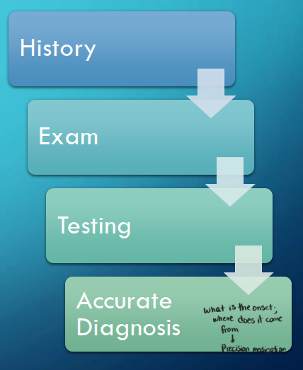
Image occlusion: name the hidden labels
2
Image occlusion: name the hidden labels
3
Image occlusion: name the hidden labels
4
Questions to Ask When Collecting History
Palliative or Provocative Factors
Quality
Region/Radiation
Severity
Timing
Quality
Region/Radiation
Severity
Timing
5
Medication History
• What is the patient’s current medication list?
• Dose
• Dosing schedule (times, relationship to other medications, relationship to meals)
• Duration
• Adherence
• Has the patient experienced adverse drug reactions
• What medications have been used in the past or recently discontinued? Why were they discontinued?
• Could the symptoms be drug-induced?
• Dose
• Dosing schedule (times, relationship to other medications, relationship to meals)
• Duration
• Adherence
• Has the patient experienced adverse drug reactions
• What medications have been used in the past or recently discontinued? Why were they discontinued?
• Could the symptoms be drug-induced?
6
Pediatric Patients
• Allow the child to provide as much history as they are developmentally able
• History may be obtained from the patient's guardians or caretakers rather than the child in most cases
• History should include
• Family history
• History of the pregnancy
• Details of labor and delivery
• Compare the child’s developmental stage to standard age-related developmental milestones
• History may be obtained from the patient's guardians or caretakers rather than the child in most cases
• History should include
• Family history
• History of the pregnancy
• Details of labor and delivery
• Compare the child’s developmental stage to standard age-related developmental milestones
7
Seven Components of Neuro Exam
1. Higher Cortical Function (Mental Status)
2. Cranial Nerves
3. Motor Function
4. Reflexes
5. Cerebellar Function
6. Sensory Function
7. Gait
2. Cranial Nerves
3. Motor Function
4. Reflexes
5. Cerebellar Function
6. Sensory Function
7. Gait
8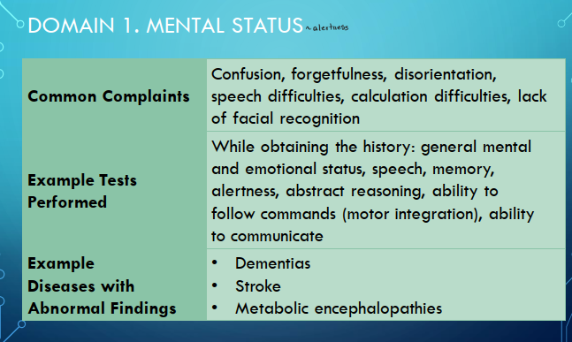
Image occlusion: name the hidden labels
9
Image occlusion: name the hidden labels
10
Image occlusion: name the hidden labels
11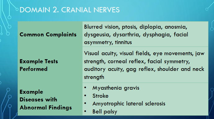
Image occlusion: name the hidden labels
12
Image occlusion: name the hidden labels
13
Image occlusion: name the hidden labels
14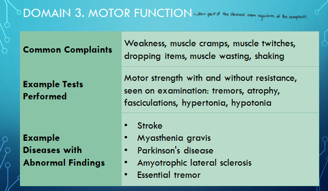
Image occlusion: name the hidden labels
15
Image occlusion: name the hidden labels
16
Image occlusion: name the hidden labels
17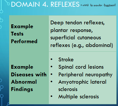
Image occlusion: name the hidden labels
18
Image occlusion: name the hidden labels
19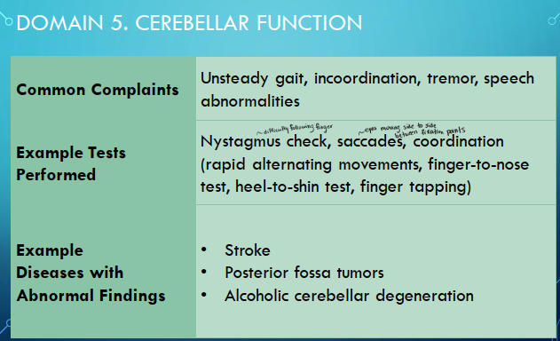
Image occlusion: name the hidden labels
20
Image occlusion: name the hidden labels
21
Image occlusion: name the hidden labels
22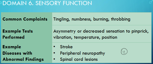
Image occlusion: name the hidden labels
23
Image occlusion: name the hidden labels
24
Image occlusion: name the hidden labels
25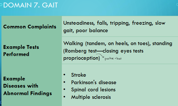
Image occlusion: name the hidden labels
26
Image occlusion: name the hidden labels
27
Image occlusion: name the hidden labels
28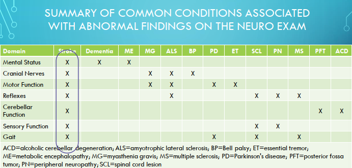
Image occlusion: name the hidden labels
29
Neurologic Exam in Pediatrics
• Adapt for age and developmental stage
• Ex. Observe walking and playing for cerebellar and motor function
• Ex. Observe facial expressions, withdrawal, and responses to tactile sensory stimulation to assess sensory function
• Assess weight, height, and head circumference
• In infants, assess fontanels for size and note if they are open or
closed
• Ex. Observe walking and playing for cerebellar and motor function
• Ex. Observe facial expressions, withdrawal, and responses to tactile sensory stimulation to assess sensory function
• Assess weight, height, and head circumference
• In infants, assess fontanels for size and note if they are open or
closed
30
Lumbar Puncture (LP)
• Diagnostic reasons:
• CNS infections
• Inflammatory processes
• Subarachnoid hemorrhage
• Therapeutic reasons:
• Relieve symptoms from increased intracranial pressure
• Deliver medications
• CNS infections
• Inflammatory processes
• Subarachnoid hemorrhage
• Therapeutic reasons:
• Relieve symptoms from increased intracranial pressure
• Deliver medications
31
Contraindications to LP
Local Skin Infection over the site
Suspected intracranial or spinal cord lesion
Raised intracranial pressure (ICP) with signs of impending herniation
Bleeding diathesis
Uncontrolled coagulopathy
Suspected intracranial or spinal cord lesion
Raised intracranial pressure (ICP) with signs of impending herniation
Bleeding diathesis
Uncontrolled coagulopathy
32
Medications Used During LP
• Medications commonly administered via LP:
• Anesthesia
• Chemotherapy
• Antibiotics
• Contrast dye
• Fluoroscopic guidance or anesthesia IF:
• Patient is uncooperative
• Patient's cardiorespiration is compromised
• Start empiric antibiotics immediately IF strong clinical suspicion exists for:
• Bacterial infection
• Herpes meningoencephalitis
• Anesthesia
• Chemotherapy
• Antibiotics
• Contrast dye
• Fluoroscopic guidance or anesthesia IF:
• Patient is uncooperative
• Patient's cardiorespiration is compromised
• Start empiric antibiotics immediately IF strong clinical suspicion exists for:
• Bacterial infection
• Herpes meningoencephalitis
33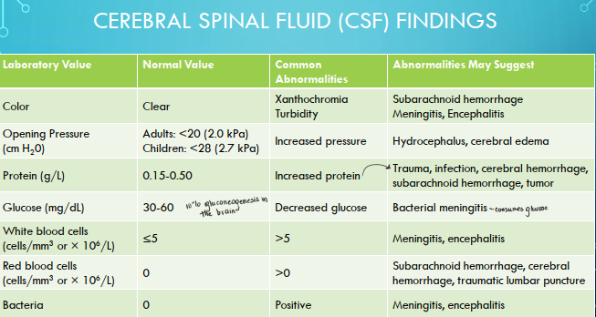
Image occlusion: name the hidden labels
34
Image occlusion: name the hidden labels
35
Image occlusion: name the hidden labels
36
Computed Tomography (CT)
• Uses x-rays to produce images
• Highly availability
• Can be done rapidly (can be usedin urgent situations)
• Used to identify strokes, tumors, hemorrhages, infarctions, hydrocephalus, atrophy
• IV contrast can aid in imaging of vessels to identify breakdown of blood-brain barrier
• Highly availability
• Can be done rapidly (can be usedin urgent situations)
• Used to identify strokes, tumors, hemorrhages, infarctions, hydrocephalus, atrophy
• IV contrast can aid in imaging of vessels to identify breakdown of blood-brain barrier
37
Magnetic Resonance Imaging
• Uses magnetic properties of hydrogen atom to produce computer-processed scans
• Used to identify lesions in white matter (e.g., plaques in multiple sclerosis, lesions in posterior fossa,
tumors, very early ischemic stroke
• Used to identify lesions in white matter (e.g., plaques in multiple sclerosis, lesions in posterior fossa,
tumors, very early ischemic stroke
38
Magnetic Resonance Angiography (MRA)
• Used to evaluate cerebral circulatory system
• Used to identify local arterial stenosis, aneurysms, vascular malformations
• Used to identify local arterial stenosis, aneurysms, vascular malformations
39
Positron Emission Tomography (PET), Single-Photon Emission Computed Tomography (SPECT), and Functional MRI (FMRI)
• Considered tests of brain function
• Studied extensively in epilepsy, cerebrovascular disorders, cerebral tumorsmovement disorders, and dementia
• Studied extensively in epilepsy, cerebrovascular disorders, cerebral tumorsmovement disorders, and dementia
40
Electroencephalogram (EEG)
• Records electrical activity of the brain
• Record is interpreted by observing the basic rhythms and waveforms, the symmetry of the recording, and abnormal electrical discharges
• Primarily used in diagnosis of seizures
• Most are about 30 minutes in length; however, long duration (>24 hours) are sometimes used
• Record is interpreted by observing the basic rhythms and waveforms, the symmetry of the recording, and abnormal electrical discharges
• Primarily used in diagnosis of seizures
• Most are about 30 minutes in length; however, long duration (>24 hours) are sometimes used
41
Evoked Potentials (EP)
• Measures response to electrical signals sent along sensory pathways
• Examines conduction along pathways
• Helpful for localizing lesions and evaluating hearing problems and multiple sclerosis
• Examines conduction along pathways
• Helpful for localizing lesions and evaluating hearing problems and multiple sclerosis
42
Nerve Conduction Velocity
• Measured by stimulating the nerve and recording the speed and amplitude of conduction of the impulse
• Used to detect presence of localized peripheral injuries, diffuse neuropathies, and neuromuscular junction disorders
• Used to detect presence of localized peripheral injuries, diffuse neuropathies, and neuromuscular junction disorders
43
Electromyography (EMG)
• A needle is placed in the muscle to measure its resting and contractual electrical activity
• Used to diagnose peripheral neuropathies, amyotrophic lateral sclerosis, radiculopathies and muscle disease
• Used to diagnose peripheral neuropathies, amyotrophic lateral sclerosis, radiculopathies and muscle disease
44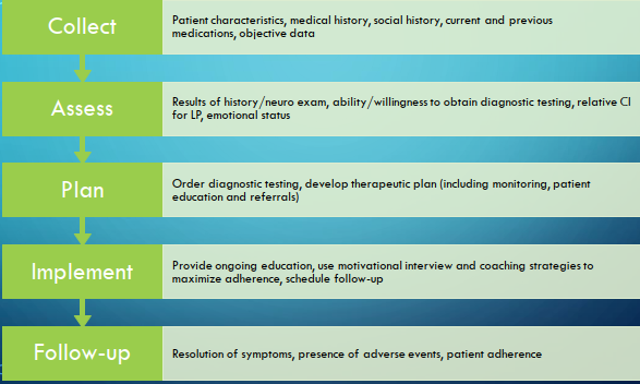
Image occlusion: name the hidden labels
45
Image occlusion: name the hidden labels
46
Image occlusion: name the hidden labels
47
Image occlusion: name the hidden labels
48
Image occlusion: name the hidden labels