BIOL 431 - LEC 4
Study these flashcardsFree · spaced repetition · iPhone, Android, Mac & web
All cards
1
What does ECG mean? and what is it?
ECG = electrocardiogram, recording of AP as they travel through heart, provieds condicyion pathway abnormalities and cardiomegaly ( enlargement)
2
What happens at each point?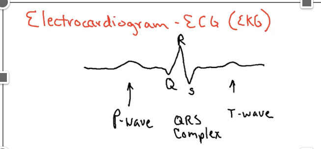
P= atrial depole, QRS= ventricular depole, T= ventricle repole
3
What wave is atrial contraction and what is significant?
P wave, SA through both atria, enlargement may indicate enlarged atrium
4
What wave is ventricular depole>?
QRS complex, begins 100msec after p wave begins, enlarged r way may mean enlarged ventricles
5
What wave is the repole of the ventricles and what happens here?
T wave, ventricles begin to relax, Flattened t wave = reduced o2 in mycardium (coronary artier disease)
6
What is the difference between interval and segment?
interval = deflection + time span, whereas segments is just the time span
7
What does the P-Q interval measure?
from p wave to beginning of QRS complex, conduction time between atrial excitation to ventricular excitation
8
What is the S-T segment?
end of S start of T, represents complete depole of contractile fibers, Elevated in acute MI and depressed in ischemia
9
What is the Q-T interval?
marks time from begging of ventricle depole to mend of ventricle repole, lengthened in mycardial damage, coronary ischemia, conduction pathway abnormalities
10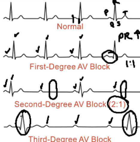
What represents the three different stages of heart block?
11
What is Systole?
contraction, act of forcing blood out, increasing pressure
12
What is diastole?
relaxation, act of filling with blood, decreased pressure
13
What happens at 1-8?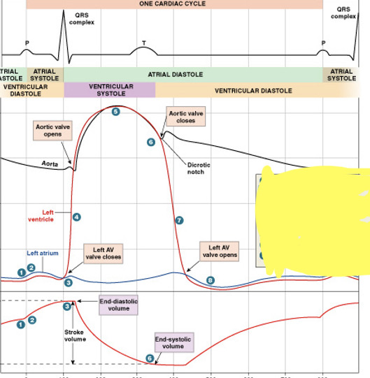
1. atrial contraction begins, 2. atria ejects blood into vent, 3. atrial systole ends:AV valves close, 4. isovolumetric ventricular contraction, 5. ventricular ejection, 6. semi lunar valve close, 7. isovolumetric relaxation, 8. AV valves open: passive atrial filling
14
What is the path of Atrial systole?
P wave starts when SA node fires, causing atrial depole, atrial contraction, increasing pressure last 30% pushed into ventricle
15
What is the path of ventricular systole?
QRS complex starts at end of atrial systole, AP goes to AV node the to bundles of his, vent depole, vent contraction, increased pressure, forced out through arteries
16
What is EDV or end diastolic volume mean?
volume f blood enters a ventricle during diastole (relaxtiation) 120-130ml
17
What is end systolic volume?
volume of blood left in the ventricle following its systole ( contraction 50-60ml) EDV - SV = ESV
18
What is stroke volume?
amount of blood ejected by vent during systole (at rest avg adult 70ml @75bpm)
19
What is the frank starlings law state?
When the HR begins to slow this will allow more blood to flow in increasing force of contraction
20
What is Cardiac output?
amount of blood eject by each ventricle/minute (stroke volume x beats per minute, 70ml/sec x 75 bpm = 5250ml/min or 5.25L/min = average)