All cards
1
metastasus of tumor cells occurs by three mechanisms...
Local spread/invasion (includes spread to coverings: pleura)
Via lymphatic channels
Via blood vessels (veins > arteries) to next capillary bed
Via lymphatic channels
Via blood vessels (veins > arteries) to next capillary bed
2
the _______ and ______ connect the thorax to the neck and abdomen
superior and inferior aperature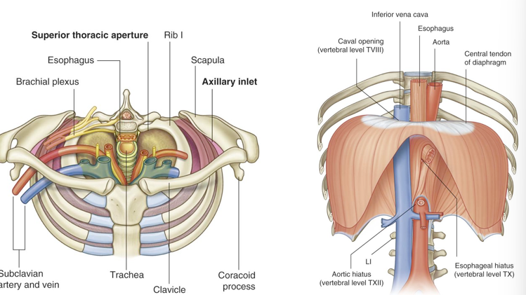
3
the costovertebral articulation is what type of joint?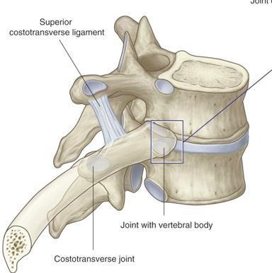
synovial plane
4
what are the true ribs
1-7
5
what are the false ribs
8-10
6
what are the floating ribs
11 and 12
7
what does this show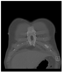
variation: sternal foramen
8
what type of joint is the costochondral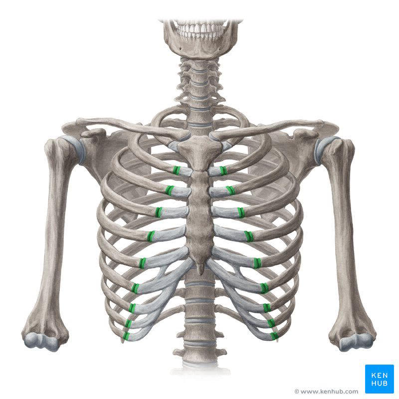
1* cartilaginous
9
what type of joint is the interchondral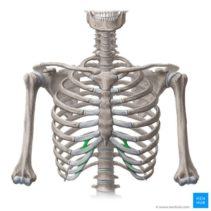
plane synovial
10
what type of joint is the sternocostal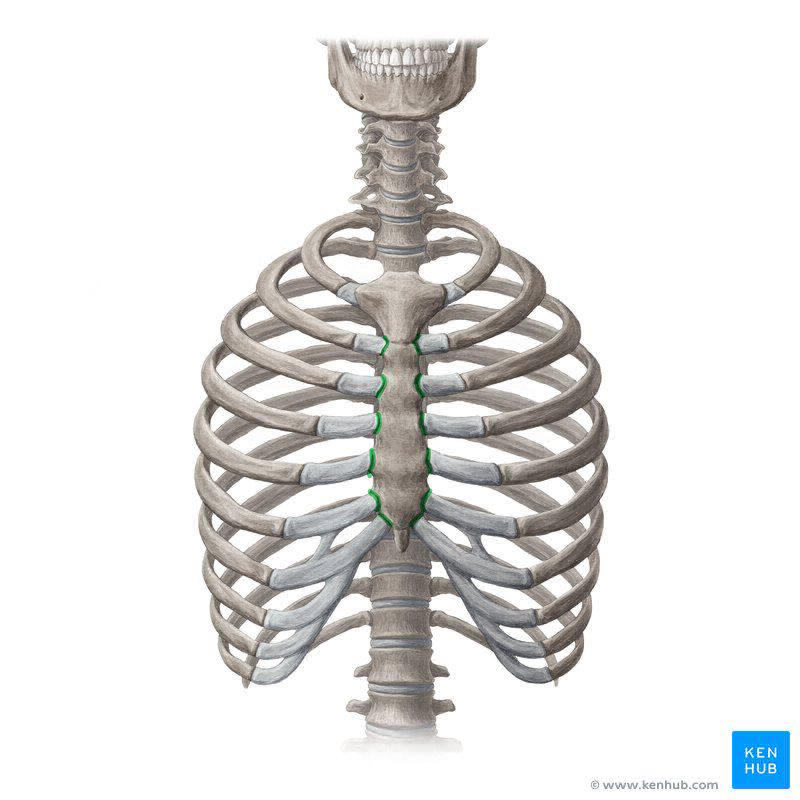
1- 1* cartilaginous
2-7: plane synovial
2-7: plane synovial
11
what type of joint is the manubriosternal?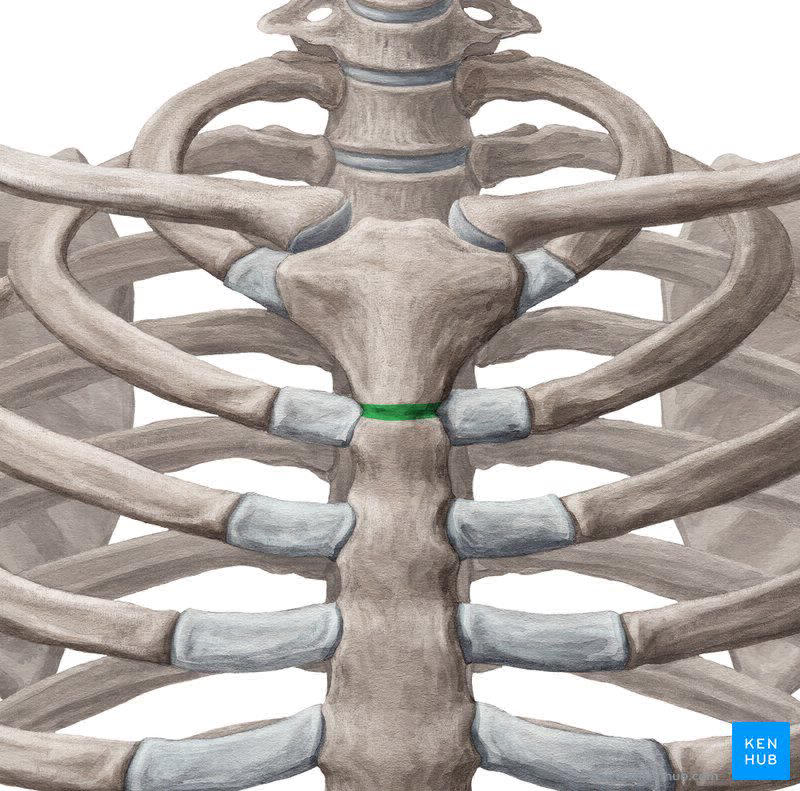
secondary cartilaginous (symphysis)
12
what does this show (red line)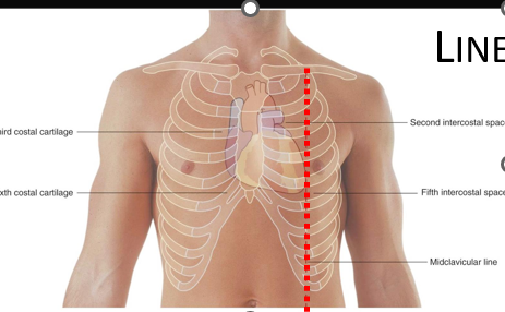
midclavicular line
13
what does this show? (red)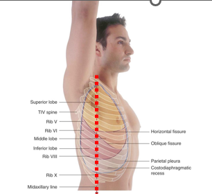
midaxillary line
14
what does this show (red)?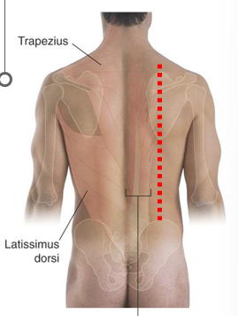
scapular line
15
innervation intercostal muscles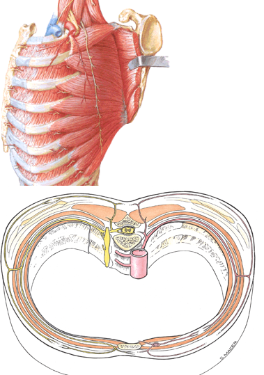
intercostal nerves from associated intercostal space
16
fx of external intercostals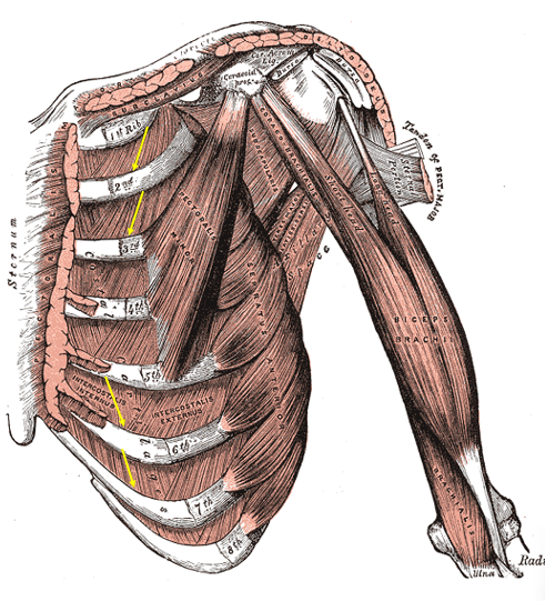
most active in inspiration, move ribs superior (elevate) (forced)
17
fx internal intercostals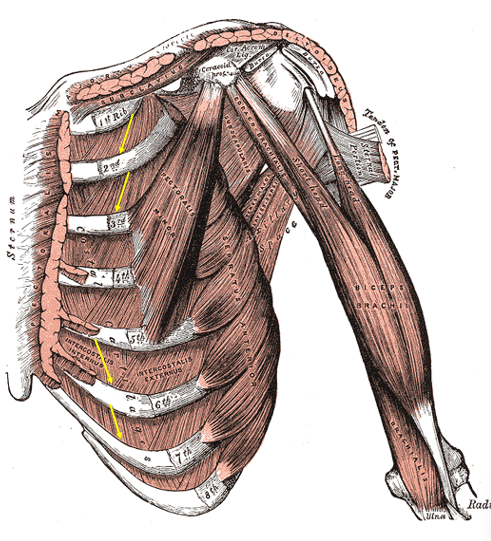
most actove in exproation, move ribs inferior (depress) (forced)
18
fx innermost intercostals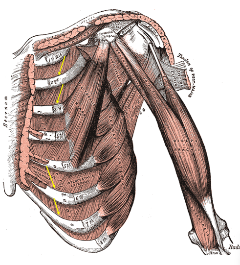
assist internal intercostals
19
O and I subcostal muscles
inner surface of ribs inserting at secoind or thoird rib below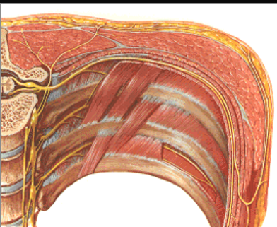
20
action subcostal muscles
depress ribs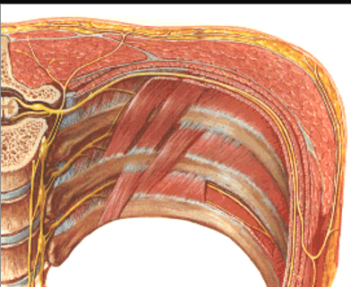
21
what innervates the subcostal muscles
intercostal nerves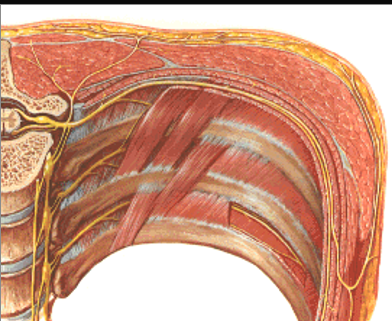
22
O and I transversus thoracis
deep surface of thoracic wall on inferior 1/3 sternum, xiphoid process, costal cartilages pf 2nd-6th rib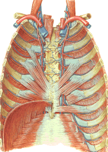
23
action transversus thoracis
draws ribs downward aiding in expiration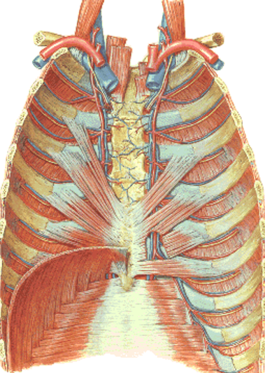
24
innervation of transversus thoracis
intercostal nerves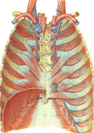
25
innervation diaphragm
motor: phrenic n: C3-5
sensory: centrally phrenic N and perpherally from intercostal and subcostal nerves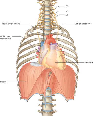
sensory: centrally phrenic N and perpherally from intercostal and subcostal nerves
26
these intercostals attach to the vertebrae but not the sternum
external intercostal m
27
these intercostals attach to the sternum but not to the vertebar
internal intercostal
28
these intercostals dont directly attach to the sternum or vertebrae
innermost intercostal m
29
what two muscles are the intercostal v, a, and n between?
internal and innermost intercostal m
30
during inhalation, diaphragm _____ and gut pressure ______
depresses; increases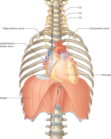
31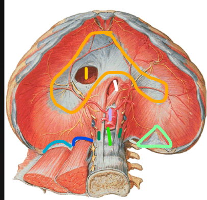
Image occlusion: name the hidden labels
32
Image occlusion: name the hidden labels
33
Image occlusion: name the hidden labels
34
Image occlusion: name the hidden labels
35
Image occlusion: name the hidden labels
36
Image occlusion: name the hidden labels
37
Image occlusion: name the hidden labels
38
Image occlusion: name the hidden labels
39
Image occlusion: name the hidden labels
40
what level is the caval hiatus, esophagea; hiatius, and aortic hiatus at
Caval: 8
esophageal: 10
aortic: 12
(I ate 10 eggs at noon)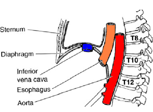
esophageal: 10
aortic: 12
(I ate 10 eggs at noon)
41
what lives in the caval hiatus (x2)
IVC and right phrenic n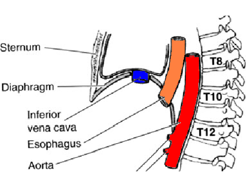
42
what lives in the esophageal hiatus (x3)
esophagus, vagal trunks, esophageal branches of left gastric a.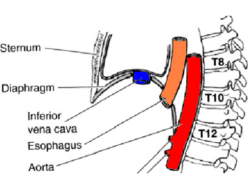
43
what lives in the aortic hiatus (x3)
aorta, thoracic duct, azygous v.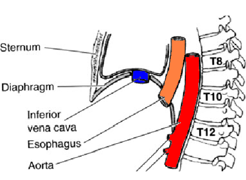
44
diaphragm action: increases volume will ___________ pressure in the thoracic cavity and decreases volume will ______ pressure in theabdominal cavity
decrease; increase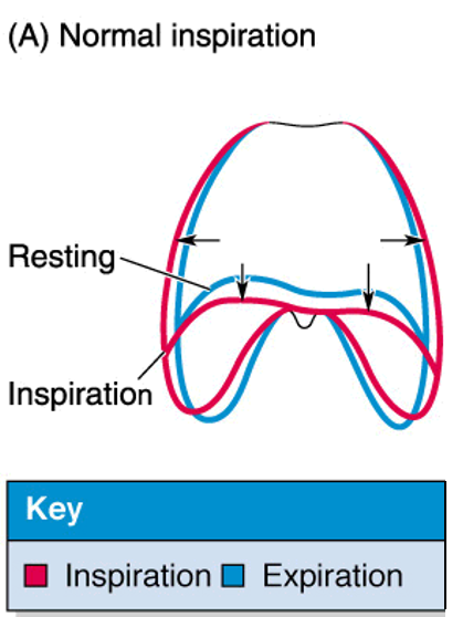
45
accessory muscles of respiration (x5)
Scalenes
Sternocleidomastoid
Pectoralis minor
Serratus anterior
Serratus posterior Superior and inferior
Sternocleidomastoid
Pectoralis minor
Serratus anterior
Serratus posterior Superior and inferior
46
what is at risk most in TOS
subclavian A, lower branch of brachial plexus
47
what does this show?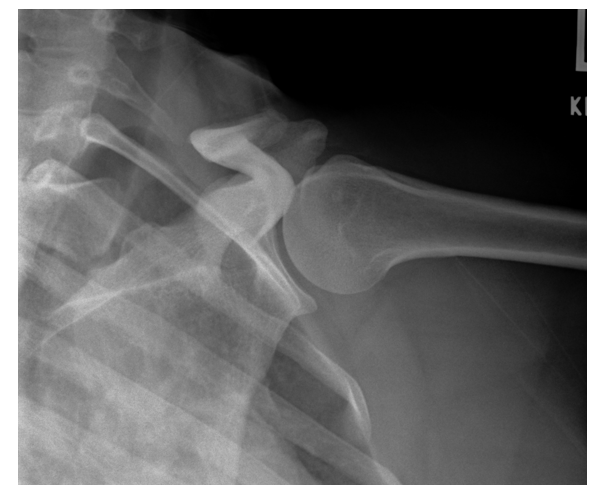
extra rib (cervical rib)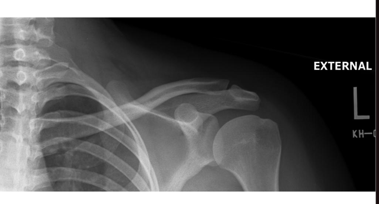
48
what provides collateral support to the thorax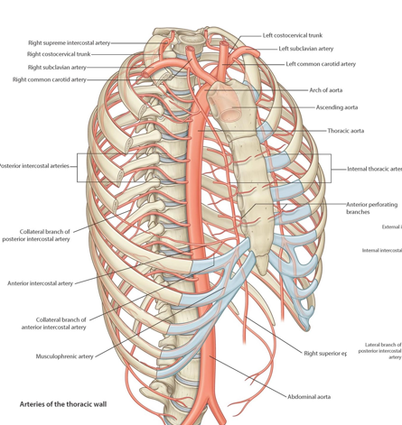
posterior and anterior intercostal arteries
49
where do the posterior and anterior intercostal veins drain
posterior interrosi to azygous veins to superoor vena cava
anterior interrosi to interal thoracic v to superior vena cava
anterior interrosi to interal thoracic v to superior vena cava
50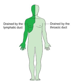
Image occlusion: name the hidden labels
51
Image occlusion: name the hidden labels
52
where would a needle be inserted for an intercostal nerve block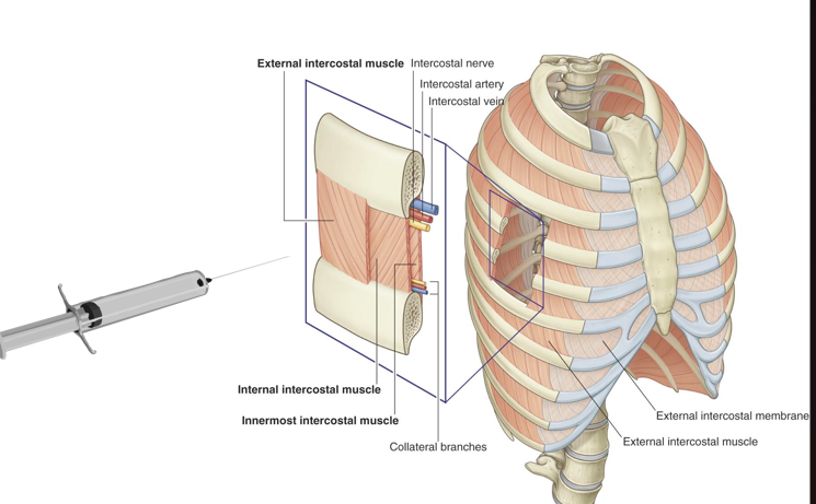
at bottom of rib/near bottom of rib
53
herniation where the gut goes through the diaphragm
diaphramatic hernia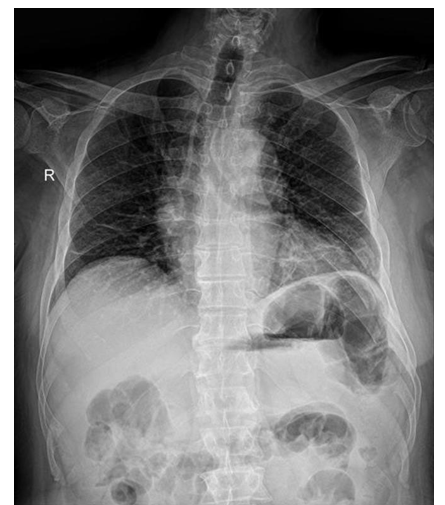
54
collapsed lung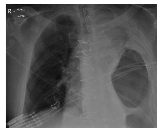
pneumothorax
55
lung tumor that can impinge on sympathetic chain and lower trunk of brachial plexus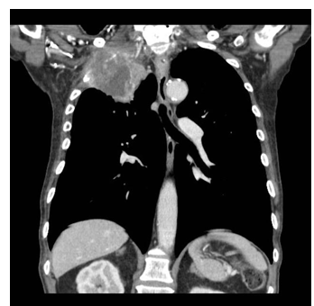
pancost tumor
56
three spaces of the thoracic cavity
right pulmonary
left pulmonary
mediastinum
left pulmonary
mediastinum
57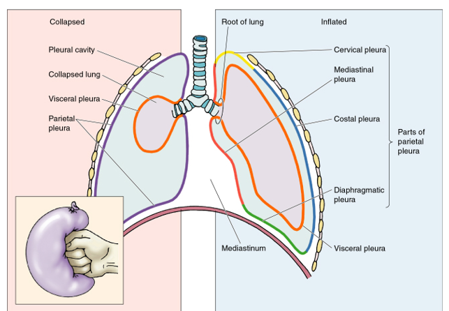
Image occlusion: name the hidden labels
58
Image occlusion: name the hidden labels
59
Image occlusion: name the hidden labels
60
Image occlusion: name the hidden labels
61
Image occlusion: name the hidden labels
62
Image occlusion: name the hidden labels
63
Image occlusion: name the hidden labels
64
inflamed pleuea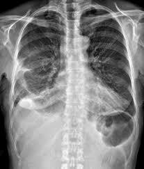
pleurisy/pleuritis
65
air into pleural cavity, lung partial or complete collapse (atelectasis); pain, difficulty breathing, shortness of breath (open, tension)
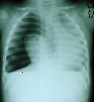
pneumothorax
66
diagnosis? location?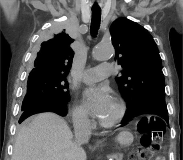
mesothelioma
parietal pleural layer of lung
parietal pleural layer of lung
67
ligament continuous between the visceral and parietal pleura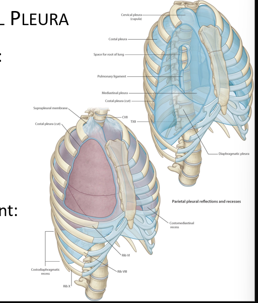
pulmonary ligament
68
thickening at the top of the ling that connects to the neck fascia
sibson's fascia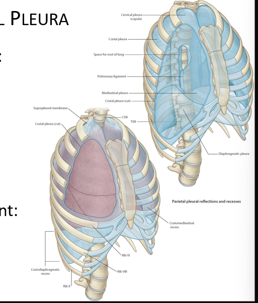
69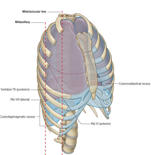
Image occlusion: name the hidden labels
70
Image occlusion: name the hidden labels
71
where is a thoracentesis done?
what layers does it go through?
what layers does it go through?
-midaxillary line at 9th intercostal space
-skin, superficial fascia, 3 layers intercostal muscles, parietal pleura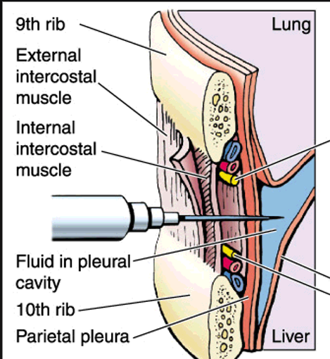
-skin, superficial fascia, 3 layers intercostal muscles, parietal pleura
72
where does the needle stop in a thoracocentesis?
costodiaphramatic recess
73
which of the pleuea is pain sensitive
parietal
74
which of the pleura receives blood from bronchiole vessels
visceral
75
which of the pleura receives blood from somatics
parietal
76
what innervates the costal and periphera; diaphragmatic pleura
intercostal n.
77
what innervates the mediastinal and central diaphragmatic pleura
phrenic n.
78
these can be removed from the lungs if they become canceroys because they are so vast
bronchiopulmonaru segments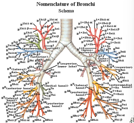
79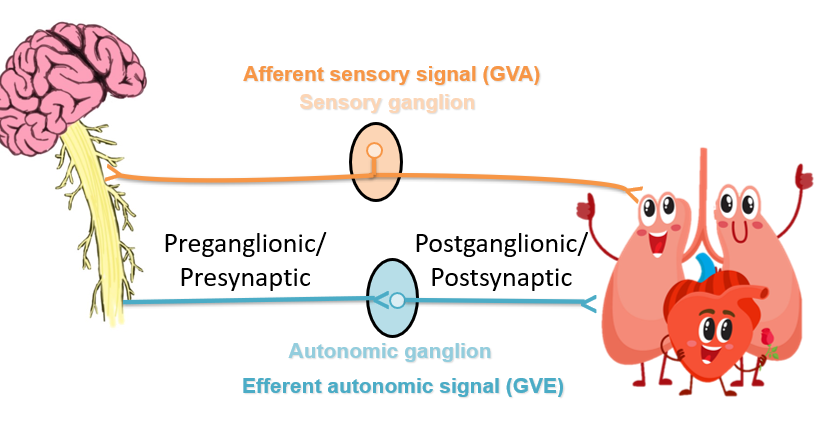
Image occlusion: name the hidden labels
80
Image occlusion: name the hidden labels
81
Image occlusion: name the hidden labels
82
Image occlusion: name the hidden labels
83
where are preganglionic sympathetic neurons situated
thoracolumbar spinal cord specifically intermediolateral (IML) cell column in the lateral horn
84
what is the end of the sympathetic trunk called
ganglion impar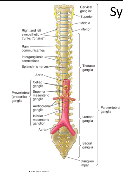
85
where is the lateral horn located (sympathetic)(also white ramus only here)
between T1-L2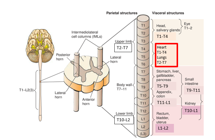
86
red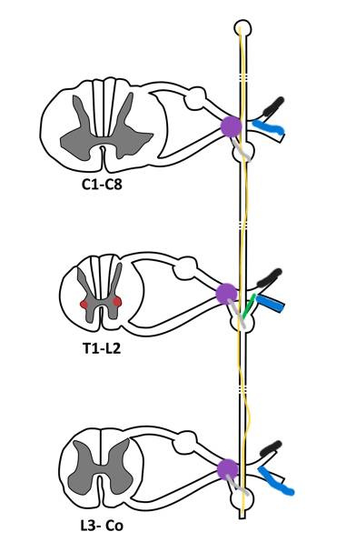
IML
87
yellow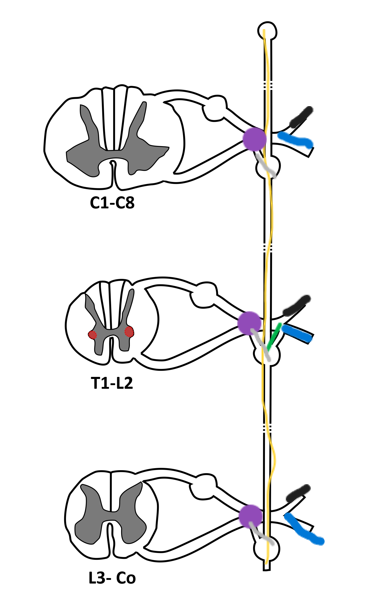
sympathetic trunk and ganglia
88
purple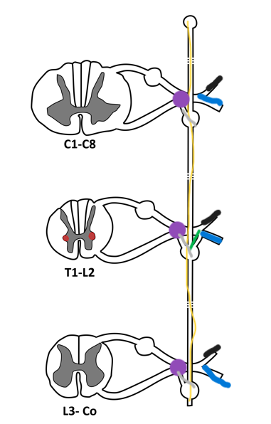
mixed spinal nerve
89
green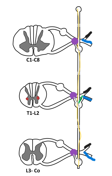
white rami (T1-L2)
90
gray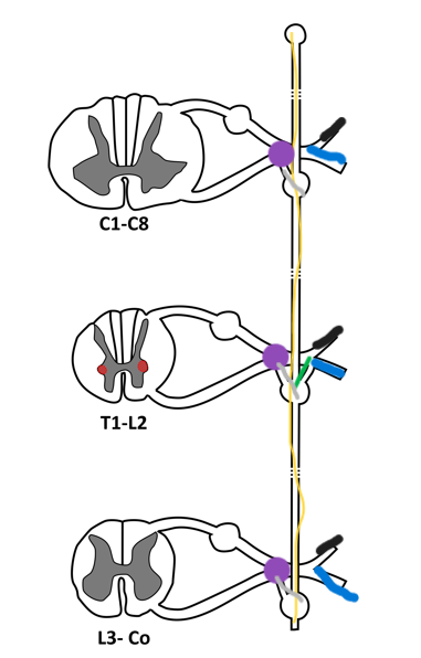
gray rami (post synaptic)
91
blue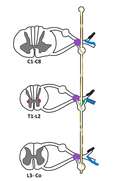
ventral ramus
92
black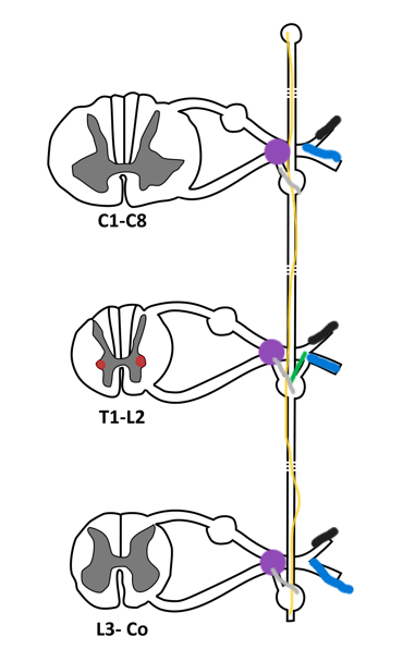
dorsal ramus
93
pathway of sympathetics to the body wall
leaves IML to white ramus and synapses in the chain with postganglionic. Cell body in the sympathetic chain that leaves throug gray rami and out dorsal and ventral rami at any level
94
pathway of sympathetics to viscera above diaphragm
pass through anterior root through white ramus and synapses in higher levels of the chain ganglia. Leave as visceral nerves
95
organs from ________________ receive sympathetic axos directly from the sympathetic chain
head, neck, and thorax
96
sympathetics to viscera below diaphragm pathway
pass through anterior root through white ramus. leaves without synapsing (thoracic splanchnic nerves). Cell body located in preaortic ganglia
97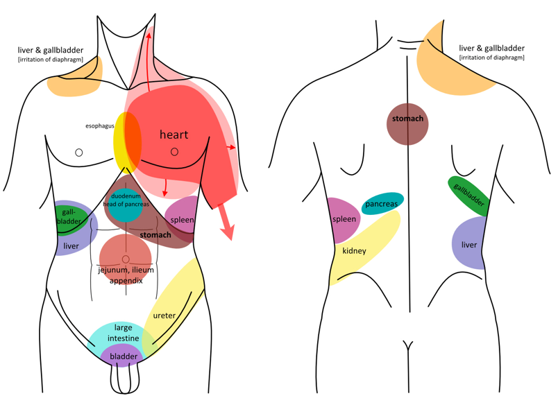
Image occlusion: name the hidden labels
98
Image occlusion: name the hidden labels
99
Image occlusion: name the hidden labels
100
Image occlusion: name the hidden labels
101
Image occlusion: name the hidden labels
102
Image occlusion: name the hidden labels
103
Image occlusion: name the hidden labels
104
Image occlusion: name the hidden labels
105
Image occlusion: name the hidden labels
106
Image occlusion: name the hidden labels
107
Image occlusion: name the hidden labels
108
Image occlusion: name the hidden labels
109
Image occlusion: name the hidden labels
110
Image occlusion: name the hidden labels
111
Image occlusion: name the hidden labels
112
where do parasympathetics distribute
external genitalia, deep strictures (organs)
113
where do parasympathetic axons reside from
craniosacral CNS
114
what does the vagus nerve provide parasympathetic innervation to
heart, lungs, foregut, hindgut
115
part of the heart that streamlines (directs) flow like a funnel through the pulmonaric valve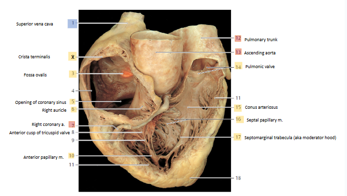
conus arteriosus
116
these arteries supply blood to the broncus and lungs
bronchial arteries
117
a condition where a blood clot travels to the lungs and blocks one or more pulmonary arteries.
-results in obstucted blood flow to lung, infarct and respiractory distress
-results in obstucted blood flow to lung, infarct and respiractory distress
pulmonary embolism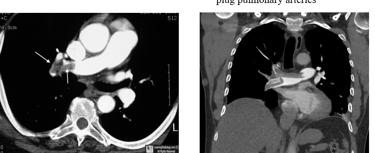
118
increased pressure in pulmonary veins, congestive heart faiulure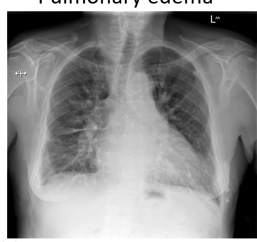
pulmonary edema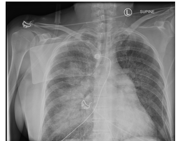
119
accumulation of fluid in the pleural cavity
pleural effusion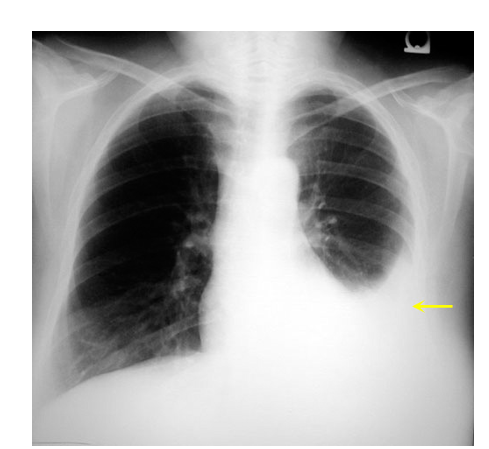
120
what does the base of the pericardiym attach to? anteriory? posteriorlu?
base: pericardiophrenic ligament
anterior: sternopericardial ligaments
posterior: posterior mediastinum by loose connecrive tissue
anterior: sternopericardial ligaments
posterior: posterior mediastinum by loose connecrive tissue
121
where does the percardium recieve its blood supply
thoracic aorta, internal thoracic a.
122
what drains the pericrdium
azygos system
123
what innervates the pericardium
Vagus nn
Fnct unknown
Phrenic nn
Sensory pain fibers only in parietal
Sympathetic trunks
Vasomotor
Fnct unknown
Phrenic nn
Sensory pain fibers only in parietal
Sympathetic trunks
Vasomotor
124
this can be used as an area to clamp if you want to cut off blood supply to the great vessels of the heart
transverse pericardial sinus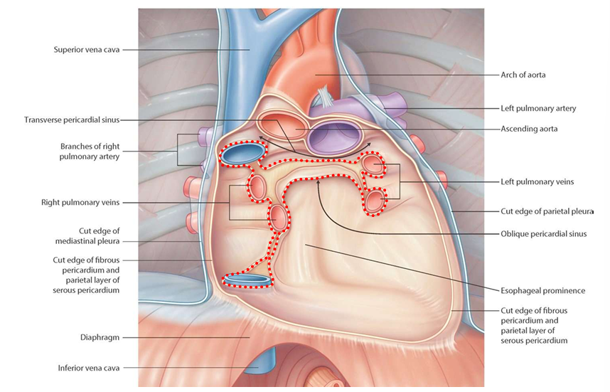
125
inflamation of the pericardium, patients present with abbrupt, pleuritic, positioned left precordial chest pain after a viran prodrome
pericarditis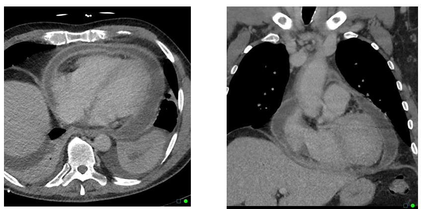
126
fluid in the pericardial sac, can restrict beating of the heart
early signs are dyspnea and reduced exercise tolerance
early signs are dyspnea and reduced exercise tolerance
pericardial efffusion (cardiac tamponade)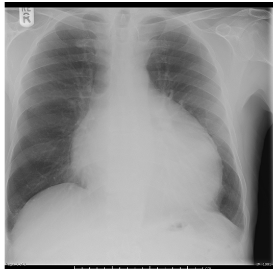
127
what is the posterior right atrium (sinus venarum) derived from
right horn of sinus venosus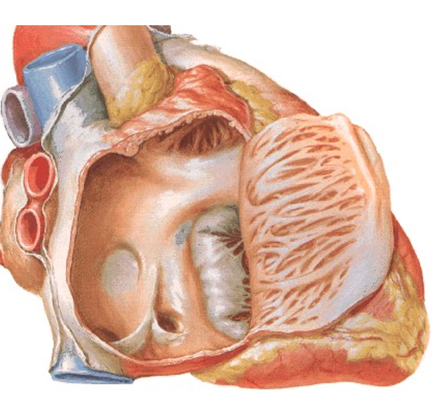
128
what is the anterior right atrium derived from
atrium proper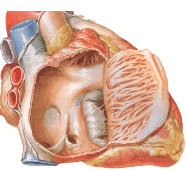
129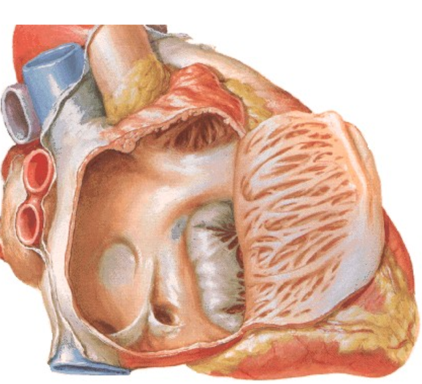
Image occlusion: name the hidden labels
130
Image occlusion: name the hidden labels
131
Image occlusion: name the hidden labels
132
this receives blood from the coronary veins of the heart and places it in the right atrium
coronary sinus
133
this is the location of the AV node and nodal bundles in the septal wall of the right atrium
triangle of koch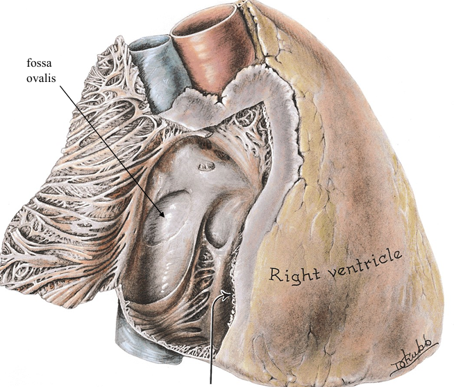
134
this is the obliterated (fetal reminant) of the foramen ovale in the septal wall of the right atrium
fossa ovalis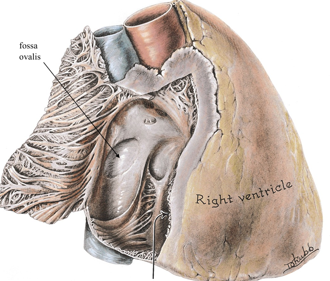
135
This is a defect in the atrium or septal wall that causes oxygenated blood to mix with deoxy blood of the heart
atrial septal defect (or ventral septal defect) caused by patent fossa ovalis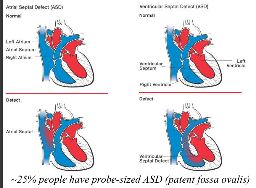
136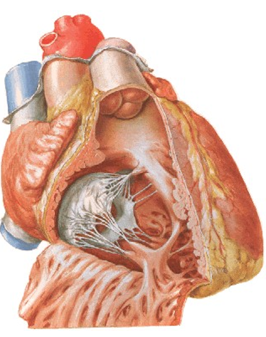
Image occlusion: name the hidden labels
137
Image occlusion: name the hidden labels
138
Image occlusion: name the hidden labels
139
Image occlusion: name the hidden labels
140
Image occlusion: name the hidden labels
141
valve is between the heart's two right chambers. It has three thin flaps of tissue, called cusps or leaflets.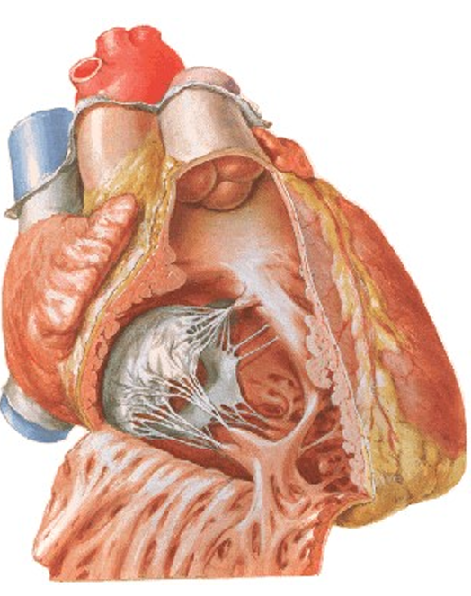
tricuspid valve
142
what is the left atrium derived from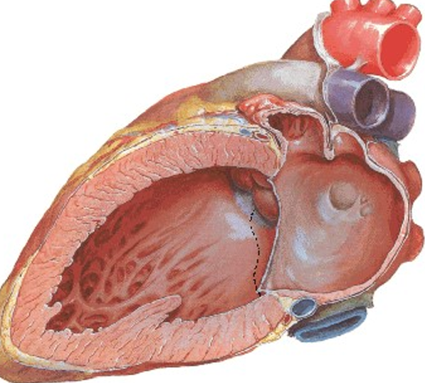
incorporation of pulmonary veins
143
what does the left atrium receive blood from
4 pulmonary veins and foramina venarum minmarum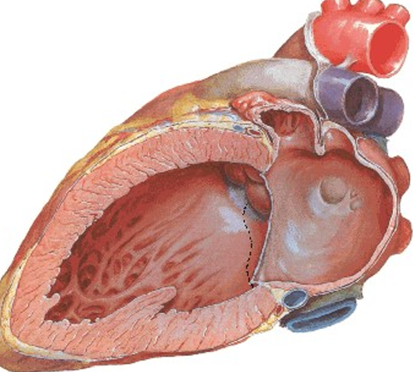
144
one of the four heart valves, located between the left atrium and left ventricle, that ensures blood flows in one direction. It has two flaps, or leaflets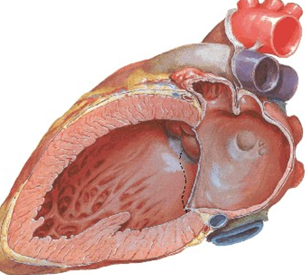
mitral valve
145
what valves are open during systole?
pulmonary and aortic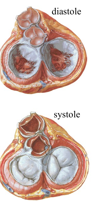
146
what valves are open during diastole
mitral and tricuspid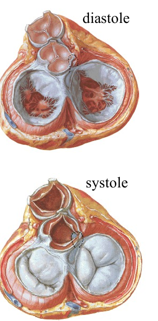
147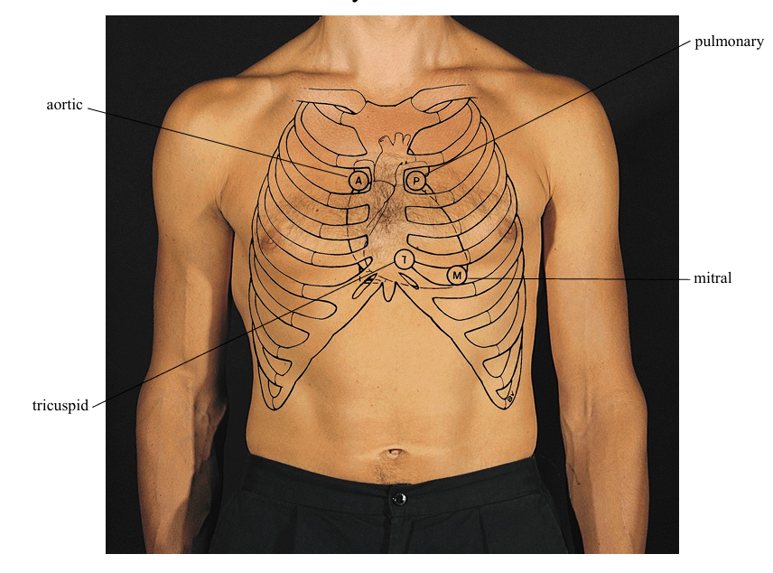
Image occlusion: name the hidden labels
148
Image occlusion: name the hidden labels
149
Image occlusion: name the hidden labels
150
Image occlusion: name the hidden labels
151
pathway of cardiac conducting
SA node
AV node
bundle of his
right and left bundle
papillary muscles and ventricular base (purkinje fibers)
AV node
bundle of his
right and left bundle
papillary muscles and ventricular base (purkinje fibers)
152
functions of the cardiac skeleton (x4)
1. Insulation – creates
an electrical
discontinuity
between atria and
ventricles
2. Attachment for
myocardium
3. Prevent bulging as
blood is pumped
4. Provides stable but
deformable base for
attachment of valve
leaflets
an electrical
discontinuity
between atria and
ventricles
2. Attachment for
myocardium
3. Prevent bulging as
blood is pumped
4. Provides stable but
deformable base for
attachment of valve
leaflets
153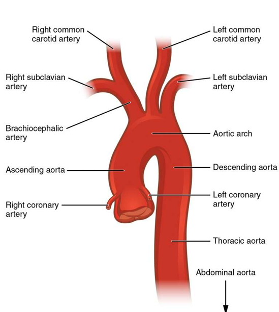
Image occlusion: name the hidden labels
154
Image occlusion: name the hidden labels
155
Image occlusion: name the hidden labels
156
Image occlusion: name the hidden labels
157
Image occlusion: name the hidden labels
158
what does the right coronary a supply
right atrium and ventricles, left AV septum
159
branches of the right coronary a
-atrial and ventricular aa.
marginal artery
posterior interventricular a. (posterior descending; a.k.a. PAD)
SA nodal artery
marginal artery
posterior interventricular a. (posterior descending; a.k.a. PAD)
SA nodal artery
160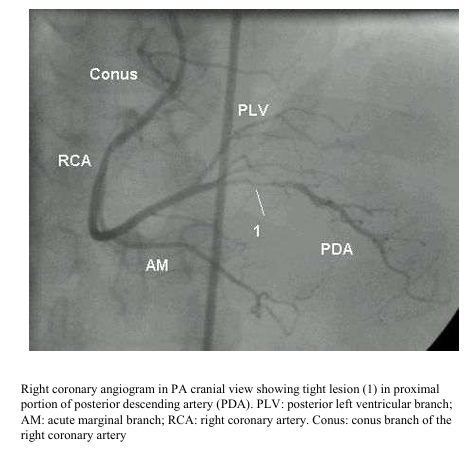
Image occlusion: name the hidden labels
161
Image occlusion: name the hidden labels
162
Image occlusion: name the hidden labels
163
Image occlusion: name the hidden labels
164
Image occlusion: name the hidden labels
165
supplies ledr atrium, centricles,, and mosr of the IV septum
left coronary a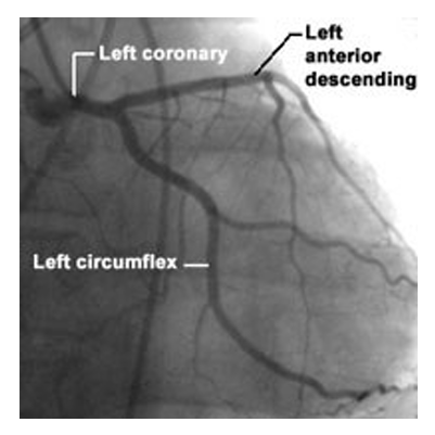
166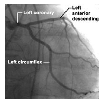
Image occlusion: name the hidden labels
167
Image occlusion: name the hidden labels
168
Image occlusion: name the hidden labels
169
what nodes drain the breasts
axillary (primary)
parasternal node
abdominal nodes
parasternal node
abdominal nodes
170
where does lymph from skin/fascia of the thoracic wall drain
axillary nodes
parasternal nodes
parasternal nodes
171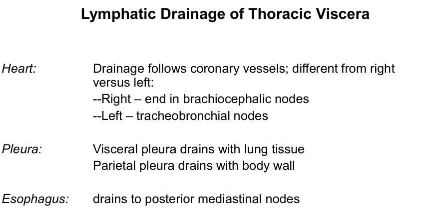
Image occlusion: name the hidden labels
172
Image occlusion: name the hidden labels
173
Image occlusion: name the hidden labels
174
Image occlusion: name the hidden labels
175
Image occlusion: name the hidden labels
176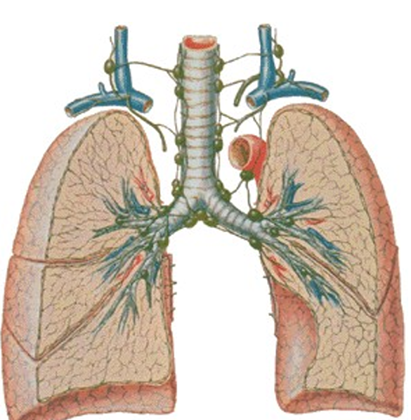
Image occlusion: name the hidden labels
177
Image occlusion: name the hidden labels
178
Image occlusion: name the hidden labels
179
Image occlusion: name the hidden labels
180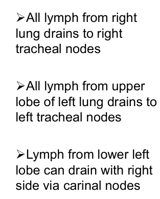
Image occlusion: name the hidden labels
181
Image occlusion: name the hidden labels
182
Image occlusion: name the hidden labels
183
greater splanchnic n spinal level
T5-T9
184
lesser planchnic n spinal level
T10 and 11
185
least splanchnic n spinal level
T12
186
what supplies the parasympathetic innervation to the heart? sympathetic?
vagus n; T1-T4
187
what defines the superior mediastinum
Tv1—Rib1
Sternal angle—Tv4-5 disc
Sternal angle—Tv4-5 disc
188
what supplies the thymus (blood)
internal thoracic and thyroid vessels
189
fx of thymus
differentiation of
lymphocytes and maintenance
of this population; essential for
the development of lymphoid
tissue
lymphocytes and maintenance
of this population; essential for
the development of lymphoid
tissue
190
branches of left branchiocepahallic v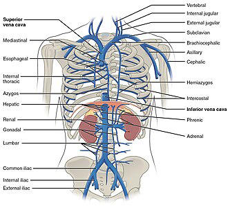
left internal jugular vein and the left subclavian vein. It also receives blood from smaller tributaries, including the left vertebral vein, left internal thoracic vein, left inferior thyroid vein, left superior intercostal vein, left supreme intercostal vein, and thymic veins.
191
branches of right brachiocephalic v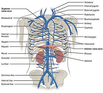
the right vertebral vein, the right internal thoracic vein, and the right inferior thyroid vein. Additionally, it can receive blood from the right supreme posterior intercostal vein of the first intercostal space and other veins like the pericardiacophrenic, sternal, and anterior mediastinal veins.
192
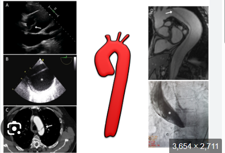
a congenital (1 in 2000),
abnormal stenosis of
aorta; typically occurs
near ligamentum
arteriosum
good collateral
circulation can develop
to deliver blood around
blockage, these
enlarged arteries will
cause notching and
erosion of bordering ribs
abnormal stenosis of
aorta; typically occurs
near ligamentum
arteriosum
good collateral
circulation can develop
to deliver blood around
blockage, these
enlarged arteries will
cause notching and
erosion of bordering ribs
coarctation of the aorta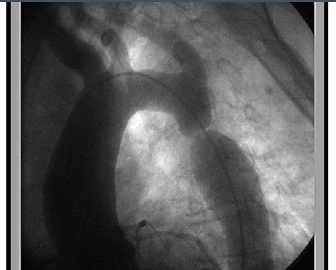
193
what acts as collateral circulation in cases of a blocked ascending aorta/stenosis/coarctation of the aorta
intercostals, vertebral, scapular, epigastric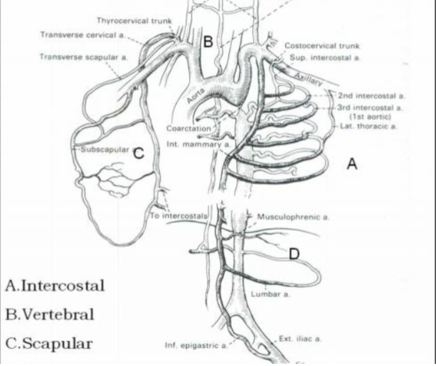
194
what are two things that are in danger od compressing the trachea
aortic arch and brachiocephallic trunk
195
what branches off the thoracic aorta (x6)
1. Bronchial
2. Pericardial
3. Intercostal/subcostal
4. Superior phrenic
5. Esophageal
6. Mediastinal
2. Pericardial
3. Intercostal/subcostal
4. Superior phrenic
5. Esophageal
6. Mediastinal
196
what structures are able to compress the esophagus (x3)
aortic arch, left main bronchus, diaphragm
197
venous channels of the posterior mediastinum that drain the body wall; highly variable
azygous system: intercostal; hemiazygous, accessory hemiazygous
198
this can provude an alterbate venous route to the heart in cause of IVC blockage
azygous veins
199
a depression of the sternum; “funnel chest”; associated with exercise
intolerance, dyspnea, chest pain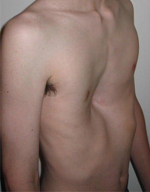
intolerance, dyspnea, chest pain
pectus excavatum
200
an anterior protrusion of the sternum; “pigeon chest”; associated with
dyspnea, frequent respiratory tract infections; commonly found with scoliosis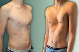
dyspnea, frequent respiratory tract infections; commonly found with scoliosis
pectus carinatum
201
what are some signs/ symptoms of thoracic outlet symdrome
a, compression of C8 and T1 roots of brachial plexus, see UL
b. vascular compression (subclavian artery) = cold fingers, color changes in hands, claudication,
pain
b. vascular compression (subclavian artery) = cold fingers, color changes in hands, claudication,
pain
202
a severe, potentially life-threatening injury caused by multiple rib fractures, where a segment of the chest wall separates and moves paradoxically during breathing, leading to difficulty breathing and impaired oxygenation
flail chest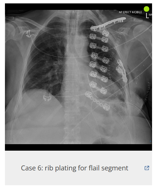
203
what nerves are the most at risk of injury when an intercostal nerve block is performed
intercostal and collateral branch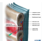
204
60% of malignant brest tumors arise from _______ of breast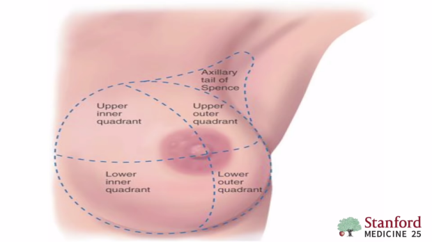
superior lateral quadrant
205
structures most at risk during masectomy or radical dissection of diseased axillary lymph nodes (x3
long thoracic nerve
intercostalbrachial n
pectoral n
intercostalbrachial n
pectoral n
206
where should the stethescope be placed to listen to breath sounds from the superior lobes
anterior wall
-R: above the 3rd rib
- L: above the 5th rib
-R: above the 3rd rib
- L: above the 5th rib
207
where should the stethescope be placed if yoy want to listen to breath sounds from the middle lobe
anterior chest wall below 4th rib
208
where should the stethescope be placed if you want to listen to breath sounds from the lower lobes
posterior chest wall (scapulae line) below 5th rib
209
where would a chest tube be inserted
purpose: drain large amount of air, fluid, etc from pleural space
purpose: drain large amount of air, fluid, etc from pleural space
4th or 5th intercostal space near midaxillary line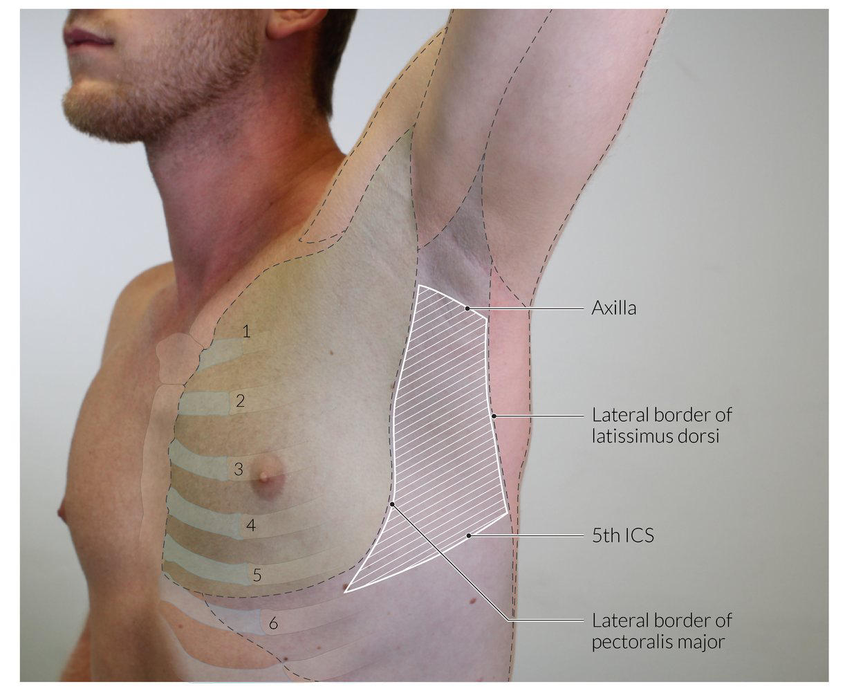
210
common sites of metastases from the lungs (x4)
central nervous system (brainstem and cerebellum)
bone (vertebrae)
liver
adrenal gland
bone (vertebrae)
liver
adrenal gland
211
collapse of lung or lobe resulting from blockage of some part of the airway; the opening to
the right middle lobe is the smallest/most narrow, as a result the R middle lobe is the most commonly
affected; obstructive atelectasis may cause mediastinal shift towards the affected lung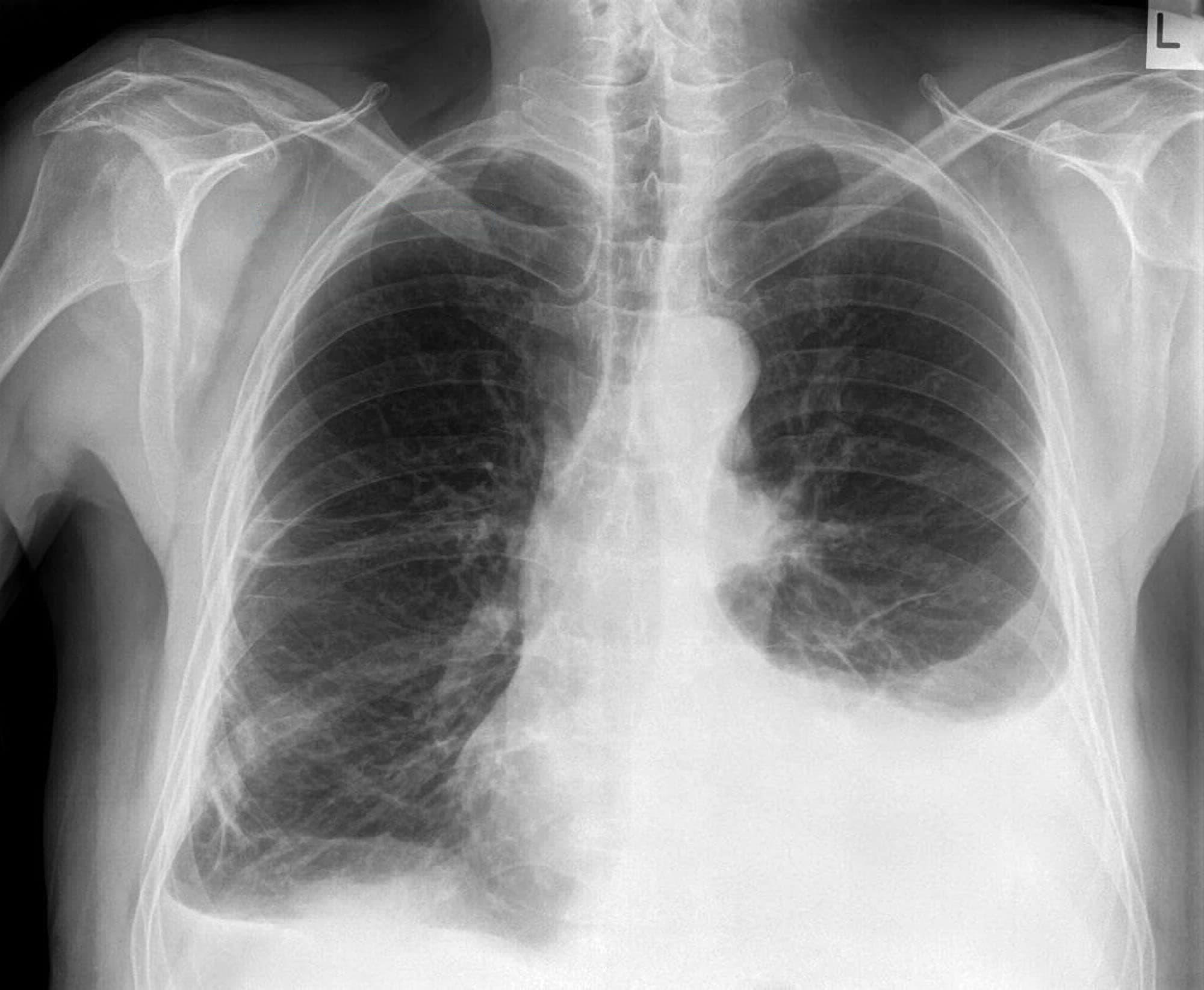
the right middle lobe is the smallest/most narrow, as a result the R middle lobe is the most commonly
affected; obstructive atelectasis may cause mediastinal shift towards the affected lung
atelectasis
212
spitting up blood”; bleeding from the airway from bronchial a >> pulmonary arteries;
hemoptysis
213
what could be damaged in pericardocentesis
internal thoracic artery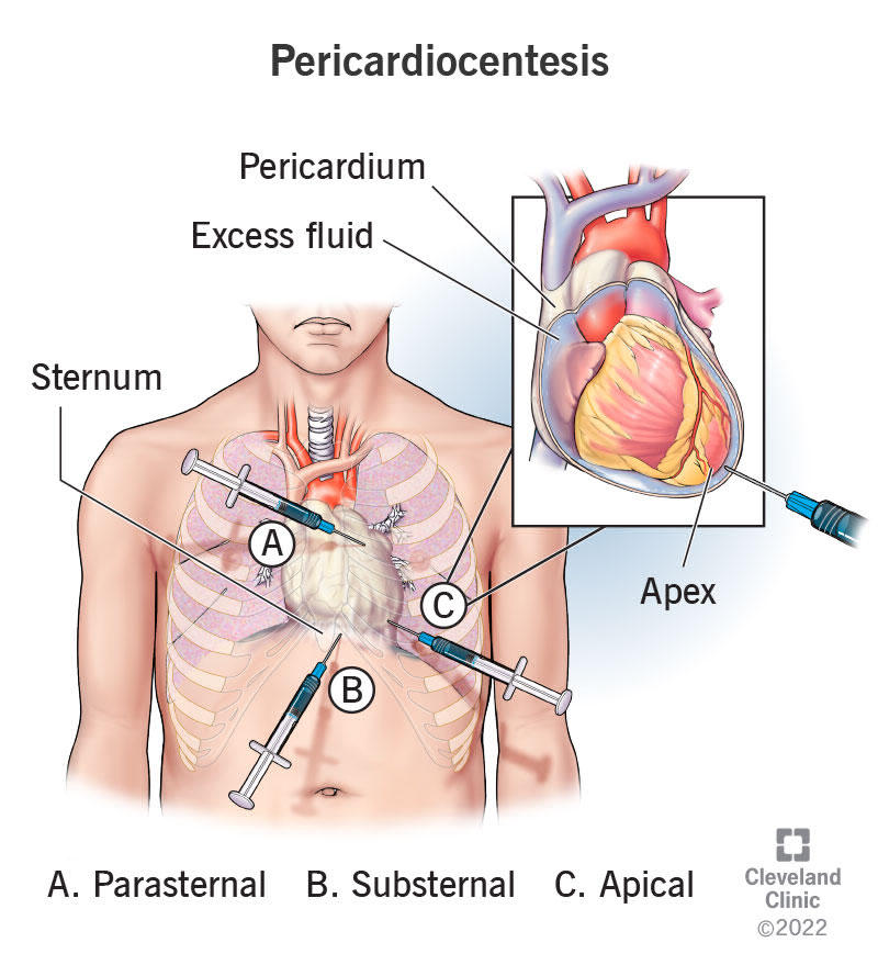
214
Compression of the heart from accumulated fluid in the pericardial sac; associated with beck's triad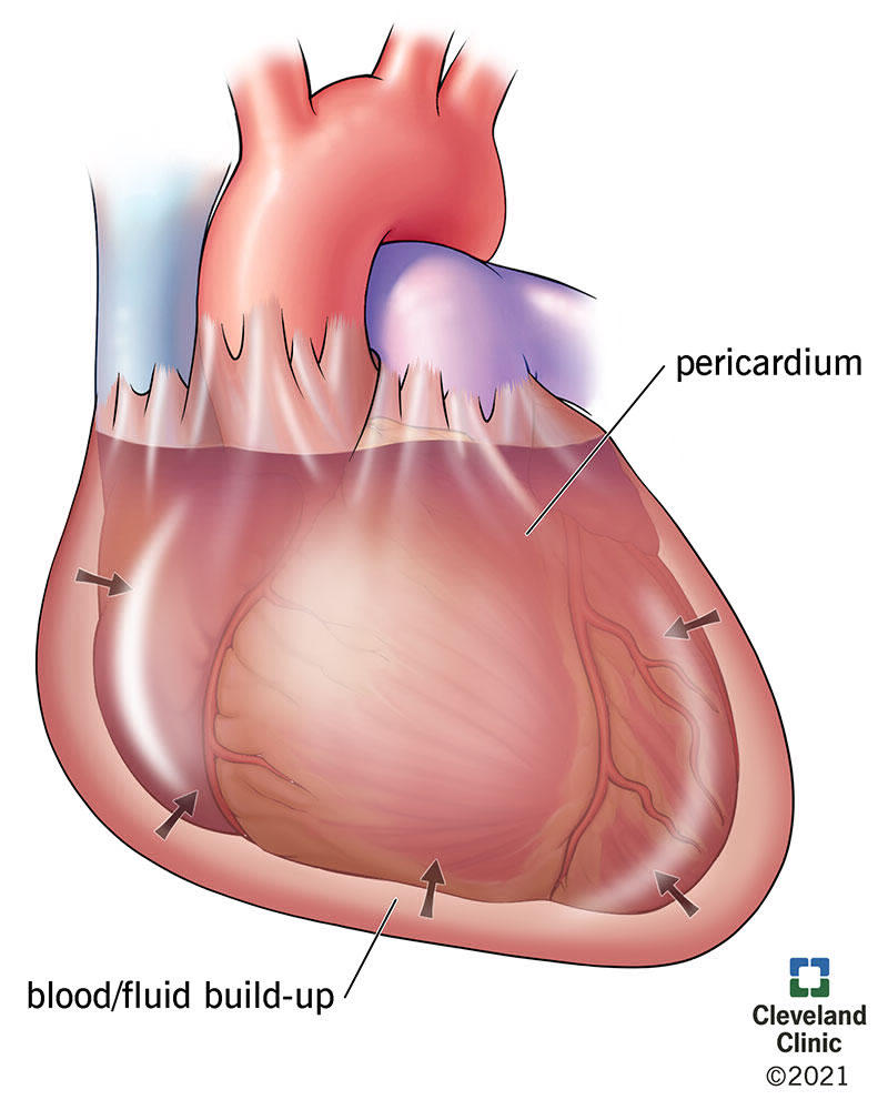
cardiac tamponade
215
what is beck's triad
-rapid heartbeat & muffled heart sounds
- distended neck veins
- hypotension/weak pulses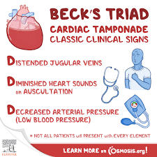
- distended neck veins
- hypotension/weak pulses
216
3 most common sites of occlusion in the coronary arteries
anterior interventrucular (LAD)
right coronary artery
left circumflex
right coronary artery
left circumflex
217
chest pain from the heart resulting from insufficient supply of oxygen to cardiac muscle
angina pectoris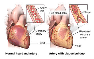
218
this results in a dissociation between the atrial and ventricylar contractions
complete bundle block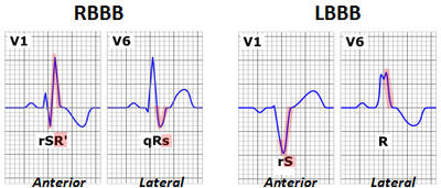
219
compression of the left recurrent laryngeal nerve from a cardiovascular etiology:
enlarged left atrium (from mitral valve stenosis), aneurysm of the aortic arch
a. Hoarseness > dysphagia > dyspnea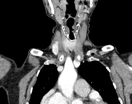
enlarged left atrium (from mitral valve stenosis), aneurysm of the aortic arch
a. Hoarseness > dysphagia > dyspnea
ortner syndrome
220
what three points is the aorta fixed at: suscepatbale to damage from MVA 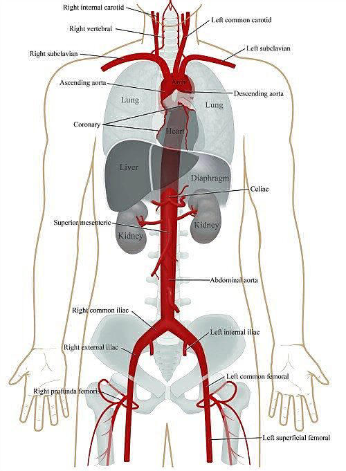
aortic valve
ligamentum arteriosum
aortic hiatus
ligamentum arteriosum
aortic hiatus
221
creation of space within the wall of the aorta; this space can accumulate significant
amounts of blood resulting in strokes and/or infarcts
a. tearing of the tunica intima and subsequent accumulation of blood volume in the wall of the vessel
b. signs and symptoms: hypotension, exsanguination, stroke, ischemia, claudication, compression (mass effect)
amounts of blood resulting in strokes and/or infarcts
a. tearing of the tunica intima and subsequent accumulation of blood volume in the wall of the vessel
b. signs and symptoms: hypotension, exsanguination, stroke, ischemia, claudication, compression (mass effect)
aortic dissection