Histo Exam 2
Study these flashcardsFree · spaced repetition · iPhone, Android, Mac & web
All cards
1
bone cells located in lacunae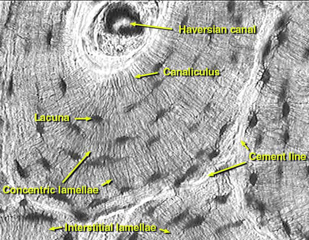
osteocyte
2
bone making cells
osteoblasts
3
bone precursor cells
osteoprogenitor cells
4
bone destroying cells
osteoclasts
5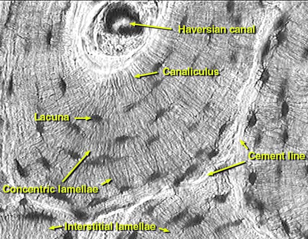
Image occlusion: name the hidden labels
6
Image occlusion: name the hidden labels
7
Image occlusion: name the hidden labels
8
Image occlusion: name the hidden labels
9
Image occlusion: name the hidden labels
10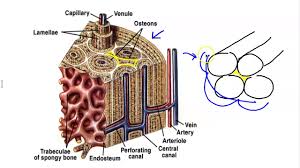
Image occlusion: name the hidden labels
11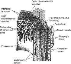
Image occlusion: name the hidden labels
12
where can immature bone be found?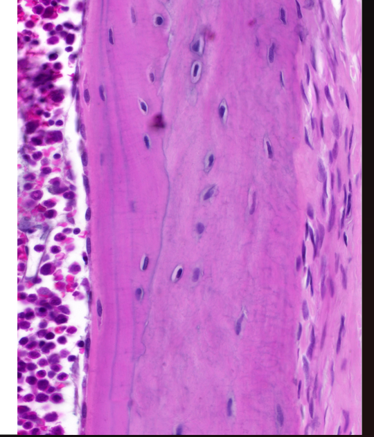
the skeleton of the developing fetus, adult alveolar sockets, tendon attachments
13
Collagenous membrane on the outer surface of the bone
Carries vessels and nerves, contains nociceptors
(right)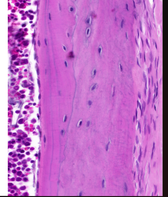
Carries vessels and nerves, contains nociceptors
(right)
periosteum
14
Covering the marrow cavity
Composed of mostly osteoprogenitor cells
(left)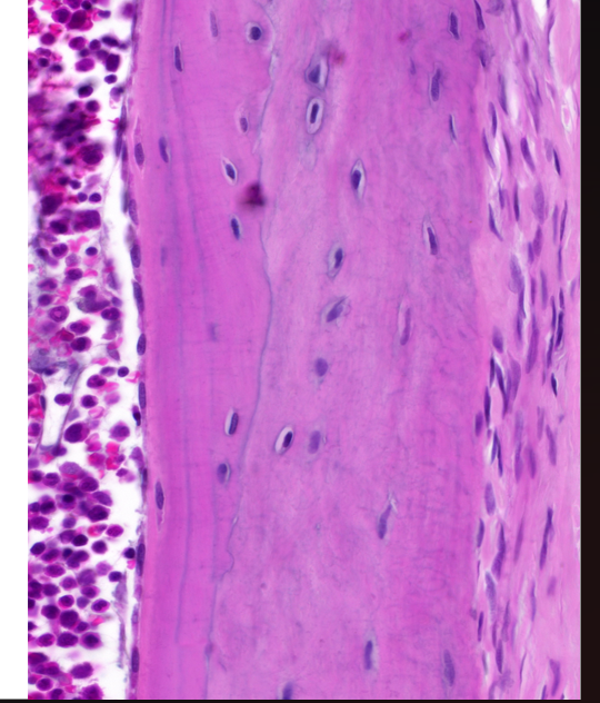
Composed of mostly osteoprogenitor cells
(left)
endosteum
15
where can osteoprogenitor cells be found?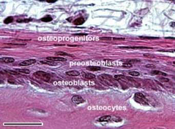
Inner layer of periosteum
Endosteum (endosteal cells)
Line the Haversian and Volkmann’s canals
Endosteum (endosteal cells)
Line the Haversian and Volkmann’s canals
16
cell shown in the upper right corner in semi cuboidal shape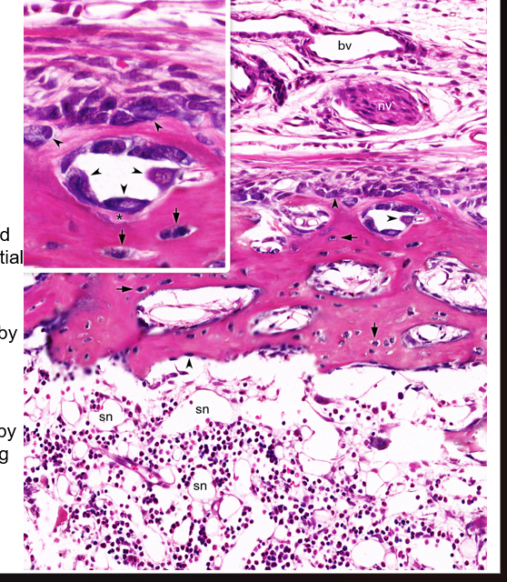
osteoblasts
17
what are osteoblasts called when surrounded by osteoid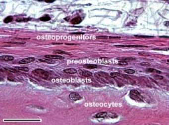
osteocyte
18
function of osteocytes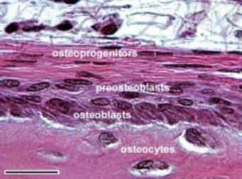
maintain matrix (secreteeabsorbs)
19
these osteocutes have few organelles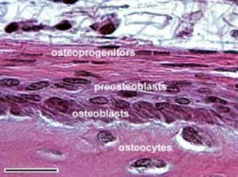
inactive
20
these osteocytes have rER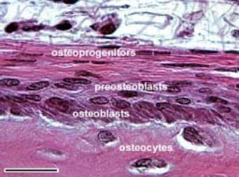
formative osteocytes
21
these osteocytes contain lyosomes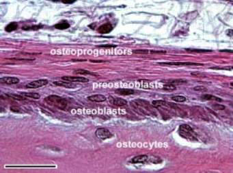
resorptive osteocytes
22
where do osteoclasts originate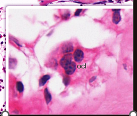
monocytes (macrophages) of blood)
23
white in image next to osteoclast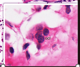
howship lacuna
24
story of intramembranous ossification
Mesenchymal cells aggregate and turn into osteoblasts
Osteoblasts secrete osteoid, mineralize it, enclose themselves into lacunae and turn into osteocytes
Remaining mesenchyme forms periosteum on the surface of the bone
Osteoblasts secrete osteoid, mineralize it, enclose themselves into lacunae and turn into osteocytes
Remaining mesenchyme forms periosteum on the surface of the bone
25
where does intramembranous ossification occur?
embryo skull, small fractures
26
what is the story of enchondral ossification?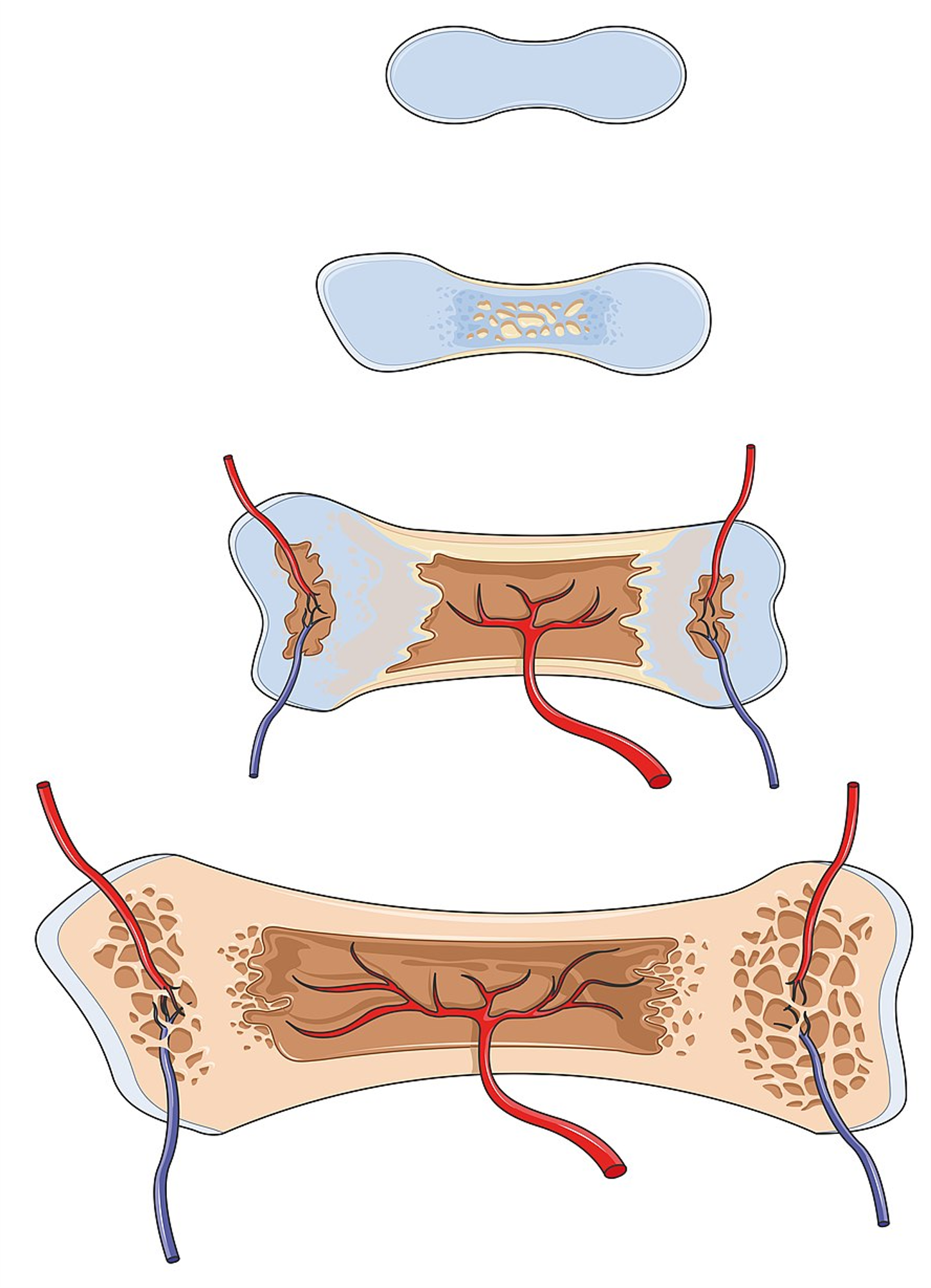
hyaline cart bone where perichondrium forms bone collar around cartilage. ossification centers develop and chondrocytes die. capilary loops break into bone, osteoblasts make bone, epiphyseal plate forms bone in opposite directions
27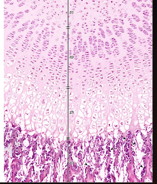
Image occlusion: name the hidden labels
28
Image occlusion: name the hidden labels
29
Image occlusion: name the hidden labels
30
Image occlusion: name the hidden labels
31
white structure in image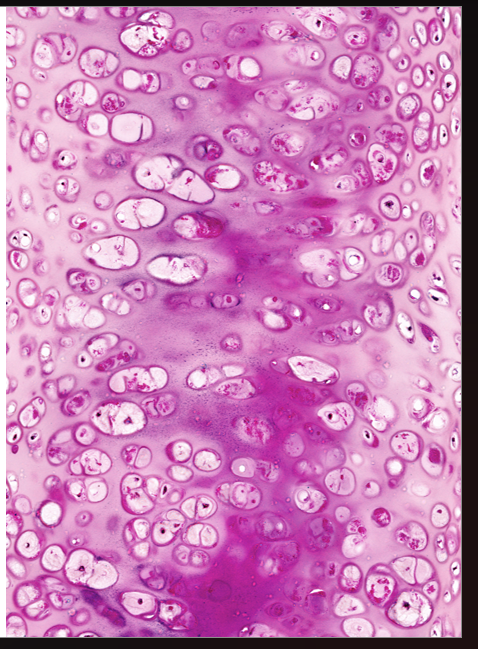
chondrocytes
32
function of chondrocytes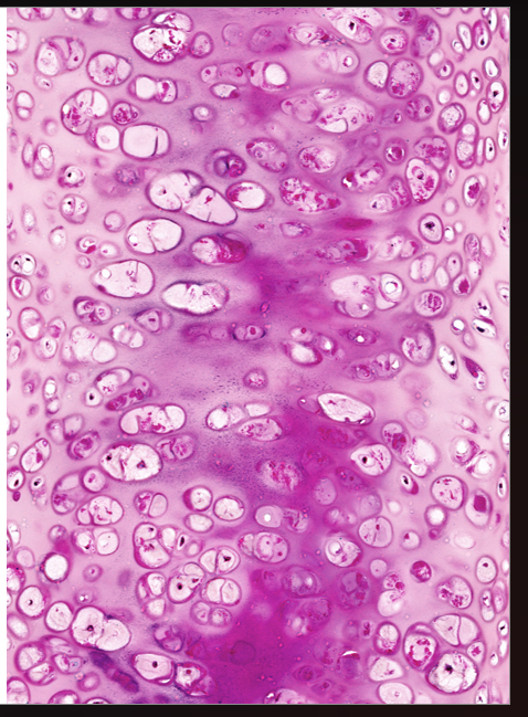
produce matrix
33
3 subtypes of cartilage matrix
hyaline cartilage, elastic fibers, fibrous cartlahe
34
what type of collagen is hyaline cart? fibrous cart?
2; 1
35
where are chondroblasts located?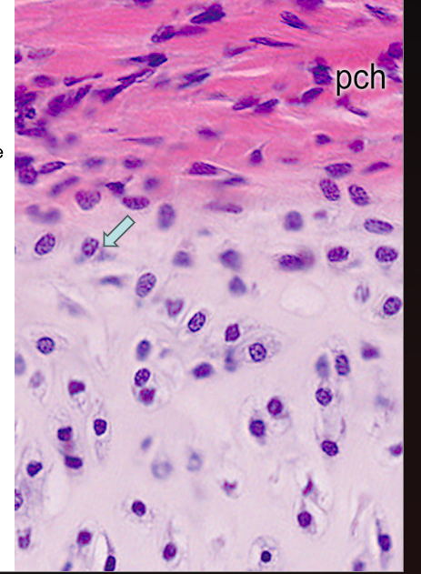
growing cartilage under the perichondrium
36
recently divided choondrocytes are sittingf in clusters in the lacunae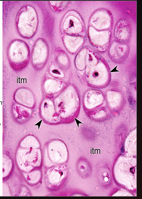
isogenous groups
37
black arrow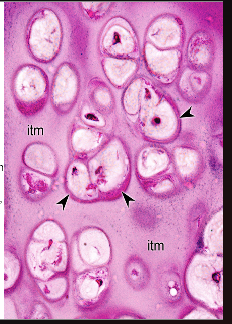
territorial matrix
38
Itm in image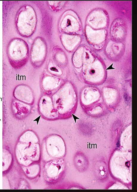
interterritorial matrix
39
fx hyaline cartilage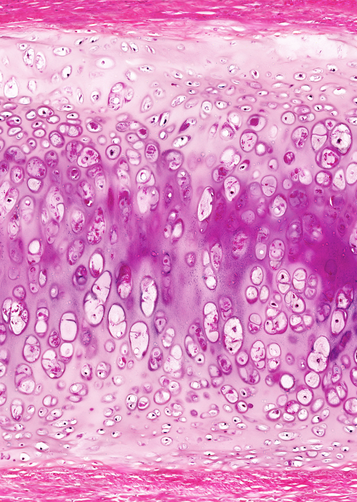
Bind bones together (synchondrosis)
Low friction surfaces for the joints
Model for the developing skeleton of the fetus (enchondral ossification)
Low friction surfaces for the joints
Model for the developing skeleton of the fetus (enchondral ossification)
40
Collagenous membrane on the surface of the cartilage
Carries vessels and nerves – essential for the avascular cartilage (destruction of the perichondrium usually results in destruction of the underlying cartilage)
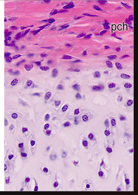
Carries vessels and nerves – essential for the avascular cartilage (destruction of the perichondrium usually results in destruction of the underlying cartilage)
perichondrium
41
where is pericondrium not present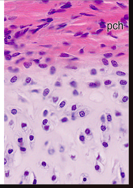
articular surfaces, when cart is in contact with bone
42
what stains elastic fibers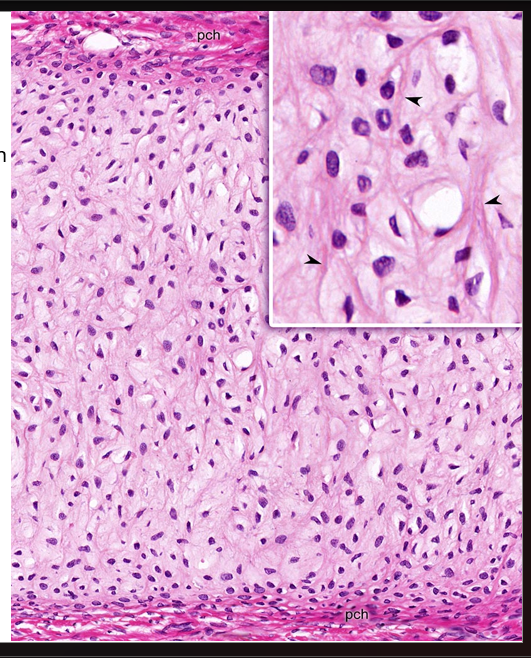
eoisin, resorcin-fuchsin, orcein
43
where are elastic fibers found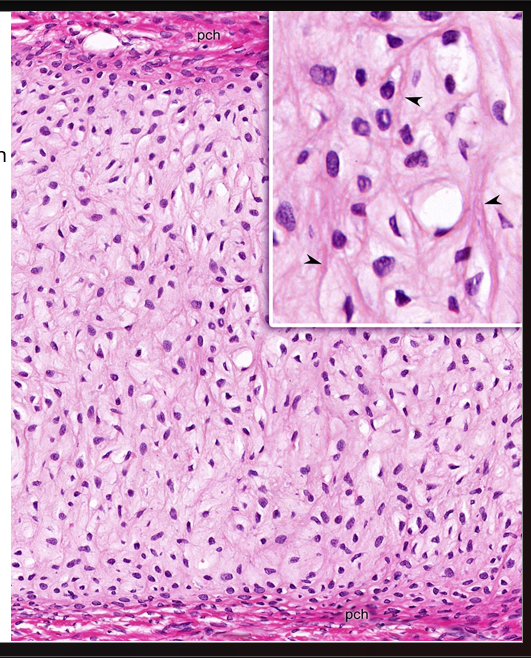
ear, cart of the larynx
44
cart type?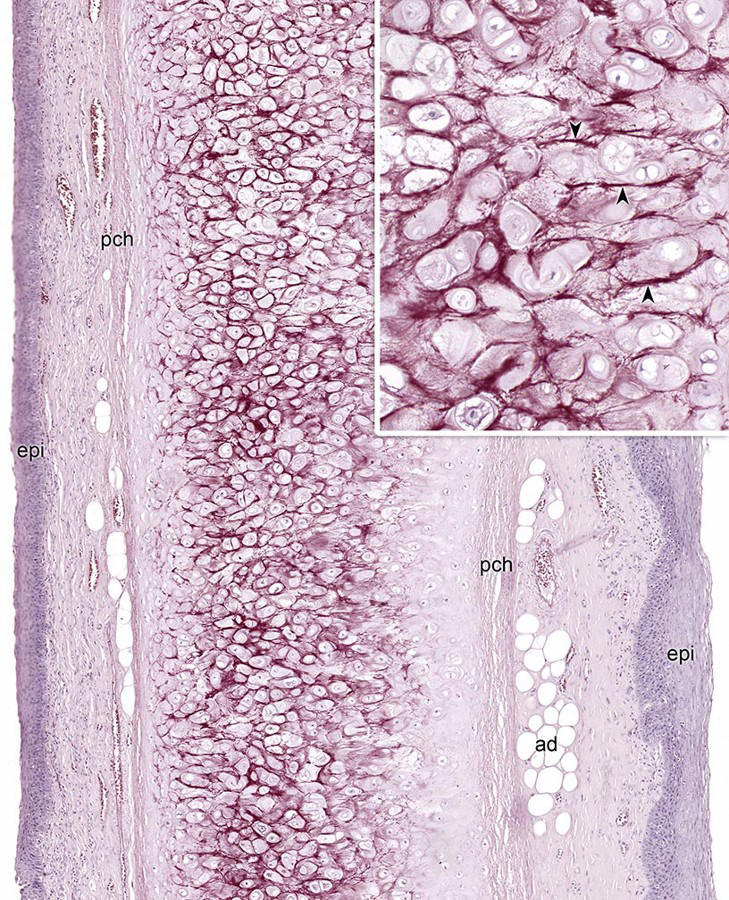
elastic cartilage
45
Combination of dense regular connective tissue and hyaline cartilage
Chondrocytes are sitting in rows (isogenous groups)
Large amount of collagen fibers between them
No perichondrium
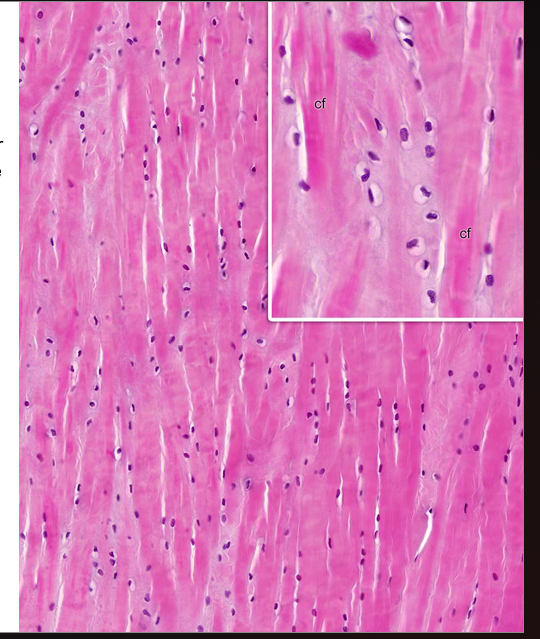
Chondrocytes are sitting in rows (isogenous groups)
Large amount of collagen fibers between them
No perichondrium
fibrocartilage
46
chondroblasts are formed by the inner layer of perichondrium (cells produce collagen type I), they produce matrix (collagen type II) and turn to chondrocytes
appositional growth
47
division of chondrocytes within the lacunae – isogenous groups
interstitial growth
48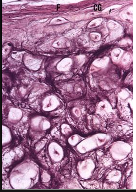
elastic cartilage
49
What is the total volume of blood in the human body?
About 6 liters (7-8% of body weight)
50
What are the two main components of blood?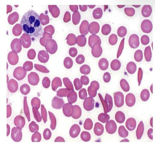
Cellular elements and plasma
51
What is hematocrit?
Percentage of cellular elements volume compared to total blood volume
52
What is the hematocrit range for males?
39-50%
53
What is the hematocrit range for females?
35-45%
54
What percentage of plasma is water?
91-92%
55
Which plasma protein maintains colloid osmotic pressure and acts as a carrier protein?
Albumin
56
Which plasma proteins are immunoglobulins?
γ-globulins
57
Which plasma protein is the largest and helps in clot formation?
Fibrinogen
58
What is the shape and size of an erythrocyte?
Biconcave disc, 7.8 μm
59
What is the lifespan of an erythrocyte?
120 days
60
Where are old erythrocytes broken down?
Spleen, bone marrow, liver
61
What is hemoglobin composed of?
Four polypeptide subunits, each with a heme group
62
What causes sickle cell anemia?
Point mutation replacing glutamic acid with valine in β-globin chain at position 6
63
Name the two major categories of leukocytes.
Granulocytes and agranulocytes
64
Which leukocyte type is most abundant?
Neutrophils (50-70%)
65
What is the typical nucleus shape of neutrophils?
Multilobed nucleus
66
Which granules in neutrophils contain myeloperoxidase?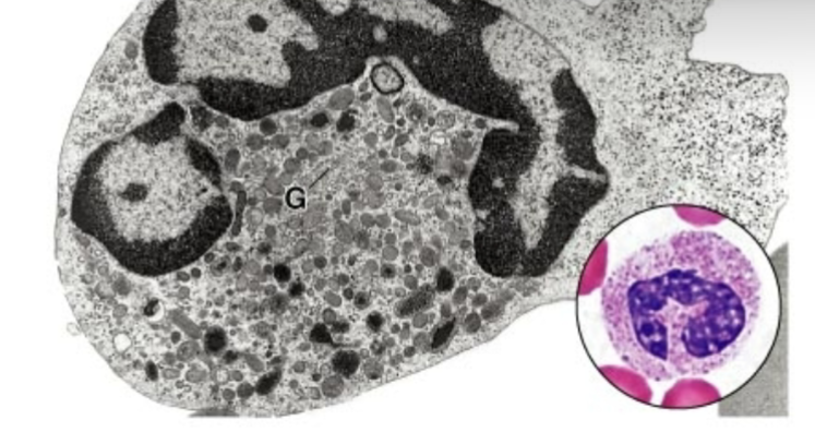
Azurophilic granules (primary granules)
67
What do neutrophil specific granules contain?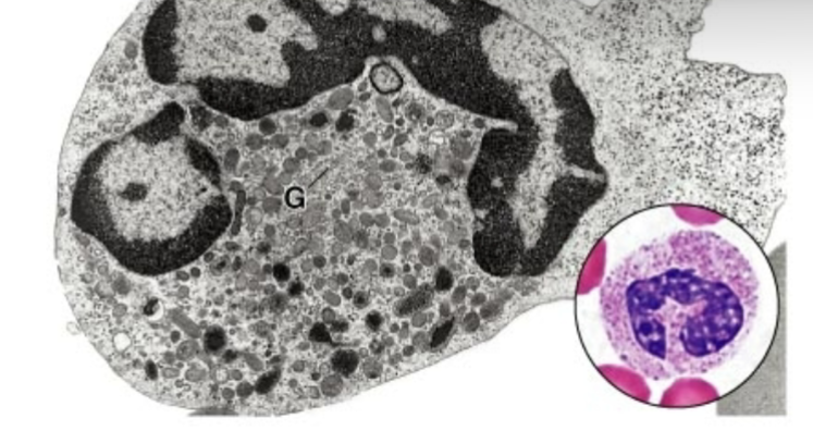
Enzymes, complement activators, bactericidal agents
68
What directs neutrophils to injury sites?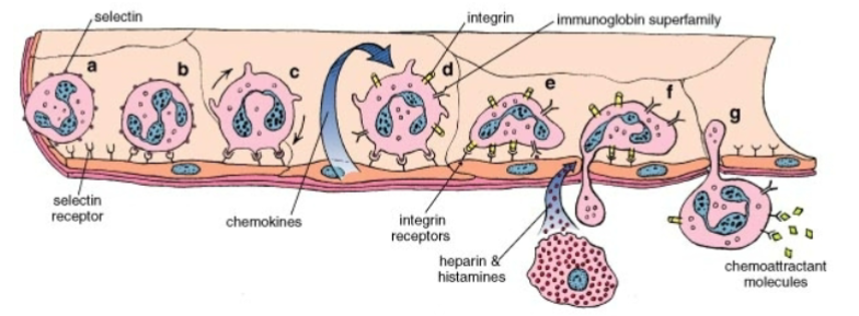
Chemoattractant molecules
69
Which leukocyte has a bilobed nucleus and large eosinophilic granules?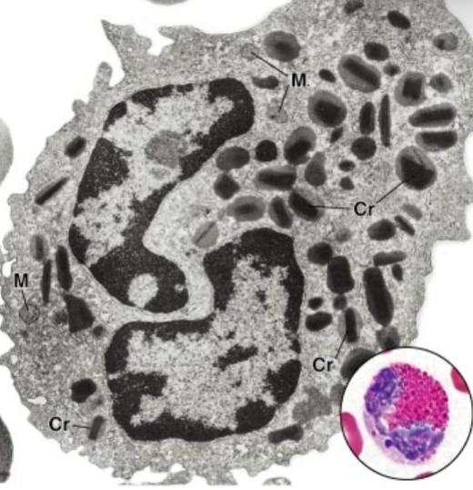
Eosinophils
70
What is the major basic protein in eosinophil granules responsible for?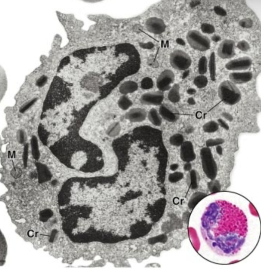
Acidophilia and cytotoxicity to parasites
71
Which leukocyte contains heparan sulphate and histamine in its granules?
Basophils
72
Which receptor on basophils binds IgE?
Fc receptor for IgE
73
What percentage of leukocytes are lymphocytes?
25-30%
74
Which lymphocyte type differentiates in the thymus?
T lymphocytes
75
Which lymphocyte type produces circulating antibodies?
B lymphocytes
76
Which lymphocyte type kills virus-infected and tumor cells?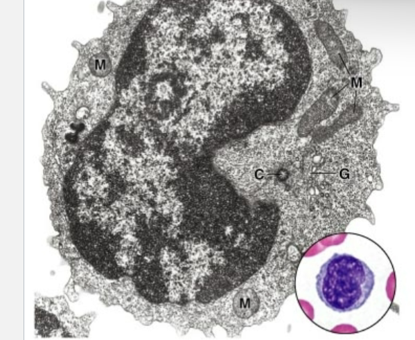
NK cells
77
What is the largest leukocyte?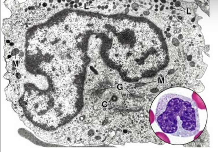
Monocyte
78
Into what cells do monocytes differentiate in tissues?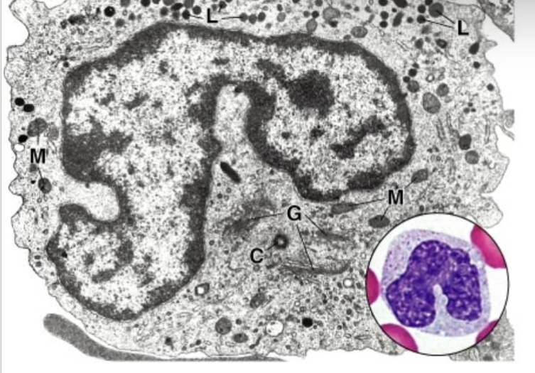
Macrophages
79
How long do monocytes remain in the blood?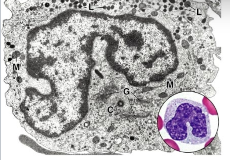
About 3 days
80
What is the shape of a monocyte nucleus?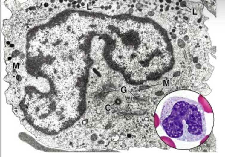
Bean-shaped
81
Which platelet granules contain fibrinogen and coagulation factors?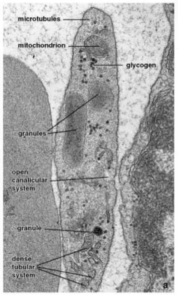
α-granules
82
Which platelet granules contain ADP, ATP, serotonin, histamine?
δ-granules
83
Which platelet granules contain hydrolytic enzymes for clot resorption?
λ-granules
84
What are the three main cell types of the nervous system?
Neurons, glia, blood vessel cells
85
What are the three classes of neurons?
Sensory, motor, interneurons
86
Which neurons are found in the dorsal root ganglion?
Pseudounipolar neurons
87
Where are Purkinje cells found?
Cerebellum
88
Which glial cells produce myelin in the CNS?
Oligodendrocytes
89
Which glial cells produce myelin in the PNS?
Schwann cells
90
What is the function of astrocytes?
Support neurons, form BBB, regulate extracellular environment
91
What is the smallest glial cell type?
Microglia
92
What is the function of microglia?
Phagocytosis and debris removal
93
Where are ependymal cells found?
Lining ventricles and central canal of CNS
94
What is the node of Ranvier?
Gap between myelin segments enabling saltatory conduction
95
What is the function of Schmidt-Lanterman clefts?
Allow cytoplasmic continuity in myelin sheaths
96
Which cells surround neuron cell bodies in ganglia?
Satellite cells
97
What is the difference between oligodendrocytes and Schwann cells?
Oligodendrocytes myelinate multiple axons in CNS, Schwann cells one axon in PNS
98
What are the three connective tissue coverings of a peripheral nerve?
Endoneurium, perineurium, epineurium
99
What are the two main divisions of the nervous system?
CNS and PNS
100
What are the two functional divisions of the nervous system?
Somatic and autonomic nervous systems
101
Which neurotransmitters open Na+ channels?
Excitatory neurotransmitters
102
Which neurotransmitters open Cl- channels?
Inhibitory neurotransmitters
103
Name the four types of synaptic contacts.
Axodendritic, axosomatic, axoaxonic, dendrodendritic
104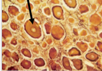
pseudounipolar
105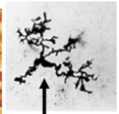
microglia
106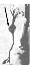
bipolar
107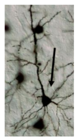
pyramidal
108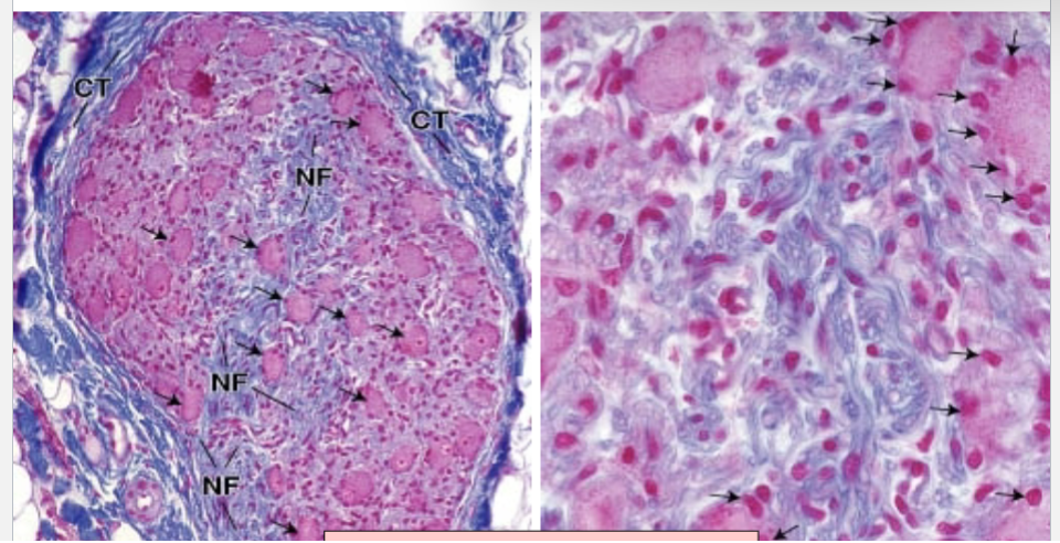
satellite cells
109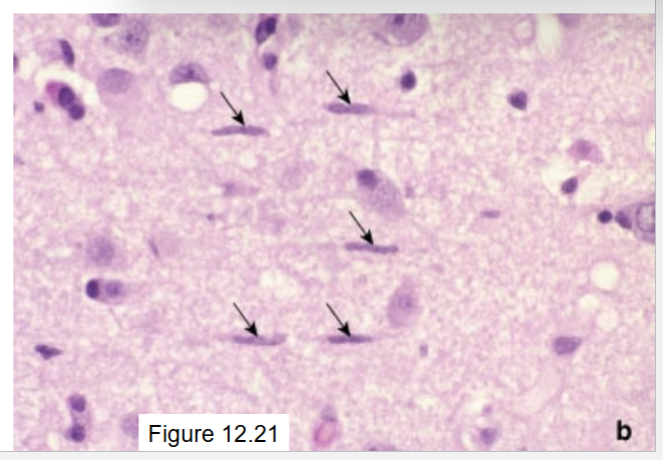
microglia
110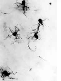
oligodendrocytes
111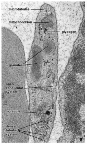
platelet
112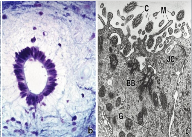
ependymal cells
113
accumulation of mainly B-lymphocytes (also some plasma cells and eosinophils) in the mucous membrane – GALT(gut) and BALT (bronchus)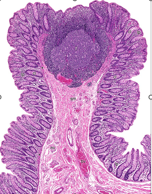
MALT
114
Round shaped lymphocytic aggregations in the malt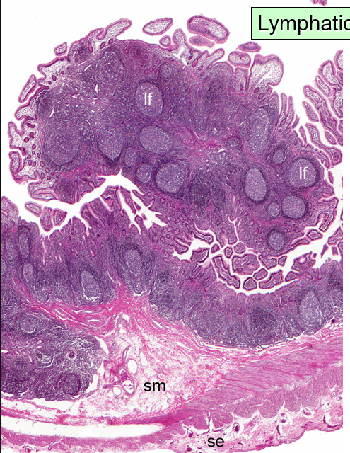
lymphatic nodules (follicles)
115
site of initial immune response-guarding body against pathogens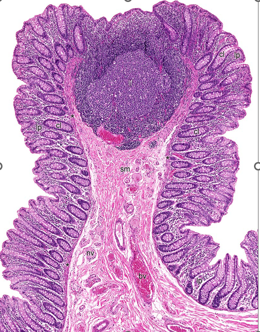
MALT
116
ring of tonsils at the entrance of the pharynx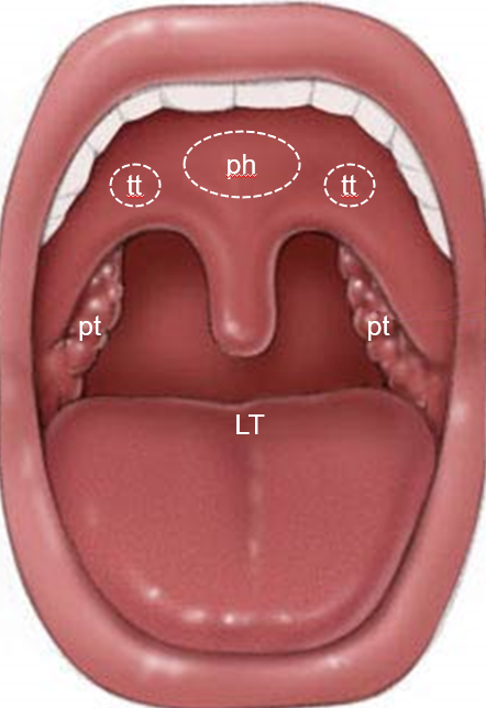
waldeyer's ring
117
what is the pharynheal tonsils and tubarian tonsils made up of (epi tissue type)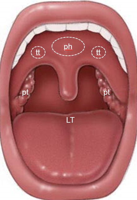
ciliated psudostratified epithelium
118
what are the palntine tonsils and lingual tonsils made up of (epi tissue type)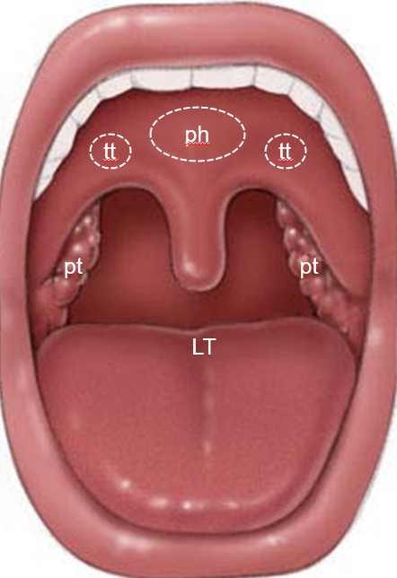
stratified squamous epithelium
119
?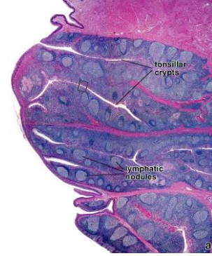
Palatine tonsils
120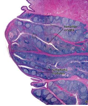
Image occlusion: name the hidden labels
121
Image occlusion: name the hidden labels
122
what structure?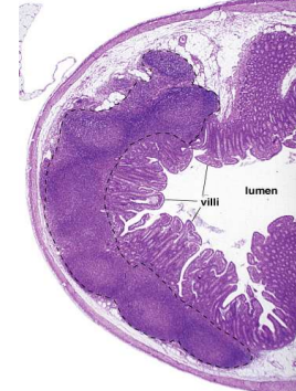
Peyer's patches and vermiform appendix
123
where are peyer's patches found?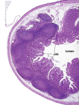
ileum
124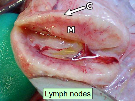
Image occlusion: name the hidden labels
125
Image occlusion: name the hidden labels
126
where does lymph enter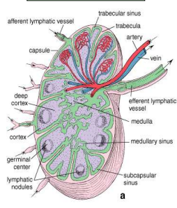
afferent lymphatic vessels
127
where does lymph leave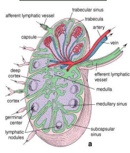
efferent lymphatic vessels
128
what lives in the superficial or nodual structure of the lymph node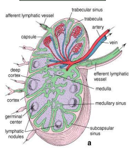
lymph nodules
129
what lives in the deep or paracortex of the lymph node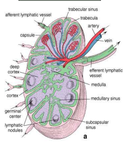
T cells
130
what lives in the medulla of the lymph node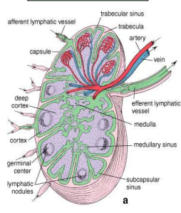
medullary cords
131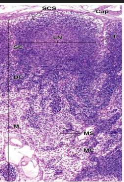
Image occlusion: name the hidden labels
132
Image occlusion: name the hidden labels
133
Image occlusion: name the hidden labels
134
Image occlusion: name the hidden labels
135
Image occlusion: name the hidden labels
136
Image occlusion: name the hidden labels
137
Image occlusion: name the hidden labels
138
Image occlusion: name the hidden labels
139
what makes up reticular tissue in lymph nodes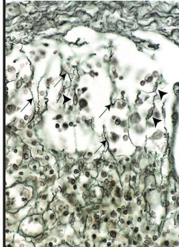
-reticular cells
-follicular dendritic cells
-follicular dendritic cells
140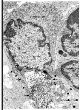
Image occlusion: name the hidden labels
141
Image occlusion: name the hidden labels
142
what is the pathway of lymph node circulation of lymphocytes from the lymph (x5)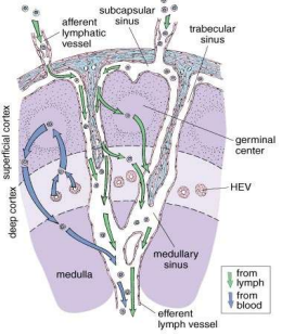
afferent lymph vessels, subscapular sinus, trabecular sinus, medullary sinus, efferent lymph vessel
143
where blood enters lymph nodes from bloodstream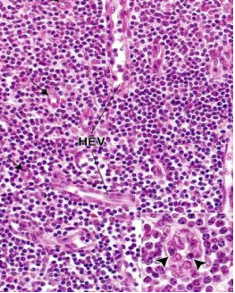
high endothelial venules
144
these cells from the bone infiltrate the thymus at embryonic age
multipotential lymphoid stem cells
145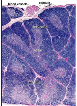
Image occlusion: name the hidden labels
146
Image occlusion: name the hidden labels
147
Image occlusion: name the hidden labels
148
Image occlusion: name the hidden labels
149
Image occlusion: name the hidden labels
150
Image occlusion: name the hidden labels
151
?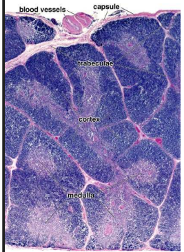
thymus
152
Between the elements of the network formed by
epithelioreticular cells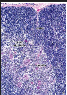
epithelioreticular cells
T cell (thymocytes)
153
Dispersed in the cortex• Phagocytose of T cells that do not fulfill thymic education
requerements
• Intensive PAS staining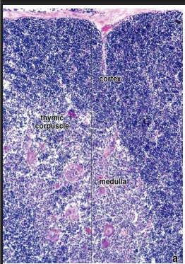
requerements
• Intensive PAS staining
macrophages
154
•Flattened cells with occluding junctions
•At the border of the cortex and the capsule or the trabeculae•Separate parenchy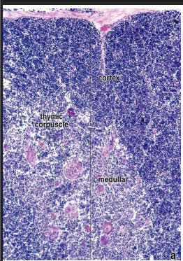
•At the border of the cortex and the capsule or the trabeculae•Separate parenchy
Type I
155
•Stellate cells with desmosomes and light (!) nucleus
•Express MHC I and II molecules
•Compartmentalize the cortex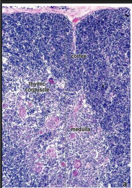
•Express MHC I and II molecules
•Compartmentalize the cortex
type II
156
•At the border of cortex and medulla
•Flat cells, occluding junctions•Form a barrier between the cortex and the medulla
•Express MHC I and II molecules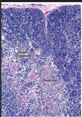
•Flat cells, occluding junctions•Form a barrier between the cortex and the medulla
•Express MHC I and II molecules
Type III
157
•Flat cells at the border of cortex and medulla close to the type III cells
•Occluding junctions (IV-IV and IV-III)
•Form a barrier between the cortex and the medulla with type III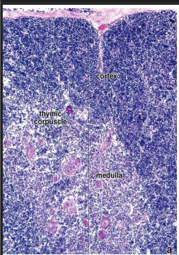
•Occluding junctions (IV-IV and IV-III)
•Form a barrier between the cortex and the medulla with type III
Type IV
158
•Stellate cells with desmosomes and light (!) nucleus
•Compartmentalize the medulla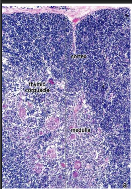
•Compartmentalize the medulla
Type V
159
•Concentrically arranged cells (looks like an onion in section)
•Center may keratinize•Keratohyalin granules, intermediate filaments, lipid (epithelial origin)
•Desmosomes
•Produce thymosin and thymopoietin hormones – promote thymic cell education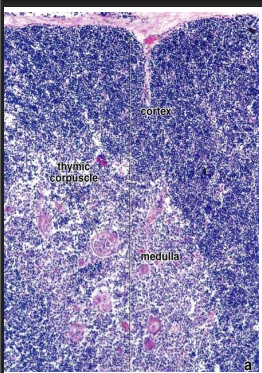
•Center may keratinize•Keratohyalin granules, intermediate filaments, lipid (epithelial origin)
•Desmosomes
•Produce thymosin and thymopoietin hormones – promote thymic cell education
Type VI
160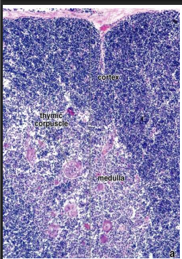
Image occlusion: name the hidden labels
161
Image occlusion: name the hidden labels
162
Image occlusion: name the hidden labels
163
Image occlusion: name the hidden labels
164
Image occlusion: name the hidden labels
165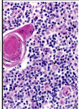
Image occlusion: name the hidden labels
166
Image occlusion: name the hidden labels
167
Protects developing lymphocytes from exposure of antigens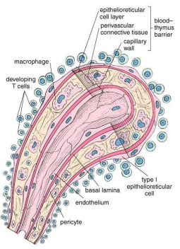
Blood-thymus barrier
168
The spleen is the largest lymph organ.
It is divided into ___________ and ___________which are the structural substrates of the two main functions
(immunological monitoring and cellular filtration of blood).Red pulp is mostly composed of blood, and white pulp is formed by lymphatic tissue.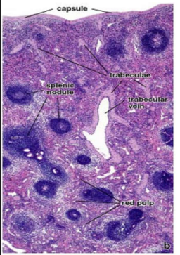
It is divided into ___________ and ___________which are the structural substrates of the two main functions
(immunological monitoring and cellular filtration of blood).Red pulp is mostly composed of blood, and white pulp is formed by lymphatic tissue.
white pulp and red pulp
169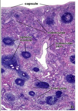
Image occlusion: name the hidden labels
170
Image occlusion: name the hidden labels
171
Image occlusion: name the hidden labels
172
Splenic artery divides into _______________. ___________ branch from the trabecular into the white pulp. Central arteries are sheathed with ______________ (PALS – T cells) and pierce a lymph follicle (Malpighian
corpuscle containing B cells). After leaving the white pulp the central artery divides into straight ____________ which communicate with the sinusoids (red pulp sinuses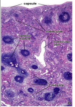
corpuscle containing B cells). After leaving the white pulp the central artery divides into straight ____________ which communicate with the sinusoids (red pulp sinuses
trabecular a.; central a.; periarterial lymphatic sheath; penicillar arterioles