Histology S1 U2
Study these flashcardsFree · spaced repetition · iPhone, Android, Mac & web
All cards
1
what tissue makes upthe fibrous rings that make up the fibrous skeleton
dense irregular connective tissue
2
what is the purpose of the fibrous trings of the fibrous skeleton
provide the attachment site for the leaflets of all four valves of the heart that allow blood flow in only one direction through the openings.
3
what makes yup the membranous part of the IV septum (tissue)
dense connective ttissue
4
what makes up the epicardium of the heart (tissue)
single layer mesothelial cells and connective and adipoise rissue
5
a condition in which excess fluid (blood or pericardial effusion) rapidly accumulates in the pericardial cavity.
cardiac tamponade
6
(a procedure to drain the fluid from the pericardial cavity).
pericardiocentesis
7
what makes up the endocardoum of the heart (tissue)
inner layer of endithelium and subendothelial connectove tissie
-middle layer connective tissue and smooth muscle cells
-deep layer of connective tissue
-middle layer connective tissue and smooth muscle cells
-deep layer of connective tissue
8
what are the three layers of the heart valves
fbrosa, spongiosa, vetricularis/atrialis
9
what does this show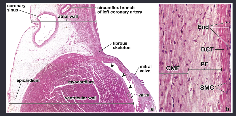
left atrial and left ventricular walls
10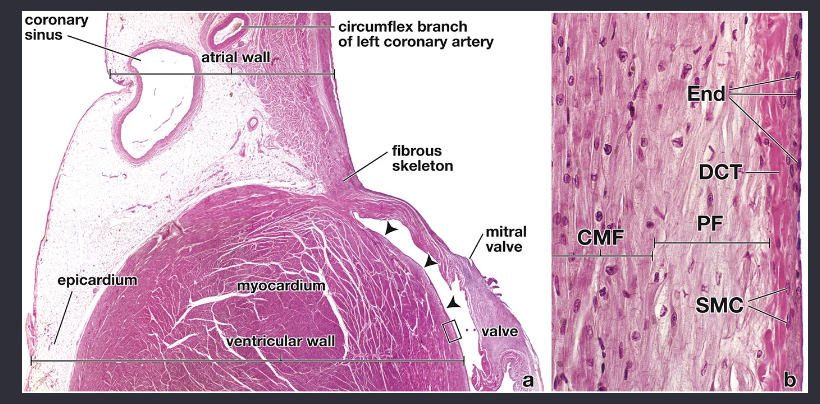
Image occlusion: name the hidden labels
11
Image occlusion: name the hidden labels
12
Image occlusion: name the hidden labels
13
Image occlusion: name the hidden labels
14
Image occlusion: name the hidden labels
15
Image occlusion: name the hidden labels
16
Image occlusion: name the hidden labels
17
Image occlusion: name the hidden labels
18
Image occlusion: name the hidden labels
19
Image occlusion: name the hidden labels
20
Image occlusion: name the hidden labels
21
what is this?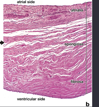
mitral valve
22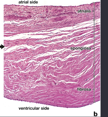
Image occlusion: name the hidden labels
23
Image occlusion: name the hidden labels
24
Image occlusion: name the hidden labels
25
what makes up the sponggiosum of heart valves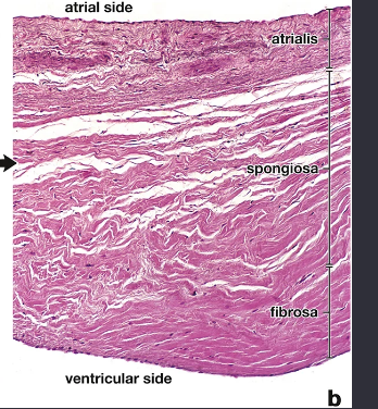
loosely arranged collagen
-elastic fibers
-ground substances (proteoglycans and glycosaminoglycans)
-elastic fibers
-ground substances (proteoglycans and glycosaminoglycans)
26
fx of spongiosum in heart valves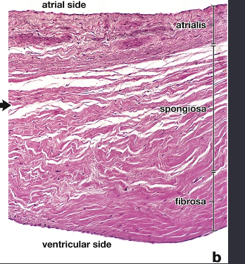
shock absorber t dampen vibrations associated with closing valve
27
what makes up the ventricularis/atrialis layer of the valves? (tissue)
collagen fobers
elastic fibers
elastic lamellae
elastic fibers
elastic lamellae
28
heart diseases are characterized by activation of ____________ cells as well as by increased expression of genes encoding extracellular matrix proteins and remodeling enzymes.
valvular interstitial cells
29
three types of path chabges to valves
1) degeneration of extracellular matrix by accumulation of pathologic proteoglycans, collagen degradation, and elastic fiber fragmentation
2) fibrosis, which is characterized by accumulation of collagen, degradation of proteoglycans, and elastic fiber fragmentation
3) nodular calcification that begins within valvular interstitial cells.
2) fibrosis, which is characterized by accumulation of collagen, degradation of proteoglycans, and elastic fiber fragmentation
3) nodular calcification that begins within valvular interstitial cells.
30
degeneration of extracellular matrix (valve disease) what does this look like
myxomatous mitral valve disease and result in a “floppy” valve that is prone to prolapse and regurgitation.
31
fibrosis Valve disease what does this look like
rheumatic heart disease and result in a thick, rigid, and inflexible valve that is prone to restricted movement and stenosis
32
nodular calcifivcation valve disease what does this look like
degenerative calcific aortic valve stenosis that is characterized by thickening of the valve leaflets and formation of calcium nodules.
33
what is the paceaker rate of the SA node
60-100b/min
34
in a cpomplete heart block, what drives depolaroization and repolarization of the heart[p
purkinje fibers
35
intrinsic spontaneous pacemaker function that generate impulses of the SA node
P cells
36
responsible for propagating impulses into the right atrium SA node
transitional cells (T cells)
37
what does this show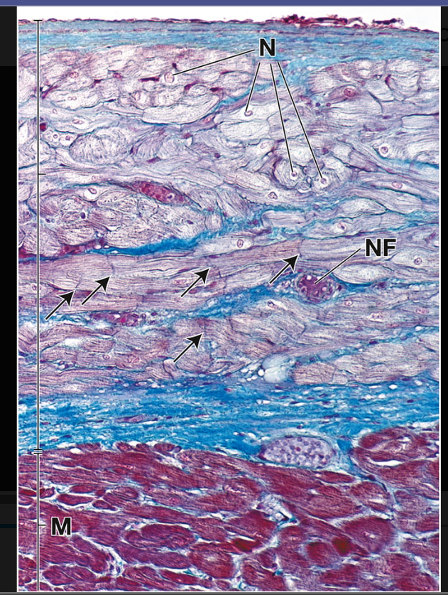
wal of the ventrivle
38
results from age-related degeneration of nodal cardiac muscle cells in the SA node that affects its ability to generate and transmit impulses to the atrial musculature
sick sinus syndrome
39
symptoms of sick sinus syndrome
palpitations (irregular heartbeat) and tissue hypoperfusion leading to fatigue, presyncope (lightheadedness, muscular weakness, blurred vision, and feeling faint), and syncope (fainting).
40
Activation of adrenergic receptors (mainly β1 type) by epinephrine and less efficiently by norepinephrine increases the force of contraction ______________________) and the heart rate ___________).
positive inotropic effect; positive chronotropic effect
41
(high-pressure receptors) sense arterial blood pressure. These receptors are located in the carotid sinus and aortic arch.
baroreceptors
42
where can you find baroreceptors
carotif sinus and aortic arch
43
(low-pressure receptors) located within the walls of the atria and ventricles sense central venous pressure and provide the CNS with information about cardiac distention.
volume receptors
44
where are volume receptors of the jeart located
walls of atria and ventricles
45
detect alterations in oxygen, carbon dioxide tension, and pH. These receptors are the carotid and aortic bodies located at the bifurcation of the common carotid arteries and in the aortic arch, respectively.
chemoreceptors
46
where are the chemoreceptors located that deal with the circulatory system
carotid and aortic bodies at the bifurcation of common carotf a. and aortic arch
47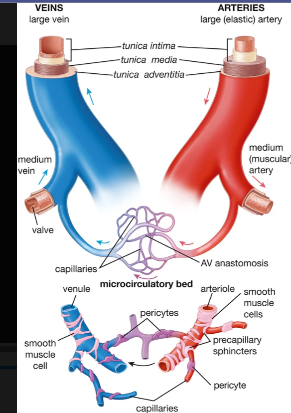
Image occlusion: name the hidden labels
48
Image occlusion: name the hidden labels
49
Image occlusion: name the hidden labels
50
what makes up the tunica intima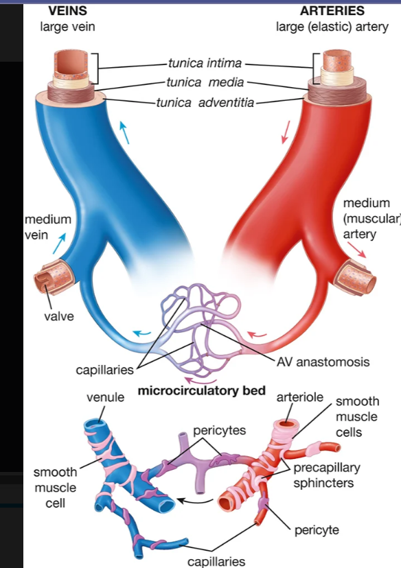
a single layer of squamous epithelial cells, the endothelium; (2) the basal lamina of the endothelial cells (a thin extracellular layer composed chiefly of collagen, proteoglycans, and glycoproteins); and (3) the subendothelial layer, consisting of loose connective tissue.
51
what makes yo the tunica adventitia (tissue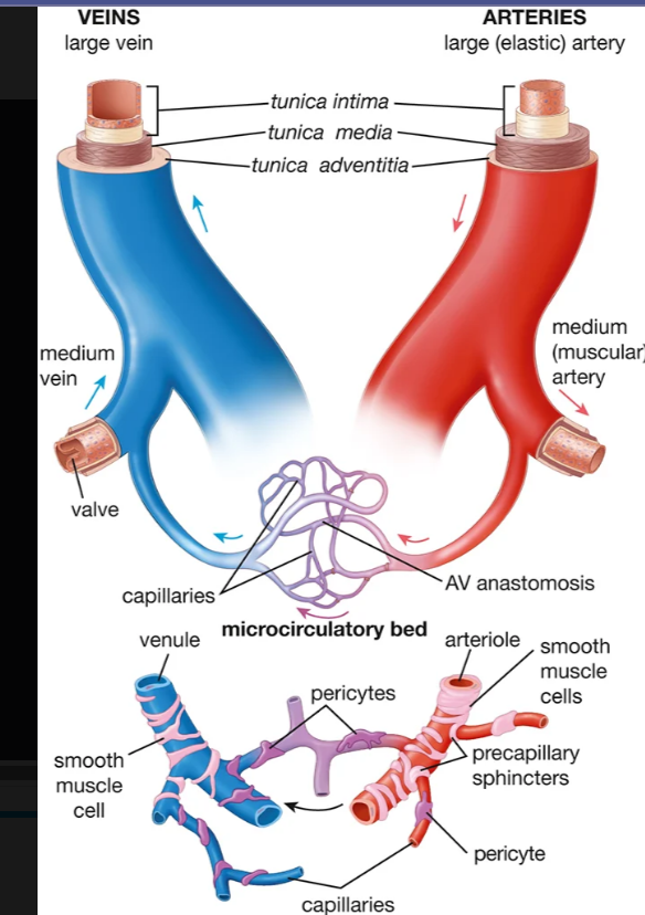
longtudinally arranged collagen
ekastic fibers
ekastic fibers
52
supplies blood to the vascular walls themselves
-in large arteries and veins
-in large arteries and veins
vasa vasorum
53
controls contraction of smooth muscle in vessel walls
-in large arteries and veins
-in large arteries and veins
nervi vasorum
54
where do lesions develop in atherosclerosis
tunica intoma of large elastic arteries
55
what happens after injury to the tinica intima (forming the initial fatty streak) (atherosclerposis)
1) monocytes enter and become macrophages
2) macrophages phangocytize LDL
3) macrophages become foam cells
4) foam cells infiltrate T lymphocytes and form fattu streak
2) macrophages phangocytize LDL
3) macrophages become foam cells
4) foam cells infiltrate T lymphocytes and form fattu streak
56
what does this show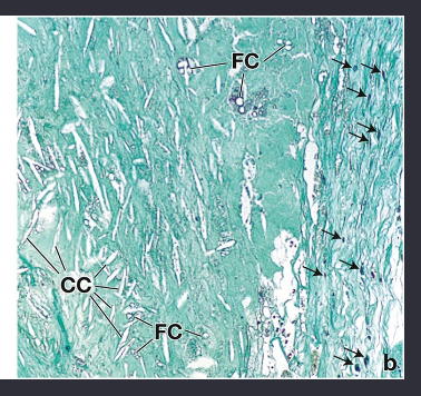
atheromatous lesion
57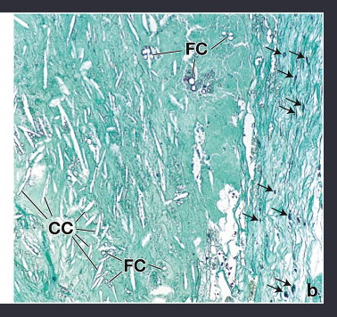
Image occlusion: name the hidden labels
58
Image occlusion: name the hidden labels
59
regulates the blood vessel diameter, inhibits monocyte adhesion to dysfunctional endothelial cells, and maintains an antiproliferative and antiapoptotic environment in the vessel wall.
nitric oxide
60
what continuously synthesixes NO in endothelial cells
endothelial nitric oxide synthase (eNOS)
61
functional pathway of NO and how it works to achieve smooth muscle relaztaion
1) NO made by endothelial cells
2) NO binds to guanylate cyclase in smooth muscle cytoplasm
3) cGMP made
4) cGMP activates PKG
5) PKG cascade decreases Ca2+ concentration
6) smooth muscle relaxation
2) NO binds to guanylate cyclase in smooth muscle cytoplasm
3) cGMP made
4) cGMP activates PKG
5) PKG cascade decreases Ca2+ concentration
6) smooth muscle relaxation
62
PGI2 pathway in endothelial cells
PGI2 made by endothelial cells
2) PGI2 vinds to receorors on smooth muscle
3) cAMP activated
4) PKA activated
5) MLCK activated
6) calcium calmodulin complex inhibited
7) smooth muscke rexation
2) PGI2 vinds to receorors on smooth muscle
3) cAMP activated
4) PKA activated
5) MLCK activated
6) calcium calmodulin complex inhibited
7) smooth muscke rexation
63
a highly toxic protein that exhibits a high degree of sequence homology with ET-1. After it enters the circulation, the toxin binds to ETA receptors and causes life-threatening, intense coronary vasoconstriction.
sarafotoxin
64
thromboxane A2 effect on NO
decreases NO prpduction or inactivates NO superoxide anion
65
defined by a sustained diastolic pressure >80 mm Hg or a sustained systolic pressure >130 mm Hg.
hypertesion
66
what is this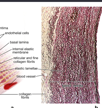
photomicrograph elastic artery
67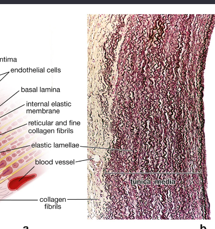
Image occlusion: name the hidden labels
68
Image occlusion: name the hidden labels
69
Image occlusion: name the hidden labels
70
what is thos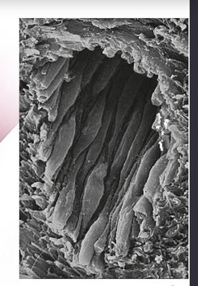
scanning electron micrograph of endotheliym
71
what does this show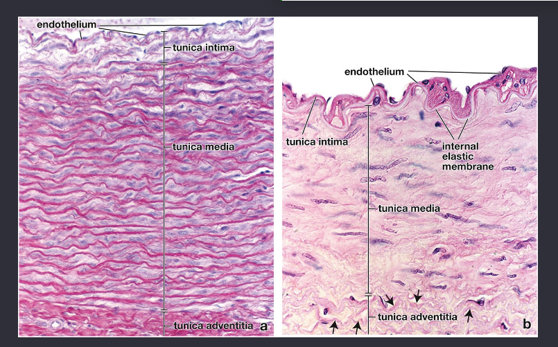
elastic and muscular artery
72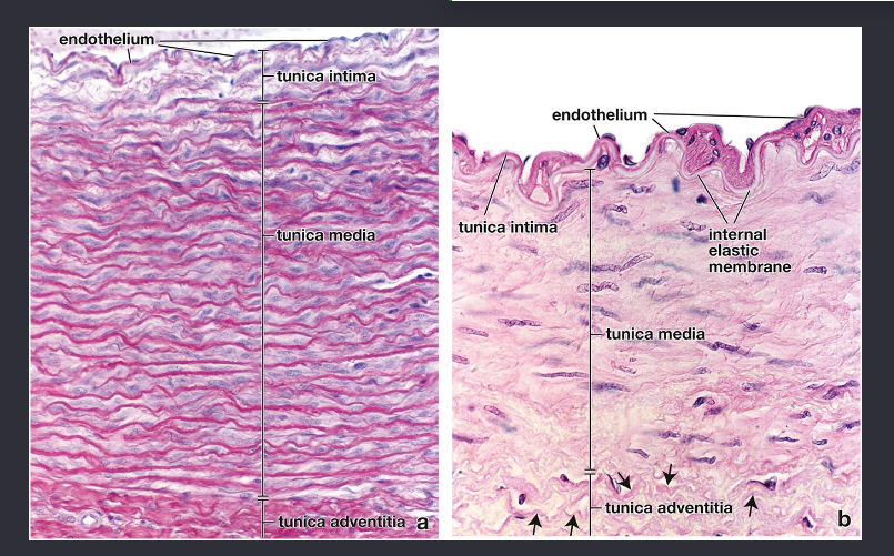
Image occlusion: name the hidden labels
73
Image occlusion: name the hidden labels
74
Image occlusion: name the hidden labels
75
Image occlusion: name the hidden labels
76
Image occlusion: name the hidden labels
77
what does this show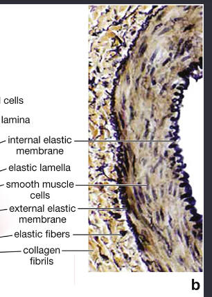
muscular artery
78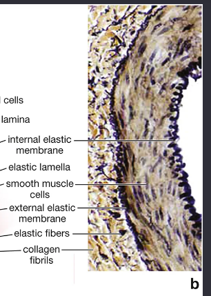
Image occlusion: name the hidden labels
79
Image occlusion: name the hidden labels
80
Image occlusion: name the hidden labels
81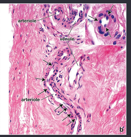
Image occlusion: name the hidden labels
82
Image occlusion: name the hidden labels
83
arterioles serve as flow regulators for this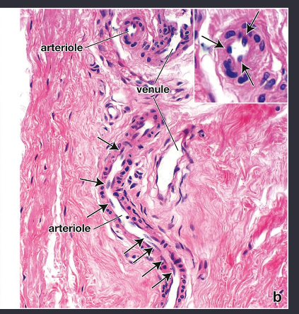
capillary beds
84
arterioles: contraction of the smooth muscle in the wall of an arteriole increases the _____________, reducing or shutting off the blood going to the capillaries. 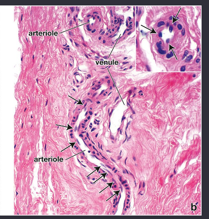
vascular resistance
85
what does this show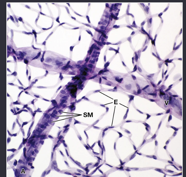
capillary network of the retina
86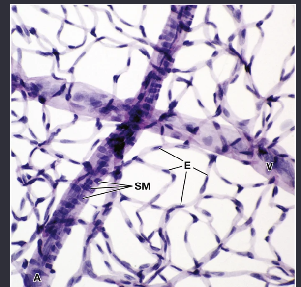
Image occlusion: name the hidden labels
87
Image occlusion: name the hidden labels
88
Image occlusion: name the hidden labels
89
where are continuous capillaries found
connective tissue
cardiac m
skeletal m
smooth m
skin
lungs
CNS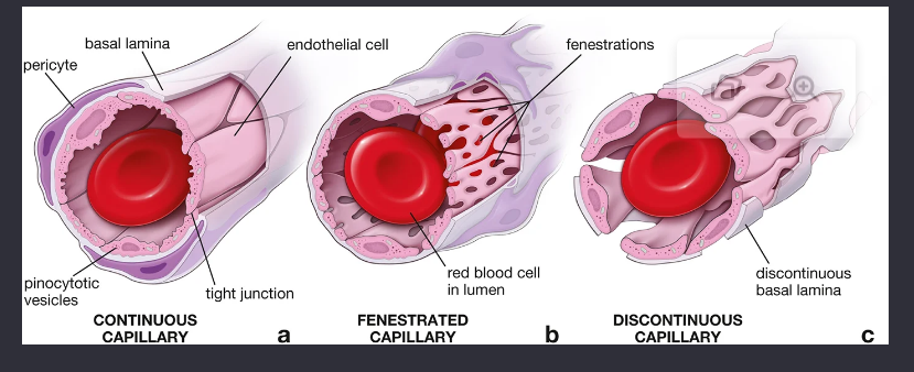
cardiac m
skeletal m
smooth m
skin
lungs
CNS
90
capillaries: characterized by an uninterrupted vascular endothelium that rests on a continuous basal lamina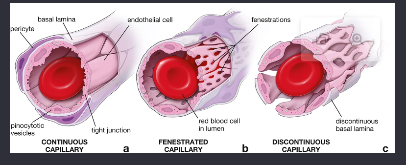
continuous
91
where are fenestrated caoillaries found
endocrine glands, sites if flkuid or metabolite absorption
92
whwere aere discontinuous capilaries dound
liver, spleen, bone marrow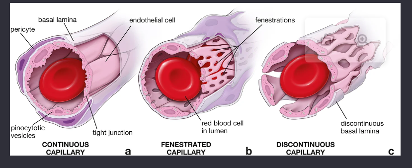
93
Vascular endothelial cells lining these capillaries have large openings in their cytoplasm, and they are separated by wide, irregular, intercellular gaps that allow for the passage of blood plasma proteins 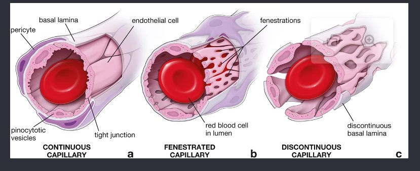
sinusoidal (discontinuous ) cappilaries
94
Closing/opening an AV shunt in the skin causes blood to flow through the capillary bed, enhancing heat loss
closing
95
Opening/closing an AV shunt in the skin reduces the blood flow to the skin capillaries, conserving body heat.
opening
96
located at their origin from either an arteriole or a metarteriole. These sphincters control the amount of blood passing through the capillary bed.
precapillary sphincter
97
what does this show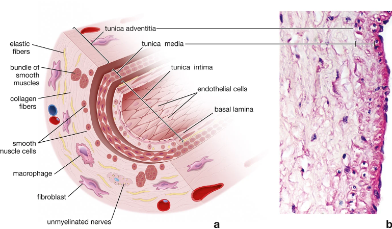
medsium sized vein
98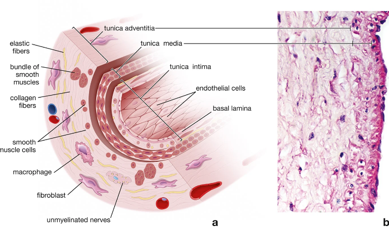
Image occlusion: name the hidden labels
99
Image occlusion: name the hidden labels
100
what does this show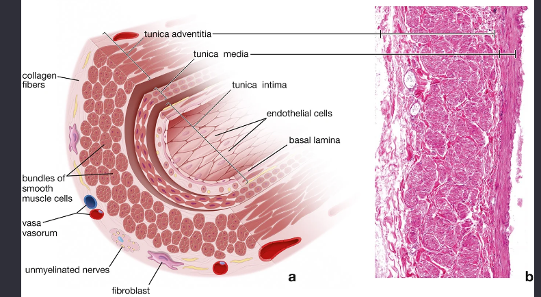
large vein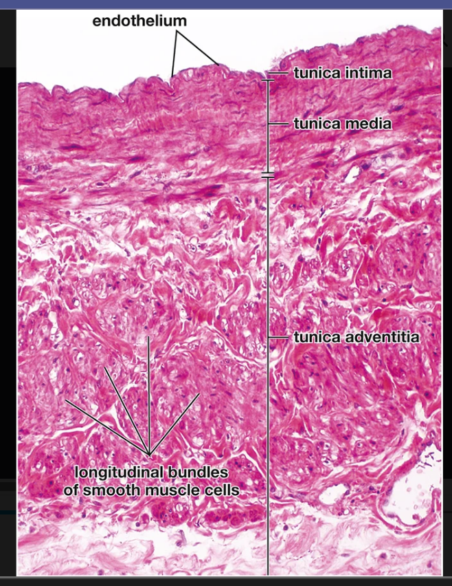
101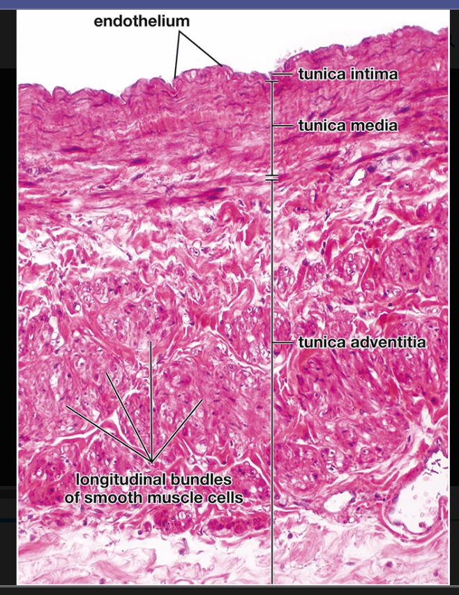
Image occlusion: name the hidden labels
102
Image occlusion: name the hidden labels
103
Image occlusion: name the hidden labels
104
Image occlusion: name the hidden labels
105
what are the tissue characteristics if the coronary atrteries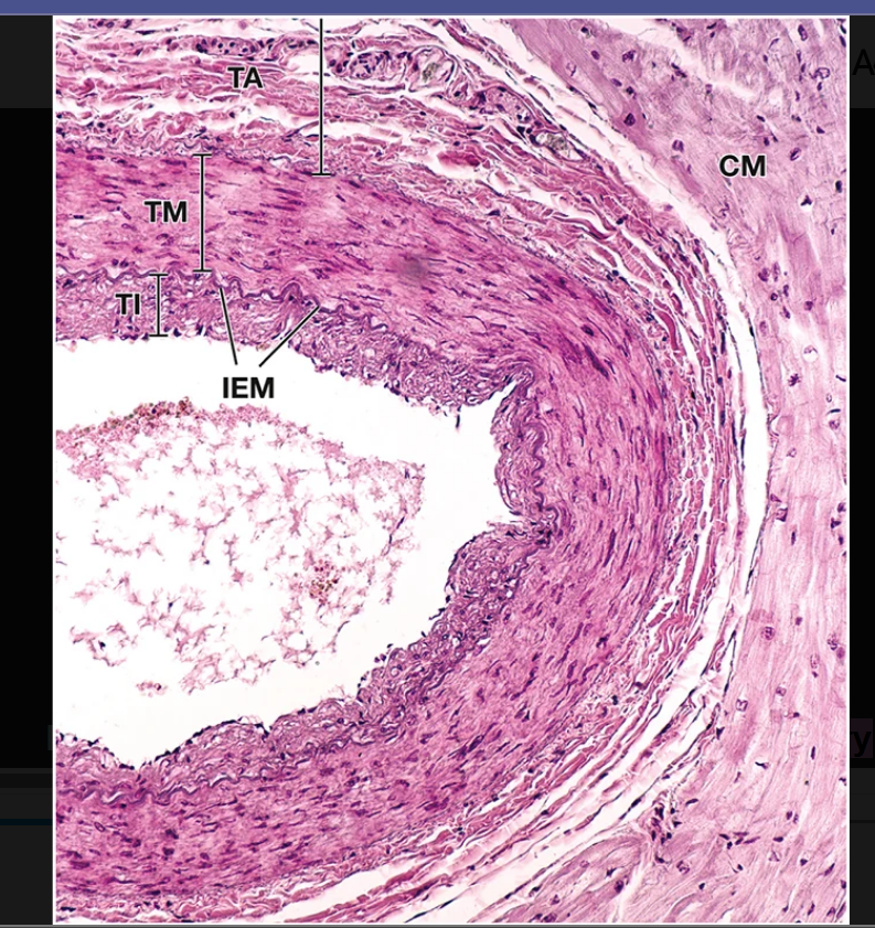
usually thicker than those of comparable arteries in the upper or lower limb because of the large amounts of circular smooth muscle layers in the tunica media.
106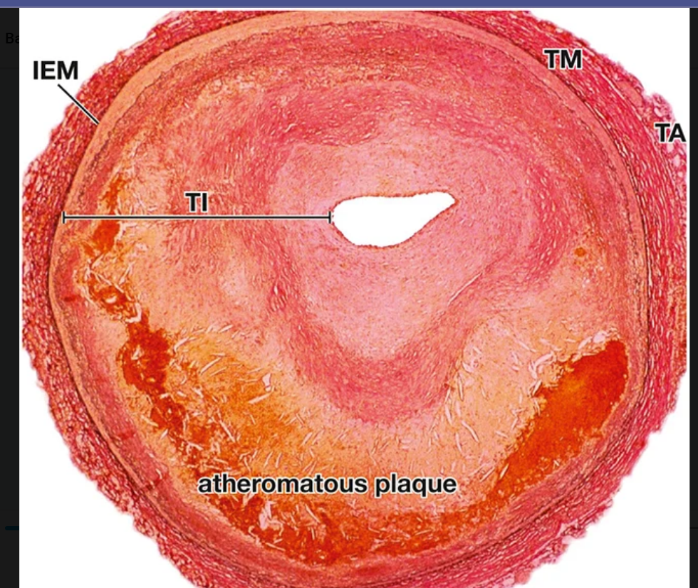
Image occlusion: name the hidden labels
107
what is this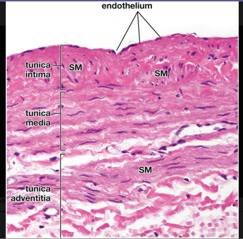
great saphenous v
108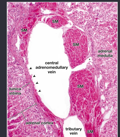
Image occlusion: name the hidden labels