Physiology S1 U2
Study these flashcardsFree · spaced repetition · iPhone, Android, Mac & web
First 300 of 499 cards
1
to transport blood under high pressure to the tissues.
-strong vascular walls
-blood flows at a high velocity through here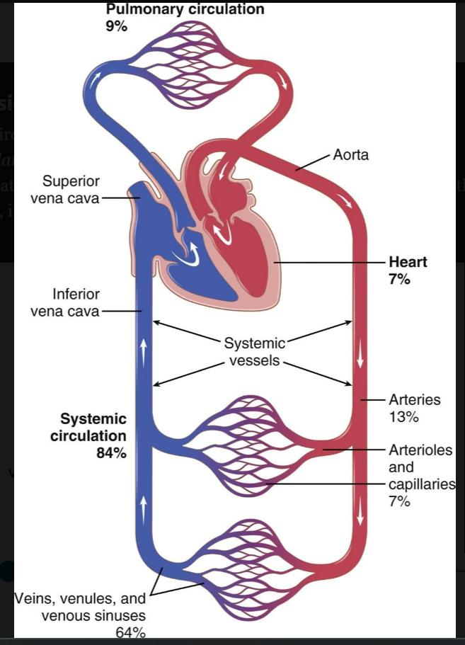
-strong vascular walls
-blood flows at a high velocity through here
arteries
2
the last small branches of the arterial system and control blood flow into the capillaries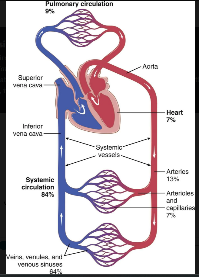
arterioles
3
fx to exchange fluid, nutrients, waste products of cell metabolism, electrolytes, hormones, and other substances between the blood and interstitial fluid. 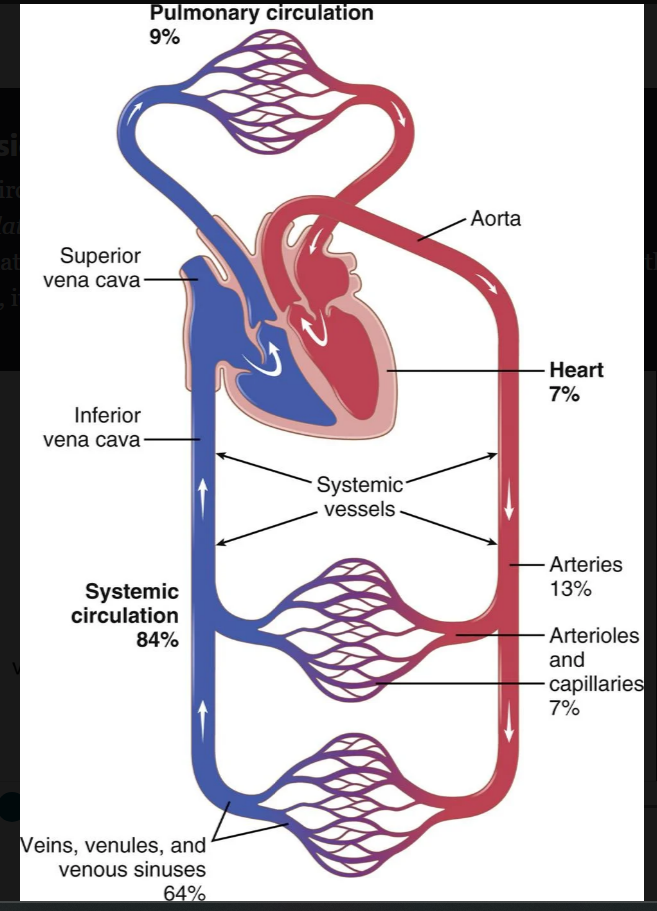
capillaries
4
collect blood from the capillaries and gradually coalesce into progressively larger veins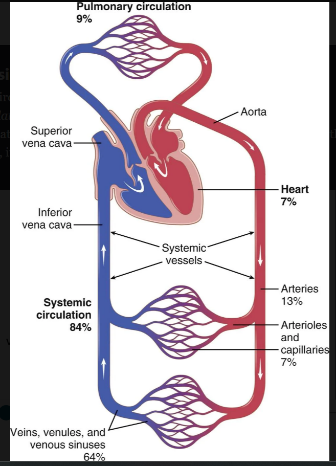
venules
5
Blood Flow Velocity Is Inversely Proportional to Vascular ______________
cross sectional area
6
velocity equation of blood flow
v=F(blood flow volume)/A (cross sectional area)
7
how do tissues get increased blood flow when they need it?
microvessels of each tissue, especially the arterioles, continuously monitor tissue needs, such as the availability of oxygen and other nutrients and the accumulation of carbon dioxide and other tissue waste products. These microvessels, in turn, dilate or constrict to control local blood flow at the level required for the tissue activity.
8
he sum of all the local tissue flows. When blood flows through a tissue, it immediately returns by way of the veins to the heart.
cardiac output
9
if at any time the pressure falls significantly below the normal level of about 100 mm Hg, a barrage of _____________ elicits a series of circulatory changes to raise the pressure back toward normal within seconds.
nervous reflexes
10
what is blood flow through blood vessels determined by (x2)
(1) pressure difference of the blood between the two ends of the vessel, also sometimes called the pressure gradient along the vessel, which pushes the blood through the vessel; and (2) the impediment to blood flow through the vessel, which is called vascular resistance.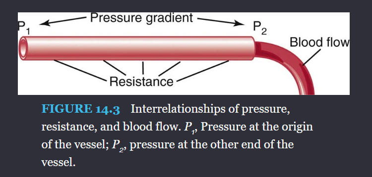
11
The flow through the vessel can be calculated by the following formula, which is called Ohm’s law:
F= pressure change/Resistance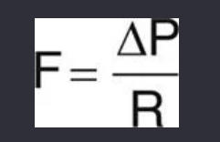
12
the quantity of blood that passes a given point in the circulation in a given period of time.
blood flow rate
13
total blood flow in the circulation is called the ________- because it is the amount of blood pumped into the aorta by the heart each minute.
cardiac output
14
how does an ultrasonic doppler flowmeter work to measure blood flow
A minute piezoelectric crystal is mounted at one end in the wall of the transducer device. This crystal, when energized with an appropriate electronic apparatus, transmits ultrasound at a frequency of several hundred thousand cycles per second downstream along the flowing blood. A portion of the sound is reflected by the red blood cells in the flowing blood. The reflected ultrasound waves then travel backward from the blood cells toward the crystal
-records the doppler effect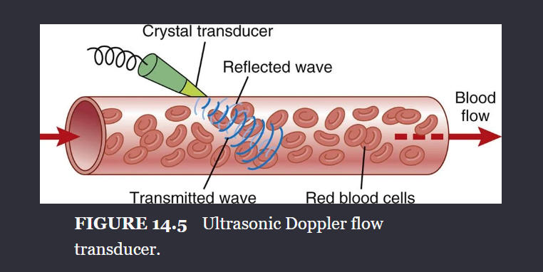
-records the doppler effect
15
blood flows at a steady rate through a long, smooth blood vessel,
laminar flow or streamline flow
16
when laminar flow occurs, the velociy of flow in the center of the vessel is far (lesserreater) than towards the outer edges
greater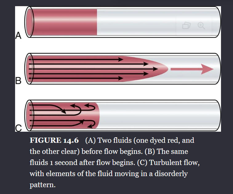
17
When the rate of blood flow becomes too great, when it passes by an obstruction in a vessel, when it makes a sharp turn, or when it passes over a rough surface, the flow may then become
turbulent flow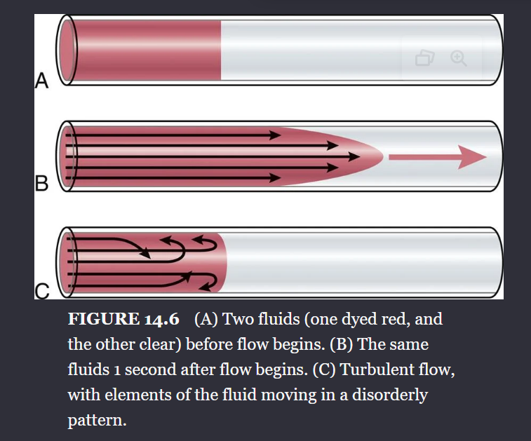
18
what is wrong with using the mercury manometer for measuring blood pressure?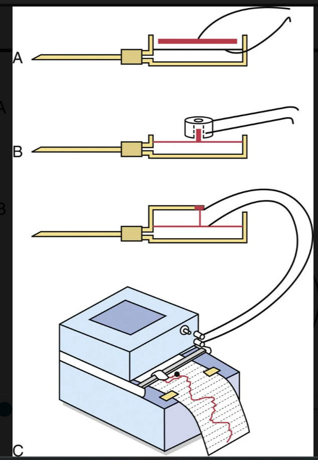
although excellent for recording steady pressures, cannot respond to pressure changes that occur more rapidly than about one cycle every 2 to 3 seconds.
19
he impediment to blood flow in a vessel, but it cannot be measured directly.
resistance
20
If the pressure difference between two points is 1 mm Hg and the flow is 1 mL/sec, the resistance is said to be _____ peripheral resistance unit, usually abbreviated PRU
1
21
a measure of the blood flow through a vessel for a given pressure difference.
conductance
22
conductance is the exact reciprocal of...
resistance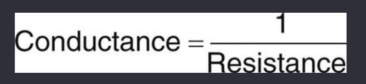
23
Poiseuille’s law
F is the rate of blood flow, ΔP is the pressure difference between the ends of the vessel, r is the radius of the vessel, l is length of the vessel, and η is viscosity of the blood.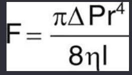
24
demonstrates once again that the_______ of a blood vessel plays the greatest role of all factors in determining the rate of blood flow through a vessel.
diameter
25
r total resistance in a series of blood vessels
26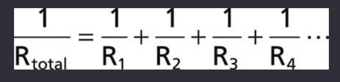
r total resistance in a series if blood vessels in paralel
27
conductance equation in parallel
28
the ______ of blood icreases as hematocrit increases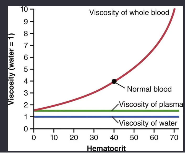
viscocity
29
When pressure falls below a critical level, called the ___________, flow ceases because the blood vessels are completely collapsed.
criticak closing pressure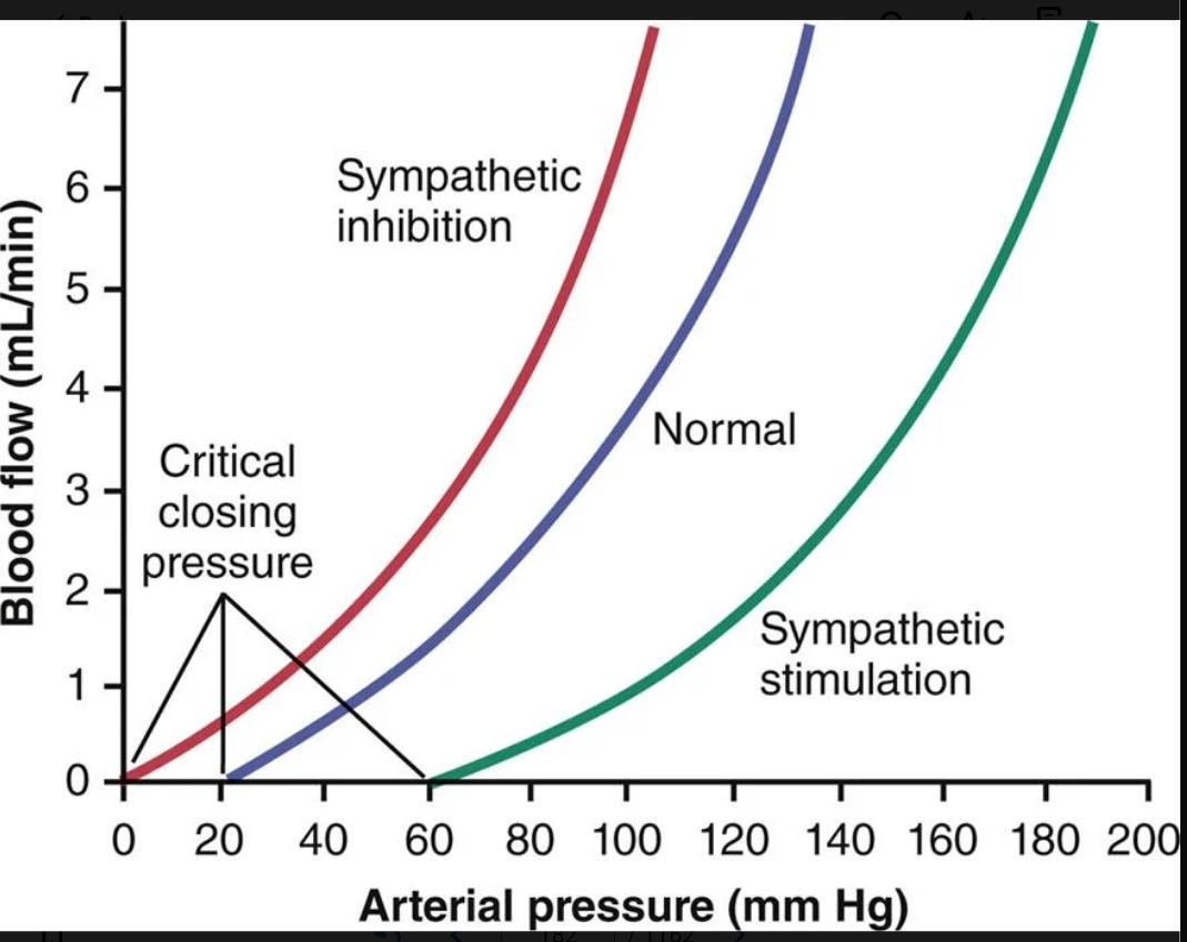
30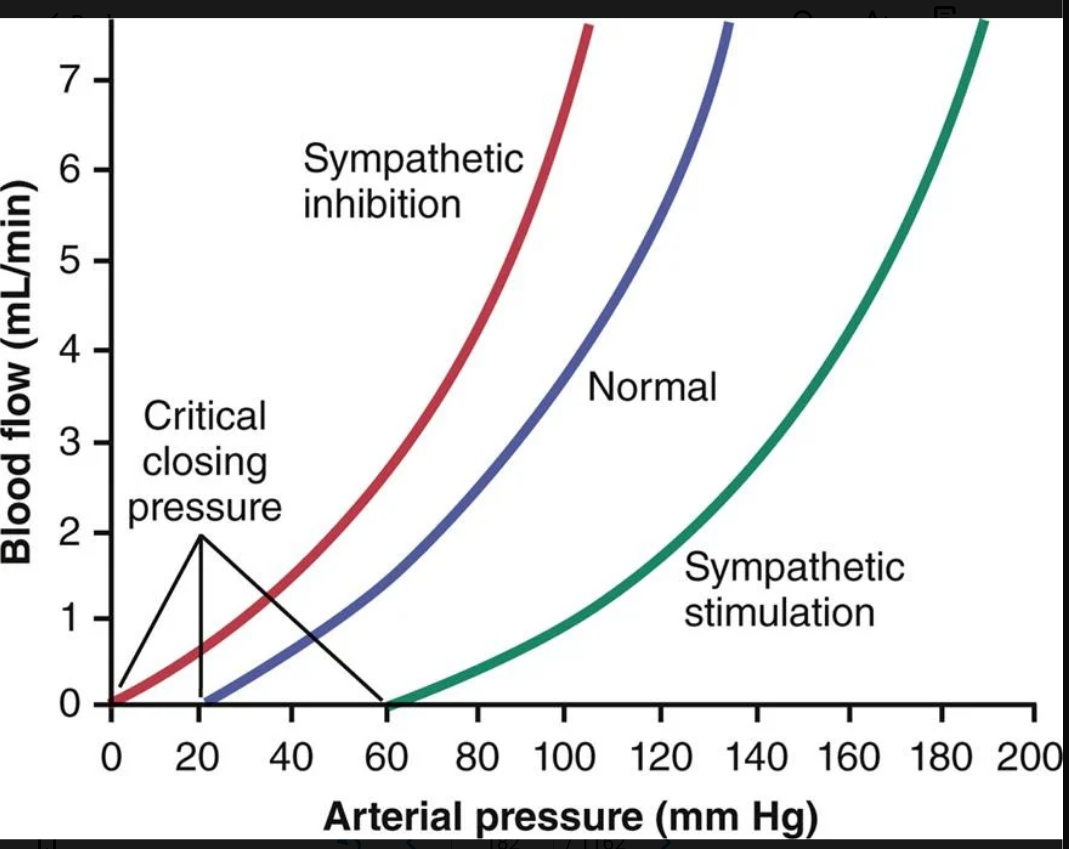
Image occlusion: name the hidden labels
31
Image occlusion: name the hidden labels
32
Image occlusion: name the hidden labels
33
larger blood vessels exposed to high pressures, such as the aorta, must have stronger ________ to withstand higher levels of tension and are generally reinforced with fibrous bands of collagen. In contrast, capillaries have a much smaller radii and therefore are exposed to much lower wall tension, permitting them to withstand pressures as high as 65 to 70 mm Hg in some organs such as the kidneys.
walls
34
As blood flows it creates a frictional force, or drag, on the endothelial cells lining the blood vessels
shear stress
35
valuable characteristic of the vascular system is that all blood vessels are__________ This of arteries allows them to accommodate the pulsatile output of the heart and to average out the pressure pulsations.
distensible
36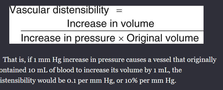
vascular distensibility equation
37
The total quantity of blood that can be stored in a given portion of the circulation for each mm Hg pressure rise
vascular compliance
38
vascular compliance equation
39
means that a vessel exposed to increased volume initially exhibits a large increase in pressure, but progressive delayed stretching of smooth muscle in the vessel wall allows the pressure to return toward normal over a period of minutes to hours
delayed compliance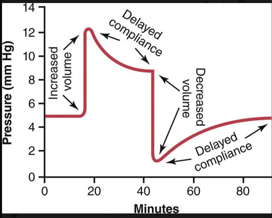
40
the pressure at the top of each pulse, called the systolic pressure, is about _____ mm Hg. At the lowest point of each pulse, called the diastolic pressure, it is about ______ mm Hg. The difference between these two pressures, about 40 mm Hg, is called the ________.
120; 80; pulse pressure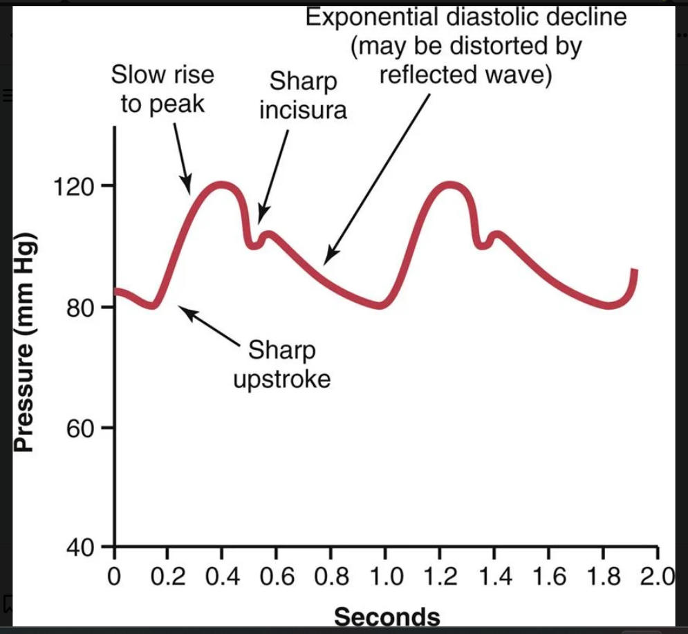
41
major factors that effect pulse pressure (x2)
(1) the stroke volume output of the heart; and (2) the compliance (total distensibility) of the arterial tree.
42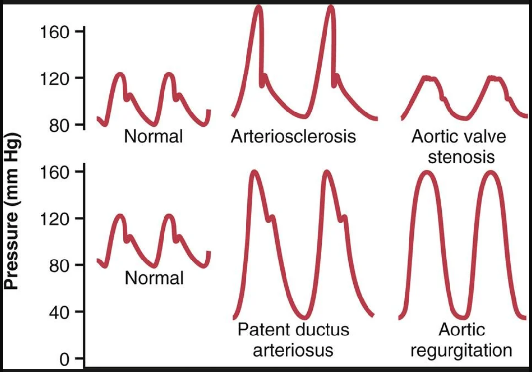
Image occlusion: name the hidden labels
43
Image occlusion: name the hidden labels
44
Image occlusion: name the hidden labels
45
Image occlusion: name the hidden labels
46
pulse pressure is roughly equivalent to
stroke volume/arterial compliance
47
the diameter of the aortic valve opening is reduced significantly, and the aortic pressure pulse is decreased significantly because of diminished blood flow outward through the stenotic valve.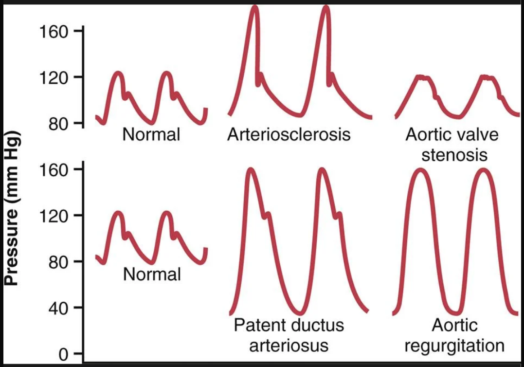
aortic valve stenosis
48
50% or more of the blood pumped into the aorta by the left ventricle flows immediately backward through the wide open ductus into the pulmonary artery and lung blood vessels, thus allowing the diastolic pressure to fall very low before the next heartbeat and increasing the pulse pressure.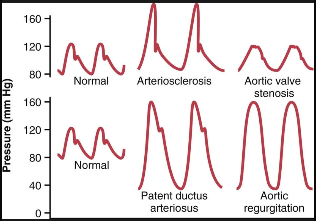
patent ductus arteriosus
49
the aortic valve is absent or does not close completely. Therefore, after each heartbeat, the blood that has just been pumped into the aorta flows immediately backward into the left ventricle. As a result, the aortic pressure can fall all the way to zero between heartbeats.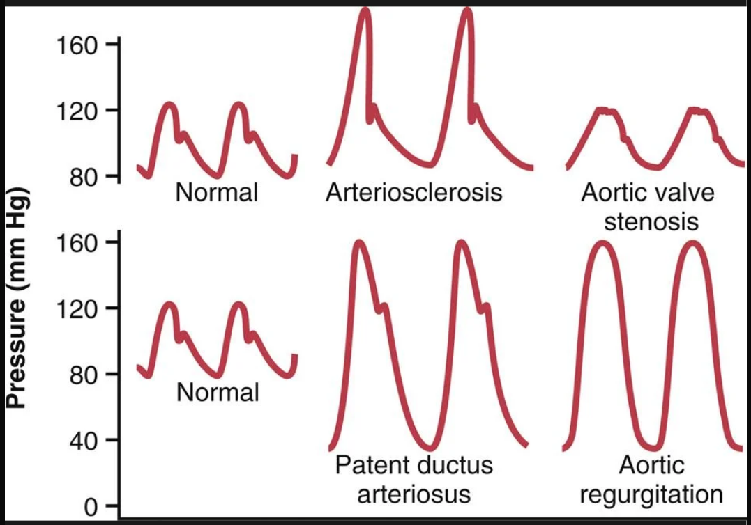
aortic regurgitation
50
what is believed to be the cause of korotkoff sounds?
caused mainly by blood jetting through the partly occluded vessel and by vibrations of the vessel wall.
51
the average of the arterial pressures measured millisecond by millisecond over a period of time.
mean arterial presusre
52
the right atrial pressure is regulated by a balance between (x2)
(1) the ability of the heart to pump blood out of the right atrium and ventricle into the lungs and (2) the flow of blood from the peripheral veins into the right atrium.
53
some factors that can increase venous return and increase rught atrial pressure (x3)
(1) increased blood volume; (2) increased large vessel tone throughout the body with resultant increased peripheral venous pressures; and (3) dilation of the arterioles, which decreases the peripheral resistance and allows rapid flow of blood from the arteries into the veins.
54
characterized by large bulbous protrusions of the veins beneath the skin of the entire leg, particularly the lower leg.
-distruction of the leaflets of the venous valves and the venous valves themselves
-distruction of the leaflets of the venous valves and the venous valves themselves
varucose veins
55
special blood reservoirs in the body (x3)
(1) the spleen, which sometimes can decrease in size sufficiently to release as much as 100 mL of blood into other areas of the circulation; (2) the liver, the sinuses of which can release several hundred milliliters of blood into the remainder of the circulation; (3) the large abdominal veins, which can contribute as much as 300 mL; and (4) the venous plexus beneath the skin, which also can contribute several hundred milliliters.
56
__________ of the spleen is a special reservoir that contains large quantities of concentrated red blood cells. These concentrated red blood cells can then be expelled into the general circulation whenever the sympathetic nervous system becomes excited and causes the spleen and its vessels to contract
red pulp
57
old red blood cells are destroyed in this organ
spleen
58
these cells digest dead rbcs in the spleen
reticuloendothelial cells
59
absence of the spleen
asplenia
60
reduced function of the spleen
hyposplenism
61
mechanisms to achieve hemostasis (x4)
(1) vascular constriction; (2) formation of a platelet plug; (3) formation of a blood clot as a result of blood coagulation; and (4) eventual growth of fibrous tissue into the blood clot to close the hole in the vessel permanently.
62
the process that prevents blood loss
hemostasis
63
hemostasis: Immediately after the blood vessel has been ruptured, the trauma to the vessel wall causes ______ in the wlal to contract; this rapidly _______ blood flow from the ruptured blood vessel
smooth muscle; decreases
64
these are responsible for much of the vasoconstriction by releasing thromboxane A2 in small blood vessels
platelets
65
this is the factor released by platelets to increase vasoconstriction in smaller blood vessels
thromboxane A2
66
if there is a very small cut in a blood vessel, it is usually sealed by this rather than a blood clot?
platelet plug
67
this chemical in platelets is what causes them to contract
thrombosthenin
68
these are i platelets and are responsibkle dor synthesis of various enzymes and store large wuantities of calcium ions
ER and golgi apparatus
69
platelets contain an enzyme system that synthesizes ________ which are local hormones that cause many vascular and other local reactions
prostaglandins
70
platelets contain ________ which causes vascular endothelial cells, vascular smooth muscle cells, and fibriplasts to multiply and grow, thus causing cellular growth and eventually helps repair damaged vascular walls
platelet derived growth factor
71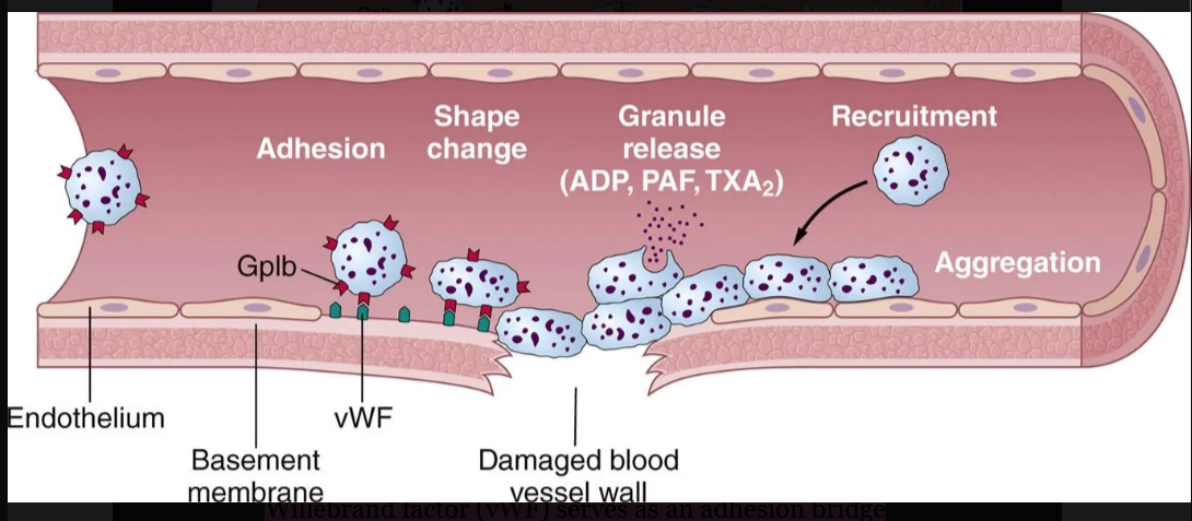
Image occlusion: name the hidden labels
72
Image occlusion: name the hidden labels
73
Image occlusion: name the hidden labels
74
Image occlusion: name the hidden labels
75
plateket plug: serves as an adhesion bridge between subendothelial collagen and the glycoprotein Ib (GpIb) platelet receptor.
vWF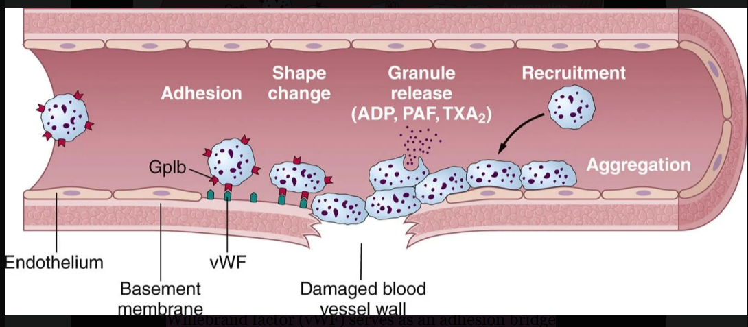
76
does a blood clot form faster if the trauma is minor or severe 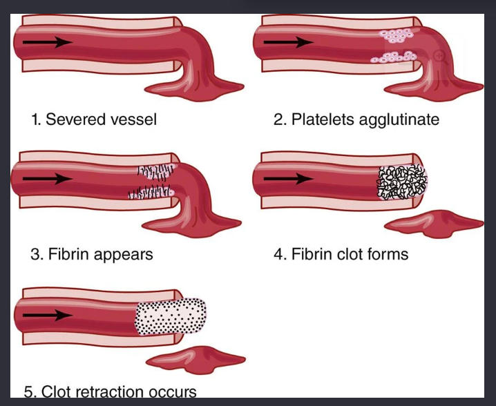
severe
77
what are the two courses once a blood clot has formed (x2)
(1) it can become invaded by fibroblasts, which subsequently form connective tissue all through the clot; or (2) it can dissolve. The usual course for a clot that forms in a small hole of a vessel wall is invasion by fibroblasts, beginning within a few hours after the clot is formed, which is promoted at least partially by growth factor secreted by platelets. This process continues to complete organization of the clot into fibrous tissue within about 1 to 2 weeks.
78
when a blood vessel is ruptured ___________ from the area of tisue damage become activated and override __________ and then the clot develops
procoagulants; anticoagulants
79
this acatlyzes the conversion of prothrombin into thrombin
prothrombin activator
80
thrombin acts as an enzyme to convert __________ that enmesh platelets, blood cells, and plasma to form trhe clot
fibrinogen into fibrin fibers
81
this is formed as a result of the rupture if bkood vessels or damage to special substances in the blood
prothrombin activator
82
prothrombin activator, in the presence of sfficient amounts of _______ causes conversion of prothrombin to thrombin
Ca2+
83
____ causes polymerization of fibrinogen moleculkes into fibrin fibers
thrombin
84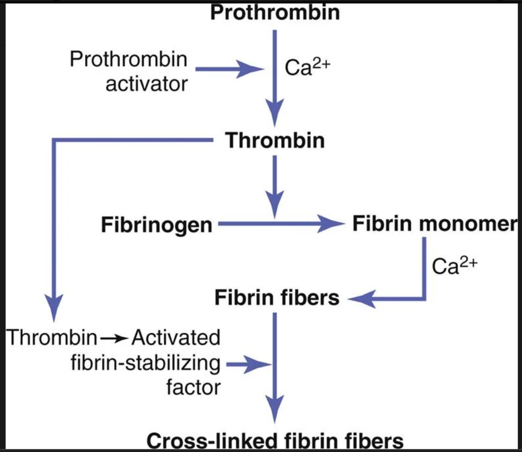
Image occlusion: name the hidden labels
85
Image occlusion: name the hidden labels
86
Image occlusion: name the hidden labels
87
Image occlusion: name the hidden labels
88
the rate-limiting factor in causing blood coagulation is usually the formation of _________ and not the subsequent reactions beyond that point because these terminal steps normally occur rapidly to form the clot.
prothrombin activator
89
prothrombin is formed continually by ______(organ) ands is continualy used y the body for clotting
liver
90
this is required by the liver for normal activation of prothrombin
vitamin K
91
Thrombin is a protein enzyme with weak proteolytic capabilities. It acts on fibrinogen to remove four low-molecular-weight _________- from each molecule of fibrinogen, forming one molecule of fibrin monomer that has the automatic capability to polymerize with other fibrin monomer molecules to form fibrin fibers.
peptidfes
92
the extrinsic blood clot formation pathway begins with
trauma to the vascular wall and surrounding tissue
93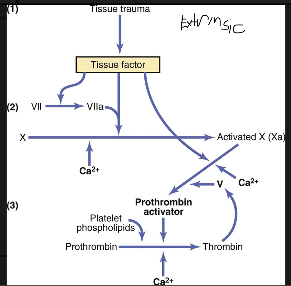
Image occlusion: name the hidden labels
94
Image occlusion: name the hidden labels
95
Image occlusion: name the hidden labels
96
Image occlusion: name the hidden labels
97
what does the intrinsic pathway for initiating clot formation begin with
trauma to the bood or exposure of the blood to collagen from traumayized blood vessel wall
98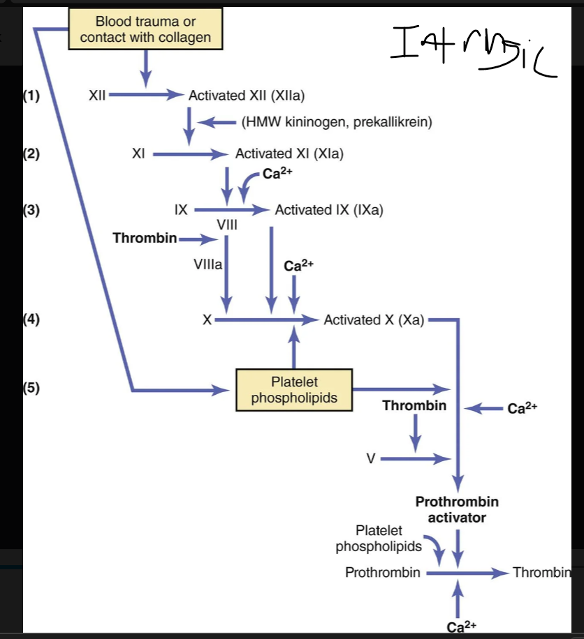
Image occlusion: name the hidden labels
99
Image occlusion: name the hidden labels
100
Image occlusion: name the hidden labels
101
Image occlusion: name the hidden labels
102
Image occlusion: name the hidden labels
103
Image occlusion: name the hidden labels
104
Image occlusion: name the hidden labels
105
Image occlusion: name the hidden labels
106
most important factors for prevening clotting in the normal vascular system: the __________ of the endothelial cell surface, which prevents contact activation of the intrinsic clotting system
smoothness
107
__________________, a mucopolysaccharide adsorbed to the surfaces of the endothelial cells, repels clotting factors and platelets, thereby preventing activation of clotting
glycocalyx
108
molecule is a highly negatively charged conjugated polysaccharide. By itself, it has little or no anticoagulant properties, but when it combines with antithrombin III, the effectiveness of antithrombin III for removing thrombin increases by 100-fold to 1000-fold and thus acts as an anticoagulant.
heparin
109
what does heparin combine with to remove free thrombin from circlatinh blood
antithroimbin III
110
what anticoagulation factors does heprin remove from the blood
activated factors IX-XII; thrombin
111
what makes heparin?
basophilic mast cells
112
these digest fibrin fibers and some other protein coagulants like fibrinogen, factor V, VIII, prothrombin, abd factor XII. it can cause a lysis of a clot by destroting many of the clotting factors
plasmin
113
this is trapped in a clot with other plasma protreins; t-PA eventyally converts this to plasmin which remobes the unnecessary remaining blood clot
plasminogen
114
most common types of bleeding tendencies (x3)
(1) vitamin K deficiency, (2) hemophilia, and (3) thrombocytopenia (platelet deficiency).
115
Vitamin K is an essential factor to a liver carboxylase that adds a c__________- to glutamic acid residues on five of the important clotting factors—prothrombin, factor VII, factor IX, factor X, and protein C.
carboxyl group
116
this enzyme reduces vitamin k back to its active form so it can be used again
vitamin k epoxde redictase complex 1 (VKORC1)
117
where is vitamin k syynthezied
intestinal tract by bacteria
118
One of the most prevalent causes of vitamin K deficiency is failure of the liver to secrete _______ into the gastrointestinal tract, which occurs as a result of liver disease or obstruction of the bile ducts.
bile
119
85% of cases, it is caused by an abnormality or deficiency of factor VIII;
hemophilia A`
120
the bleeding tendency is caused by deficiency of factor IX
hemophilia B`
121
where is the genetic defect in hemophilia
x chromosopme
122
results from loss of the large component of factor VIII.
von willebrand disease
123
The smaller component is most important in the intrinsic pathway for clotting, and it is deficiency of this part of factor VIII that causes....
hemophilia a
124
means the presence of very low numbers of platelets in the circulating blood
thrombocytopenia
125
red or purplish blotches on the skin
petechiae
126
major causes of thrombocytopenia (x5)
(1) decreased platelet production in the bone marrow due to infections or sepsis, nutrient deficiencies, or myelodysplastic disorders, which usually also reduce production of other cells (red blood cells [RBCs] and white blood cells); (2) peripheral platelet destruction by antibodies; (3) sequestration (pooling) of platelets in the spleen, especially in individuals with portal hypertension and excessively large spleens (splenomegaly); (4) consumption of platelets in thrombi; and (5) dilution of the blood from fluid resuscitation or massive transfusion.
127
abnormal clot that develops in a blood vessel
thrombus
128
free flowing clots
emboli
129
Emboli that originate in the venous system or in the right side of the heart generally flow into
lungs
130
this is delivered via catheter to remove a thrombus from the blood
t-PA
131
the clotting mechanism becomes activated in widespread areas of the circulation
-This condition often results from severe infection, septicemia, or the presence of large amounts of traumatized or dying tissue in the body that releases great quantities of tissue factor into the blood, initiating the coagulation cascade, as previously discussed.
-This condition often results from severe infection, septicemia, or the presence of large amounts of traumatized or dying tissue in the body that releases great quantities of tissue factor into the blood, initiating the coagulation cascade, as previously discussed.
disseminated intravascular coagulation (DIC)
132
this enzyme destroys heparin in the blood
heparinase
133
warfarin inhibits this enzyme and decreasesthe available form of voitamin k
VKORC1
134
Any substance that (ionization)_________ the blood calcium will prevent coagulation.
deionizes
135
time required for coagulation to take place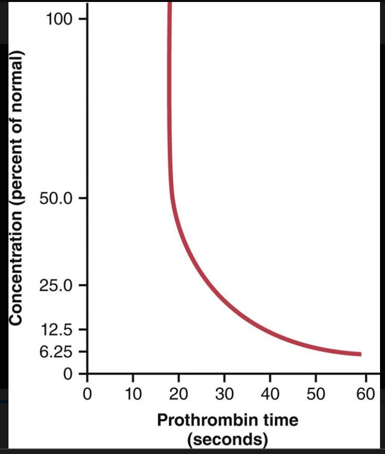
prothrombin time
136
A high INR level (______) indicates a high risk of bleeding, whereas a low INR (e.g., 0.5) suggests that there is a chance of having a clot. Patients undergoing warfarin therapy usually have an INR of 2.0 to 3.0.
4 or 5; `
137
circulation supplies systemic arterial blood to the trachea, bronchial tree (including the terminal bronchioles), supporting tissues of the lung, and outer coats (adventitia) of the pulmonary arteries and veins.
high presure, low flow circulation
138
circulation supplies venous blood from all parts of the body to the alveolar capillaries, where oxygen (O2) is added and carbon dioxide (CO2) is removed
low pressure, high flow circulation
139
right and left main branches that supply blood to the two respective lungs.
pulmonary blood vessels
140
left ventricular output are about 1% to 2% _______ than that of the right ventricular output.
greater
141
Particulate matter entering the alveoli is partly removed by these __________, and plasma protein leaking from the lung capillaries is also removed from the lung tissues, thereby helping to prevent pulmonary edema.
lymph vessels
142
When the left atrial pressure rises to high values, the ________________ also rises. Therefore, wedge pressure measurements can be used to estimate changes in _____________ and left atrial pressure in patients with congestive heart failure
pulmonary wedge pressure
143
failure of the _____ side of the heart, or increased resistance to blood flow through the ________ valve, causes blood to dam up in the pulmonary circulation, sometimes increasing the pulmonary blood volume as much as 100% and causing large increases in the pulmonary vascular pressures and, potentially, pulmonary edema
left; mitral
144
Blood flow through the lungs is essentially equal to the ____________. Therefore, the factors that control _________—mainly peripheral factors, as discussed in Chapter 20—also control pulmonary blood flow
cardiac output
145
When the concentration of O2 in the alveolar air decreases below normal, especially when it falls below 70% of normal (i.e., <73 mm Hg PO2), the adjacent blood vessels_____________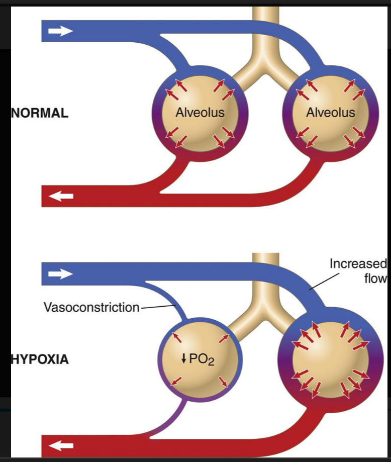
constrict
146
why do alveoli contract under low O2 conditions?
important function of distributing blood flow to where it is most effective. That is, if some alveoli are poorly ventilated and have a low O2 concentration, the local vessels constrict. This constriction causes the blood to flow through other areas of the lungs that are better aerated, thus providing an automatic control system for distributing blood flow to the pulmonary areas in proportion to their alveolar O2 pressures.
147
Note that in the standing position at rest, blood flow at the bottom of the lung is about 5 times as (lessreat) as in the top of the lung
great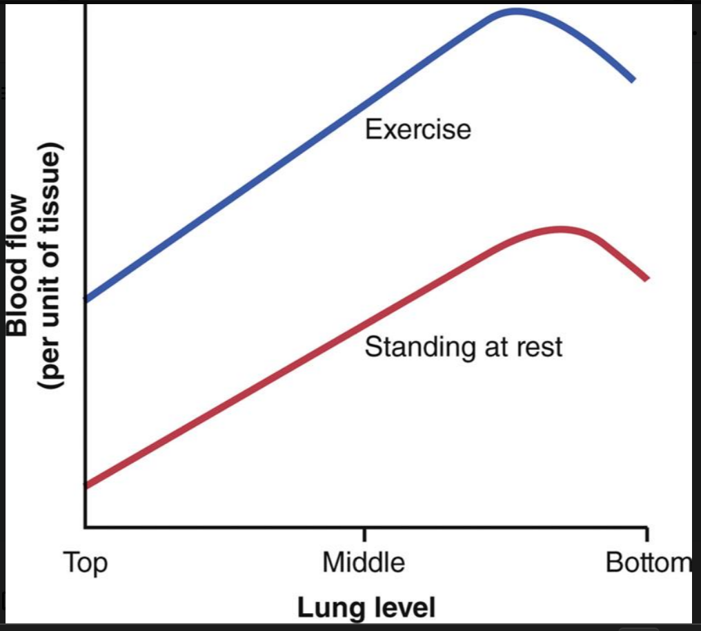
148
No blood flow during all portions of the cardiac cycle because the local alveolar capillary pressure in that area of the lung never rises higher than the alveolar air pressure during any part of the cardiac cycle
zone 1
149
Intermittent blood flow only during the peaks of pulmonary arterial pressure because the systolic pressure is then greater than the alveolar air pressure, but the diastolic pressure is less than the alveolar air pressure
zone 2
150
Continuous blood flow because the alveolar capillary pressure remains greater than alveolar air pressure during the entire cardiac cycle
zone 3
151
what zones do the lungs opperate under when they have normal conditions
zone 2 and 3
152
Zone 1 blood flow, which means no blood flow at any time during the cardiac cycle, occurs when the pulmonary systolic arterial pressure is _______ or the alveolar pressure is _________ to allow flow.
low; high
153
A major reason for increased blood flow is that the pulmonary vascular pressures rise enough during exercise to convert the lung apices from a zone 2 pattern into a zone ___- pattern of flow.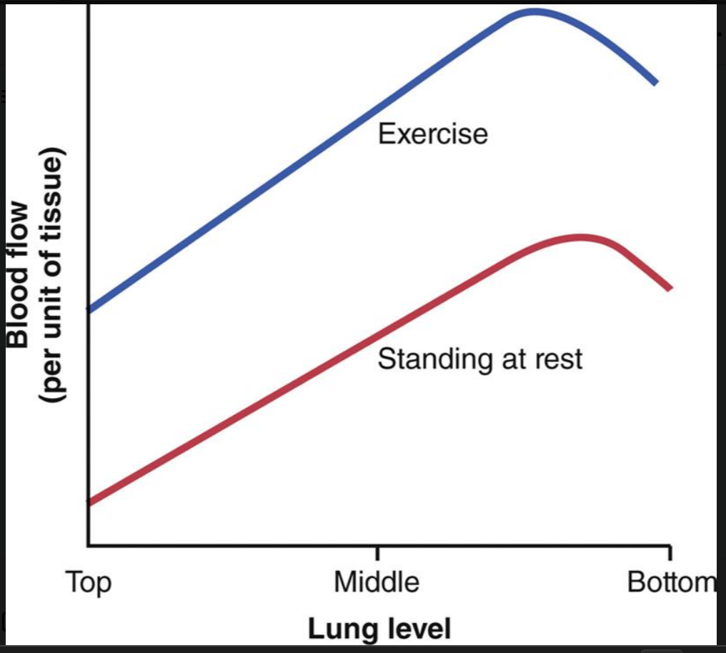
3
154
how is extra flow in the lungs accomplished (x3)
(1) by increasing the number of open capillaries, sometimes as much as 3-fold; (2) by distending all the capillaries and increasing the rate of flow through each capillary more than 2-fold; and (3) by increasing the pulmonary arterial pressure.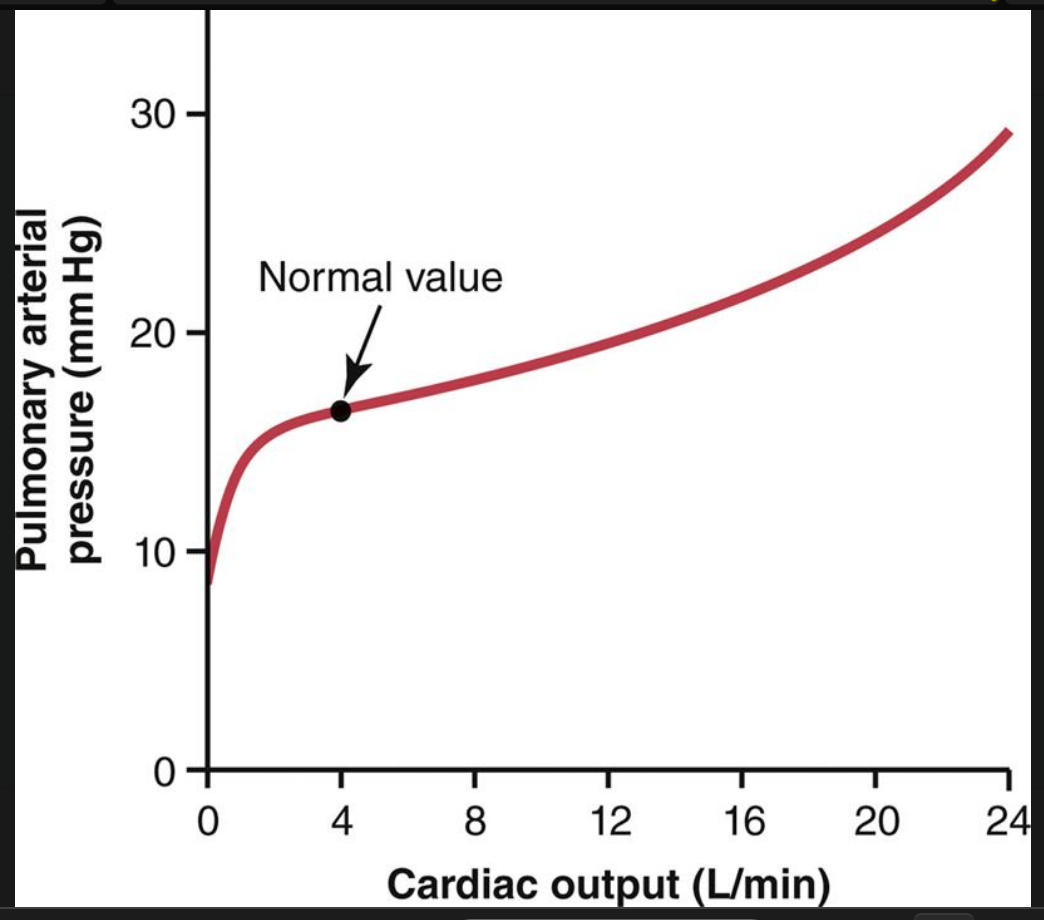
155
When the left atrial pressure rises above 30 mm Hg, causing similar increases in capillary pressure, ____________ is likely to develop
pulmonary edema
156
From histological studies of the total cross-sectional area of all the pulmonary capillaries, it can be calculated that when the cardiac output is normal, blood passes through the pulmonary capillaries in about 0.8 second. When the cardiac output ______, this time can shorten to as little as 0.3 second
increases
157
does pulmonary or peripheral cappilaroies have a higher pressure
periphera;
158
The interstitial fluid pressure in the lung is slightly more _ than that in peripheral subcutaneous tissue
negative
159
does the pulmonary interstitial fluid or peripheral tissue have a higher colloid osmotic ptessure
pulmonary interstitial fluid
160
filtration pressure causes a slight continual flow of fluid from the pulmonary __________ into the __________ and, except for a small amount that evaporates in the alveoli, this fluid is pumped back to the circulation through the pulmonary lymphatic system.
cappilaries; interstitial spaces
161
factor that increases fluid filtration out of the pulmonary capillaries or that impedes pulmonary lymphatic function and causes pulmonary interstitial fluid pressure to _______ from the negative range into the positive range will tend to cause filling of the pulmonary interstitial spaces and alveoli with free flu
rise
162
what are the most common causes of pulmonary edema? (x2)
1. Left-sided heart failure or mitral valve disease, with consequent great increases in pulmonary venous pressure and pulmonary capillary pressure, and flooding of the interstitial spaces and alveoli
2. Damage to the pulmonary blood capillary membranes caused by infections such as pneumonia or by breathing noxious substances such as chlorine gas or sulfur dioxide gas
2. Damage to the pulmonary blood capillary membranes caused by infections such as pneumonia or by breathing noxious substances such as chlorine gas or sulfur dioxide gas
163
When pulmonary capillary pressure remains elevated chronically (for at least 2 weeks), the lungs become even more resistant to pulmonary edema because the __________ expand greatly, increasing their capability of carrying fluid away from the interstitial spaces perhaps as much as 10-fold.
lymph vessels
164
The pleural membrane is a ____________ membrane through which small amounts of interstitial fluid transude continually into the pleural space
pourous, mesenchymal, serous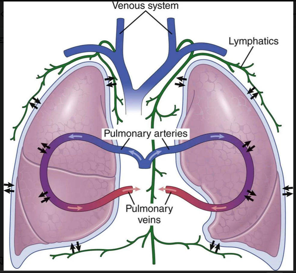
165
A _______ force is always required on the outside of the lungs to keep the lungs expanded. This force is provided by negative pressure in the normal pleural space
negative
166
Because the normal collapse tendency of the lungs is about −4 mm Hg, the ___________ pressure must always be at least as negative as −4 mm Hg to keep the lungs expanded
pleural fluid
167
the negativity of the pleural fluid pressure keeps the normal lungs pulled against the __________ of the chest cavity, except for an extremely thin layer of mucoid fluid that acts as a lubricant.
parietal pleura
168
analogous to edema fluid in the tissues and can be called edema of the pleural cavity.
pleural effusion
169
terminal arterioles
metarterioles
170
venules are (smallet/larger) than arterioles and have (stronger/weaker) muscular cpat) the pressure is (greater/lesser)
larger; weaker; lesser
171
means intermittent contraction of the metarterioles and precapillary sphincters (and sometimes even the very small arterioles).
vasomotion
172
Despite the fact that blood flow through each capillary is intermittent, so many capillaries are present in the tissues that their overall function becomes a_____________
averaged
173
Lipid-Soluble Substances Diffuse Directly Through ______________ of the Capillary Endothelium
cell membranes
174
Water-Soluble, Non–Lipid-Soluble Substances Diffuse Through ------------ in capillart membrane
intracellular pores
175
fluid in the interstitium is derived by filtration and diffusion from the capillaries. It contains almost the same constituents as plasma except for much lower concentrations of _______
proteins
176
osmotic pressure caused by the plasma proteins (called _____________________) tends to cause fluid movement by osmosis from the interstitial spaces into the blood
colloid osmotic pressure
177
returns to the circulation the small amounts of excess protein and fluid that leak from the blood into the interstitial spaces.
lymphatic system
178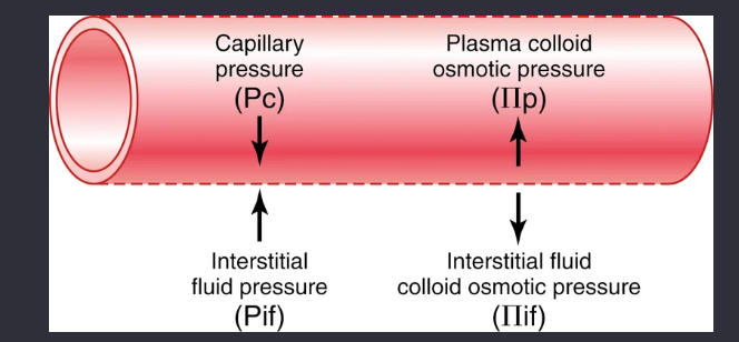
four primary forces that determine whether fluid will move out of the blood into the interstitial fluid or in the opposite direction. (starling forces)
179
if the sum of the starling pressures is positive (net filtration pressure) there will be
a net fluid filtration across the capillaries
180
if the sum of the starlking fptces is negative (net filtration pressure) there will be
a net fluid absorton from the interstitial spaces into the cappilaries
181
80% of the total colloid osmotic pressure of the plasma results from the
albumin
182
a 20-mm Hg rise in mean capillary pressure causes an increase in net filtration pressure from 0.3 to 20.3 mm Hg, which results in 68 times as much net filtration of fluid into the interstitial spaces as normally occurs. To prevent accumulation of excess fluid in these spaces would require 68 times the normal flow of fluid into the lymphatic system, an amount that is 2 to 5 times too much for the lymphatics to carry away. As a result, fluid will begin to accumulate in the interstitial spaces and _________ will result
edema
183
the capillary pressure falls very low, net reabsorption of fluid into the capillaries will occur instead of net filtration, and the __________ will increase at the expense of the interstitial fluid volume.
blood volume
184
any factor that increases interstitial fluid pressure also increases lymph flow if the lymph vessels are functioning normally. Such factors include the following:
• Elevated capillary hydrostatic pressure
• Decreased plasma colloid osmotic pressure
• Increased interstitial fluid colloid osmotic pressure
• Increased permeability of the capillaries
• Decreased plasma colloid osmotic pressure
• Increased interstitial fluid colloid osmotic pressure
• Increased permeability of the capillaries
185
any external factor that intermittently compresses the lymph vessel can also cause pumping. In order of their importance, such factors are as follows:
• Contraction of surrounding skeletal muscles
• Movement of the parts of the body
• Pulsations of arteries adjacent to the lymphatics
• Compression of the tissues by objects outside the body
• Movement of the parts of the body
• Pulsations of arteries adjacent to the lymphatics
• Compression of the tissues by objects outside the body
186
what determines lymph flow (equation)
lymh flow= interstitial fluid pressure x activity of the lymphatic pump
187
inflow and outflow of air between the atmosphere and the lung alveoli
pulmonary ventilation
188
During heavy breathing, however, the elastic forces are not powerful enough to cause the necessary rapid expiration, so extra force is achieved mainly by contraction of the __________, which pushes the abdominal contents upward against the bottom of the diaphragm, thereby compressing the lungs.
abdominal muscles
189
all the muscles that elevate the chest cage are classified as muscles of ________, and the muscles that depress the chest cage are classified as muscles of _________.
inspiration; expiration
190
most important muscles that raise the rib cage are the _________, but others that help are the following: (1) _____________ muscles, which lift upward on the sternum; (2) __________, which lift many of the ribs; and (3) ________, which lift the first two ribs.
external intercostals; sternocleidomastoid; anterior serrati; scaleni
191
the pressure of the fluid in the thin space between the lung pleura and chest wall pleura (negative pressure)
pleural pressure
192
the pressure difference between that in the alveoli and that on the outer surfaces of the lungs (pleural pressure)
transpleural pressure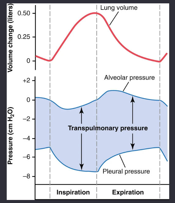
193
measure of the elastic forces in the lungs that tend to collapse the lungs at each instant of respiration
recoil pressure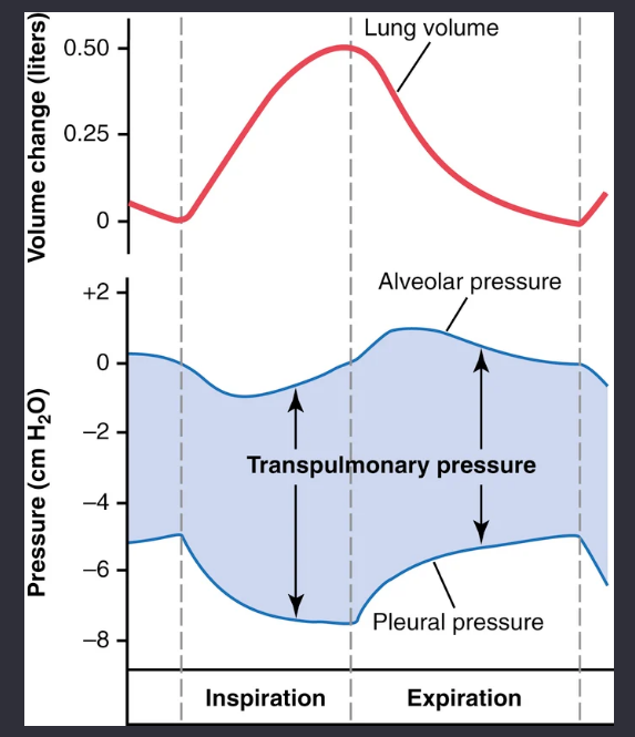
194
The extent to which the lungs will expand for each unit increase in transpulmonary pressure (if enough time is allowed to reach equilibrium) is called
lung compliance
195
what determines the elastic forces of lung tissue (x2)
elastin and collagen fibers
196
what cells secrete surfacant?
type II alveolar epithelial cells
197
in surfacant; helps to reduce surface tension (specific phospholipid)
dipalmitoyl phosphatidylcholine
198
the surface tension in the alveoli tends to collapse the alveoli. This collapse creates ________ in the alveoli, attempting to push the air out.
positive presure
199
how to generate amount of pressure generated in an alveolus
pressure=(2x surface tension)adius of alveolus
200
the smaller the alveolus, the greater the alveolar ___________ caused by the surface tension.
pressure
201
work that required to expand the lungs against the lung and chest elastic forces,
compliance work
202
work required to overcome the viscosity of the lung and chest wall structures
tissue resistance work
203
work required to overcome airway resistance to movement of air into the lungs
airway resistance work
204
what is the energy limitation and how that corresponds to respiration
the intensity of exercise performed can only be reflected on the amount of enerfy (ATP) produced
205
how is pulmonary ventilation recorded
spirometry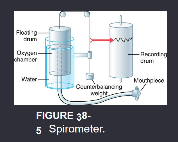
206
the volume of air inspired or expired with each normal breath; it amounts to about 500 ml in the average healthy man.
tidal volume
207
the extra volume of air that can be inspired over and above the normal tidal volume when the person inspires with full force
inspiratory reserve volume
208
the maximum extra volume of air that can be expired by forceful expiration after the end of a normal tidal expiration
expiratory reserve volume
209
the volume of air remaining in the lungs after the most forceful expiration
residual volume
210
This capacity is the amount of air (≈3500 ml) that a person can breathe in
inspiratory capacity
211
inspiratory capacity equation
inspiratory capacity= tidal volume+inspiratory reserve volume
212
functional residual volume equation
FRV= expiratory reseve volume+residual volume
213
This capacity is the amount of air that remains in the lungs at the end of normal expiration
functional residual capacity
214
vital capacity equation
VC= inspiratory reserve volume+tidal volume+expiratory reserve volume
215
capacity is the maximum amount of air a person can expel from the lungs after first filling the lungs to their maximum extent and then expiring to the maximum extent
vital capacity
216
the maximum volume to which the lungs can be expanded with the greatest possible effort
total lung capacity
217
total lung capacity equation
total lung capacity=vital capacity+residual volume
218
the volume of air that remains in the lungs at the end of each normal expiration; value changes markedly in some pulmonary diseases
functional residual capacuty (FRC)
219
the total amount of new air moved into the respiratory passages each minute
minute respiratory volume
220
minute respiratory volume equation
minute respiratory volume+ tidal volumeXrespiratory rate/min
221
air that does not make it to the lungs where gas exchange occurs
dead space air
222
these sympathetic NT are important in the bronchial tree and cuse dialation or nondialation
norepinephrine and epinephrine
223
what releases histamoine and slow reactive substance of anaphylaxis in the lung tissue during allergc reacytion
mast cells
224
what is the initiating stimulus in the sneeze reflex
irritation in the nasal reflex
225
this function of speech is achieved by the laryn
phonation
226
this function of speech is achieved by the mouth
articulation
227
The heart is composed of three major types of cardiac muscle— (x3)
atrial muscle
ventricular muscle
excitatory and conductive muscle fibers
ventricular muscle
excitatory and conductive muscle fibers
228
how does the nbase of the heart move during systole
toward the apex
229
how does the lefdt ventrivle move dyuring diastole
relaxation, recoild and untwists to allow blood to enter the pumping chaombers rapidly
230
intrercalated discs what are they?
cell membranes that separate individual cardiac muscle cells from one another.
231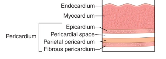
Image occlusion: name the hidden labels
232
Image occlusion: name the hidden labels
233
Image occlusion: name the hidden labels
234
Image occlusion: name the hidden labels
235
Image occlusion: name the hidden labels
236
Image occlusion: name the hidden labels
237
intracellular potential of tje heart (resting)
85-
238
what happens at 0?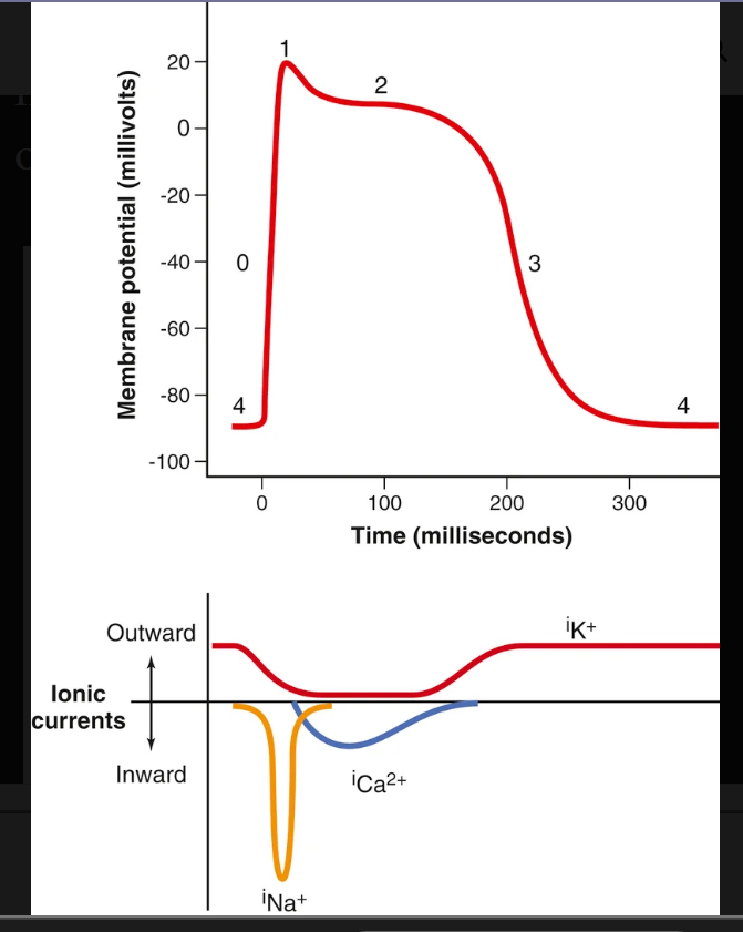
Voltage-gated sodium channels (fast sodium channels) open and permit sodium to rapidly flow into the cell and depolarize it. The membrane potential reaches about +20 millivolts before the sodium channels close.
239
what happens at 1?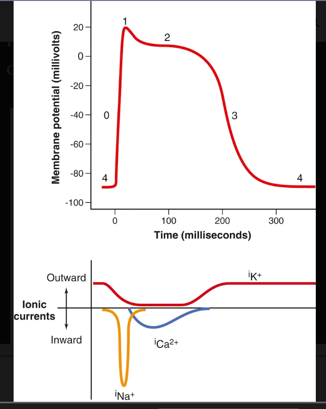
The sodium channels close, the cell begins to repolarize, and potassium ions leave the cell through open potassium channels.
240
what happens at 2?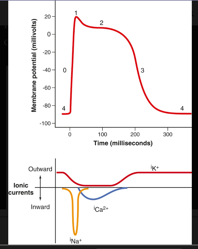
plateaus as a result of increased calcium ion permeability and decreased potassium ion permeability. The voltage-gated calcium ion channels open slowly during phases 1 and 0, and calcium enters the cell. Potassium channels then close, and the combination of decreased potassium ion efflux and increased calcium ion influx causes the action potential to plateau.
241
what happens at 3?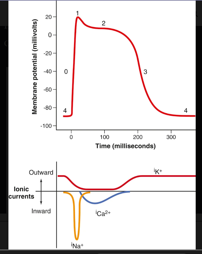
The closure of calcium ion channels and increased potassium ion permeability, permitting potassium ions to exit the cell rapidly,
242
cardiac cells: T tubule action potentials then act on the membranes of the _________________ to cause release of calcium ions into the muscle sarcoplasm from the sarcoplasmic reticulum.
LONGITUDINAL sarcoplasmuc tub;les
243
cardiac cells: Calcium entering the cell activates calcium release channels, also called ____________, in the sarcoplasmic reticulum membrane, triggering the release of calcium into the sarcoplasm.
ryanodine receptor channels
244
what transports CA2+ vack to the reticulum?
calcium–adenosine triphosphatase (ATPase) pump
245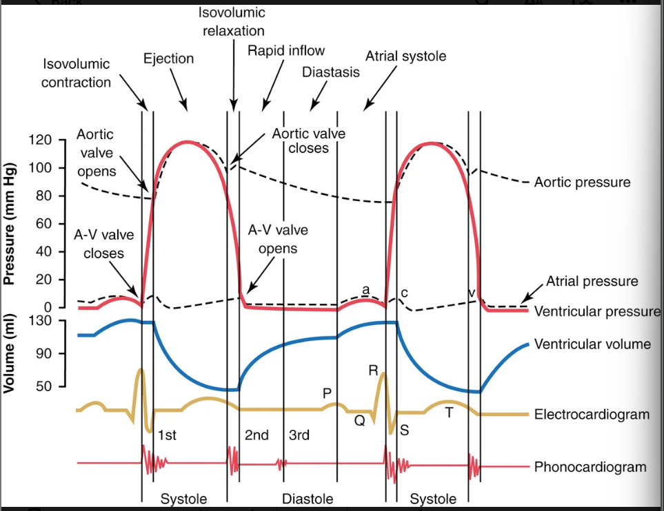
Image occlusion: name the hidden labels
246
Image occlusion: name the hidden labels
247
Image occlusion: name the hidden labels
248
Image occlusion: name the hidden labels
249
Image occlusion: name the hidden labels
250
Image occlusion: name the hidden labels
251
Image occlusion: name the hidden labels
252
Image occlusion: name the hidden labels
253
Image occlusion: name the hidden labels
254
Image occlusion: name the hidden labels
255
what causes the p wave
spread of depolarization through the atria
256
what causes QRS waves
electrical depolarization of the ventricles
257
what causes t waves
stage of repolarization of the ventricles when the ventricular muscle fibers begin to relax.
258
when the atria fail to function, the difference is unlikely to be noticed unless a person _________________; then, symptoms of heart failure occasionally develop, especially shortness of breath.
exercises
259
what causes the a wave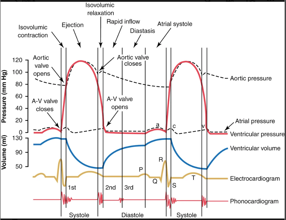
atrial cpntraction
260
what causes the c wave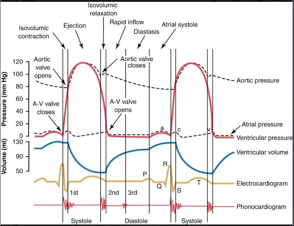
ventricles begin to contract; it is caused partly by slight backflow of blood into the atria at the onset of ventricular contraction,
261
what causes the v wave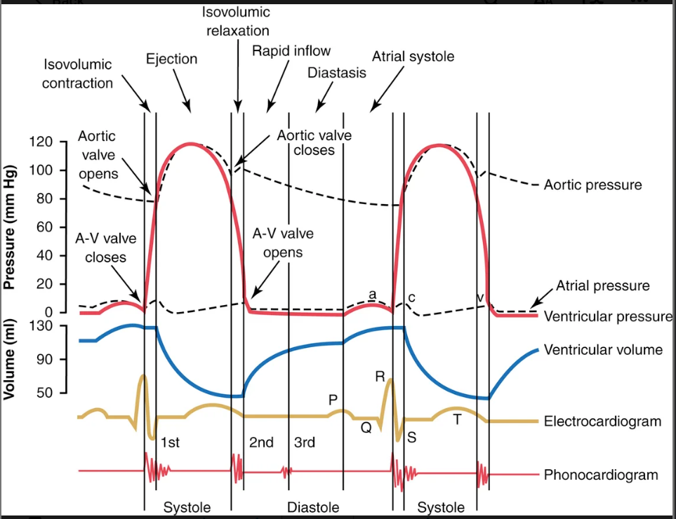
slow flow of blood into the atria from the veins while the A-V valves are closed
262
the moderately increased pressures that have developed in the atria during ventricular systole immediately push the____________ valves open and allow blood to flow rapidly into the ventricles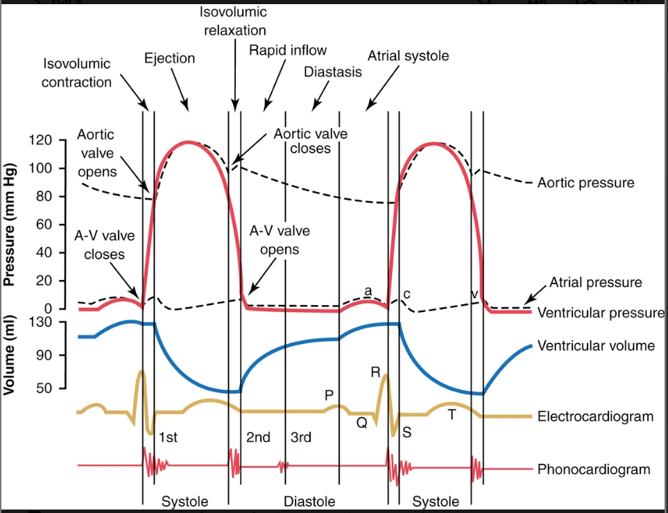
AV
263
Immediately after ventricular contraction begins, the ventricular pressure (increases/decreases) causing the AV valves to close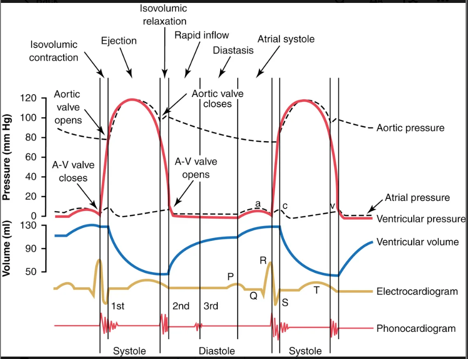
increases
264
the ventricles need to develop more pressure after closing the AV valves to push these valves open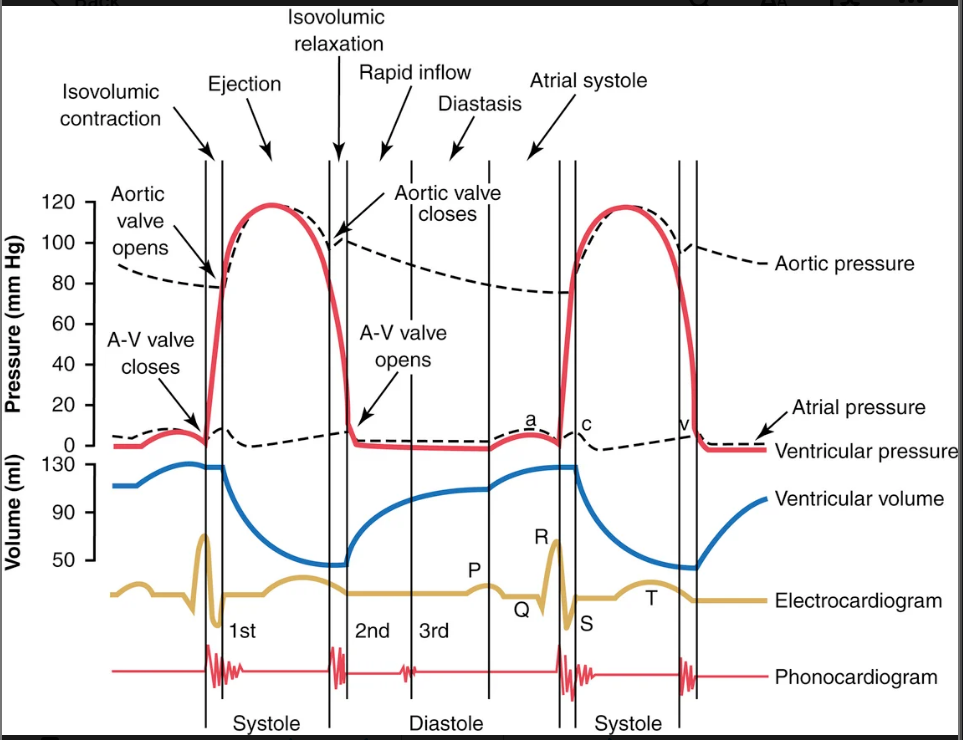
aortic and pulmonary (semilunar)
265
at the end of systole, ventricilar ________ begins allowing the right and left IV pressures to (increase/decrease)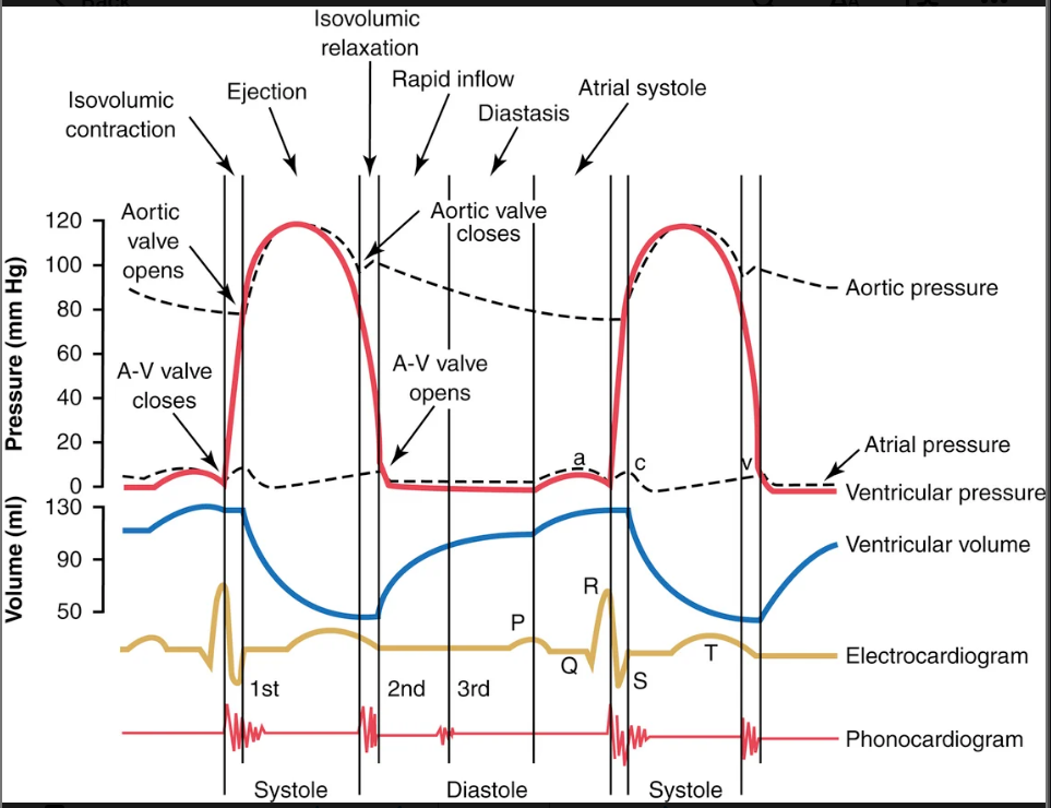
relaxation; decrease
266
how does the passive opening of the valves of the left ventricle work
they close when a backward pressure gradient pushes blood backward, and they open when a forward pressure gradient forces blood in the forward direction
267
how do the papillary muscles help the valves close
dont close the valves directlt but they pull the vanes of the valves inward toward the ventricles to prevent their bulging too far backward toward the atria during ventricular contraction.
268
the high pressures in the arteries at the end of systole cause the semilunar valves to (open/close)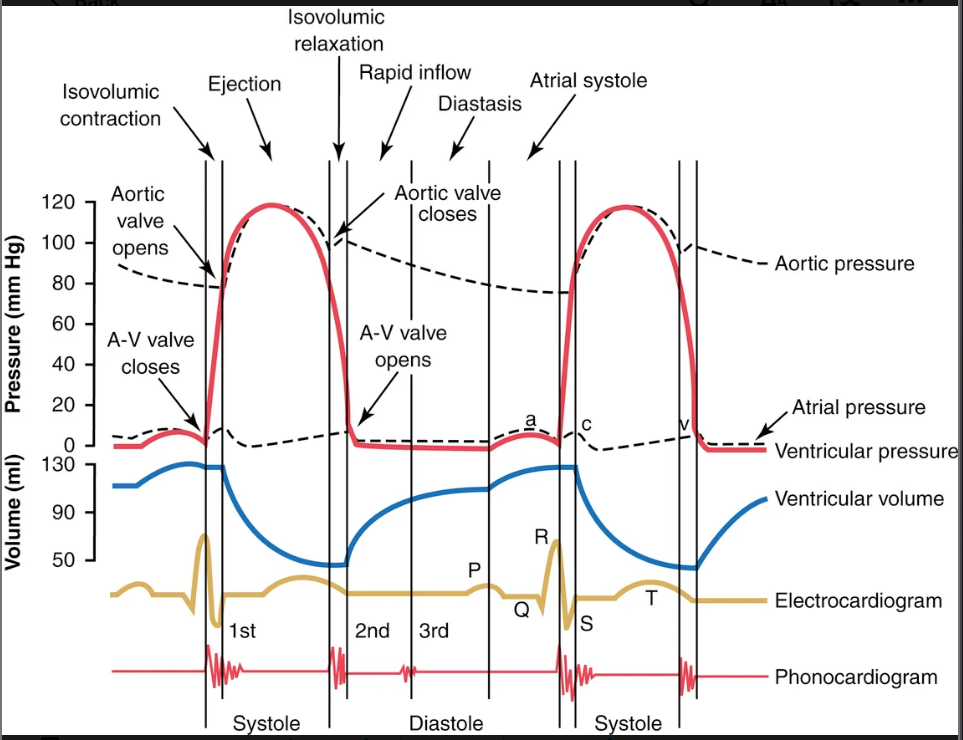
close
269
which valves have a greater ejection velocity semilunar of av
semilunar
270
what causes the S1 sound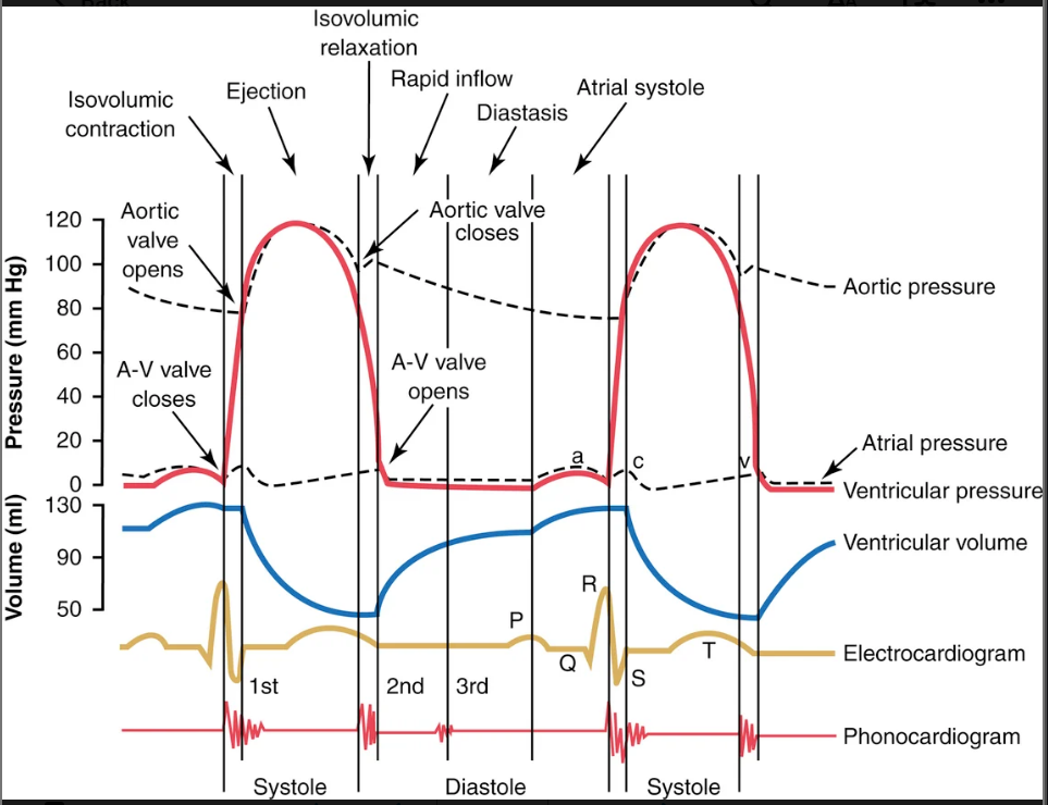
closure of the A-V valves. The vibration pitch is low and relatively long-lasting
271
what causes the S2 sounds
aortic and pulmonary valves close at the end of systole, one hears a rapid snap because these valves close rapidly
272
the amount of energy that the heart converts to work during each heartbeat while pumping blood into the arteries
stroke work output (heart)
273
major proportion is used to move the blood from the low-pressure veins to the high-pressure arteries.
volume presure work (external work)
274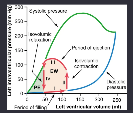
Image occlusion: name the hidden labels
275
Image occlusion: name the hidden labels
276
Image occlusion: name the hidden labels
277
Image occlusion: name the hidden labels
278
Image occlusion: name the hidden labels
279
Image occlusion: name the hidden labels
280
what do the red lines show in this diagram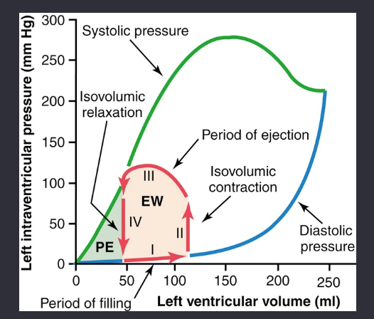
volume pressure diagram of the cardiac cycle for ormal function of left ventricle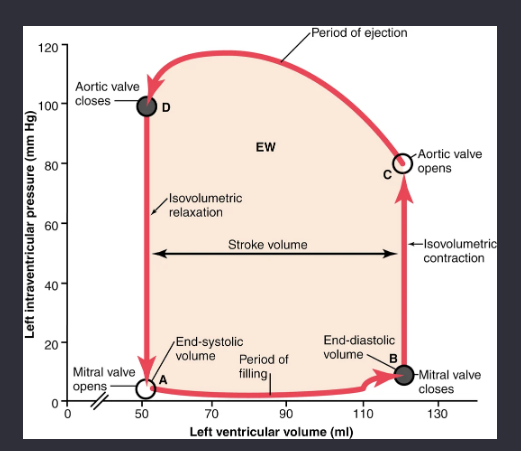
281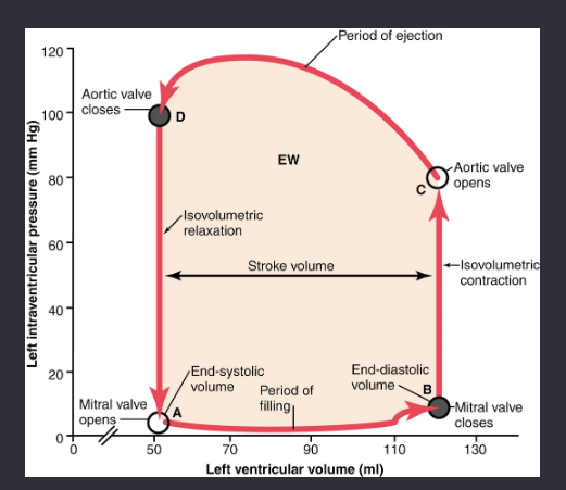
Image occlusion: name the hidden labels
282
Image occlusion: name the hidden labels
283
Image occlusion: name the hidden labels
284
Image occlusion: name the hidden labels
285
Image occlusion: name the hidden labels
286
Image occlusion: name the hidden labels
287
Image occlusion: name the hidden labels
288
Image occlusion: name the hidden labels
289
Image occlusion: name the hidden labels
290
Image occlusion: name the hidden labels
291
Image occlusion: name the hidden labels
292
considered to be the end-diastolic pressure when the ventricle has become filled.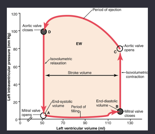
preload
293
pressure in the aorta leading from the ventricle.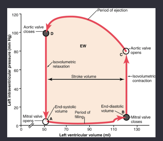
afterload
294
_____________consumption has also been shown to be nearly proportional to the tension that occurs in the heart muscle during contraction multiplied by the duration of time that the contraction persists; this is called the tension-time index
oxygen
295
law of laplace ventricilar wall tension equation
ventricular wall tension= left ventricular pressure x radius
296
Because tension is high when systolic pressure (and therefore left ventricular pressure) is high, correspondingly (more/less) oxygen is used.
more
297
When systolic pressure is chronically elevated, these two things are increased chronically
wall stress and cardiac workload
298
the ratio of work output to total chemical energy used to perform the work.
cardiac efficiency
299
what is the maximum cardiac efficiency of the noprmal heart
20-25%
300
the more the heart muscle is stretched during filling, the (greater/less) the force of contraction and the (greater/less) blood pumped into the aorta
greater x2
199 more cards in OutLearn. Open the full deck