Exam 1 lecture
Study these flashcardsFree · spaced repetition · iPhone, Android, Mac & web
All cards
1
a comprehensive system of
healthcare based upon a specific philosophy and distinctive
art. Our philosophy and principles embrace holistic
evaluation and treatment of patients to obtain the best
possible outcomes in medical care.
healthcare based upon a specific philosophy and distinctive
art. Our philosophy and principles embrace holistic
evaluation and treatment of patients to obtain the best
possible outcomes in medical care.
osteopathic medicine
2
a science which consists of such exact
exhaustive and variable knowledge of the structure and
functions of the human mechanism, anatomy, physiology,
psychology, including the chemistry and physics of its known
elements
exhaustive and variable knowledge of the structure and
functions of the human mechanism, anatomy, physiology,
psychology, including the chemistry and physics of its known
elements
oseopathy
3
what is the DO MD difference?
• Extra training in the neuromusculoskeletalExtra training in the neuromusculoskeletal
system* (at least 200 hours)system* (at least 200 hours)
•• Osteopathic Manipulative Treatment*Osteopathic Manipulative Treatment*
•• Osteopathic PHILOSOPHY*Osteopathic PHILOSOPHY
system* (at least 200 hours)system* (at least 200 hours)
•• Osteopathic Manipulative Treatment*Osteopathic Manipulative Treatment*
•• Osteopathic PHILOSOPHY*Osteopathic PHILOSOPHY
4
what are the 4 main tenets of OM in order?
The body is a unit; the person is a unit of body, mind, and
spirit.
The body is capable of self-regulation, self-healing, and
health maintenance
Structure and function are reciprocally interrelated.
Rational treatment is based upon an understanding of the
basic principles of body unity, self-regulation, and the
interrelationship of structure and function
spirit.
The body is capable of self-regulation, self-healing, and
health maintenance
Structure and function are reciprocally interrelated.
Rational treatment is based upon an understanding of the
basic principles of body unity, self-regulation, and the
interrelationship of structure and function
5
researcher in oseopathic practice and proving it
Irvin M Korr
6
Therapeutic
use of the hands to restore
normal Structure (anatomy)
and Function
use of the hands to restore
normal Structure (anatomy)
and Function
OMT/OMM
7
when was osteopathy founded?
June 22, 1874
8
when was the first school of osteopathy founded
1892
9
When was the AMA established
1847
10
what does the AMA do?
regulate medical school education
11
what were DOs very successful in treating over MDs, giving them more credibility in the 1910s
influenza
12
when did Do schools start integrating pharmacology into the curriculum
1930s (early)
13
when did AOA and ACGME merge completely
2020
14
when was lecom started
1993
15
what is a SOAP note? what makes it up?
a soap note ois a subjective objective assessment plan, It is where you collect data, evaluate and synthesize that data, create a differential diagnosis, make a management plan, then reevaluate at the next visit and find health for the patient you are caring for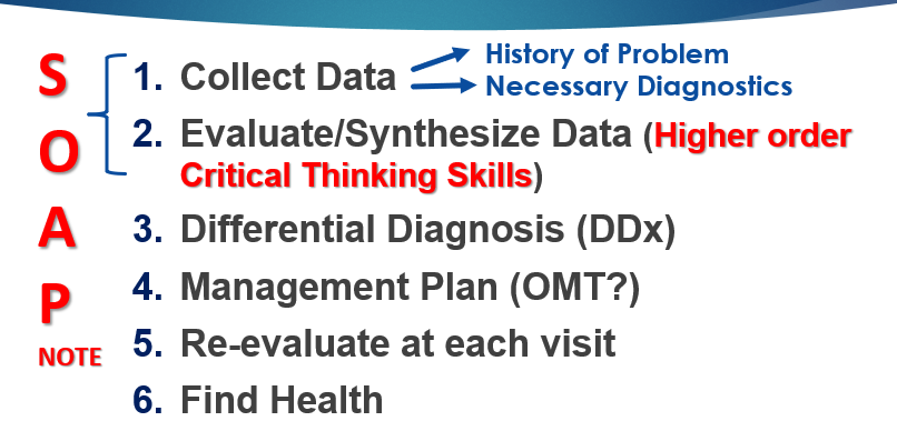
16
what are the parts of data collection in osteopathic medicibe?
observations, patient history, physical exam (integrated neuromusculoskeletal exam), palpations, motion testing (gross and segmental), and special testing including labs, radiology, or others
17
what are you evaluating for in a NMS exam?
what is normal or not normal
18
What is part of a palpatory NMS exam?
TART changes/evaluation
19
what is TART?
Tissue texture abnormalities
assymetry of tissue/joint motions
restricted ROM of tissue/joints
Tenderness
assymetry of tissue/joint motions
restricted ROM of tissue/joints
Tenderness
20
what is somatic dysfunction
impaired or altered function of related components of the somatic system: includes skeltal, arthrodial and myofasical structures, and related vascu;lar, lymphatic and neural elements
21
How do you treat Somatic dysfunction?
OMT
22
What are the 6 osteopathic models
Classics: biomechanical structura;, respiratory circulatory, metabolic nutritional, neurological, behavioral biophysical
Bioenergetics
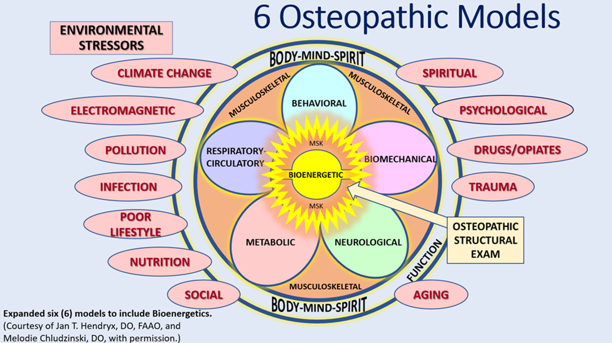
Bioenergetics
23
what is the biomechanical model
anatomy of muscles, spine, extremities, posture, motion, tenegrity
OMT: normalize mechanical somatic dysfunction, structural integrity, physiological function, homeostasis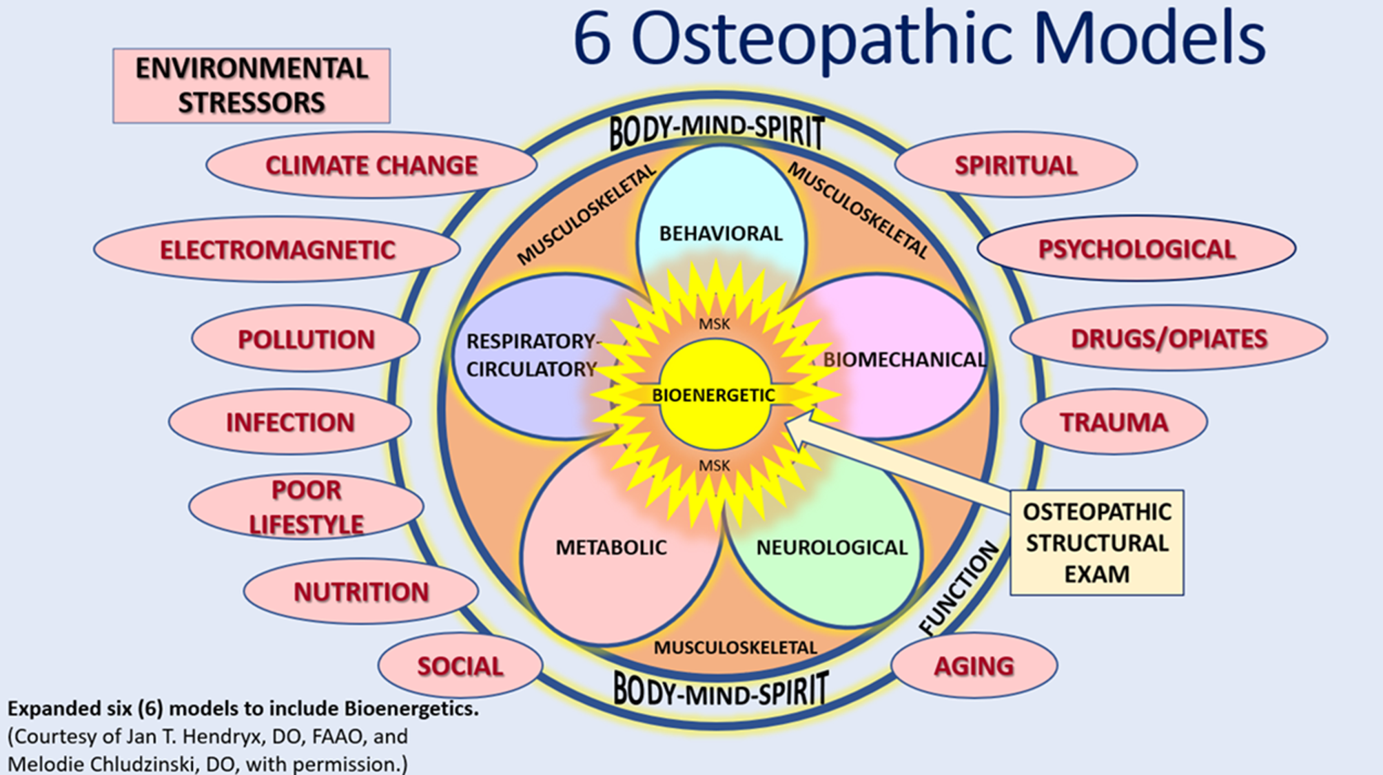
OMT: normalize mechanical somatic dysfunction, structural integrity, physiological function, homeostasis
24
what is the neurological model
model emphasizes the CNS, PNS, and ANS, that control coordinate and integrate body functions
OMT is directed ypwatfs normalizing neurological functioninh, balancing the autonomic nervous system, proprioce[tion, and homeostasis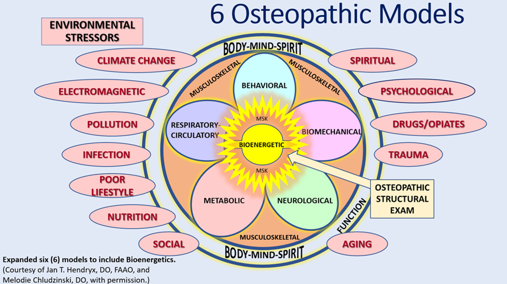
OMT is directed ypwatfs normalizing neurological functioninh, balancing the autonomic nervous system, proprioce[tion, and homeostasis
25
What is the respiratory circulatory approach
emphsizes pulmonary circulatory and fluid lymphatic systems
OMT directed towards normalizing blood flow and lymphatic flow and diaphagm function and tissue oxygenation
OMT directed towards normalizing blood flow and lymphatic flow and diaphagm function and tissue oxygenation
26
what is the metabolic nutrional model?
emohasizes homeostatic adaptive mechanisms through + and feedback loops to regulate energy exchange and conservation through metabolic processes
OMT lymphatic pump and visceral techniques
treatment includes lifestyle changes and nutritional counseling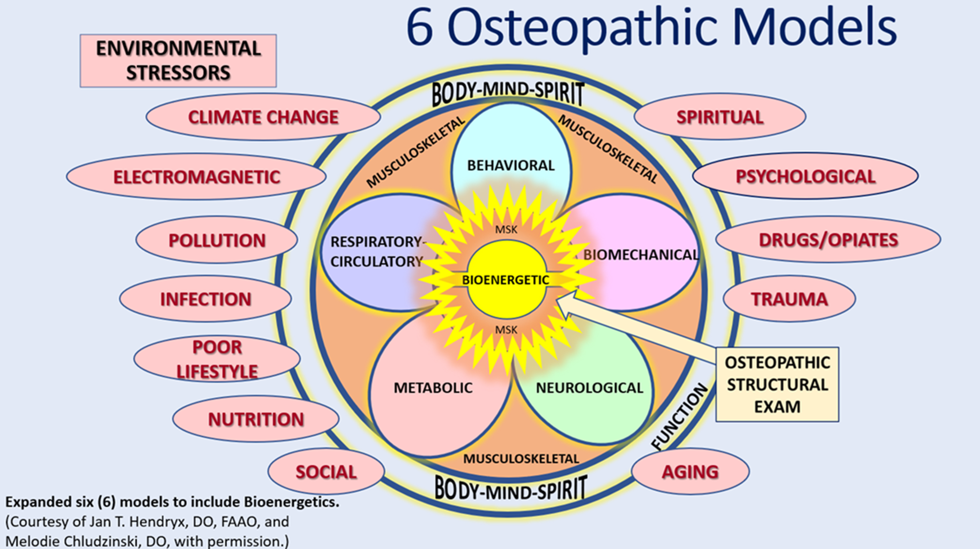
OMT lymphatic pump and visceral techniques
treatment includes lifestyle changes and nutritional counseling
27
What is the behavioral biopsychosocial model
mental emotional and spiritual dementions to health ad disease
OMT would approach gently guided imagery, breeathing exercises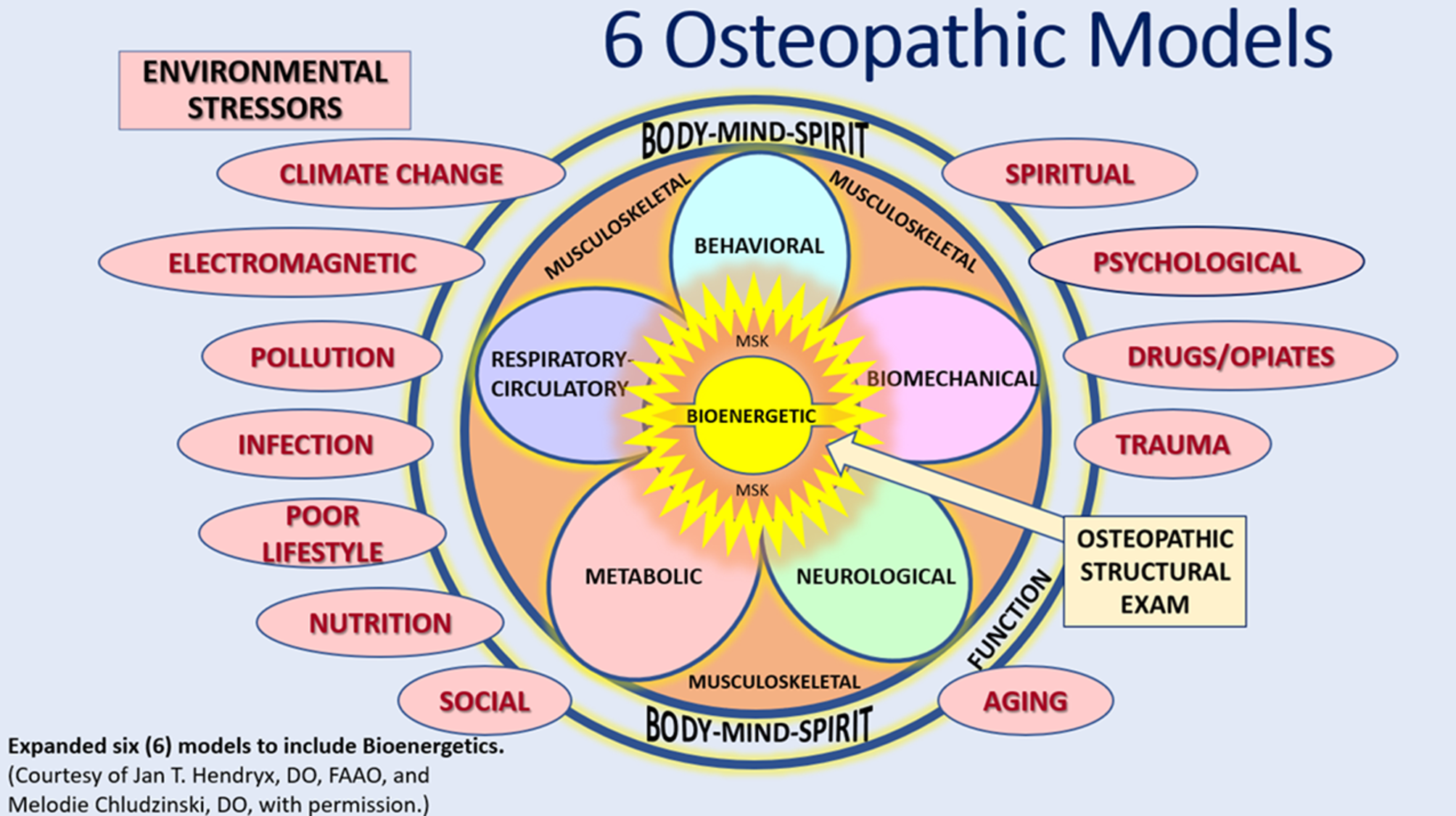
OMT would approach gently guided imagery, breeathing exercises
28
what is palpation
The application of variable manual pressures to the surface of the body for the purpose of determining the shape, size, consistency, position, inherent motility and health of tissues beneath
29
what info do you get from palpation
Normal vs abnormal:
Tissues: soft or hard
Motions: ranges of motion, barriers, ease
Rhythms: arterial pulse, respiratory
Tissues: soft or hard
Motions: ranges of motion, barriers, ease
Rhythms: arterial pulse, respiratory
30
3 types of sensory nerve receptors
mechanoreceptors, thermoreceptors, nociceptors
31
what do mechanoceptors sense
touch and proprioception
32
what do thermoreceptors sense
cooling and heating
33
what do nociceptors sense
pain, itch, tickle, tingling to stabbinh
34
what are the 4 different types of mechanoreceptors used in palpation?
merkel disc, ruffini corpuscle, meissner corpuscle, pacinian corpuscle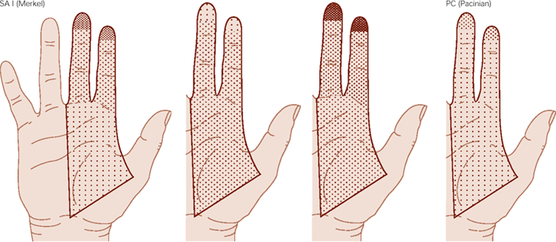
35
what is the palp purpose of merkel discs
monitor pulse, sensing end points, and sensing skin deformations
36
what do merkel discs sense
degree of pressure
37
what type of mechanoreceptor are merkel discs
slow adapting
38
what are ruffini corpuscles used for palpation
lumps and bumps under the skin by stretching it, enlarged lymph nodes
39
what type of mechanoreceptor are ruffini corpuscles
slow adapting
40
what are meissner corpuscles used for in palpation
2 point discrimiation
41
what type of mechanoreceptors are meissner corpuscles
rapid adapting
42
what are pacinian corpuscles used for in palpatipon
vibratory sense like while using a tuning fork or thrill when sensing for a quiver in someone's pulse
43
what type of mechanorecepors are pacinian corpuscles?
rapid adapting
44
what do you use to sense temp changes in palpation
dorsolateral aspect of hand ulnar surface)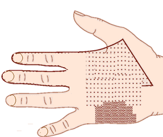
45
what are the phases of basic palpation
1) detection
2) internal amplifcation: see the structure yo are palpating and have some internal knowledge of the anatomy
3) analysis and interoretation: know the difference between normal and adnormal
2) internal amplifcation: see the structure yo are palpating and have some internal knowledge of the anatomy
3) analysis and interoretation: know the difference between normal and adnormal
46
are these somatic dysfunctions: fractures, degenerative processes, inflamatory processes
nay nay
47
how are somatic dysfunctions named
for the freedom of motion
48
acute/chronic: recent history (tissue tecture chages)
acute
49
acute/chronic (tissue texture changes): history remote
chronic
50
acute/chronic (tissue texture changes): tenderness sharp, localized
acute
51
acute/chronic (tissue texture changes): tenderness dull ache burning
chronic
52
acute/chronic (tissue texture changes): temp increased warmth
acute
53
acute/chronic (tissue texture changes): tempo coolness
chronic
54
acute/chronic (tissue texture changes): texture boggy
acute
55
acute/chronic (tissue texture changes): texture smooth
chronic
56
acute/chronic (tissue texture changes): tension increasedigid
acute
57
acute/chronic (tissue texture changes): tension slightly increaed (ropey)
chronic
58
acute/chronic (tissue texture changes): edema oresent
acute
59
acute/chronic (tissue texture changes): edema not present
chrnic
60
acute/chronic (tissue texture changes): moisture increaed
acute
61
acute/chronic (tissue texture changes): moisture decreaed
chronic
62
acute/chronic (tissue texture changes): skin color erythematous
acute
63
acute/chronic (tissue texture changes): skin color pale
chronic
64
acute/chronic (tissue texture changes): erthema test redness lasts
acute
65
acute/chronic (tissue texture changes): erthema test redness fades
chronic
66
what is viscerosomatic dysfunctin
localized visceral stimuli producing oatterns of reflex response in segmentally related somatic structures
67
point at which the patient may actively move any point, further movement possible with passive movement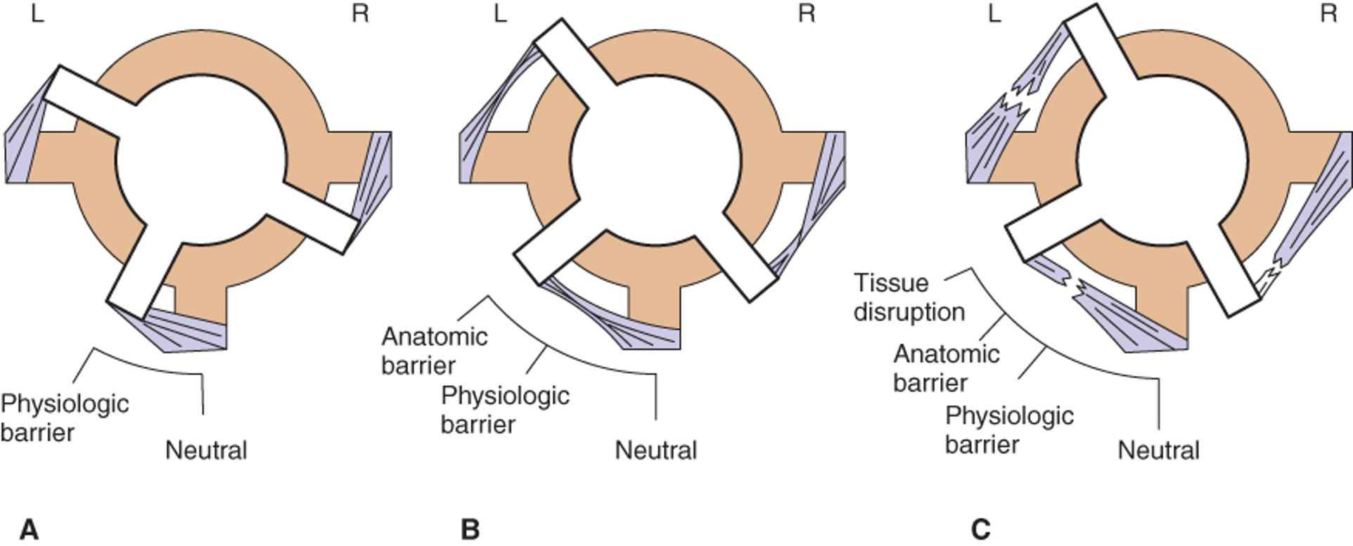
physioloigical movement
68
limit to which a joint may be mooved beyong the physiological barrier, further may cause damage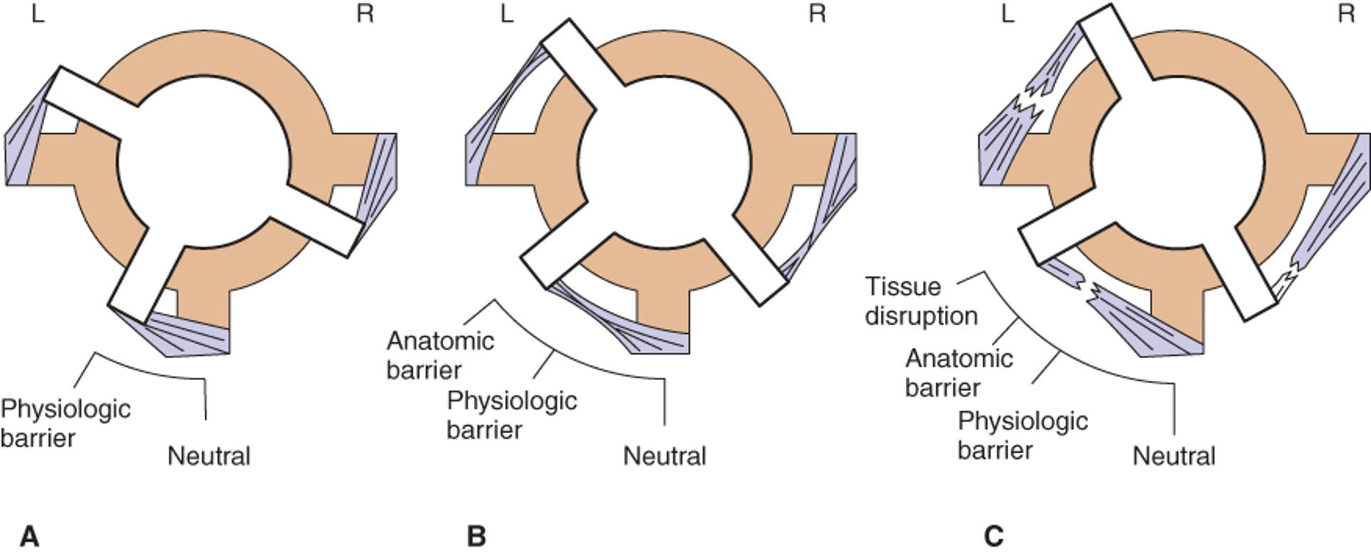
anatomical movement
69
barrier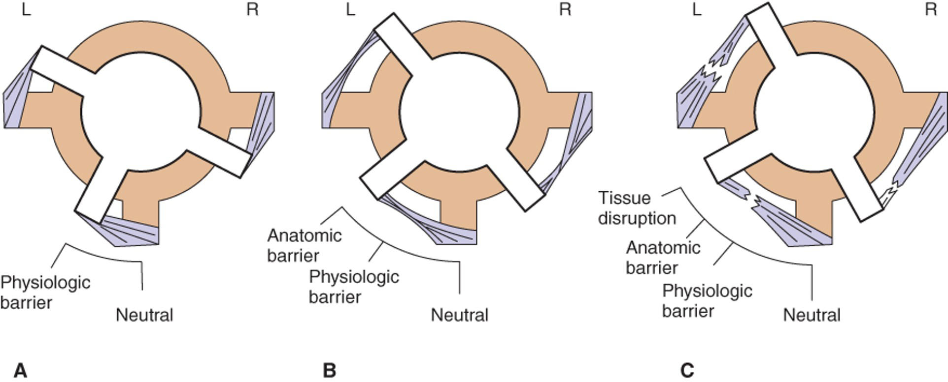
an abnormality preventing equal movement on both sides of the body
70
what is the new neutral position in this image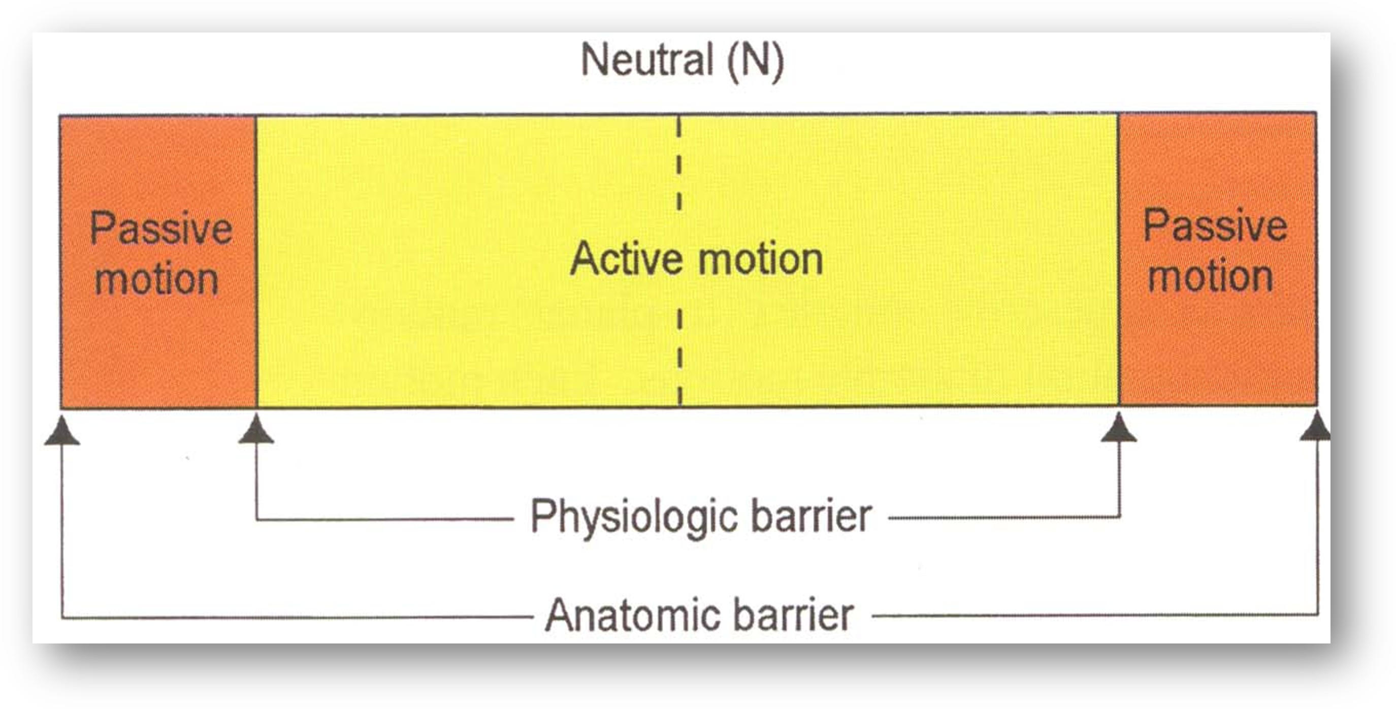
blue line because the range of motion has been changed/limited due to motion loss
71
manipulation done by the patient guided by the hysician
active
72
manipulation done by the ohysicoian pn the patient
passive
73
manipulation where the joint or tissue is taken into the direction of restriction
direct
74
manipulation in which the joint or tissue is take away from the restriction, into theease of motion
indirect
75
what are the indirect techniques (4)
counterstrain, balanced ligamentous tension, facilitated positional release, myofascal release (can b either)
76
how is counterstrain done
Diagnose a specific tenderpoint
Passively place patient into a position of ease
Hold for 90 seconds
Passively return to neutral
Reassess
Passively place patient into a position of ease
Hold for 90 seconds
Passively return to neutral
Reassess
77
what does counterstrain act apon
resetting of the golgi tendon organ
78
what are counterindications of counterstrain
Absolute – severe illness/trauma, instability, vascular/neurologic syndromes, severe spondylosis
Relative – inability to relax, patient non-compliance/intolerance, exacerbation of underlying condition
Relative – inability to relax, patient non-compliance/intolerance, exacerbation of underlying condition
79
what is the technique of Balanced ligamentous tension
Disengagement (Compression or Traction)
Exaggeration (into the ease/freedom of motion)
Balance
Exaggeration (into the ease/freedom of motion)
Balance
80
what is the technique of facilitated positional release
neutral, compress (palpate deep tp level of muscle and squeeze), ease
(basically fast counterstrain)
(basically fast counterstrain)
81
continual palpatory feedback to alleviate restriction of the somatic dysfunction and its related fascia and musculature”
myofasical release
82
what are the direct techniques (x7)
soft tissue technique
inhibitory technique
articulatory trchnique
muscle energy
high velocity low amplitude
still technique (sometimes both)
osteopathy in cranial field(sometimes both)
inhibitory technique
articulatory trchnique
muscle energy
high velocity low amplitude
still technique (sometimes both)
osteopathy in cranial field(sometimes both)
83
involve lateral stretching, linear stretching, deep pressure, traction and/or separation of muscle origin and insertion while monitoring tissue response and motion changes by palpation
(Passive)
(Passive)
soft tissue technique
84
Steady pressure is applied to soft tissues to reduce reflex activity and promote tissue relaxation
Similar to Soft Tissue Technique, but usually more localized and aggressive
(passive)
Similar to Soft Tissue Technique, but usually more localized and aggressive
(passive)
inhibitory technique
85
Passive
Joint or area placed into restriction and then a moderate-to-high velocity, low amplitude “springing” force is used, placing the patient into the barrier (at the end range of motion)
Joint or area placed into restriction and then a moderate-to-high velocity, low amplitude “springing” force is used, placing the patient into the barrier (at the end range of motion)
articulatory technique
86
active technique
golgi tendon reflex
a form of osteopathic manipulative diagnosis and treatment in which the patient’s muscles are actively used on request, from a precisely controlled position, in a specific direction, and against a distinctly executed physician counterforce.”
golgi tendon reflex
a form of osteopathic manipulative diagnosis and treatment in which the patient’s muscles are actively used on request, from a precisely controlled position, in a specific direction, and against a distinctly executed physician counterforce.”
muscle energy
87
cracking technique
“An osteopathic method in which the restrictive barrier is engaged in one of more planes of motion and a rapid, therapeutic force of brief duration traveling a short distance is applied within the anatomic range of motion.” (ECOP)
“An osteopathic method in which the restrictive barrier is engaged in one of more planes of motion and a rapid, therapeutic force of brief duration traveling a short distance is applied within the anatomic range of motion.” (ECOP)
HVLA technique
88
what are some contraindications and precautions for HVLA?
Contraindications
Fracture
Neurological symptoms present with localization or testing
Severe rheumatoid arthritis (RA), particularly in the cervical spine
Cancer in region (primary or metastatic)
Medicolegal situation
Precautions
Once the pathologic barrier is found, do NOT disengage
Localization of force
Avoid excessive thurst
Hypermobility concerns
Don’t become a crack addict!
Fracture
Neurological symptoms present with localization or testing
Severe rheumatoid arthritis (RA), particularly in the cervical spine
Cancer in region (primary or metastatic)
Medicolegal situation
Precautions
Once the pathologic barrier is found, do NOT disengage
Localization of force
Avoid excessive thurst
Hypermobility concerns
Don’t become a crack addict!
89
what are the combo techniques (x2)
still technique and osteopathy in cranial field
90
indirect to direct
Place the joint or segment in the ease (freedom) of motion (INDIRECT)
Add a distracting force
May be compression or traction
While maintaining distraction, place dysfunction into the barrier (DIRECT)
Remove distracting force, return to neutral
Reassess
Place the joint or segment in the ease (freedom) of motion (INDIRECT)
Add a distracting force
May be compression or traction
While maintaining distraction, place dysfunction into the barrier (DIRECT)
Remove distracting force, return to neutral
Reassess
still technique
91
passive treatment that diagnoses strain patterns that develop between the bones of the skull and sacrum
osetopathy on the cranial field
92
towards the head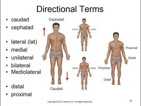
cephalad
93
caudad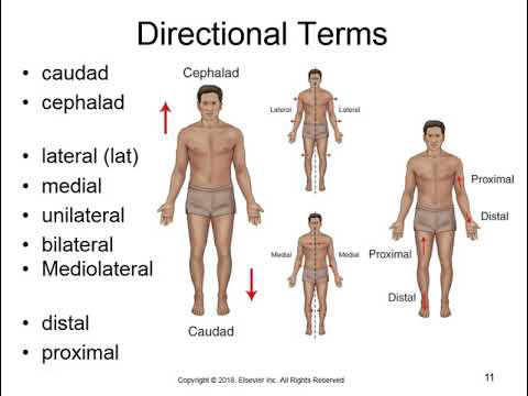
towards the tail
94
rotation occurs in this plane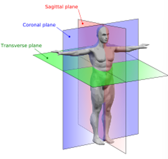
transverse
95
sidebending occurs in this plane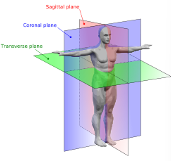
coronal
96
flexion/extension occurs in this plane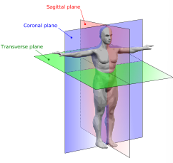
sagittal
97
Areas where curves reverse are commonly susceptible to somatic dysfunction
transition zones
98
what are the locations of transition zones (x4)
Occipito-cervical junction
Cervico-thoracic junction
Thoraco-lumbar junction
Lumbo-sacral junction
Cervico-thoracic junction
Thoraco-lumbar junction
Lumbo-sacral junction
99
what is the spinal seg that corresponds with the suprasternal notch 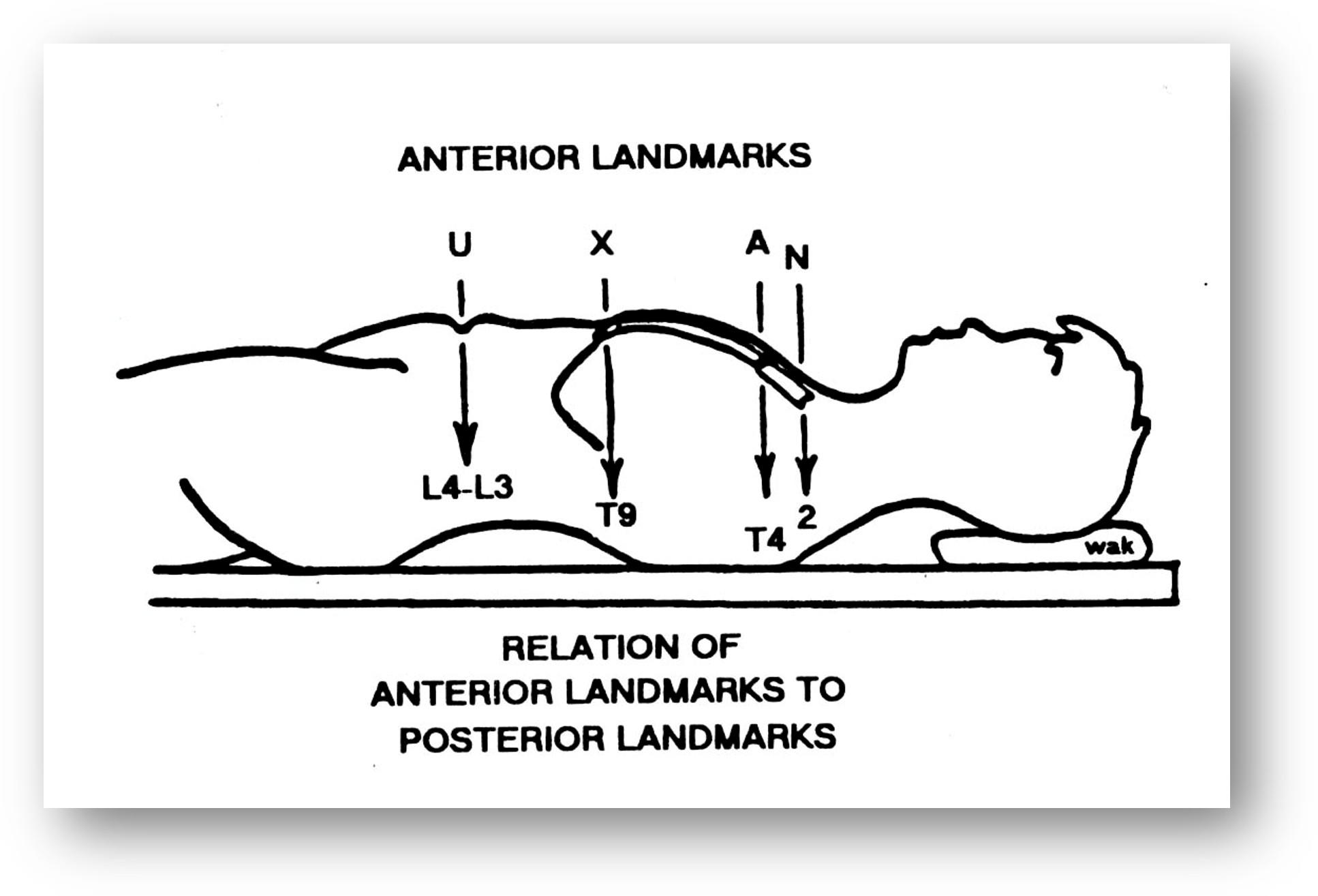
T2
100
what is the spinal seg that corresponds with the angle of louis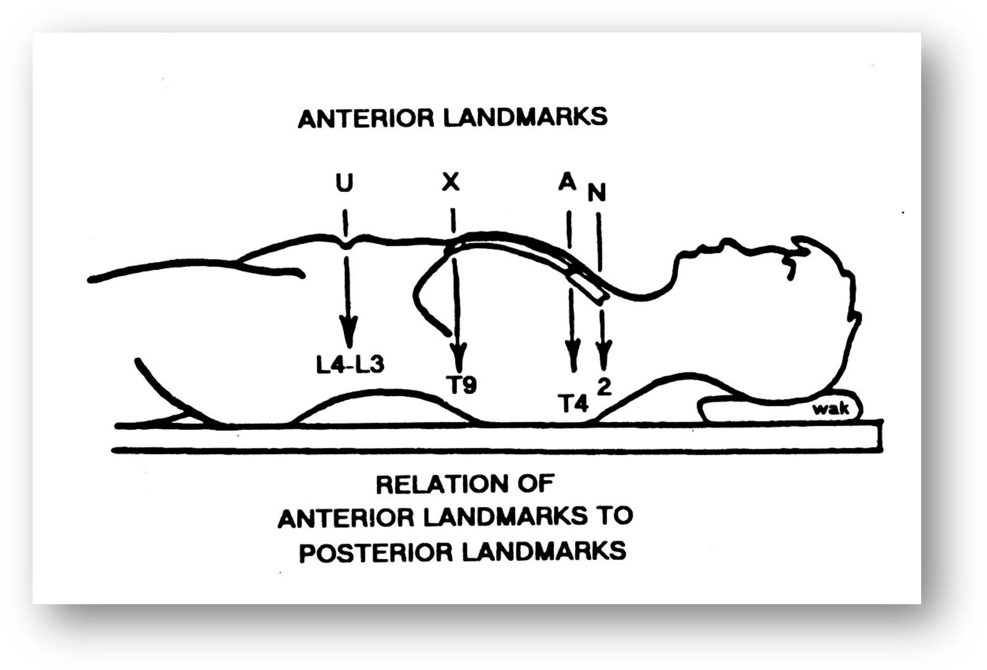
T4
101
what is the spinal seg that corresponds with te xiphoid process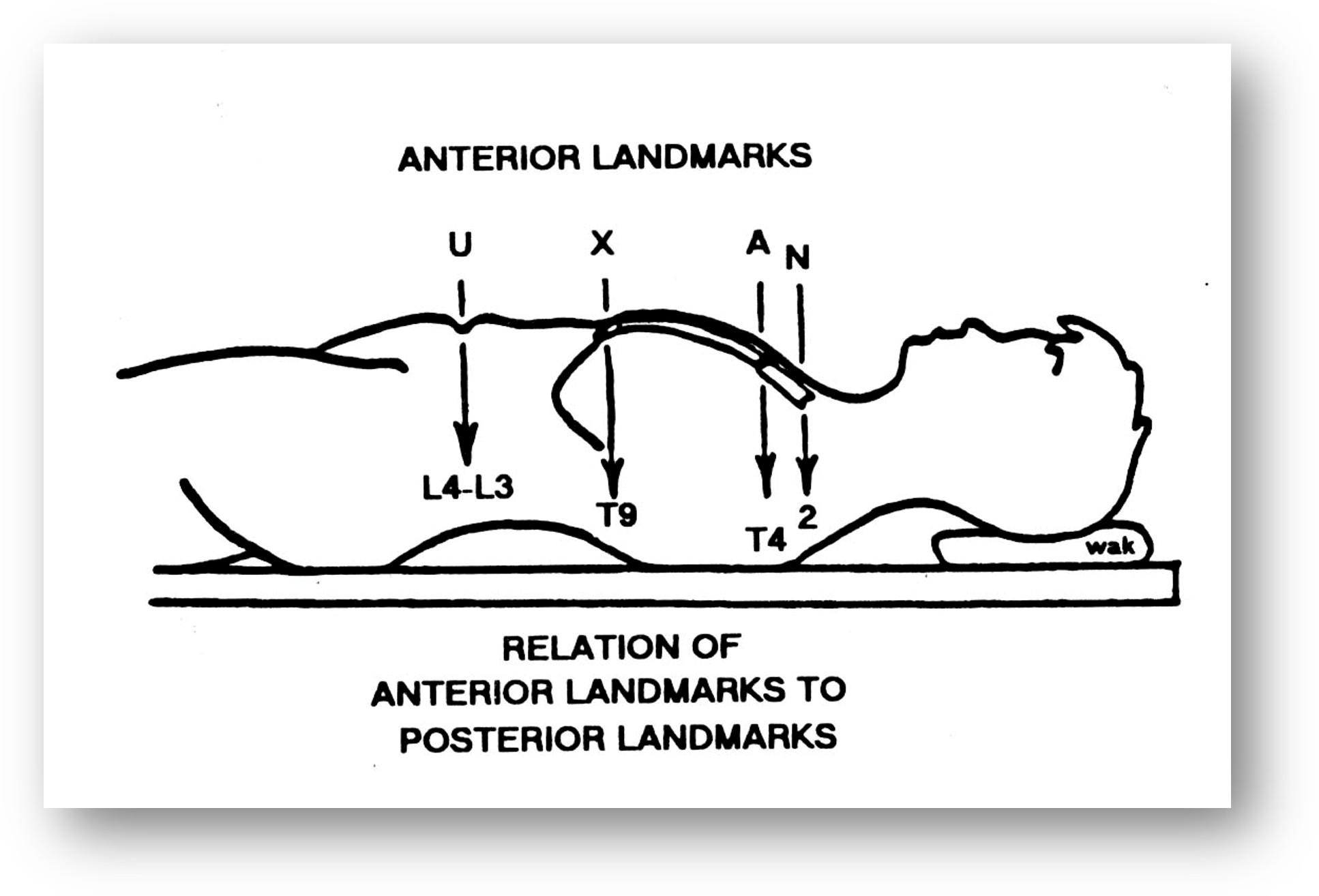
T9
102
what is the spinal cord seg that corresponds with the umbilicus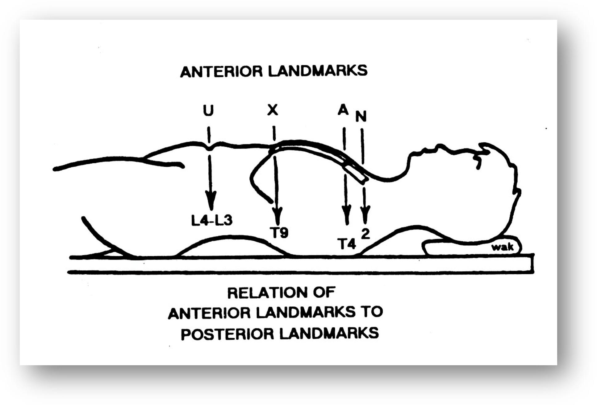
L4
103
what is the landmark that lines up with the spine of the scapula; inferior border?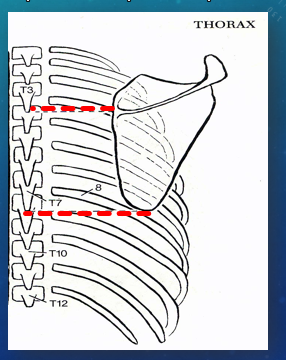
T3; T7
104
which spinal segments have the greatest ROM and are most vulnerable to injury
cervical
105
which spinal segments have least ROM but greatest protection
thoracic spine
106
what are the functions of the anterior vertebral column (x4)
Support
Weight bearing
Shock absorbing
Protection of the spinal cord
Weight bearing
Shock absorbing
Protection of the spinal cord
107
What are the functions of the posterior vertebral column (x2)
Directs joint motion
Protects spinal cord
Protects spinal cord
108
what is the facet orientation of cervical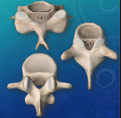
backward, upward, medial
109
what is the facet orientation for thoracic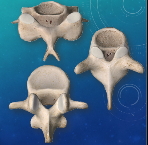
backward, upward, lateral
110
what is the facet oruentation for lumbar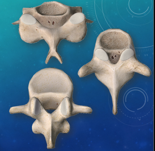
backward upward medial OR baclkward medial
111
this joint is responsible for 50% of flexion and extension of c spine
occipito atlantal
112
this joint is responsile fpr 50% of rotation of the C spine
atlanto axial
113
this is responsible for 50% of flexion and extension of cervical spine (bone)
C2-C7
114
what is the greatest to least ROM of the thoracic spine
Rotation
Sidebending
Flexion
Extension
Sidebending
Flexion
Extension
115
where is the spine for the T1-T3 vert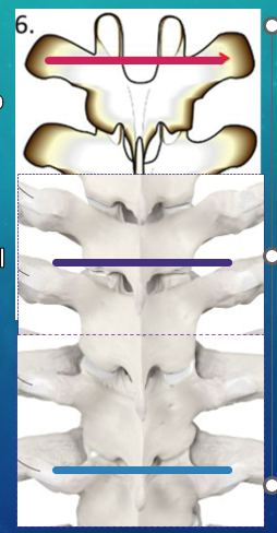
same level as the tip of transverse process
116
where is the spine for the T4-T6 vert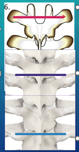
sp is 1/2 vert level below the tip of TP
117
where are the spinous processes of T7-T9 vert?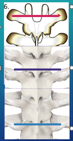
sp one vert level below tip of TP
118
where is spinous process T10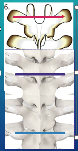
sp is one vert level below tip of TP
119
where is the spinous process T11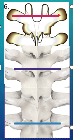
sp 1/2 vert level below tip TP
120
where is the spinous process T12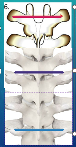
sp is same level as tip of TP
121
what are fryette's principles of physiological spine motion in order
1st Principle
When sidebending is attempted from neutral position, rotation of vertebral bodies follows to the opposite direction.
2nd Principle
When sidebending is attempted from non-neutral position, rotation must precede sidebending to the same side.
3rd Principle
Motion introduced in one plane limits and modifies motion in the other planes.
When sidebending is attempted from neutral position, rotation of vertebral bodies follows to the opposite direction.
2nd Principle
When sidebending is attempted from non-neutral position, rotation must precede sidebending to the same side.
3rd Principle
Motion introduced in one plane limits and modifies motion in the other planes.
122
scoliosis reversibility: Curve reduced with side bending, rotation or forward bending
functional scoliotic curve
123
scoliosis reversibility: Curve fixed and not reduced with side bending, rotation or forward bending
structural scoliotic curve
124
Lateral (sideways) curvature of the spine (coronal plane); aka rotoscoliosis
Cobb angle >10 degrees
Cobb angle >10 degrees
scoliosis
125
left scoliosis
levoscoliosis
126
right scoliosis
dextroscoliosis
127
how is scoliosis named
acording to the convexity
128
idopathic scoliosis that may be genetic and is diagnosised in preteen years
strictural
129
scoliosis where muscles are hypertonic and can be caused by short leg syndrome with compensation and weak musculature
functional scoliosis
130
what are the types of structural scoliosis
congenital, idiopathic, acquired
131
unknown etilogy with >20 degree curve starting at <12yrs age mostly in females
adolescent idopathic scoliosis
132
excessive kyphosis of the thoracic spine, can run in families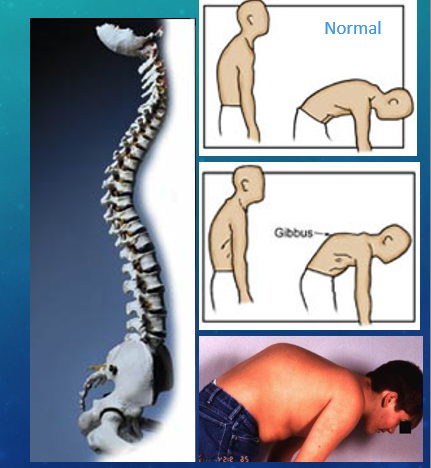
scheuermann's kyphosius
133
mild scoliosis severity
5-15*
134
moderate scoliosis severity
20-40*
135
severe scoliosis severity
>50*
136
what is part of an IPASSS physical exam?
Inspection
palpation
active passive rom
strength
sensation
special tests
palpation
active passive rom
strength
sensation
special tests
137
scapula congenitally doesnt place right 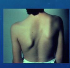
sprengel's deformity
138
sequence of diagnosis
observation, palpation, ROM, intersegmental ROM
139
how is somatic dysfunction named?
type of dysfunction (I or II)
neutral N, flexed F or extended E
left L or rght R
roated R or sidebent S
neutral N, flexed F or extended E
left L or rght R
roated R or sidebent S
140
important palpatable posterior landmarks! (x6)
mastoid processes, shoulder AC joints, inferior angle of scapula, illiac crest, PSIS, greater trochanter
141
what is the scoliosis test?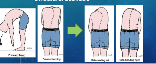
forward bend test, if shows curve have tem do sidebending test, if curve disappears here functional scoliosis
142
type I somatic dysfunction
Keys
• Observe
• Look for an
area the is
more posterior
(group curve)
• Palpate
• Check for
group
• Segmental Testing
• Check each
level
• Should not
change with
F/E
• Observe
• Look for an
area the is
more posterior
(group curve)
• Palpate
• Check for
group
• Segmental Testing
• Check each
level
• Should not
change with
F/E
143
type II somatic dysfunction
Keys
• Palpate
• Look for transition
areas
• Check at beginning,
end, apex
• Feels like a “speed
bump”
• Segmental testing
• Observe & palpate
for worsening or
improvement of the
rotation
• Named the direction of
improvement (Ease)
• Palpate
• Look for transition
areas
• Check at beginning,
end, apex
• Feels like a “speed
bump”
• Segmental testing
• Observe & palpate
for worsening or
improvement of the
rotation
• Named the direction of
improvement (Ease)
144
an architectural system in which
structures stabilize themselves by balancingstructures stabilize themselves by balancing
counteracting forces of compression and tension.counteracting forces of compression and tensi
structures stabilize themselves by balancingstructures stabilize themselves by balancing
counteracting forces of compression and tension.counteracting forces of compression and tensi
tensegrity
145
drains lymph from your right arm and the right sides of your head, neck and chest.
right lymphatic duct
146
drains from the following:
-The left sides of your head, neck and chest
-Your belly (abdominal) area
-Both of your legs
-The left sides of your head, neck and chest
-Your belly (abdominal) area
-Both of your legs
left thoracic duct
147
what is the true shoulder joint
glenohumeral joint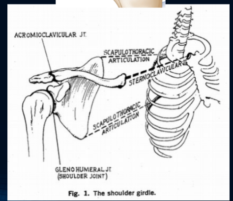
148
what makes up the anatomical inlet
manubrium, 1st ribs bilkaterally and t1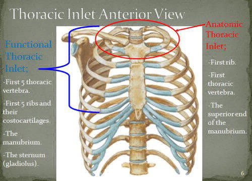
149
what makes up the functional inlet
manubrium w/ang;e of louis, 1st and 2nd ribs B/L, T1-T4
150
what makes up the thoraci outlet
clavicle, 1st ribs, scapula
151
what is contained in the interscalene triangle
brachial plexus and subclavian a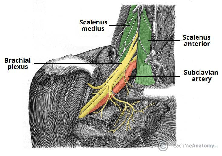
152
what is in the costoclavicular space
brachial plexus and subclavian a and v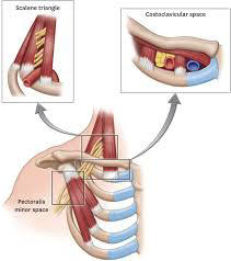
153
what makes up the subcoracoid space
brachial plexus and azillary a and v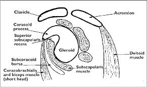
154
where can entrapment occur in thoracic outlet syndrome? (x3)
Between the Scalenes - anterior
and medius
2. Between the Clavicle and the first
rib
3. Between Pectoralis minor tendon
and ribs
and medius
2. Between the Clavicle and the first
rib
3. Between Pectoralis minor tendon
and ribs
155
what is the true elbow joint
humerus and ulna hinge joint
156
how many articilations does the trochlea have ad what aree they
trochlear notch of the ulna
157
how many artoculations does the capitulium have
cupped proximal radial head
158
The measurement of deviation
• Of the ulna in relationship to the long axis
of the humerus
• Of the ulna in relationship to the long axis
of the humerus
carrying angle
159
normal carrying angle
5-15* valgus
160
why would people have an increased carying angle (valgus)
women, anductiuon somatic dysfunction
161
why would people have a decreased carrying angle (varus)
gunshock deformity, adduction somatic dysfunction
162
ADDuction of the ulna(#1)
will result in ______________
(Radial Deviation) of the
wrist (#3
will result in ______________
(Radial Deviation) of the
wrist (#3
abduction
163
abduction of the ulna results in __________ of the wrist (ulnar devoation)
adduction
164
the radial head moves _____- with supination and ______ with pronation
anterior; posterior
165
true wrist joint
radiocarpal
166
elevation of clavicle: distal clavicle (sup/inferior), proximal clavicle (sup/inf), Axis:_____, CCL: _______, ICL______
superior
inferior
AP
tightens
relaxes
inferior
AP
tightens
relaxes
167
flexion clavicle: distal clavicle (posterior/anterior), posterior clavicle ___, Axis:____- capsular liugament ___
anterior
posterior
vertical through sternum
tightens
posterior
vertical through sternum
tightens
168
extension clavicle:
distal clav____
prox clav ______
axis______
capsular ligament _____
distal clav____
prox clav ______
axis______
capsular ligament _____
posterior
anterior
verticle through sternum
tightens
anterior
verticle through sternum
tightens
169
range of motion in order shoulder joint
abduction(180), adduction(45), flexion(180) ecxtension (45, int rotation (55), ext rotation (45)
170
what joints does shoulder abduction use
GH and ST
171
ROM elbow
flexion (135), extension (0/-5), supination (90), pronation (90)
172
ROM wrist
fkexiom (80), extension (70), radial deviation (20), ulnar deviation (30), supination/pronation (90)
173
women>M, 2 types unilateral where EGM+, upper brachial plexus up to 6yrs or B/L where emg-, lower brachial plexys
nTOS
174
M>W, athetic, dpominant side, unliuateral
vTOS
175
W=M, young adultrs more likely, unliateral disfunction
aTOS
176
Proximal Scalene Triangle
• Patient – seated
• DOc – stand same side of patient being tested
• DOc - locate radial pulse
• Instruct patient
• Extend elbow
• Look toward side being tested, extend neck
• DOc - ABduct their shoulder 30 degrees, & extend
humerus/shoulder toward DOc
• Reassess Pulse
• Positive – diminished pulse
• Patient – seated
• DOc – stand same side of patient being tested
• DOc - locate radial pulse
• Instruct patient
• Extend elbow
• Look toward side being tested, extend neck
• DOc - ABduct their shoulder 30 degrees, & extend
humerus/shoulder toward DOc
• Reassess Pulse
• Positive – diminished pulse
Adson's test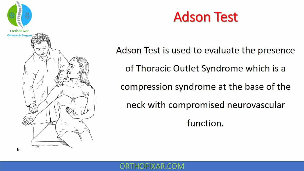
177
when is an adson's test ositive
dimiished pulse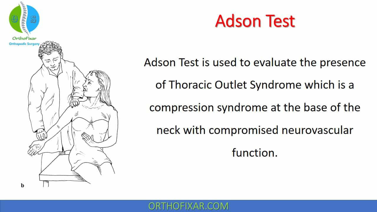
178
Costoclavicular (Halsted)
maneuver
• Patient – Seated
• DOc – standing on same side testing
• DOC - Locate radial pulse
• Instruct the patient:
• Performs an Exaggerated military posture
• Scapula retracted and depressed
• Chest protruding
• Narrows the costoclavicular space
• Extend and Rotate head away
• DOc - Apply downward traction on arm
• Reassess Pulse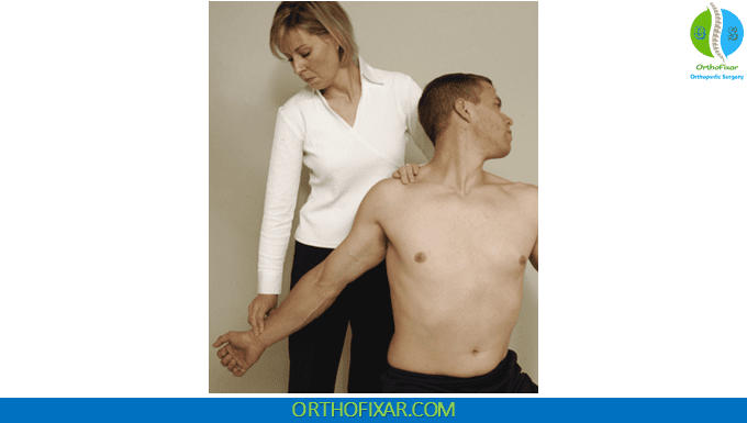
maneuver
• Patient – Seated
• DOc – standing on same side testing
• DOC - Locate radial pulse
• Instruct the patient:
• Performs an Exaggerated military posture
• Scapula retracted and depressed
• Chest protruding
• Narrows the costoclavicular space
• Extend and Rotate head away
• DOc - Apply downward traction on arm
• Reassess Pulse
halsted maneuver; positive when diminised pulse
179
Sub-coracoid Space (Pec Minor)
• Patient – seated or supine
• DOc – standing on the same side testing
• DOc - locate radial pulse
• DOc - Passively ABduct Shoulder up to 180 degrees
• May flex elbow up to 90 degrees
• Reassess Pulse
• Patient – seated or supine
• DOc – standing on the same side testing
• DOc - locate radial pulse
• DOc - Passively ABduct Shoulder up to 180 degrees
• May flex elbow up to 90 degrees
• Reassess Pulse
Wright's test; positive when diminished pulse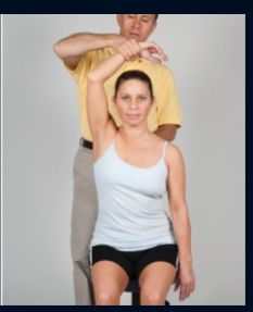
180
special tests for rotator cuff injury
drop arm test and empty can test
181
what does the drop arm test assess
supraspunatous tendon injury/inflammation/tear
positive if weakness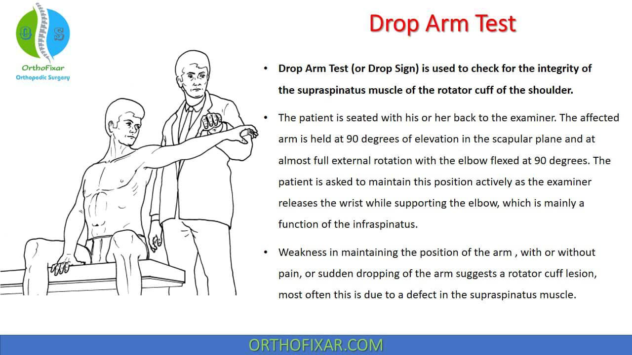
positive if weakness
182
what does the empty can test assess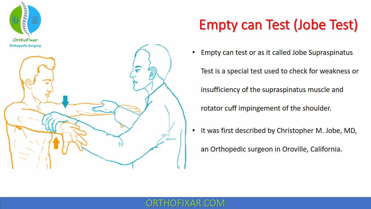
supraspiarus weakness, positive if weakness
183
special tests for sub-acromial impingement
neer test and hawkins test, positive if pain in subacromial space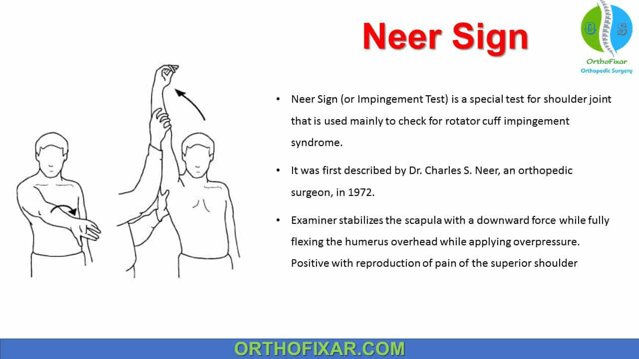
184
Pain is located at the anterior aspect of the
shoulder, radiating down into the biceps
muscle.
• The is pain over the long head of the
biceps tendon in the bicipital grove.
• The tendon becomes prominent when the
humerus is externally rotated.
shoulder, radiating down into the biceps
muscle.
• The is pain over the long head of the
biceps tendon in the bicipital grove.
• The tendon becomes prominent when the
humerus is externally rotated.
biceps tendonitis
185
special test for long head biceps tendonitis
yergson's test; posutive if pain in bicipital groove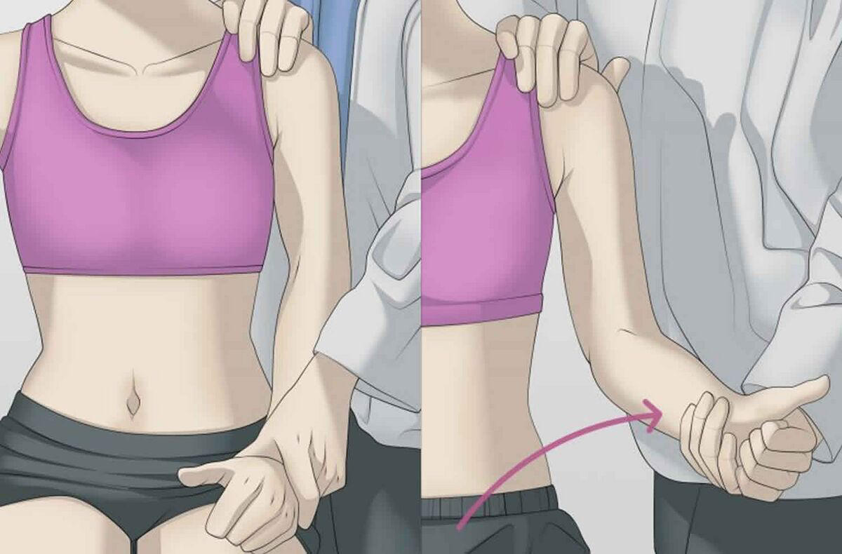
186
Lateral Epicondyle
• Origin of Extensor
Carpi Radialis
Longus & Brevis
(ECRB)
• Pain with active wrist
extension against
resistance
• Origin of Extensor
Carpi Radialis
Longus & Brevis
(ECRB)
• Pain with active wrist
extension against
resistance
tennis elbow
187
Medial Epicondyle
• Origin of Flexor Carpi
Ulnaris and Pronator
Teres
• Pain with active wrist
flexion against resistance
(forearm pronated)
• Origin of Flexor Carpi
Ulnaris and Pronator
Teres
• Pain with active wrist
flexion against resistance
(forearm pronated)
golfer's elbow
188
special tests for elbow
resisted flexion test, resisted extension test, tinel test at elboiw
189
what does the resisted flexion test, test fpr
medial epicondylitis (golfer's elbow) positive if pain in medial epicondyle
190
what does the resisted extension test test for
lateral epicondylitis 9tennis elbow), positive if pain in lateral epicondyle
191
what does the tinel test at elbow test for
ulnar nerve neuritis or entrapment within the cubital tinnel
positive: pain or tingling in ulnar nerve distribution
positive: pain or tingling in ulnar nerve distribution
192
what are the special tests for capal tunnel
tinels sign at wrist, phalen's sign
193
what does the tinel sign at wrist test
median nerve neuritis or entrapment at carpal tunnel
psoitive if pain or tingling in the median nerve distribution
psoitive if pain or tingling in the median nerve distribution
194
what does the phalen's sign test
median nerve neuritis or entrapment within carpal tunnel
posiutive if pain or tingling in the median nerve distribution
posiutive if pain or tingling in the median nerve distribution
195
where is dense regular connective tissue found in the body
tendons and ligaments
196
layers of fascia
pannicular (superficial): subcutaneous
investing (deep): axial o appendicular
visceral
meningeal
investing (deep): axial o appendicular
visceral
meningeal
197
fibrous or myofascical partitions acting as tension countertension sheets, connected to fascial cables, anchored to skeletal structures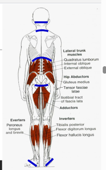
horizontal diaohragms
198
what are the horizontal diaphragms in the body (x5)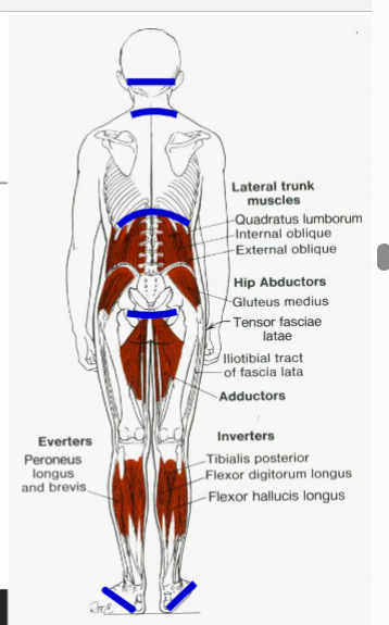
tentorium cerebelli
thoracic inlet/outlet
respiratory diaphragm
pelvic diaphragm
plantar fascia
thoracic inlet/outlet
respiratory diaphragm
pelvic diaphragm
plantar fascia
199
Dural sleeve to S2
Longitudinal ligaments Occiput-S2
Pre-vertebral, alar, buccopharyngeal fascia
Psoas major to lower extremity
-Rectus abdominus, q. lumborum,
internal/external obliques
-Trachea, esophagus, pericardium on central
tendon of diaphram
A/P Lower extremity fascia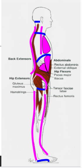
Longitudinal ligaments Occiput-S2
Pre-vertebral, alar, buccopharyngeal fascia
Psoas major to lower extremity
-Rectus abdominus, q. lumborum,
internal/external obliques
-Trachea, esophagus, pericardium on central
tendon of diaphram
A/P Lower extremity fascia
longitudinal cables
200
contradictions to myofascial tratment
absolute: no somatic dysfunction, poor patuent cooperation
relative: wpunds, burns, abscesses, infections, aortic aneurysm, cancer
relative: wpunds, burns, abscesses, infections, aortic aneurysm, cancer
201
3 main categories of OMT techniques
direct action
indirect action
bioenergetics
indirect action
bioenergetics
202
a DIRECTDIRECT
technique that usually involves
lateral stretching, linear stretching,
deep pressure, traction and/or
separation of muscle origin and
insertion while monitoring tissue
response and motion changes by
palpation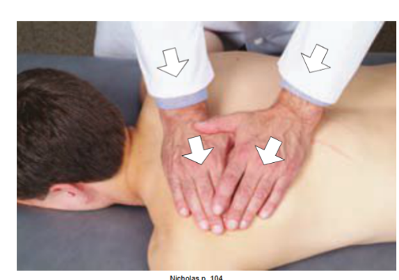
technique that usually involves
lateral stretching, linear stretching,
deep pressure, traction and/or
separation of muscle origin and
insertion while monitoring tissue
response and motion changes by
palpation
soft tissue technique
203
System of
diagnosis and treatment first
described by A.T. Still and his
early students, which engages
continual palpatory feedback
to achieve release of
myofascial tissues.
• Direct or Indirect
• Unwinding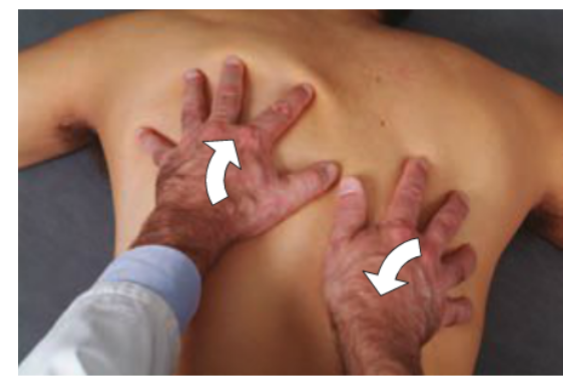
diagnosis and treatment first
described by A.T. Still and his
early students, which engages
continual palpatory feedback
to achieve release of
myofascial tissues.
• Direct or Indirect
• Unwinding
myofascial release.
204
System of OMT
that is utilized to normalize
hypertonic voluntary
muscles (developed by Stanley schiowitz)
•• Non-traumatic
• Easily utilized
• Highly effective
• Efficient (rapid!)
that is utilized to normalize
hypertonic voluntary
muscles (developed by Stanley schiowitz)
•• Non-traumatic
• Easily utilized
• Highly effective
• Efficient (rapid!)
FPR
205
study of how endogenous and exogenous
energy sources/forms influence and control living systems and their environment
energy sources/forms influence and control living systems and their environment
bioenergetics
206
energy produced endogenously by living systems
bioenergy
207
an interdisciplinary science
using methods of, and theories from, physics
to study biological systems. Spans all scales
of biological organization, from the
molecular to whole organisms to ecosystems.
using methods of, and theories from, physics
to study biological systems. Spans all scales
of biological organization, from the
molecular to whole organisms to ecosystems.
biophysics
208
traits of connective tissue (x4)
adaotability, plastcity, elasticity, viscocity
209
ability to respond to stresses to meet needs of the system
adaptability
210
ability to be formed and molded
plasticity
211
recoverability after stretching
elasticity
212
rate of defirmation under load and capabikity to yield under stress
viscocity
213
a non precioitating suspension (ECM)
colloid
214
specialized properties of fascia (x3)
tensegrity, piezoelectric, non neuro communication
215
Mechanical force acting
on a cell membrane
• Opening of
mechanosensitive pores
• Distortion of
mechanosensitive
proteins
on a cell membrane
• Opening of
mechanosensitive pores
• Distortion of
mechanosensitive
proteins
mechanotransduction
216
Mechanical
stress is transformed into
electrical potentials and
electrical potentials are
transformed into
mechanical motion
stress is transformed into
electrical potentials and
electrical potentials are
transformed into
mechanical motion
piexoelectric phenomenon
217
bioelectric ptroperties of fascia (x3)
compression/stretching
EMF frequencies
lazer like vibrations 2* to molecular array PAG oscillations
EMF frequencies
lazer like vibrations 2* to molecular array PAG oscillations
218
these are responsible for change of tissue viscocity frim sol gen vixe versa, influened by EMFs
arrays
219
a non-invasive medical treatment using red and near-infrared light to stimulate cellular processes, accelerating healing, reducing pain, and promoting tissue regeneration.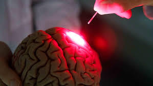
photobiomodulation
220
“Every change
in the function of a bone is
followed by changes in internal
architecture and external
conformation in accordance with
mathematical laws.
in the function of a bone is
followed by changes in internal
architecture and external
conformation in accordance with
mathematical laws.
wollf's law
221
electrical communoication through the fascia outsie the nervous system
non neurological communication
222
complex,
extremely weak EMF of
the organism
hypothesized to involve
electromagnetic
bioinformation for
regulating
homeodynamics
extremely weak EMF of
the organism
hypothesized to involve
electromagnetic
bioinformation for
regulating
homeodynamics
biofield
223
study of the interaction
between electromagnetic
fields and biological living
systems
between electromagnetic
fields and biological living
systems
bioelectromagnetics
224
inherent ability of living cells,
tissues, and organisms to
produce and emit electrical
and magnetic fields, and the
response of cells to
electromagnetic fields
tissues, and organisms to
produce and emit electrical
and magnetic fields, and the
response of cells to
electromagnetic fields
bioelectromagnetism
225
If you have type 1 somatic dysfunction, how is R and S named?
R= same side as disfunction
S=opposite side as R
S=opposite side as R
226
if ypou have a type 2 somatic dusfunction, how is R and S named
R is same side as disfunction, S is same side of R
227
what is non newtonian fluid
viscocity of the flluid changed where increased stress increases viscocity
228
Layers of fascia in order from superficial to deep
pannivular fascia (superficial)
axial and appendicular fascia
meningeal fascia
visceral fascia
axial and appendicular fascia
meningeal fascia
visceral fascia
229
Entrapment of Lateral Femoral Cutaneous nerve
Symptoms
Pain, numbness, tingling, burning over outer thigh (not radiating below the knee)
Usually, Unilateral
Worse walking or standing, Better with sitting
Symptoms
Pain, numbness, tingling, burning over outer thigh (not radiating below the knee)
Usually, Unilateral
Worse walking or standing, Better with sitting
meralgia paresthetica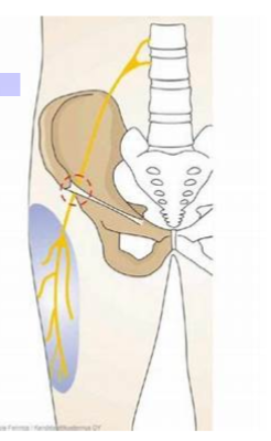
230
This muscle can compress the sciatic nerve and cause somatic dysfuncion
piriformis m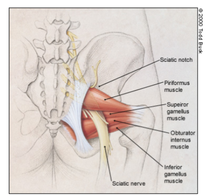
231
This muscle is the strongest flexor of the thigh
iliopsoas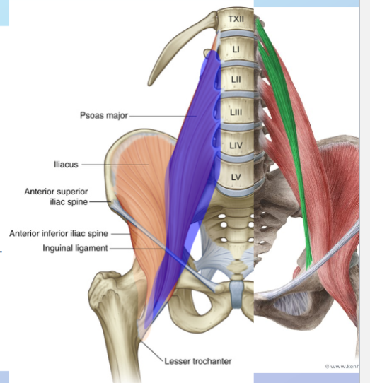
232
What muscles insert into the iliotibial band (x2)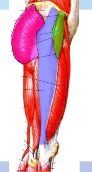
tensor fasciae latae (green)
gluteus maximus (pink)
gluteus maximus (pink)
233
this is the location of chapman's reflexes that are assocoiated with bowl dysfunction (visceralsomatic)
iliotibial band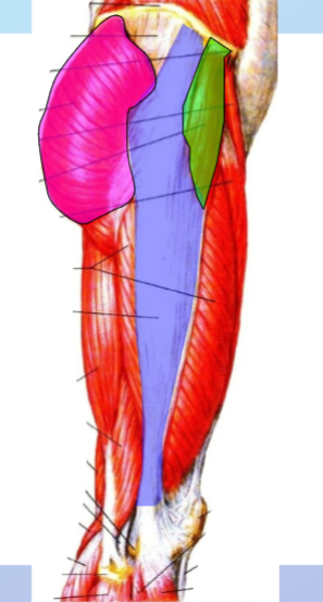
234
This is the strongest lgament in the body and prevcents hyperextension of the hip
iliofemoral ligament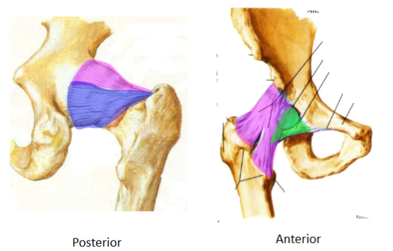
235
when does the iliofemoral ligament tense?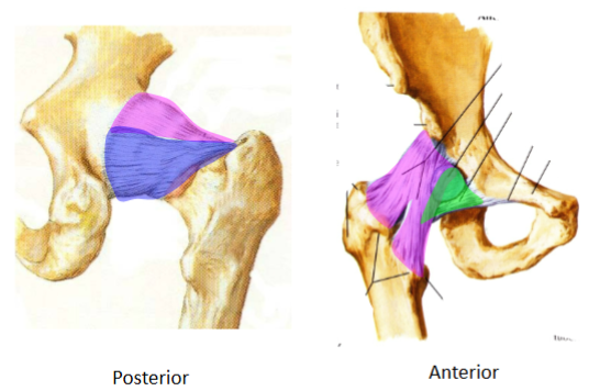
full hip extension
236
What is so special abt the condylpoid joint at the knee
doubke condylar, complex synovial articulations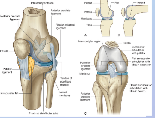
237
This ligament limits lateral glifde of the knee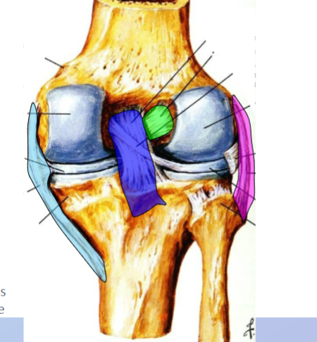
lateral collateral liugament (pink)
238
this ligament limits medial glide of the knee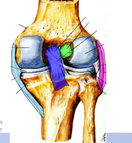
medial collateral ligament (blue)
239
this ligament inhibits anterior glide of the knee/tibia
anterior cruciate ligament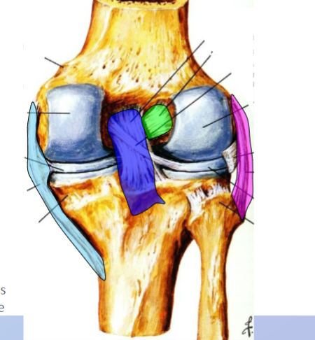
240
this ligament limits posterior glide of the knee/tibia
posterior cruciate ligament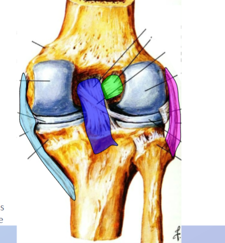
241
what does the terriblke triad of the knee joint consist of?(x3)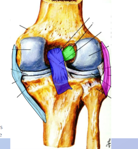
ACL, MCL, medoal meniscus
242
what is the femoroacetablar joint motions and range
flexion: 135 d
extension 30d
abduction 45-50
adduction 20-30
internal rotation 35
external rotation 45
extension 30d
abduction 45-50
adduction 20-30
internal rotation 35
external rotation 45
243
what is the ROM of the knee
fkexion 135
extension 0
internal rotation 10
external rotation 10
extension 0
internal rotation 10
external rotation 10
244
what is the normal motion of the fibular head while it is anterior
Dorsiflexion
Eversion
External Rotation
with ANTlers: Anterior
Eversion
External Rotation
with ANTlers: Anterior
245
Fibular head normal motion posterior
Posterior
Inversion
Plantarflexion
Internal rotation
(Pi^2)
Inversion
Plantarflexion
Internal rotation
(Pi^2)
246
ankle ROM
dorsiflexion 20
plantarflexion 50
inversion 35
eversion 25
plantarflexion 50
inversion 35
eversion 25
247
with dorsiflexiuon, the talus experiences (post/anterior) glide; with plantarflexion the talus experiences (post/anterior) glide
posterior; anterior
248
hip dysfunction is commonly perceived as pain here
knee
249
Trendelenburg test (muscle, test for, positive test)
-gluteus medius
-Insufficiency of hip abductor
mechanism. Most associated with
incompetence/weakness of the
Gluteus Medius muscle
-Testing standing leg, the controlateral hip pelvis drops beklow the pelvus
-Insufficiency of hip abductor
mechanism. Most associated with
incompetence/weakness of the
Gluteus Medius muscle
-Testing standing leg, the controlateral hip pelvis drops beklow the pelvus
250
Thomas test (muscle, testing for, positive test)
iliopsoas
hypertonicty of one of both m\
positive if distal thigh off table on side testing-unable to return to neutral
or
lumbar lordosis retuns from flattened starting position
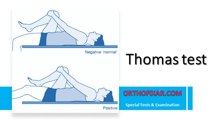
hypertonicty of one of both m\
positive if distal thigh off table on side testing-unable to return to neutral
or
lumbar lordosis retuns from flattened starting position
251
Ober test (muscles, tetsing for, whats posiutive)
IT band and TFL
-hypertonicoity of muscle or fascia
-if ipsilateral leg is held in abdiction, the knee will not addict and cross mudline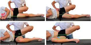
-hypertonicoity of muscle or fascia
-if ipsilateral leg is held in abdiction, the knee will not addict and cross mudline
252
what ligaments injured in lateral/everson sprain of ankle
ATF, CF, PTF
253
what ligaments are damaed in a medial/eversion ankle sprain
Tibianavicular
Anterior Tibiatalor
Tibiocalcaneal
Posterior Tibiotalor
Anterior Tibiatalor
Tibiocalcaneal
Posterior Tibiotalor
254
what is in danger of damage in a high ankle sprain?
ant tibiofibular
post tibiofibular
interosseus membrane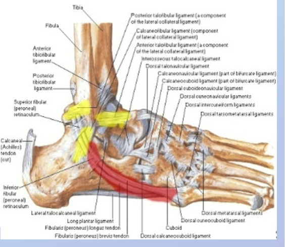
post tibiofibular
interosseus membrane
255
anterior drawer test (muscle, testing for, positive test)
anterior talofibular ligament
-laxity injury, tear partial or full to ATFL
-laxity from normal or laxity compared to other side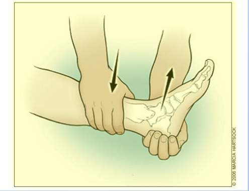
-laxity injury, tear partial or full to ATFL
-laxity from normal or laxity compared to other side
256
posterior drawer test (site, testing fdor, positive test)
posterior talofibular ligament
-laxity injury, tear partial or full to PTFL
-laxity from normal or laxity compared to other side
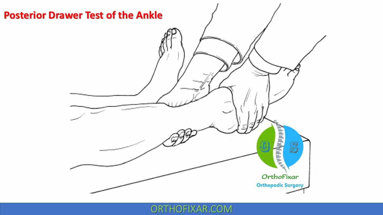
-laxity injury, tear partial or full to PTFL
-laxity from normal or laxity compared to other side
257
talar tilt test (site, testing for, what is positive)
calcaneal fibular ligament
-laxity injury, tear partial or fiull CFL
-laxity from normal or laxity compared to other side
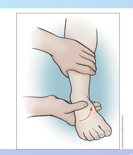
-laxity injury, tear partial or fiull CFL
-laxity from normal or laxity compared to other side
258
valgus stress test (site, tetsing for, positive test)
deltoid ligament
laxity: injury, tear partial or full to deltoid ligament
-laxity from normal or laxity compared to other side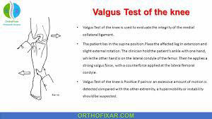
laxity: injury, tear partial or full to deltoid ligament
-laxity from normal or laxity compared to other side
259
when to order xray pittsburgh knee rules (x3)
Hx of trauma (blunt) or fall
Age <12yr or >50 yrs
Inability to bear weight (4 steps)
Age <12yr or >50 yrs
Inability to bear weight (4 steps)
260
ottawa knee rules xrays (x5)
Hx of trauma/injury
Age > 55yrs
Point tenderness (Specific
locations)
Unable to flex knee to 90 degrees
Inability to bear weight (4 steps)
Age > 55yrs
Point tenderness (Specific
locations)
Unable to flex knee to 90 degrees
Inability to bear weight (4 steps)
261
when to order xray: ottawa ankle rules (x4)
-Ankle & Midfoot Injuries
Hx of trauma/injury
Point tenderness (specific locations)
Inability to bear weight (4 steps)
Hx of trauma/injury
Point tenderness (specific locations)
Inability to bear weight (4 steps)
262
how do you diagnose compartment syndrome (x2)
-History & Physical Exam
Direct measurement of compartment
pressures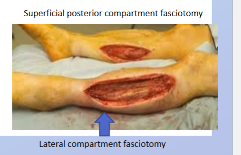
Direct measurement of compartment
pressures