EKG Workshops
Study these flashcardsFree · spaced repetition · iPhone, Android, Mac & web
All cards
1
A wave of depolarization traveling
towards a positive EKG electrode
causes a/an ________ deflection on
the EKG tracing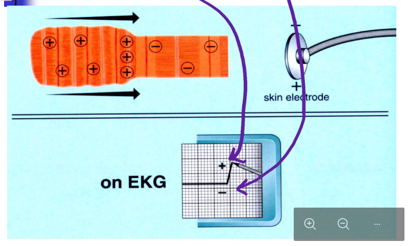
towards a positive EKG electrode
causes a/an ________ deflection on
the EKG tracing
upward
2
A wave of depolarization traveling
away from a positive EKG electrode
causes a/an ________ deflection on
the EKG tracin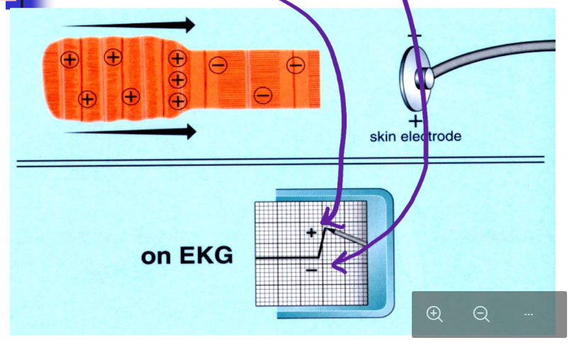
away from a positive EKG electrode
causes a/an ________ deflection on
the EKG tracin
downward
3
A wave of repolarization traveling towards a positive EKG electrode
causes a/an ________ deflection
on the EKG tracin
causes a/an ________ deflection
on the EKG tracin
downward
4
A wave of repolarization traveling away from a positive EKG electrode
causes a/an ________ deflection
on the EKG tracin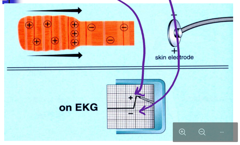
causes a/an ________ deflection
on the EKG tracin
upward
5
this causes the p wave
atrial depolarization
6
this cases the QRS wavw
ventricular depolarozation
7
distance (time) from beginning of
P wave to beginning of QRS complex
P wave to beginning of QRS complex
PR interval
8
distance (time) from beginning of
QRS complex to end of T wav
QRS complex to end of T wav
QT interval
9
distance (time) from end of QRS
complex to end of T wave
(includes the T wave but not the QRS complex)
complex to end of T wave
(includes the T wave but not the QRS complex)
ST interval
10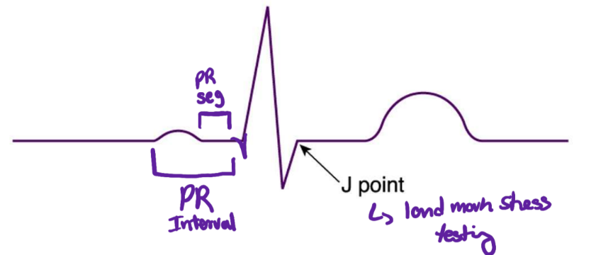
Image occlusion: name the hidden labels
11
Image occlusion: name the hidden labels
12
Image occlusion: name the hidden labels
13
what causes the PR segment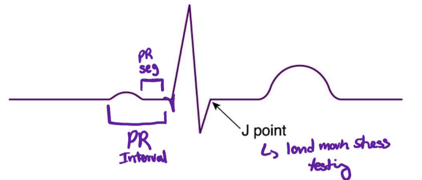
delay of AV node
14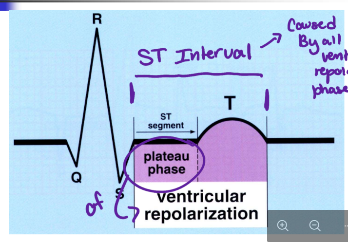
Image occlusion: name the hidden labels
15
Image occlusion: name the hidden labels
16
Image occlusion: name the hidden labels
17
Image occlusion: name the hidden labels
18
Image occlusion: name the hidden labels
19
Image occlusion: name the hidden labels
20
plateu phase of evntricular repolarization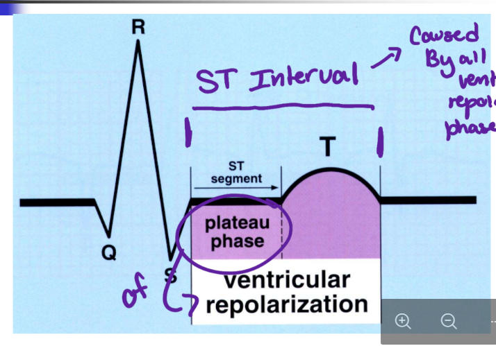
ST segment
21
entire ventricular repolarization phase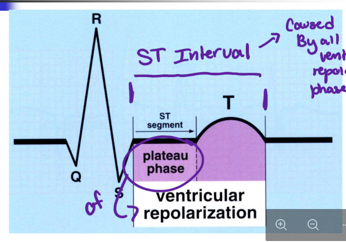
ST interval
22
rapid phase of ventricular repolarozation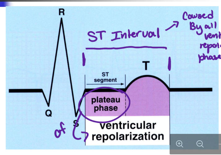
Twave
23
what occurs during the QT interval?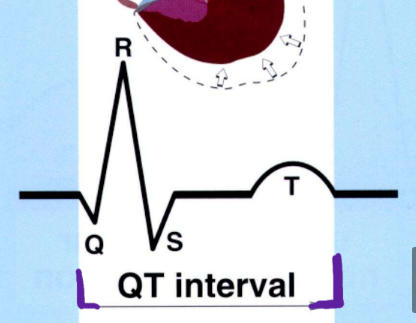
ventricular contraction
24
what occurs during the U wave?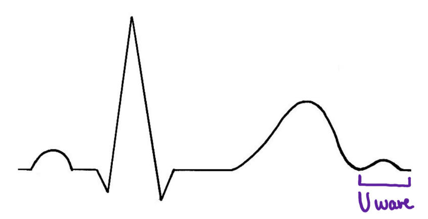
repolarzatoom of purkinje fibers
25
the vertical axis of an EKG represents voltage and at standard cal is ______
0.1mV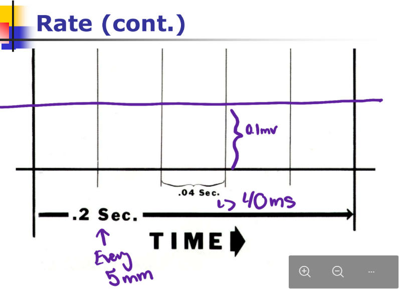
26
the horixontal axis of the EKG represents time and at standard cal is _________
0.04 sec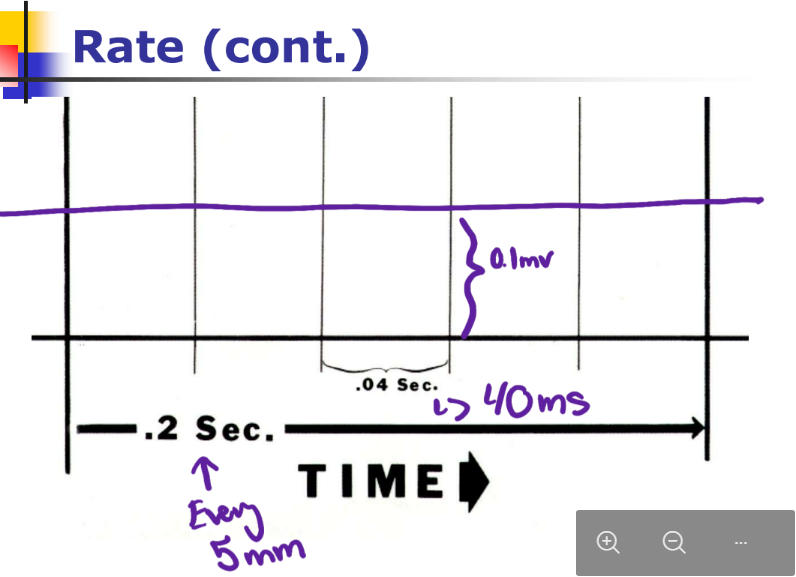
27
equation for determining b/min on regular EKG rythms
300/# big blocks
28
how do you determine b/min for irregular rythmns
use 3 second markers 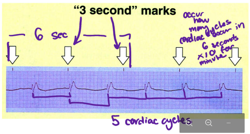
29
the 6 limb leads allow determination of axis in this plane
frontal plane
30
what are the limbleads of the frontal plane
I,II, III, aVR, aVL, aVF
31
what are the chest leads that allow deter,ination of axis in the horizontal plane
V1-6
32
fr the limb leads, there are 3 ________ and 3 _________-
bipolar; augmented unipolar
33
the bipolar ;limb leads have these electrodes
1 positive and 1 negative
34
the augmeted unipolar limb leads have these electrodes
1 positive and 1 compound reference electrode
35
which of the limbs is the groundinh lomb where the grounding electrode is placed
rght leg.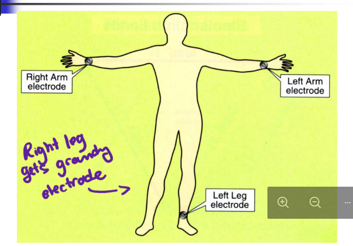
36
where are the limb leads placed and wgere is the positive node placed? (bipolar)
RA-LA: lead I; LA +
RA-LL: lead 2: LL+
LA-LL : lead 3: LL +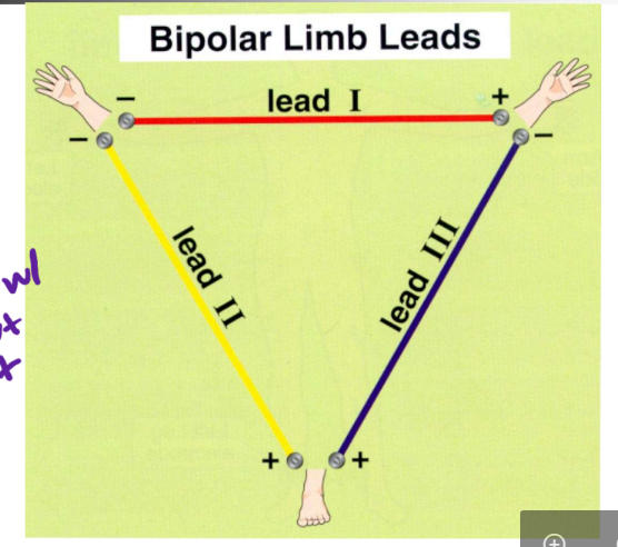
RA-LL: lead 2: LL+
LA-LL : lead 3: LL +
37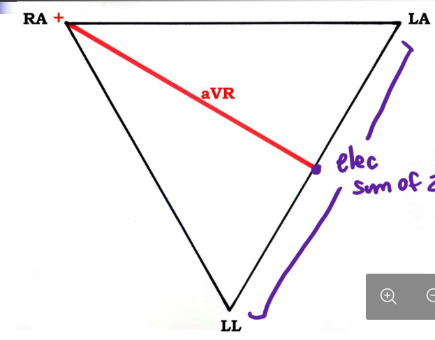
Image occlusion: name the hidden labels
38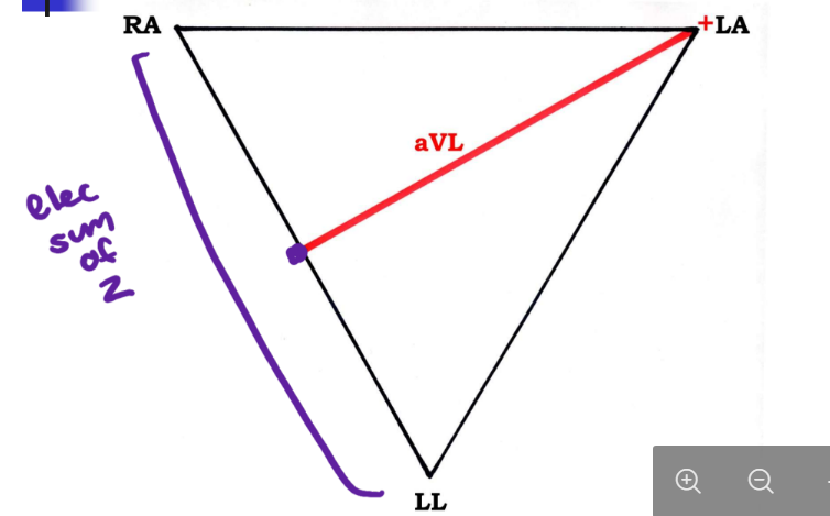
Image occlusion: name the hidden labels
39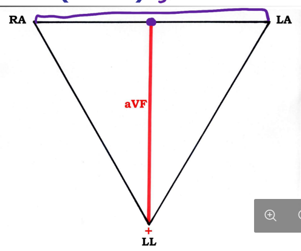
Image occlusion: name the hidden labels
40
which are the lateral leads
I and aVL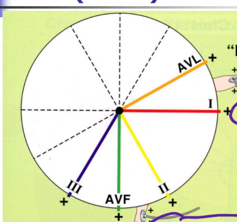
41
which are the inferior leads
II, III, aVF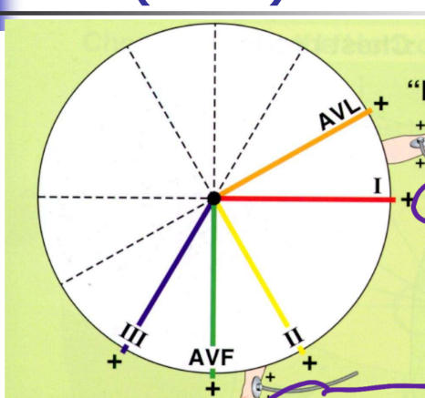
42
how do you quickly determine QRS axis in the frontal plane (what lkeads)
I and aVF, look at positive or negative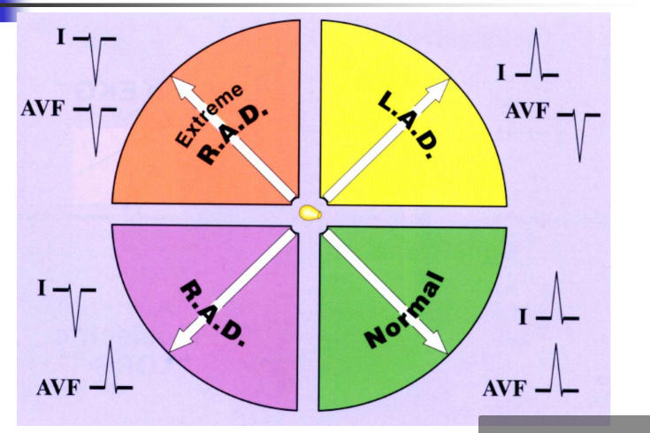
43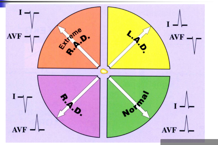
Image occlusion: name the hidden labels
44
Image occlusion: name the hidden labels
45
Image occlusion: name the hidden labels
46
Image occlusion: name the hidden labels
47
A shift in the electrical axis out of
the normal range is referred to as:
________” in the frontal plan
"_________" in the horizontal plane
the normal range is referred to as:
________” in the frontal plan
"_________" in the horizontal plane
devation
rotation
rotation
48
Simply stated, in determining the mean
QRS axis in the horizontal plane:
If the R to S ratio changes from <1 to >1:
After V4 (i.e. V5, V6, or never), then this
is referred to as “______”
At or before V2 (i.e. V1, or V2), then this
is referred to as “_______________on”
After V2 and at or before V4, then the
axis in the horizontal plane is referred to
as “___________
QRS axis in the horizontal plane:
If the R to S ratio changes from <1 to >1:
After V4 (i.e. V5, V6, or never), then this
is referred to as “______”
At or before V2 (i.e. V1, or V2), then this
is referred to as “_______________on”
After V2 and at or before V4, then the
axis in the horizontal plane is referred to
as “___________
leftward axis rotation; rightward axis rotation; normal
49
horizontal plane if R to S ratio Starts < 1 and stays < 1, then this is
referred to as
referred to as
leftward acis rotation
50
horizontal plane R to S ratio starts >1 and stays > 1 then it is
rightward axis rotation
51
horizontal plane if starts >1 ad becomes <1 by V6
refered to as leftward axis rotation
52
QRS between -45 and -90 left axis devoation
fascicular block left anterior
53
QRS between 90 to 180 right axis deviation
fascicular block left posterior
54
Ability of specialized cells to
spontaneously generate electrical
impulses which may then spread
throughout surrounding tissue
spontaneously generate electrical
impulses which may then spread
throughout surrounding tissue
automaticity
55
Potential pacemakers within the heart
automaticity foci
56
utomaticity foci in the heart
atrial foci , functional fpoci, ventricular foci
57
conduction rate atrial foci
60-80 per minte
58
conduction rate junctional foci
40-60 per minute
59
conduction rate vetricular foci
20-40 per minute
60
A protective mechanism whereby an automaticity focus with the next highest inherent rate begins pacing (escapesoverdrive suppression) in the event of a pause or cessation of pacing activity of the previously dominant pacemaker.
escape
61
Transient escape of an automaticity focus (from overdrive suppression) to generate one beat.
This occurs when there is a pausein pacing activity in the previously
dominant pacemaker.
This occurs when there is a pausein pacing activity in the previously
dominant pacemaker.
escape beat
62
Escape of an automaticity focus (from overdrive suppression) with subsequentpacing by that automaticity focus, at its inherent rate.
This occurs when there is a cessation(or arrest) of pacing activity in the
previously dominant pacemaker.
This occurs when there is a cessation(or arrest) of pacing activity in the
previously dominant pacemaker.
escape rhythm
63
normal rhythm of the heart
sinus rhyth
64
criteria of a sunus rhythm
Each QRS complex is preceded by a P wave.
The P waves must be positive in lead II, and negative in lead aVR
The P waves must be positive in lead II, and negative in lead aVR
65
normal sinus rhythm criteria
Sinus rhythm with a heart rate
between 60 and 100 beats per
minute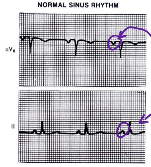
between 60 and 100 beats per
minute
66
Sinus rhythm with a heart rate less than 60 beats per minute
sinus bradycardia
67
Sinus rhythm with a heart rate
greater than 100 beats per minute
greater than 100 beats per minute
sinus tachycardia
68
Same criteria as sinus rhythm andthere is greater than 0.16 sec.
difference between the shortest and the longest PP intervals within the same EKG tracing
difference between the shortest and the longest PP intervals within the same EKG tracing
sinus arrhythmia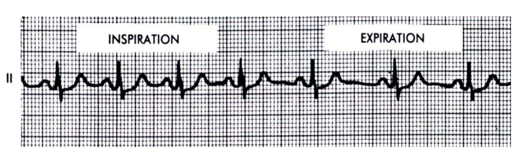
69
what is this?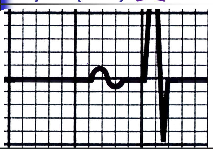
atrial rhythm
70
what is this?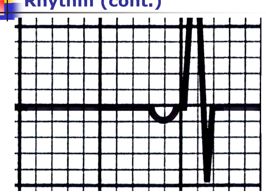
junctional rhytm
71
what is this?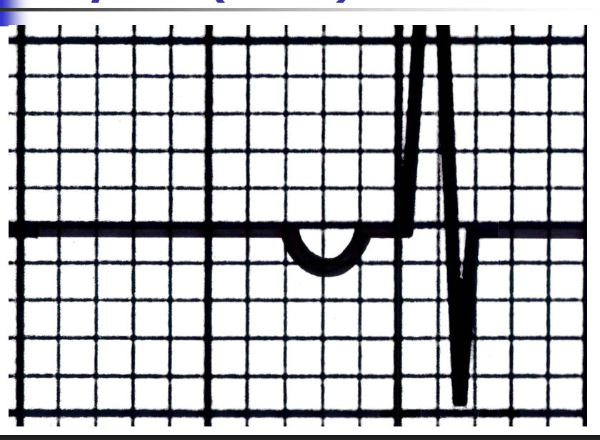
atrial rhythm
72
what is this?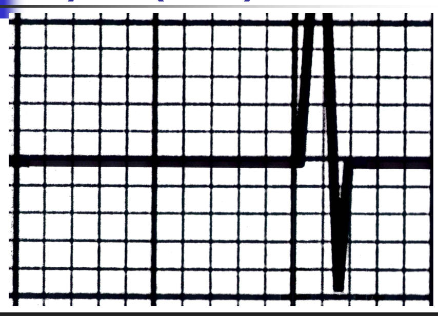
junctional rhytm
73
what is this?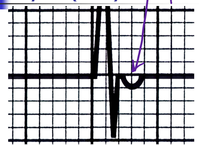
junctional rhythm
74
(positive in lead aVR and negative in lead II) immediately preceding (PR interval <120 msec) the QRS complex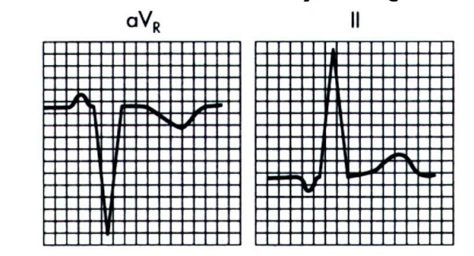
junctional retrograde p wave
75
(buried in the QRS complex or absent due to lack of retrograde AV conduction):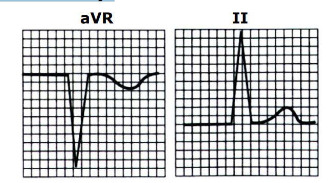
junctional absent p wave
76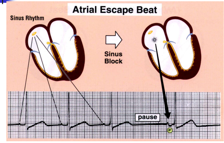
Image occlusion: name the hidden labels
77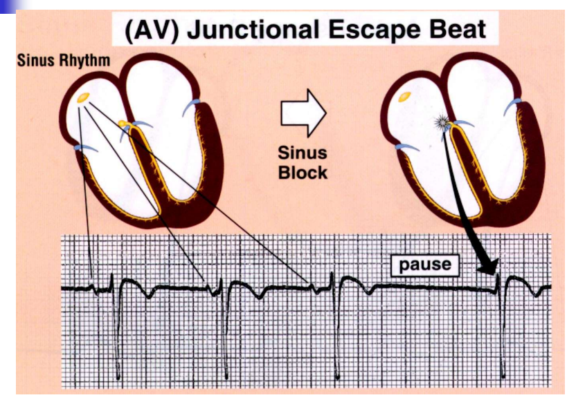
Image occlusion: name the hidden labels
78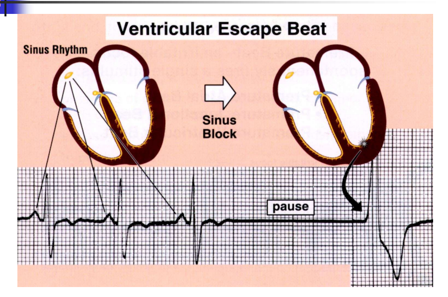
Image occlusion: name the hidden labels
79
If the QRS complex is not prolonged (not >120 msec.), ventricular depolarization must have occurred via
the his-purkinje system
80
An increase in the tendency for
automaticity foci to spontaneously
generate electrical impulses
automaticity foci to spontaneously
generate electrical impulses
irritability
81
Occurs when an irritable automaticity focus spontaneously generates a single electrical impulse prior to the next
expected beat from the dominant
pacemaker
expected beat from the dominant
pacemaker
premature beat
82
escape of an automaticity focus (from overdrive suppression) due to failure of the previously dominant pacemaker
escape beats
83
irritability of automaticity focus
premature beats
84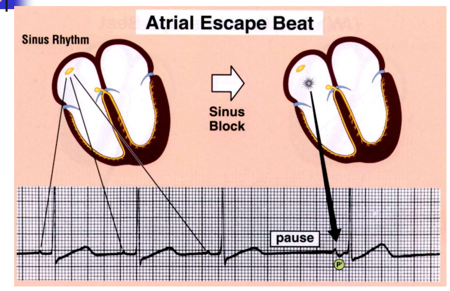
Image occlusion: name the hidden labels
85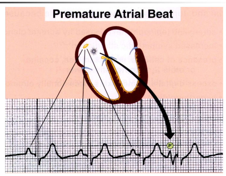
Image occlusion: name the hidden labels
86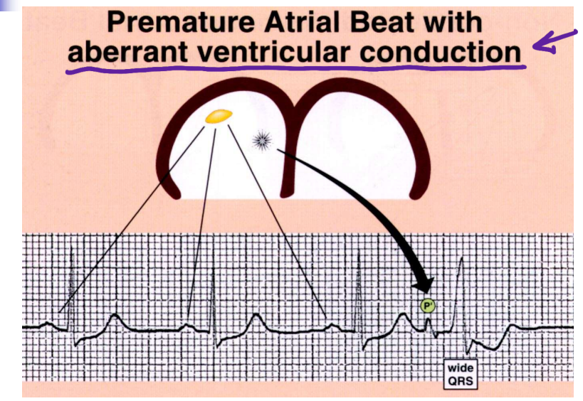
Image occlusion: name the hidden labels
87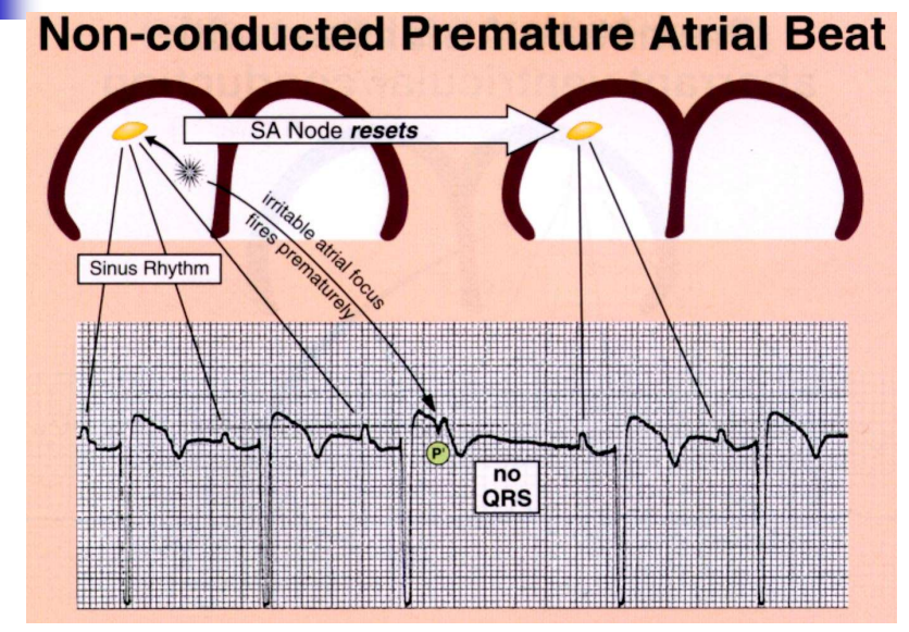
Image occlusion: name the hidden labels
88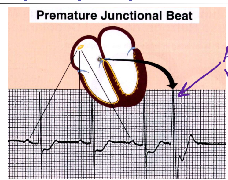
Image occlusion: name the hidden labels
89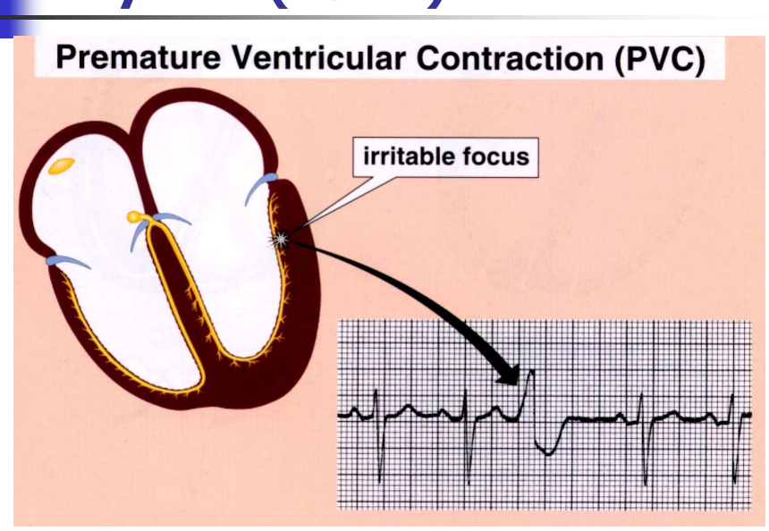
Image occlusion: name the hidden labels
90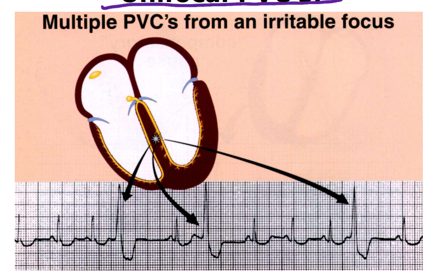
Image occlusion: name the hidden labels
91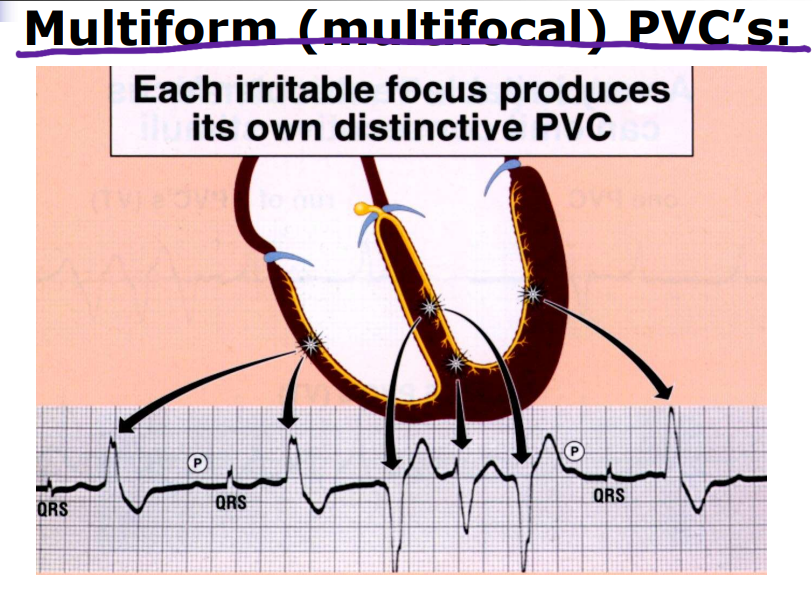
Image occlusion: name the hidden labels
92
– every other beat is premature
bigeminy
93
every third beat is premature
trigeminy
94
every fourth beat is premature
quadrigeminy
95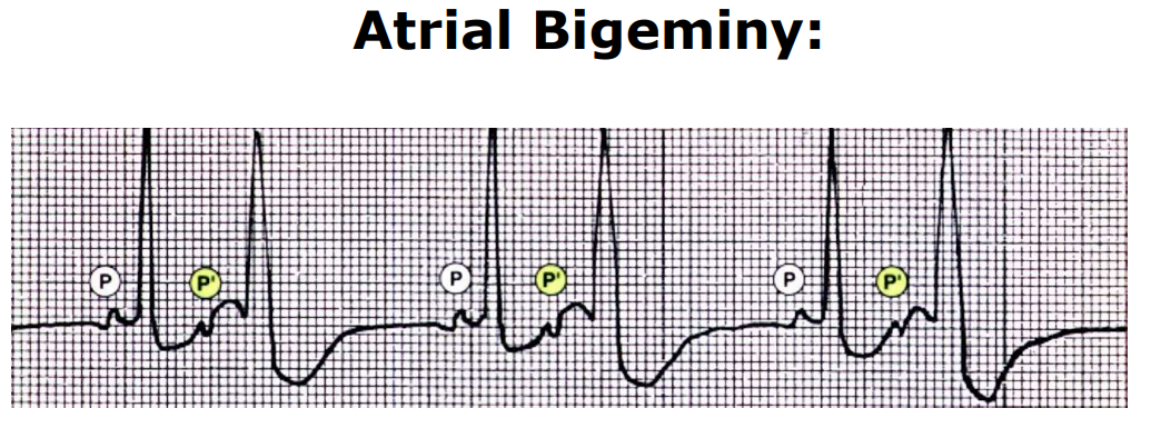
Image occlusion: name the hidden labels
96
sudden rhytm change
paroxysmal
97
Heart rate < 60 beats per minute (bpm)
bradycardia
98
Heart rate > 100 bpm
tachycardia
99
Rate between 250 and 350 bpm
flutter
100
Rate between 350 and 450 bpm
fibrillation
101
A “generic” term for any tachycardia originating above the ventricles
supraventricular tachycardia
102
Polymorphic ventricular tachycardia (VT) which is preceded by marked prolongation of the QT interval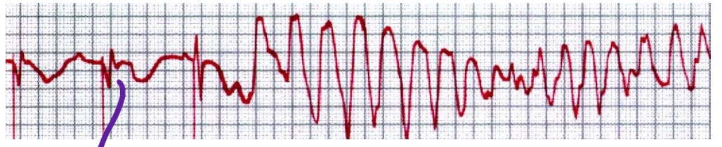
torsades de pointes
103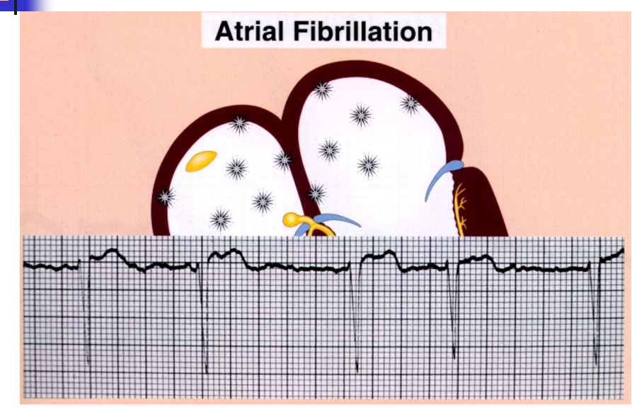
Image occlusion: name the hidden labels
104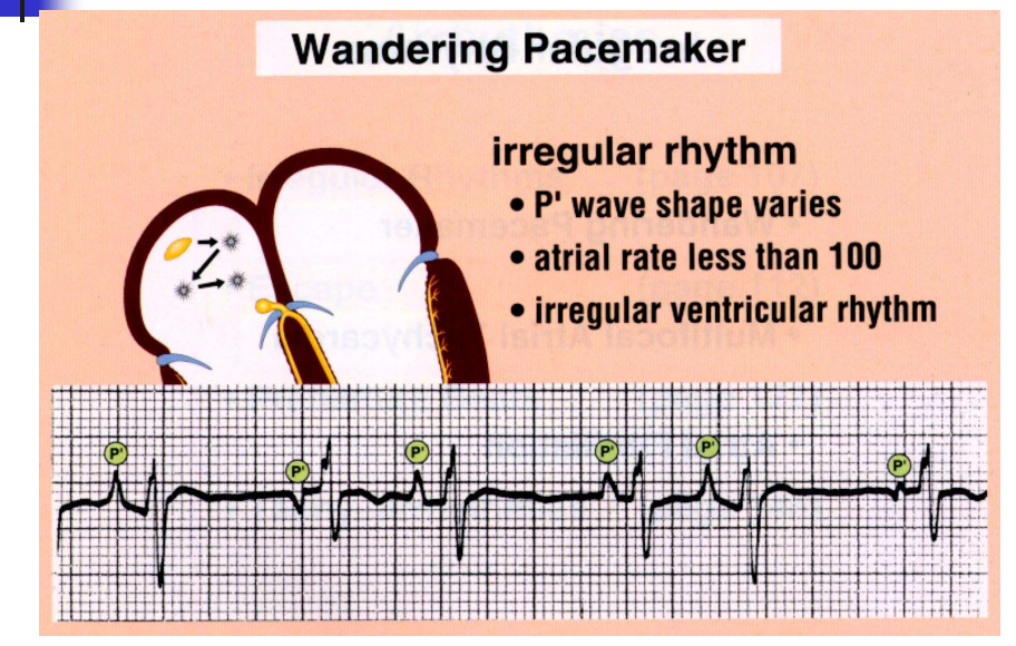
Image occlusion: name the hidden labels
105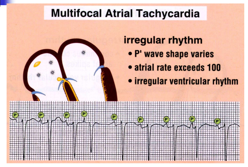
Image occlusion: name the hidden labels
106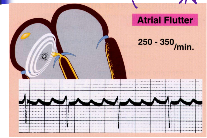
Image occlusion: name the hidden labels
107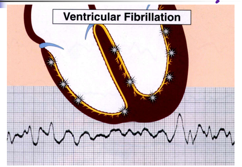
Image occlusion: name the hidden labels
108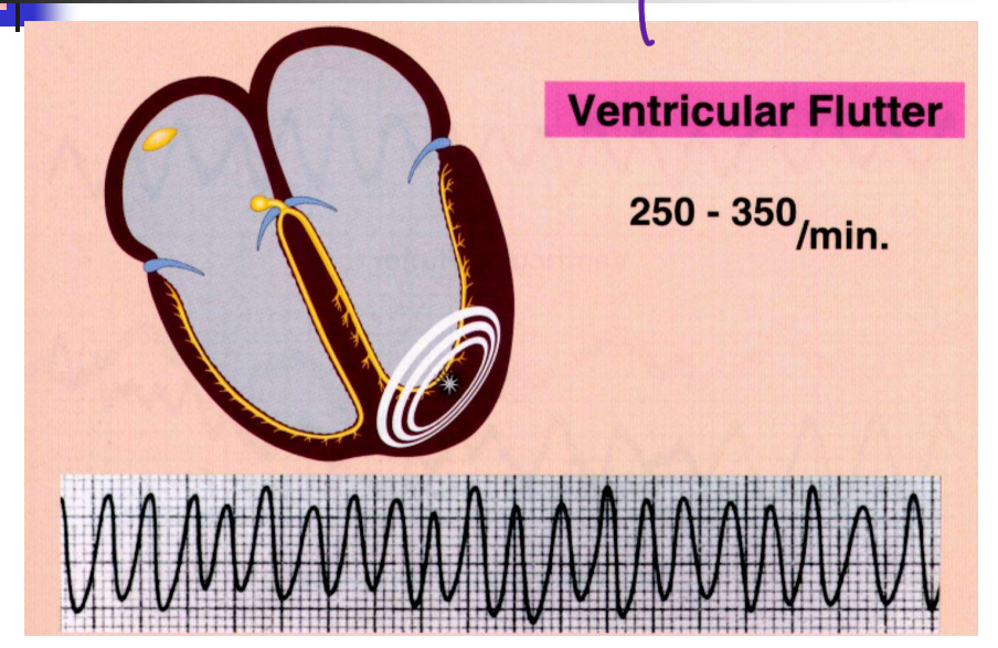
Image occlusion: name the hidden labels
109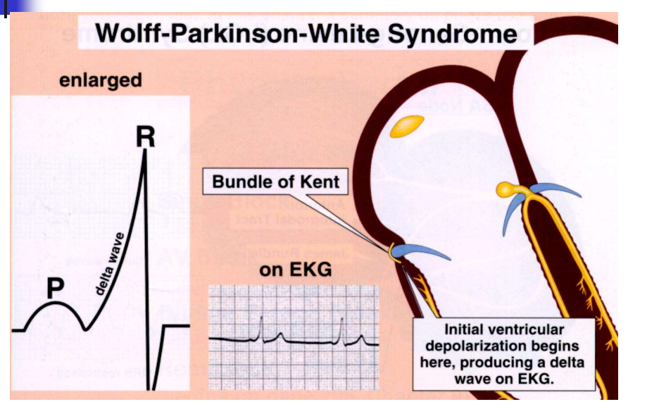
Image occlusion: name the hidden labels
110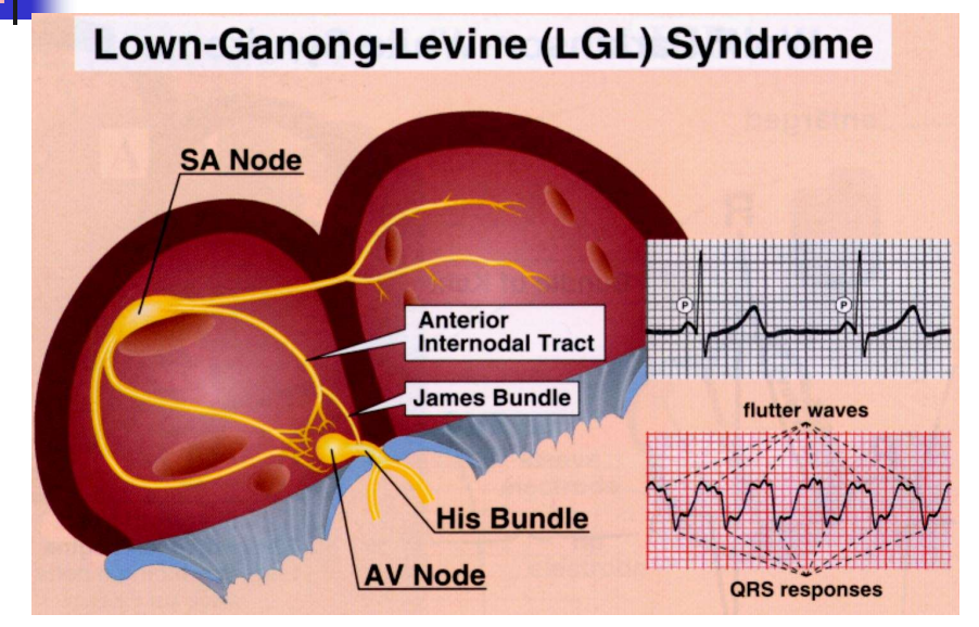
Image occlusion: name the hidden labels
111
bradyarrhythmias can de disorders of (x2)
impulse formation or impulse propigation
112
Encompasses several SA nodal abnormalities:
Chronotropic incompetence
Sinus pause, arrest or exit block
Bradycardia-tachycardia syndrome:
Paroxysms of atrial tachyarrhythmias
alternating with slow atrial and ventricular
rates
Chronotropic incompetence
Sinus pause, arrest or exit block
Bradycardia-tachycardia syndrome:
Paroxysms of atrial tachyarrhythmias
alternating with slow atrial and ventricular
rates
sick sinus syndrome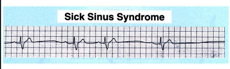
113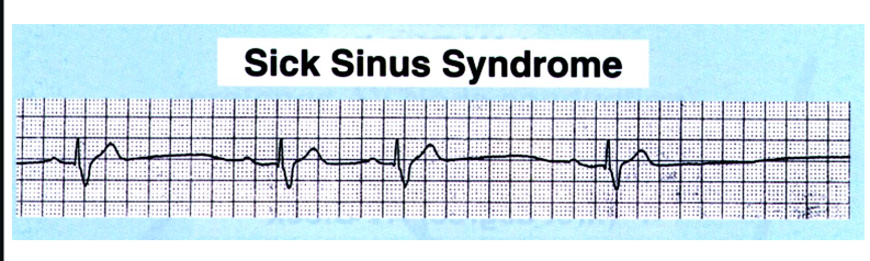
Image occlusion: name the hidden labels
114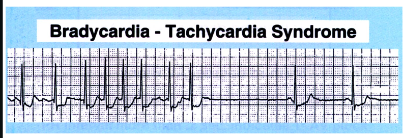
Image occlusion: name the hidden labels
115
what types of AV blocks can there be
first degree
second degree: type I and II
third degree
second degree: type I and II
third degree
116
what needs to be true to have a first degree AV block
1-to-1 ratio of P waves to QRS complexes
PR interval is fixed (consistent)
PR interval exceeds 200 msec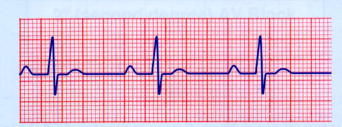
PR interval is fixed (consistent)
PR interval exceeds 200 msec
117
what does this show?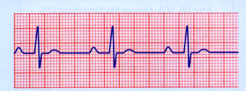
first degree AV block
118
Grouped beats
Groups or clusters of QRS complexes separated by “missed” single beats
Two P waves for every QRS complex, without groups or clusters of beats
All PR intervals will be the same (fixed/consistent)
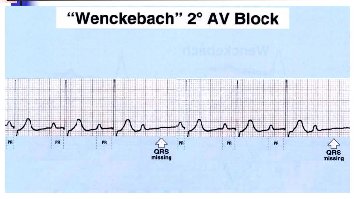
Groups or clusters of QRS complexes separated by “missed” single beats
Two P waves for every QRS complex, without groups or clusters of beats
All PR intervals will be the same (fixed/consistent)
type I second degree AV block
119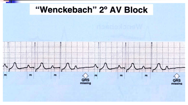
Image occlusion: name the hidden labels
120
There are more P waves than QRS complexes
All P waves that immediately precede QRS complexes have equal PR intervals
At times, there may be a fixed ratio of P waves to QRS complexes (e.g. 2-to-1*, 3-to-1, or 4-to-1, etc.)
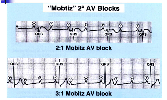
All P waves that immediately precede QRS complexes have equal PR intervals
At times, there may be a fixed ratio of P waves to QRS complexes (e.g. 2-to-1*, 3-to-1, or 4-to-1, etc.)
type II second degree AV blocj
121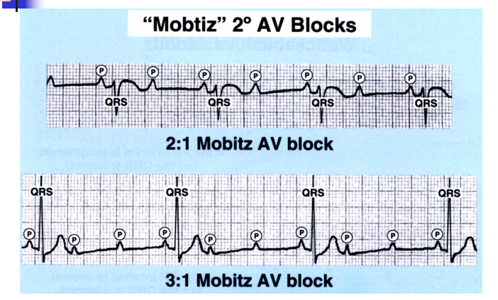
Image occlusion: name the hidden labels
122
Image occlusion: name the hidden labels
123
Image occlusion: name the hidden labels
124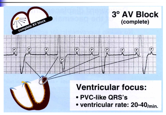
Image occlusion: name the hidden labels
125
Slowing of the previously dominant pacemaker, with subsequent junctional or ventricular escape rhythm
default AV dissocociation
126
Acceleration of a latent pacemaker which usurps control of the ventricles
usurpation av dissociation
127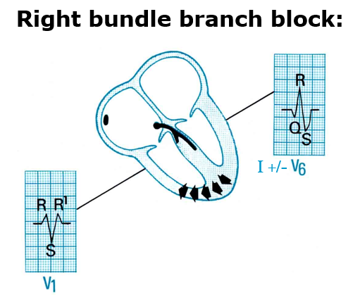
Image occlusion: name the hidden labels
128
Image occlusion: name the hidden labels
129
Image occlusion: name the hidden labels
130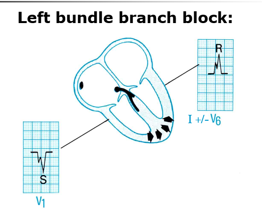
Image occlusion: name the hidden labels
131
Image occlusion: name the hidden labels
132
QRS duration > 120 msec.
Criteria for RBBB are not met
Criteria for LBBB are not met
Criteria for RBBB are not met
Criteria for LBBB are not met
nonspecific intraventricular conduction defect (nonspecific IVCD)
133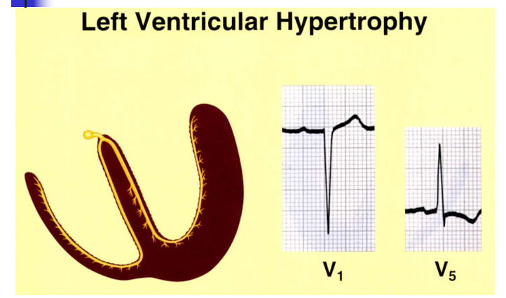
Image occlusion: name the hidden labels
134
Image occlusion: name the hidden labels
135
how do you determine if its a left ventricular hypertrophy? (x2)
V2 or V6 s is greater than 35mm
-only can use this method if individual is oilder than 40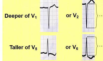
-only can use this method if individual is oilder than 40
136
what does the T wave look like in a Left ventricular hypertrophy (left chest leads)
inverted T wave (asymetrcal)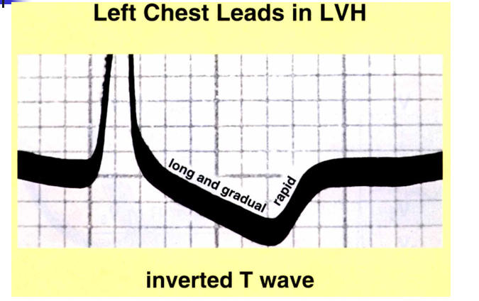
137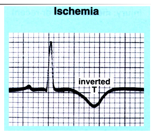
Image occlusion: name the hidden labels
138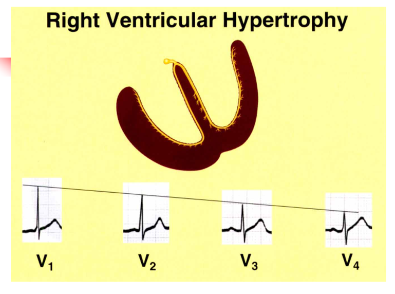
Image occlusion: name the hidden labels
139
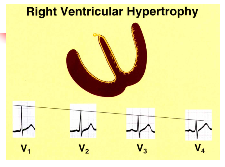
what 3 criteria strongly suggest rught ventricular hypertropy?
Right axis deviation (> 110°)
(for our test, simply use RAD or extreme RAD)
R/S > 1 in lead V1
Deep S waves in lead V5 or V6
(for our test, simply use RAD or extreme RAD)
R/S > 1 in lead V1
Deep S waves in lead V5 or V6
140
Image occlusion: name the hidden labels
141
Image occlusion: name the hidden labels
142
Image occlusion: name the hidden labels
143
Image occlusion: name the hidden labels
144
Image occlusion: name the hidden labels
145
Image occlusion: name the hidden labels
146
Image occlusion: name the hidden labels
147
Image occlusion: name the hidden labels
148
Image occlusion: name the hidden labels
149
Image occlusion: name the hidden labels
150
Image occlusion: name the hidden labels
151
Image occlusion: name the hidden labels
152
Image occlusion: name the hidden labels
153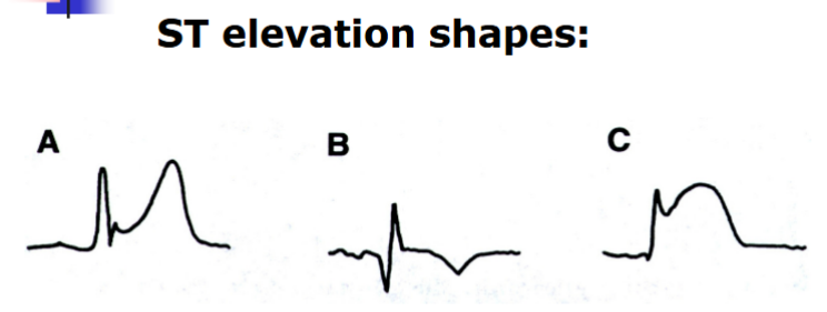
Image occlusion: name the hidden labels
154
what must be present in early repolarization St elevations
Distribution: Diffuse (usually)
Reciprocal ST depression: Absent
ST segment shape: Concave upwards
PR depression: Absent
Reciprocal ST depression: Absent
ST segment shape: Concave upwards
PR depression: Absent
155
clinical significance early repolarization ST elevation
Asymptomatic; usually an incidental
finding on a routine EKG; often present in
young males
finding on a routine EKG; often present in
young males
156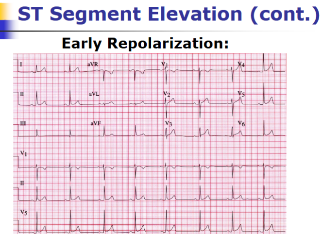
Image occlusion: name the hidden labels
157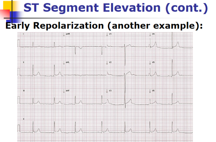
Image occlusion: name the hidden labels
158
what is required fir acute pericarditis ST elivation
Distribution: Diffuse (usually)
Reciprocal ST depression: Absent
ST segment shape: Concave upwards
PR depression: Present (in leads that have
ST elevation)
Reciprocal ST depression: Absent
ST segment shape: Concave upwards
PR depression: Present (in leads that have
ST elevation)
159
clinical cause of acute pericarditis ST elivation
gradual onset of sharp,
pleuritic chest discomfort, which is often
relieved or improved by sitting up;
sometimes preceded or accompanied by
fever or other flu-like symptoms
pleuritic chest discomfort, which is often
relieved or improved by sitting up;
sometimes preceded or accompanied by
fever or other flu-like symptoms
160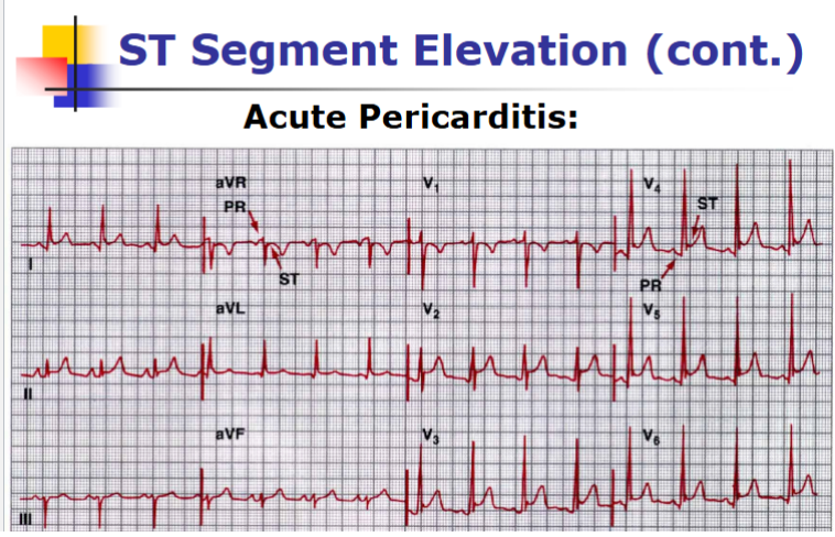
Image occlusion: name the hidden labels
161
what is required for acute MI St elevation
Distribution: Regional (vascular)
Reciprocal ST depression: Present (if MI
involves inferior or high lateral wall)
ST segment shape: Not as reliable
Concave down (if “textbook” case)
PR depression: Absent
Reciprocal ST depression: Present (if MI
involves inferior or high lateral wall)
ST segment shape: Not as reliable
Concave down (if “textbook” case)
PR depression: Absent
162
clinical cause of acute MI St elevation
(usually) Sudden onset of non-
sharp, non-pleuritic chest discomfort or
dyspnea; sometimes accompanied by
nausea, vomiting, and/or diaphoresis
sharp, non-pleuritic chest discomfort or
dyspnea; sometimes accompanied by
nausea, vomiting, and/or diaphoresis
163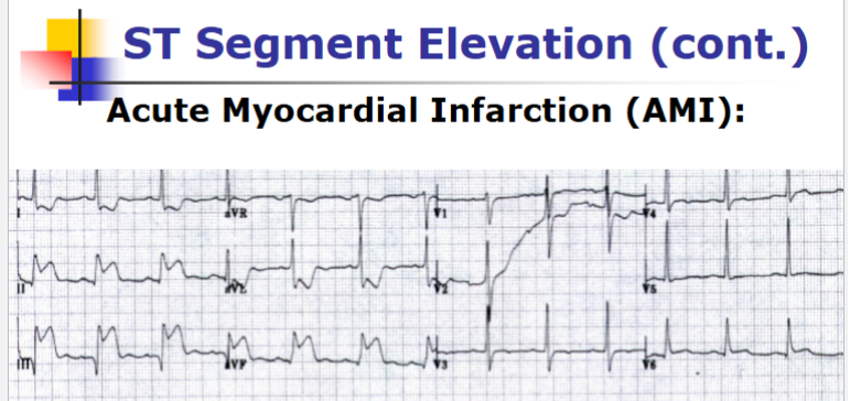
Image occlusion: name the hidden labels
164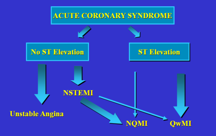
Image occlusion: name the hidden labels
165
Image occlusion: name the hidden labels
166
Image occlusion: name the hidden labels
167
Image occlusion: name the hidden labels
168
what can cause deep symmetrically inverted T waves
non st elevation MI (acute or recent, myocardial ischemia, ICH
169
what can cause significant Q waves
age indeterminate or a previous MI
170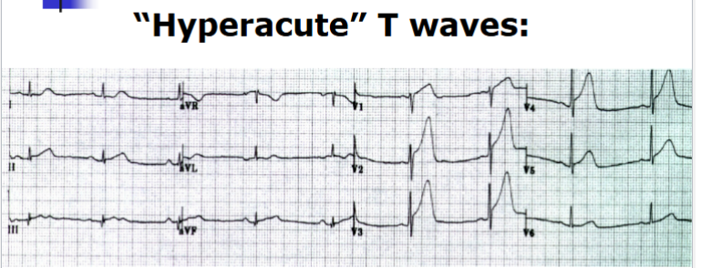
Image occlusion: name the hidden labels
171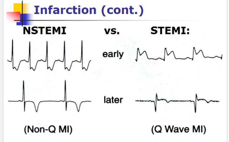
Image occlusion: name the hidden labels
172
Image occlusion: name the hidden labels
173
what signifies a significant Q wave
a q wave greater than 1mm wide; a q wave 1/3 QRS amplitude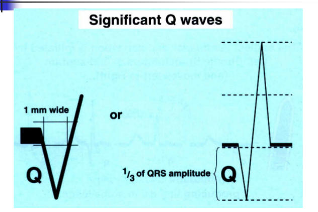
174
Always order______________-sided chest
leads when there is evidence of
an acute inferior wall MI on the
standard 12-lead EKG!
leads when there is evidence of
an acute inferior wall MI on the
standard 12-lead EKG!
right