embryo exam 3
Study these flashcardsFree · spaced repetition · iPhone, Android, Mac & web
All cards
1
the pharyngeal apparatus consists of (x4) parts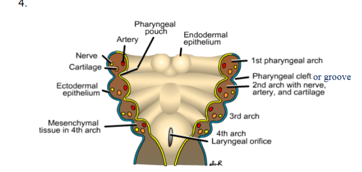
arches, cleft, pouch, membrane (ectoderm outside, endoderm inside)
2
components of the pharyngeal arch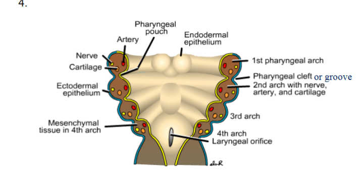
nerve, artery, cartilageone, mesenchyme (mesoderm and neural crest)
3
the first pharyngeal arch forms 2 prominences...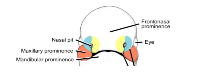
maxillary prominence (cheeks and upper jaw)
mandibular prominence (lower jaw)
mandibular prominence (lower jaw)
4
what arch: malleus
first
5
what arch incus
first
6
what arch maxilla
first
7
meckel's cartilage what arch
first
8
stapes what arch
2nd arch
9
styloid process what arch
2nd
10
stylohyoid ligament what arch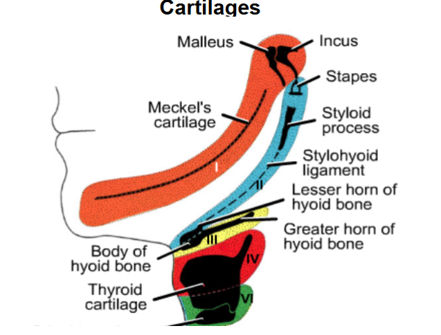
2nd
11
lesser horn of hyoid bonr what arch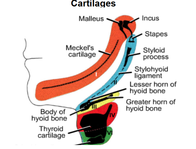
2nd
12
arch: greater horn of hyoid bone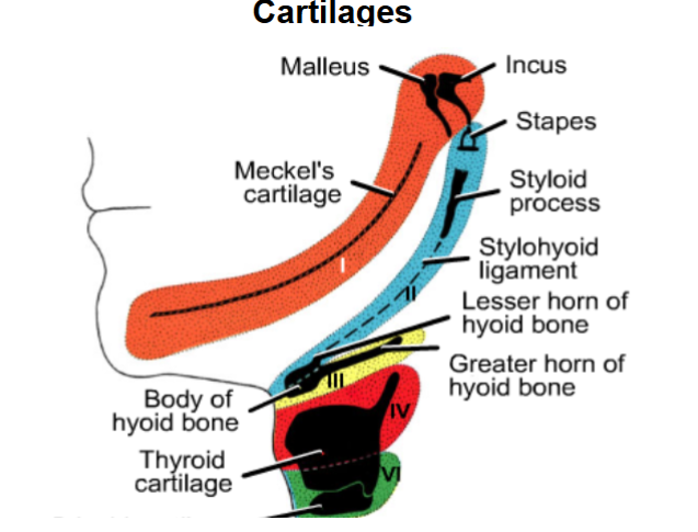
3rd
13
body of hyoid, what arch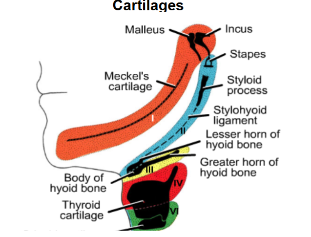
2nd and 3rd arch
14
thyoid cartilage, what arch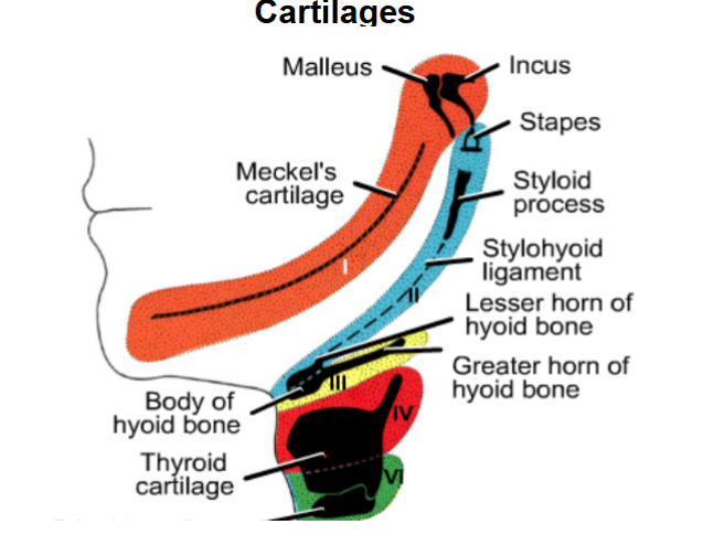
4th and 5th
15
cricoid cartilage, what arch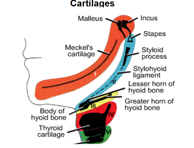
5th
16
trigeminal n, what arch
1st
17
muscles of mastication, what arch
1st
18
mylohyoid and anterior belly of digastric, wjat arch
1st
19
tensor tympani, what arch
1st
20
tensor veli palatini, what arch
1st
21
facial n, what arch`
2nd
22
muscles of facial ex[pression, arch
2nd
23
stapedius, what arch
2nd
24
stylohyoid, what arch
2nd
25
posterior belly of dogastric, what arch
2nd
26
glossopharyngeal n, what arch
3rd
27
stylopharyngeus, what arch
3rd
28
severe inflamation of the trigeminal n
trigeminal neuralgia
29
laryngeal m, what arch
4th and 6th
30
pharyngeal constrictors, what arch
4th and 6
31
levator veli palatini what arch
4th and 6th
32
vagus n what arch
4th and 6th
33
what syndrome; what arch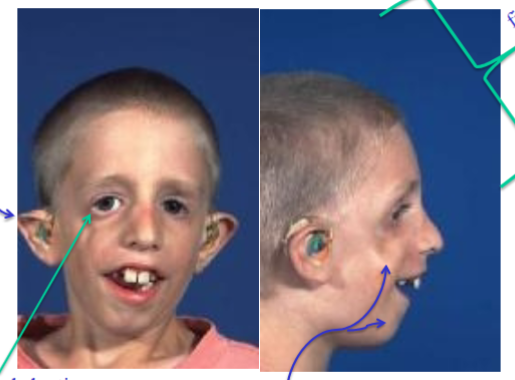
treacher collins syndrome; first arch
34
what disorder; what arch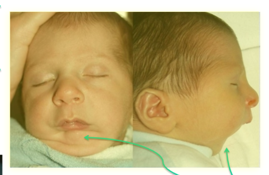
pierre robin sequence; first arch
35
what comes from the 1st pharyngeal pouch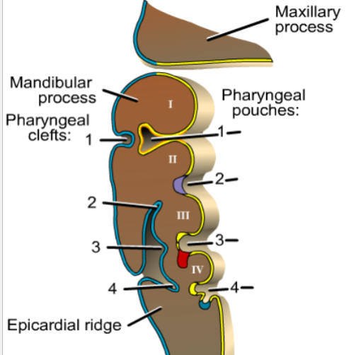
pharyngeal tympanic tube
36
what does the pharyngeal tympanic tube become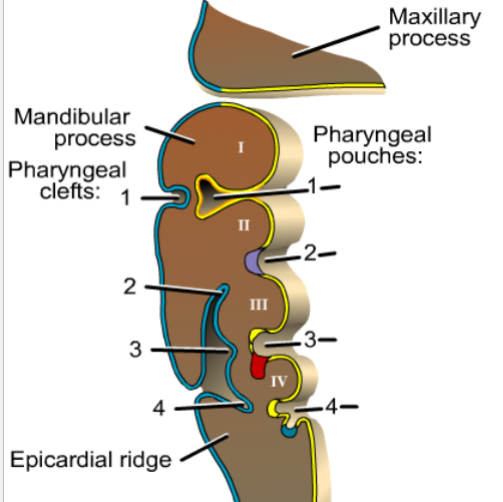
tympanic membrane
37
what does the 2nd pharyngeal pouch form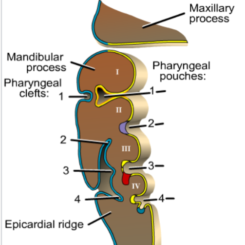
palantne tonsils and fossa
38
whatforms from the 3rd pharyngeal poucg
superior: inferior portion of parathyrid gland
inferior: thymus
inferior: thymus
39
ewhat forms the 4th pharyngeal pouch
superior: superior part of parathyroud gland
iferior: UH pharygeal bodies (become C cells of thyroid)
iferior: UH pharygeal bodies (become C cells of thyroid)
40
what forms from the 1st pharyngeal cleft
external auditory meatus
41
what forms from the 2-4th pharyngeal clefdts
obliterated
42
what can happen if the 2nd-4th pgaryngeal clefts arent oblitered
fistula or cyst can iccyr here
43
cyst formed when 2nd to 4th pharyngeal pouches dont obliterate
lateral cervical cyst
44
lateral cervical vyst with an opening to the world
external branchial fistula
45
what is this?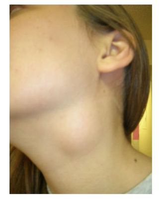
lateral cervocal cyst
46
what is this?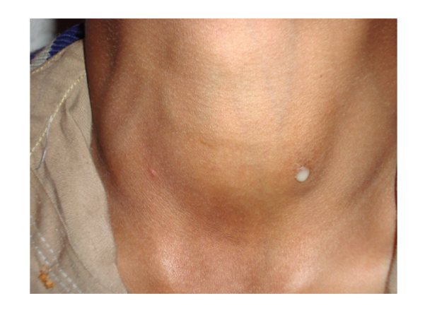
lateral cervical fistula
47
where does the thyroiud start development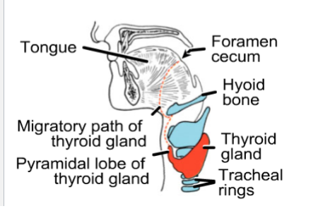
foramen cecum near tomgue
48
what does this show?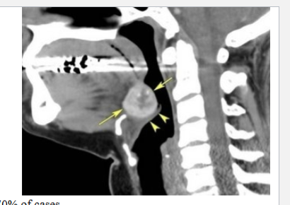
ectopic thyroid
49
what does this show?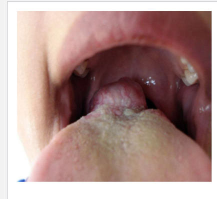
ectopic thyroid
50
what does this show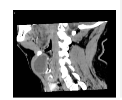
thyroglossal duct cyst
51
what does thus show?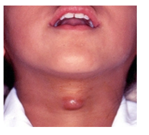
thyroid duct fistula
52
what does this shjow?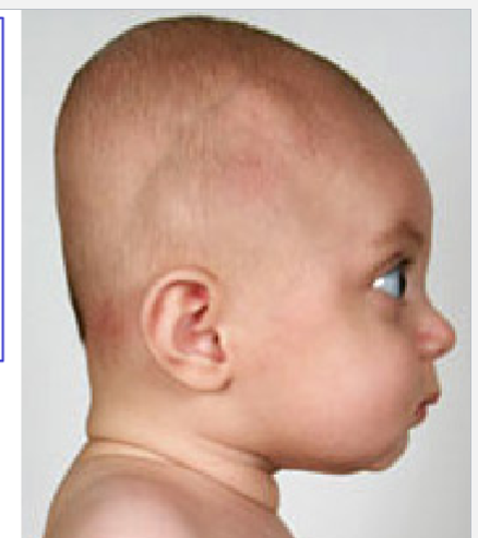
plagiocoplady: flat head
53
cranial part of skull
neurocranium
54
facial patrt of skull
viscerocraniym
55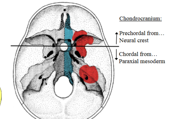
Image occlusion: name the hidden labels
56
Image occlusion: name the hidden labels
57
what makes up the viscerocranium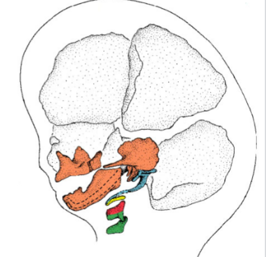
single frontonasal
paired maxillary (2)
paired mandibular (2)
paired maxillary (2)
paired mandibular (2)
58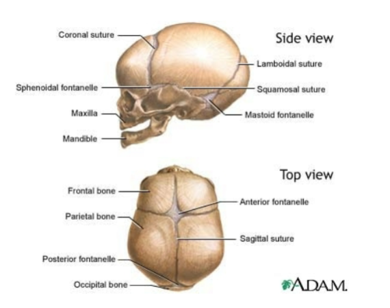
Image occlusion: name the hidden labels
59
Image occlusion: name the hidden labels
60
Image occlusion: name the hidden labels
61
Image occlusion: name the hidden labels
62
Image occlusion: name the hidden labels
63
Image occlusion: name the hidden labels
64
Image occlusion: name the hidden labels
65
Image occlusion: name the hidden labels
66
what does this show; what suture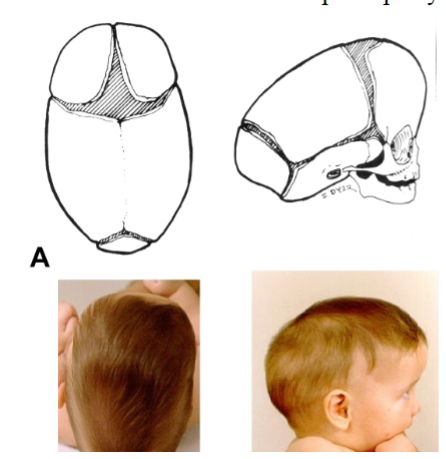
scaphocepgaly; saggital
67
what does this show; what suturee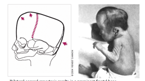
bilateral coronal synostosis; coronal sutures
68
what does thus show? what suture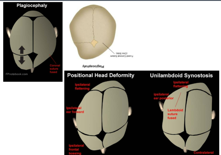
plagiocephaly; unilateral lambdoid suture
69
what does this show. what suture?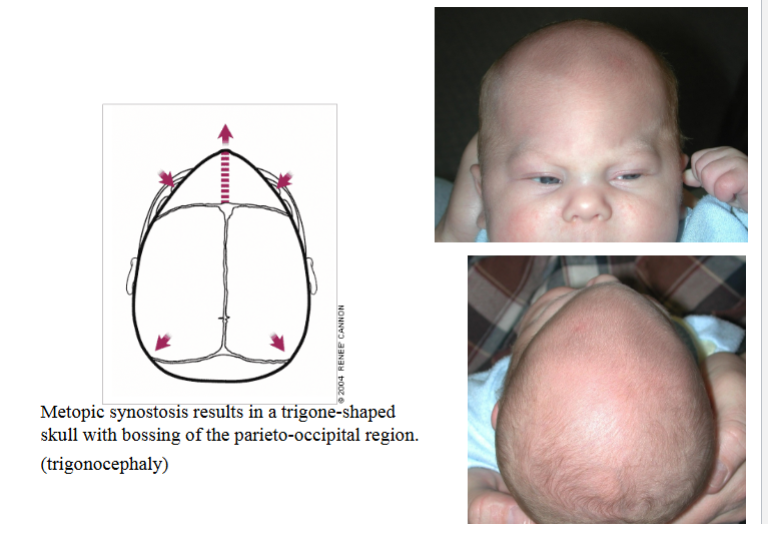
metopic synostosis; metopic suture bilateral
70
diagnosis?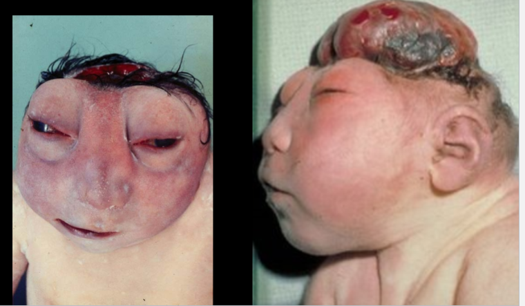
acrania
71
diagnosis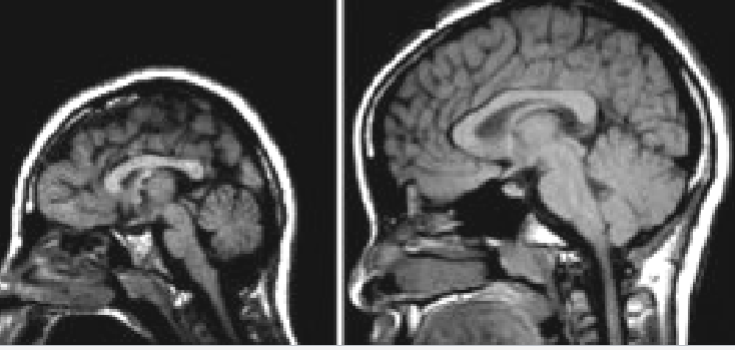
microcephally
72
ddx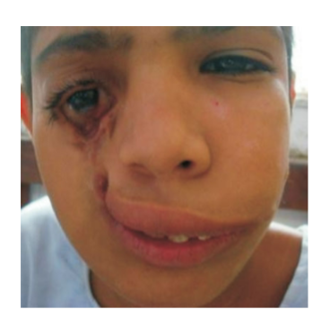
oblique cleft
73
ddx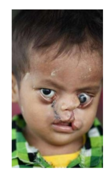
bilateral facial clefts
74
ddx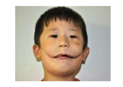
macrostomia; maxillaru and mandibular arches failed to fuse correctlt
75
minor abnormalities are present in _____ pf newborns (birth defects) major are present in _____-
15%; 3%
76
anomalies account for _____- pf infanyt deayh
25%
77
what causes birth defects (general breakdown)
55% are multifactorial
- 30% are genetic
- 15% are environmental
- 30% are genetic
- 15% are environmental
78
result in morphologic alterations of already formed structures and are caused by destructive processes.
disruptions
79
result from mechanical forces that mold a part of the fetus over a prolonged period. Clubfeet, for example, are caused by compression in the amniotic cavity
-may be reversible postnatally
-may be reversible postnatally
deformations
80
a group of anomalies occurring together that have a specific common cause. This term indicates that a diagnosis has been made and that the risk of recurrence is known.
syndrome
81
congenotal malformatons causesd by rubella exposure
Cataracts, glaucoma, heart defects, hearing loss, tooth abnormalities
82
congenital malformations caused by cytomegalovirus exposure
Microcephaly, visual impairment, intellectual disability, fetal death
83
congenital abnormaloities xaused by herpes exposure
Microphthalmia, microcephaly, retinal dysplasia
84
congenital abnormalities cxaused by toxoplasmosis exposure
Hydrocephalus, cerebral calcifications, microphthalmia
85
congenital malformations caused by isotretinoin (accutane) exposure)
Isotretinoin embryopathy: small, abnormally shaped ears, mandibular hypoplasia, cleft palate, heart defects
86
congenita malformaions caused by opioids exposure
Neural tube defects, heart defects, gastroschisis
87
congenital malformations cxaused by SSRi exposire
Heart malformations, neural tube defects, anal atresia, facial clefts, and many other defects
88
congenital malformations caused by valproic acid exposure
Neural tube defects; heart, craniofacial, and limb anomalies
89
congenital malfornmations caused by amphetamines exposure
Cleft lip and palate, heart defects
90
conenital defects caused by alcohol exposure
Cleft lip and palate, heart defects
91
congenital defects caused by DES
Malformation of the uterus, uterine tubes, and upper vagina; vaginal cancer; malformed testes
92
congenital defects caused by ,maternal diabetes
Various malformations; heart and neural tube defects most common
93
congenital defects caused by maternal obesity
Neural tube defects, heart defects, omphalocele
94
congenitak defects caused by organic mercury
Neurologic symptoms similar to those of cerebral palsy, prematurity, intrauterine growth retardation (IUGR), stillbirths
95
The skin is the largest organ in the body and has a dual origin: (1) A superficial layer, the epidermis, develops from the surface _________________. (2) A deep layer, the dermis, develops from the underlying ______________
ectoderm; mesenchyne
96
What are the endoderm-lined structures of the branchial apparatus?
Pharyngeal pouches
97
What is the ectoderm-lined structure of the branchial apparatus?
Pharyngeal grooves
98
What structure forms where a pharyngeal pouch meets a groove?
Pharyngeal membrane
99
What two prominences does the first pharyngeal arch form?
Maxillary and mandibular prominence
100
What nerve innervates the first pharyngeal arch?
Trigeminal nerve (CN V)
101
What skeletal derivatives come from the cartilage of the First Pharyngeal Arch?
Malleus, Incus, & Meckel's Cartilage
102
What skeletal derivative comes from the dorsal end of the Second Pharyngeal Arch cartilage?
Stapes and the styloid process of the temporal bone
103
What muscle of the Second Pharyngeal Arch is innervated by CN VII?
Muscles of facial expression
104
What does the artery of the third pharyngeal arch persist as?
The common carotid and internal carotid arteries
105
What structures form the tympanic membrane and the pharyngotympanic tube?
The First Pharyngeal Pouch growing outward and contacting the ectoderm
106
What structure remains of the Second Pharyngeal Pouch after the growth of the palatine tonsils?
The tonsillar fossa
107
What gland develops from the dorsal part of the Third Pharyngeal Pouch?
Inferior parathyroid gland
108
What two parts of the ear are formed by the otic vesicle?
Utricle and Saccule
109
What structure forms the Laryngeal Cartilages?
Fourth and Sixth Pharyngeal Arches
110
What is the first endocrine gland to form, starting on about day 24?
Thyroid gland
111
What is the superior opening of the thyroglossal duct that remains open on the dorsum of the tongue?
Foramen cecum
112
What condition is characterized by an abnormally short frenulum anchoring the tongue to the floor of the oral cavity?
Ankyloglossia ("tongue-tie")
113
What two parts form the skull?
Neurocranium (around the brain) and Viscerocranium (skeleton of the face)
What are the three components that derive the
What are the three components that derive the
114
tympanic membrane germ layers?
Endoderm (pouch), ectoderm (groove), and mesenchyme (mesodermal in origin)
115
What are the three middle ear ossicles?
Malleus, incus, and stapes
116
What defect results from improper closing of the choroid fissure in the eye, creating a gap in the iris or iris and retina?
Coloboma
What layer of the embryonic skin proliferates to form the superficial layer called the
What layer of the embryonic skin proliferates to form the superficial layer called the
117
periderm?
Surface ectoderm
118
What layer is often associated with anencephaly and means "no calvaria"?
Acrania (cranioschisis)
What is the time frame for the
What is the time frame for the
119
Embryonic Period?
Weeks 3 through 8
When does
When does
120
Gastrulation occur?
Day 16
When does the
When does the
121
Neural tube begin to close and the heart begin to beat?
Day 22
122
the pharyngeal pouches are ___________ lined and the pharyngealk grooves are __________ lined
endoderm; ectoderm
123
Between the two parts of the first arch there develops a thin membrane. this membrane has two layers: an outer layer of ectoderm and an inner layer of
endoderm→this membrane eventually ruptures and produces and opening from the pharynx
to the amniotic cavity
endoderm→this membrane eventually ruptures and produces and opening from the pharynx
to the amniotic cavity
oropharyngeal membrane
124
develop (during the 7th week) from solid cords growing from the oral cavity. Neural crest cells form the stroma
and the oral epithelium forms the parenchyma.
and the oral epithelium forms the parenchyma.
salivary glands
125
results from persistence of the pharyngeal
grooves; branchial fistulas occur when the second arch fails to grow over the third and fourth
arches. Such a defect results in the formation of a...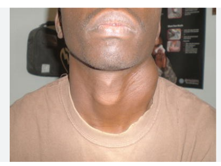
grooves; branchial fistulas occur when the second arch fails to grow over the third and fourth
arches. Such a defect results in the formation of a...
branchial cyst/fistula
126
normally pharyngeal cartilages disappear; remnants of cartilage or
bony elements under the skin anterior to the inferior 1/3 of the sternocleidomastoid persis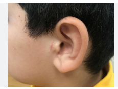
bony elements under the skin anterior to the inferior 1/3 of the sternocleidomastoid persis
branchial vestiges
127
nvolves abnormalities of the heart, parathyroid gland, face, and
thymus gland (the degree to which the immune system is affected varies). Affected
individuals will have congenital heart disease, unusual facial features with low-set ears, a
small receding mandible, wide-set eyes and are born without parathyroid glands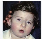
thymus gland (the degree to which the immune system is affected varies). Affected
individuals will have congenital heart disease, unusual facial features with low-set ears, a
small receding mandible, wide-set eyes and are born without parathyroid glands
DiGeorge Anomaly
128
abnormal development of 1st and 2nd pharyngeal arches
• characteristic features include microtia, mandibular hypoplasia, microstomia, skin tags
and vertebral anomalies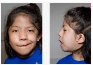
• characteristic features include microtia, mandibular hypoplasia, microstomia, skin tags
and vertebral anomalies
Goldehar syndrome
129
most of the tongue muscles are derived from ___________ these are occompanied by this n
occipital somites; hypoglossal n
130
somatic sensation from the anterior 2/3 tongue from ...
CN V
131
visceral sensation from the anterior 2/3 tongue is from...
CN VII
132
_- provides sensory inervation to the posterior 1/3 tongue
CN IX
133
__________- supplies the palatoglossus and carries taste from the root of the tongue
CN X
134
abnormally short frenulum that anchors the tongue to the floor of the oral cavity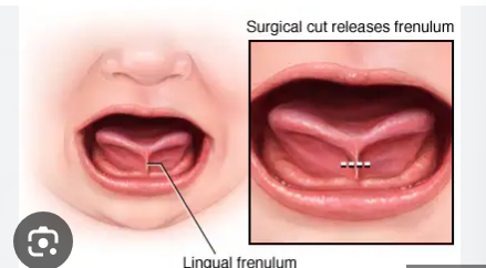
ankyloglossia
135
paired and fuses with olfactory capsule, contributes to the
ethmoid bone
ethmoid bone
prechordial cartilage
136
forms around the cranial end of the notochord and fuses with
cartilage from occipital somites→forms the base of occipital bone
cartilage from occipital somites→forms the base of occipital bone
parachordal cartilage
137
forms around the developing pituitary gland (forms the body
of the sphenoid
of the sphenoid
hypopysial cartilage
138
The development of the face begins in the 4th week around the ____________________ and relies
on organizing centers related to the forebrain
on organizing centers related to the forebrain
stomadeum
139
nasal cavity is not continuous with the pharynx, blocked by nasal
epithelium (persistent buccopharyngeal membrane?); part of the CHARGE association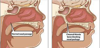
epithelium (persistent buccopharyngeal membrane?); part of the CHARGE association
choanal atresia
140
• C = coloboma of the iris, choroid
• H = heart defect
• A = atresia of choanae
• R = retarded growth
• G = genitourinary anomaly
• E = ear defect
Charge associatio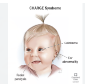
• C = coloboma of the iris, choroid
• H = heart defect
• A = atresia of choanae
• R = retarded growth
• G = genitourinary anomaly
• E = ear defect
141
type?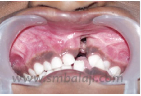
unilateral alveolar cleft
142
type?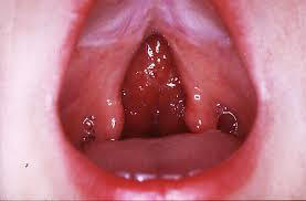
posterior cleft palate with cleft uvula
143
these migrate into the skin and give rise to melanocyres
neural crest cells
144
the dermis is derived from...
mesoderm underlying the ectoderm
145
Beginning in the 6th week there are thickenings of the oral ectoderm (dental
lamina) from which tooth buds form and grow into the underlying mesenchyme. The
permanent teeth form from the deep parts of the tooth buds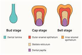
lamina) from which tooth buds form and grow into the underlying mesenchyme. The
permanent teeth form from the deep parts of the tooth buds
bud stage
146
Mesenchyme grows into each bud and gives it a cap shaped appearance. The
ectodermal portion is called the enamel organ. There are two layers to the enamel organ:
and inner and an outer layer. They are continuous at the epithelial root sheath. The deepest
part of the enamel organ invaginates and in this space the mesenchyme forms the dental
papilla that will give rise to the dental pulp. This entire structure is surrounded by a layer of
mesenchyme (dental sac). The dental sac will form the periodontal ligament and cementum
(holds the tooth in the socket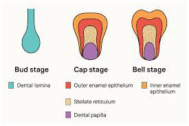
ectodermal portion is called the enamel organ. There are two layers to the enamel organ:
and inner and an outer layer. They are continuous at the epithelial root sheath. The deepest
part of the enamel organ invaginates and in this space the mesenchyme forms the dental
papilla that will give rise to the dental pulp. This entire structure is surrounded by a layer of
mesenchyme (dental sac). The dental sac will form the periodontal ligament and cementum
(holds the tooth in the socket
cap stage
147
The developing tooth becomes bell-shaped. The mesenchyme of the dental
papilla forms odontoblasts (these cells produce predentin → dentin). The cells of the inner
enamel layer form ameloblasts that produce enamel. The root sheath regresses and become
associated with the root canal where it continues to form dentin. As a result the root canal is
reduced to a narrow cavity and the tooth has an outer, superficial covering of enamel and a
deeper layer of dentin. The enamel only covers the apex of the tooth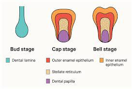
papilla forms odontoblasts (these cells produce predentin → dentin). The cells of the inner
enamel layer form ameloblasts that produce enamel. The root sheath regresses and become
associated with the root canal where it continues to form dentin. As a result the root canal is
reduced to a narrow cavity and the tooth has an outer, superficial covering of enamel and a
deeper layer of dentin. The enamel only covers the apex of the tooth
bell stage
148
This disorder results from excessive keratinization of the skin, whereby the skin
appears as very dry and scaly. A harlequin fetus has an extreme form of this disorder where
the skin is abnormally thick and cracked; they typically die within a week of birth. A collodion
infant is covered in a thick membrane that cracks and eventually is shed revealing normal
skin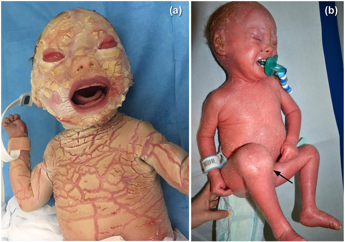
appears as very dry and scaly. A harlequin fetus has an extreme form of this disorder where
the skin is abnormally thick and cracked; they typically die within a week of birth. A collodion
infant is covered in a thick membrane that cracks and eventually is shed revealing normal
skin
ichthyosis
149
These anomalies are not true tumors but may appear as a mass within the skin.
They result from the persistence or surplus of blood or lymphatic vessels. Nevus flammeus
refers to a flat pink or red blotch that appears on the posterior aspect of the neck. A
hemangioma (port wine stain) also appears red but is found on the face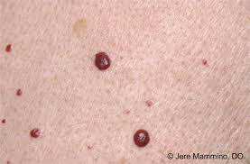
They result from the persistence or surplus of blood or lymphatic vessels. Nevus flammeus
refers to a flat pink or red blotch that appears on the posterior aspect of the neck. A
hemangioma (port wine stain) also appears red but is found on the face
angiomas
150
excessive hairiness; localized hypertrichosis is associated with spinal
bifida occulta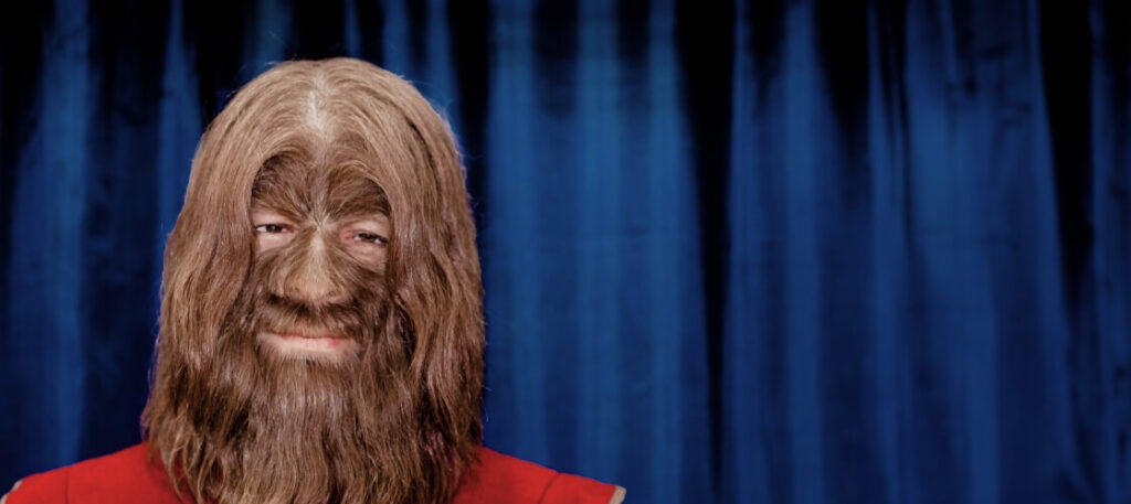
bifida occulta
hypertrichosis
151
In this condition the enamel is improperly formed and is soft
and friable and the teeth are yellow or brown because of exposed dentin. Dentinogenesis
imperfecta is a common condition where the odontoblasts fail to differentiate and dentin is
not calcified.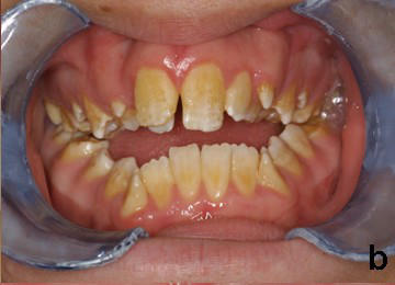
and friable and the teeth are yellow or brown because of exposed dentin. Dentinogenesis
imperfecta is a common condition where the odontoblasts fail to differentiate and dentin is
not calcified.
amelogenesis imperfecta
152
abnormal development of the parts of the inner or middle ear
congenital deafness
153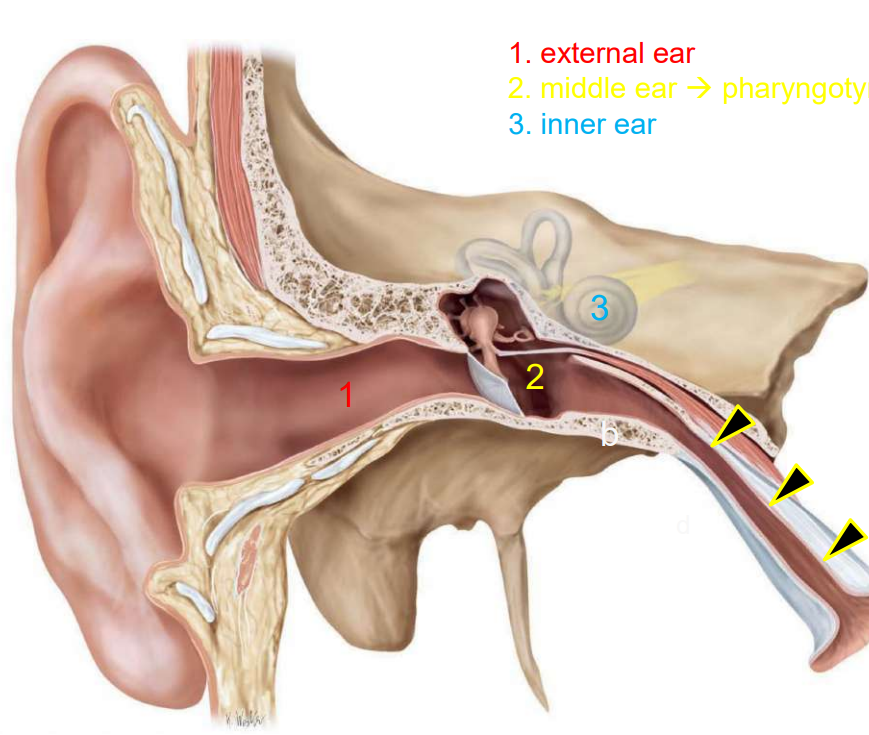
Image occlusion: name the hidden labels
154
Image occlusion: name the hidden labels
155
Image occlusion: name the hidden labels
156
this gives rise to the membranous labarynth of the inner ear
otic placode
157
cells from the otic placode and neural crest form the
vestibulocochlear ganglion
vestibulocochlear ganglion
statoacostic ganglion
158
the external and middle ear are formed by...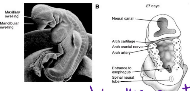
pharyngeal arch: external ear
1sy pharngeal pouch: eustacian tube and lines ear cavity
1sy pharngeal pouch: eustacian tube and lines ear cavity
159
the tympanic membrane is derived from...
endoderm (inside), ectoderm (outside), mesoderm
160
bumps for cartilage and connective tissue; form certain parts of the ear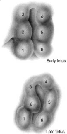
auricular hillpcks
161
external ear defects are frequently occuring with _____________
chromosomal syndromes`
162
most children with _______ have some degree of auditory dysfunction
autidm spectrum disorder
163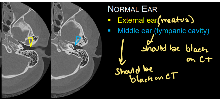
Image occlusion: name the hidden labels
164
Image occlusion: name the hidden labels
165
a congenital condition where one or both ears are abnormally small or missing, sometimes with an absent ear canal (aural atresia)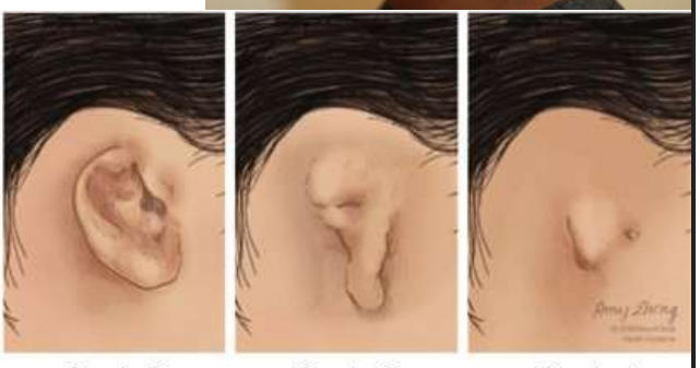
microgria
166
– incudostapedial deformity
– stapes fixation, hypoplasia or aplasia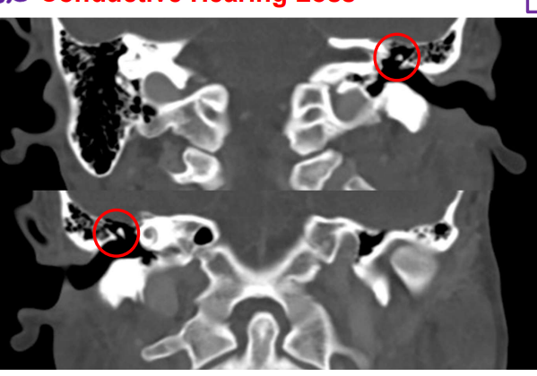
– stapes fixation, hypoplasia or aplasia
klippel feil anomaly
167
retina, posterior layers of iris and optic nerve tissue derivitive
neuroectoderm
168
lens and corneal epithelium tissue derivitive
surface ectoderm
169
fibrous and vascular coats tissue derivitive
mesoderm
170
choroid, sclera and corneal endothelium tissue derivitive
neural crest
171
the ends of the optic cup will form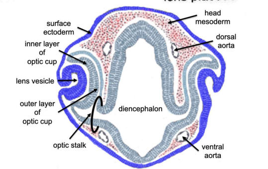
retina
172
the outer layer of the optic cup becomes? and the inner layer becomes?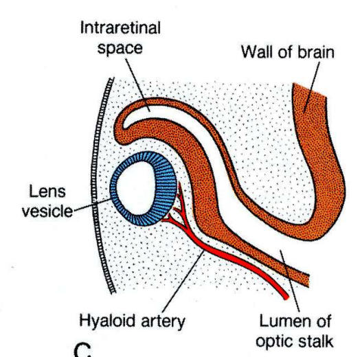
pigmented layer; neural retina
173
this space of the eye is associated with retinal detachment
intraretinal space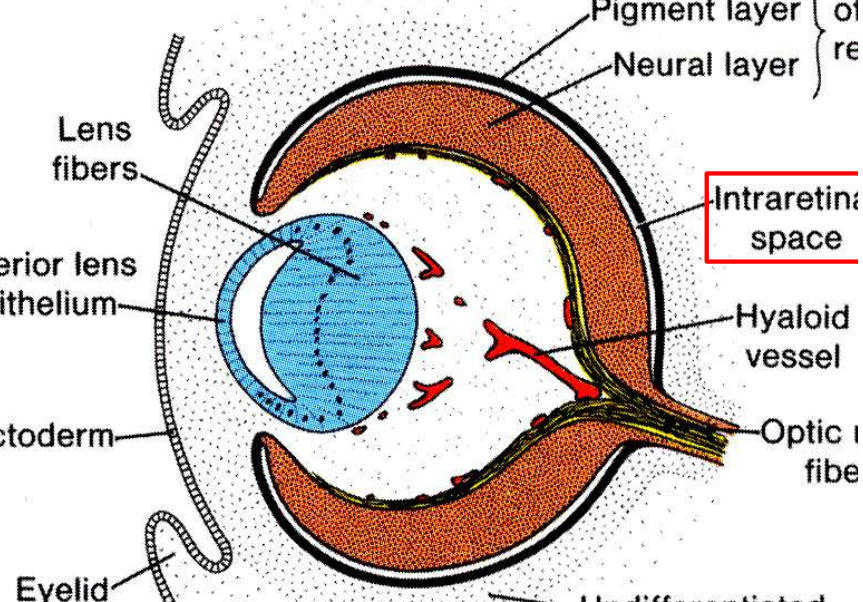
174
the cillary bpody is derived from the _ and ______
choroid mesenchyme and optic cup
175
the iris is formed at _____ and the gap becomes...
edge if oiptic cup; pupil
176
this supplies the developing lens and normally atrophies andthe lebs becomes avascular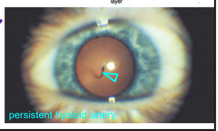
hyaloid artery
177
the cornea is derived from (x3)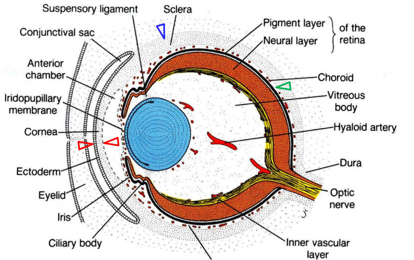
1. Surface ectoderm
2. Mesoderm – CT
3. Neural crest – corneal endothelium
2. Mesoderm – CT
3. Neural crest – corneal endothelium
178
misalignment of the eyes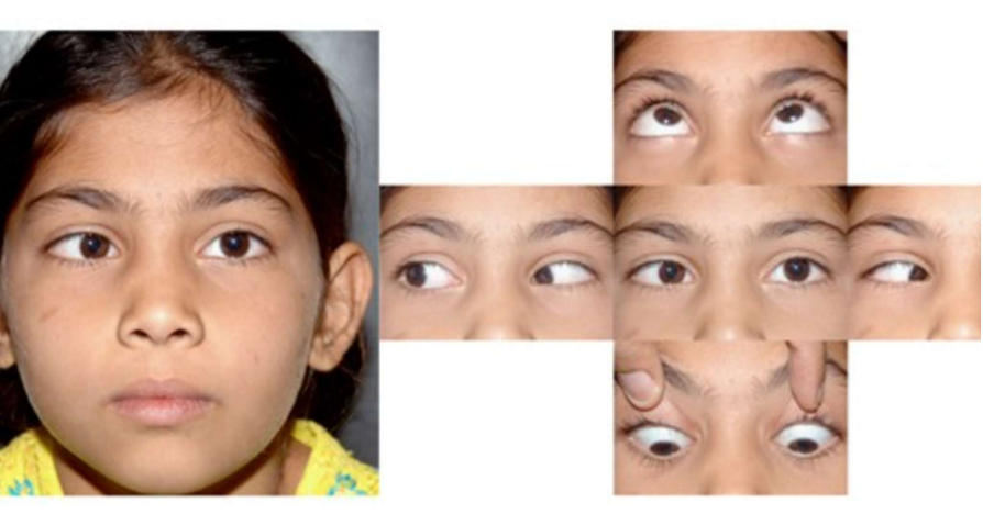
strabismus
179
what causes strabismus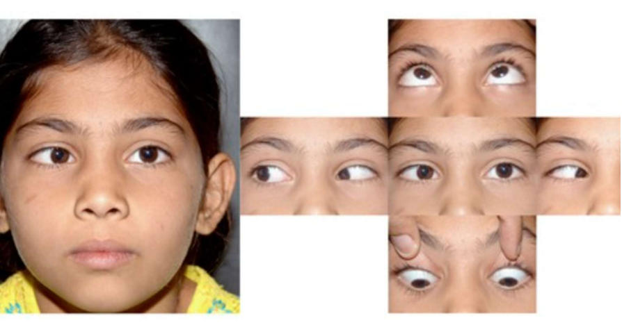
improper development or innervation of extraocular muscles
180
a single eye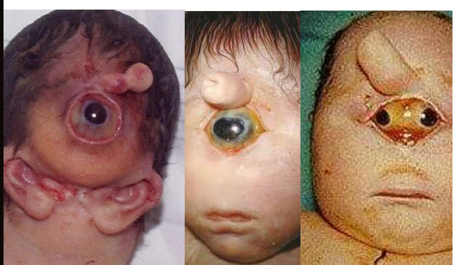
cyclopia
181
synophthalmia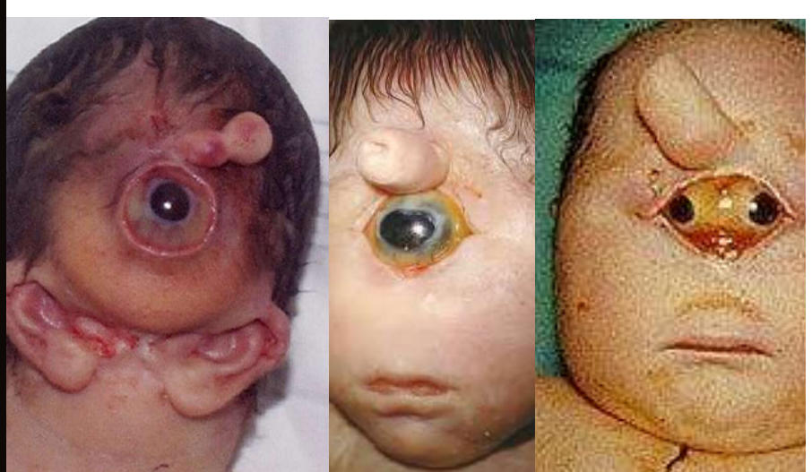
incomplete fusion of the eyes
182
teardrop shaped pupils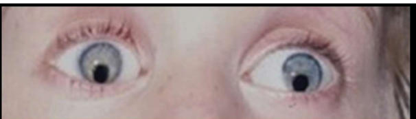
coloboma
183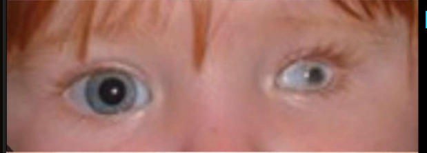
micropthalmia
184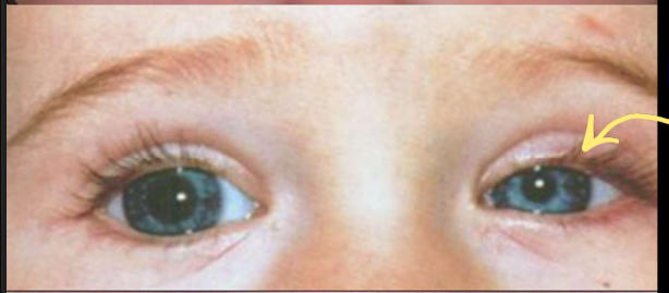
congenital horner syndrome
185
no iris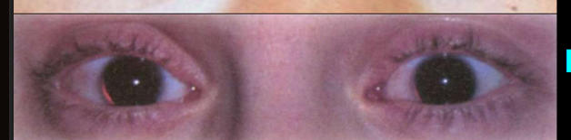
aniridiata
186
no eyes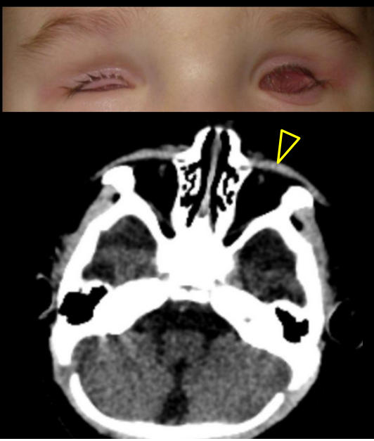
anophthalmia
187
what causes congenital cataracts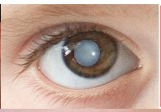
ToRCH infections:
• Toxoplasmosis• other (syphilis, varicella, etc) • Rubella
• Cytomegalovirus• Herpes
• Toxoplasmosis• other (syphilis, varicella, etc) • Rubella
• Cytomegalovirus• Herpes
188
The most common primary intraocular
malignancy in children (1:100,000)
• Derived from neural progenitors
(photoreceptor lineage) with mutation
in RB
• “white reflex”, poor eyesight,
strabismus, pain, glaucoma, retinal
mass on ophthalmic exam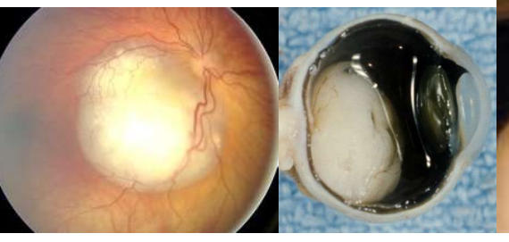
malignancy in children (1:100,000)
• Derived from neural progenitors
(photoreceptor lineage) with mutation
in RB
• “white reflex”, poor eyesight,
strabismus, pain, glaucoma, retinal
mass on ophthalmic exam
retinoblastoma
189
Indicated by an increase in intraocular pressure caused by a defect or blockage of the draining system (scleral venous sinus)
Causes: genetic mutations (NF1), rubella infection, other eye defects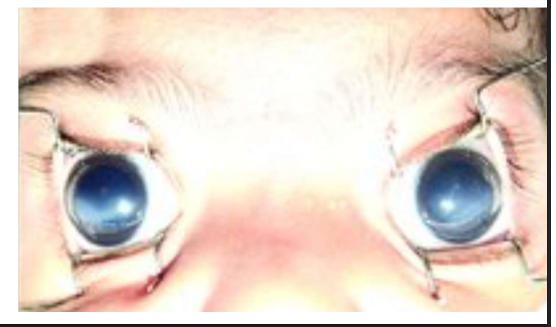
Causes: genetic mutations (NF1), rubella infection, other eye defects
congenital glaucoma
190
c;left lip what failed to fuse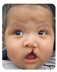
Maxillary – medial nasal
191
median cleft lip what failed tp fuse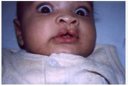
Medial nasal – medial nasal
192
olique facial cleft what failed tp fuse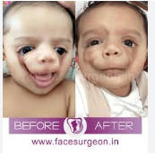
Lateral nasal – maxillary
193
cleft palat ewhat failed to fuse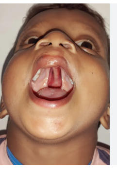
Palatine shelves (secondary palate)
194
macrostomia, what failed to fuse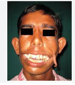
Maxillary – mandibular (too little fusion)