All cards
1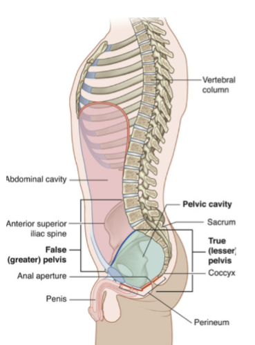
Image occlusion: name the hidden labels
2
Image occlusion: name the hidden labels
3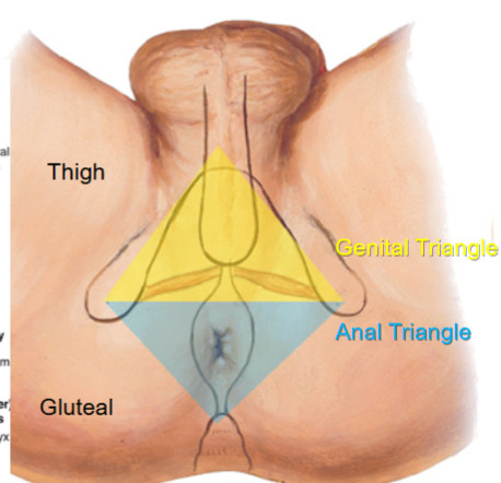
Image occlusion: name the hidden labels
4
Image occlusion: name the hidden labels
5
what are the borders of the genital triangle
pubic symphysis to ischial tuberocity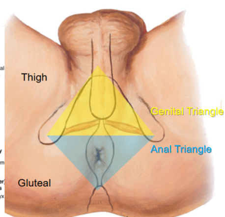
6
what are the borders of the anal triagle
iscial tuberocity to coccyx tip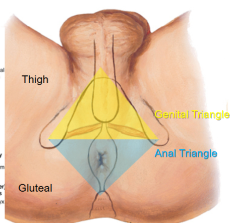
7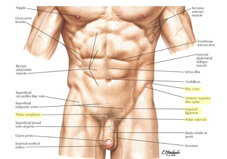
Image occlusion: name the hidden labels
8
Image occlusion: name the hidden labels
9
Image occlusion: name the hidden labels
10
Image occlusion: name the hidden labels
11
Image occlusion: name the hidden labels
12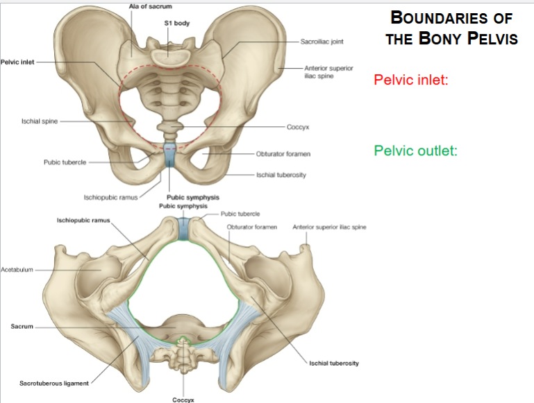
Image occlusion: name the hidden labels
13
Image occlusion: name the hidden labels
14
Differences between the male and female pelvis (x2)
greater subpubic angle
increased width of pelvic inlet and outlet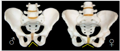
increased width of pelvic inlet and outlet
15
from the bottom of the pubic symphysis to the sacral promontoru; the only diameter that can be measured per vaginam
diagonal conjugate (blue line)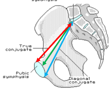
16
at inlet and outlet measuring across width
-outlet between ischial tuberocities
-outlet between ischial tuberocities
transvcerse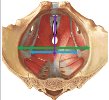
17
these ligaments prevent forward tilt of the sacrum; creates stable base for walking
sacrospinous and sacrotuberous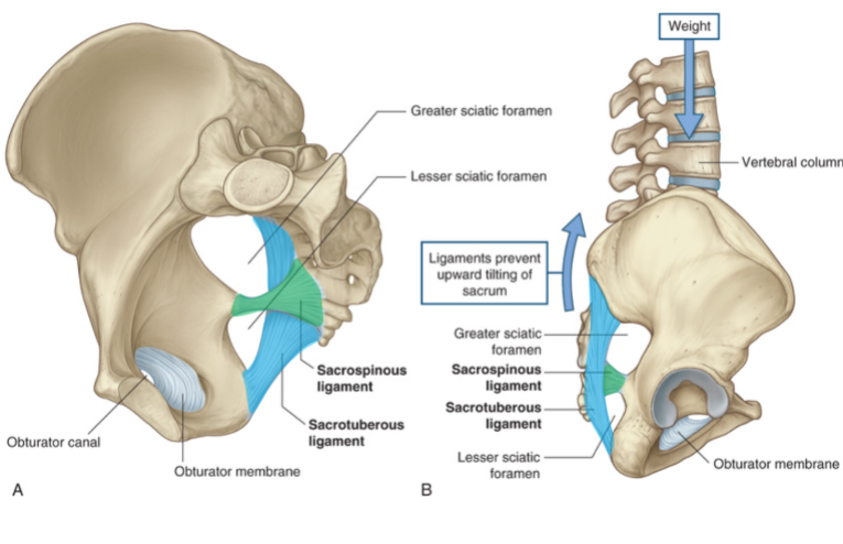
18
what passes through the greater sciatic foramen
Pudendal nerve, internal
pudendal artery and vein,
superior gluteal artery and vein,
inferior gluteal artery and vein,
piriformis m.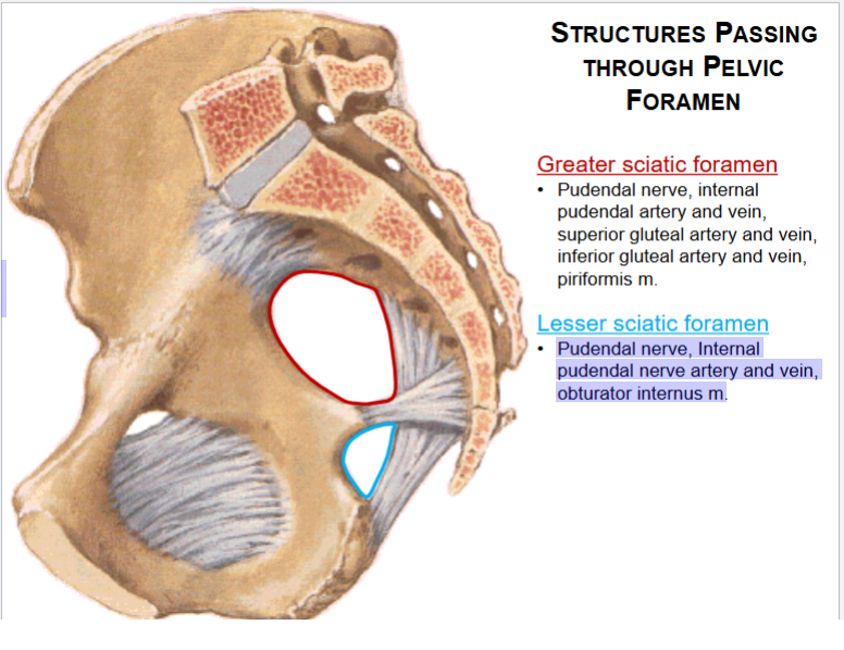
pudendal artery and vein,
superior gluteal artery and vein,
inferior gluteal artery and vein,
piriformis m.
19
what passes through the lesser sciatic foramen
Pudendal nerve, Internal
pudendal nerve artery and vein,
obturator internus m.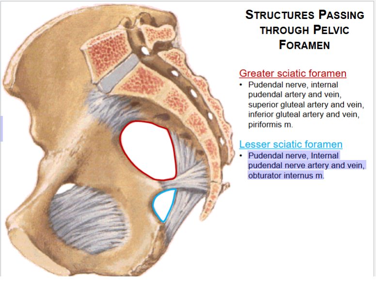
pudendal nerve artery and vein,
obturator internus m.
20
borders of the true pelvis
anterior: pubic symphysis
lateral: piriformis and obtyrator internus
posterior: sacrum
floor: levator anti m. and coccygeous m.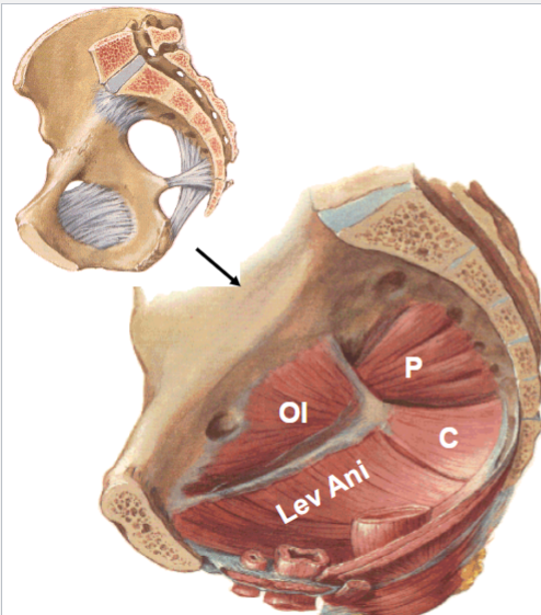
lateral: piriformis and obtyrator internus
posterior: sacrum
floor: levator anti m. and coccygeous m.
21
fx of levator anti m
lifts anus and holds stuff in place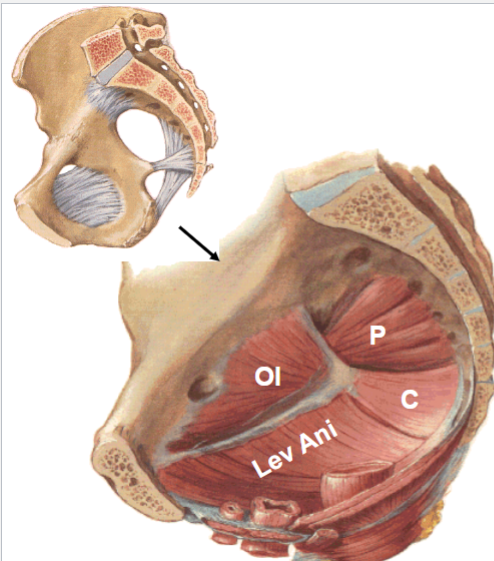
22
fx coccygeous m
assist levator anti m.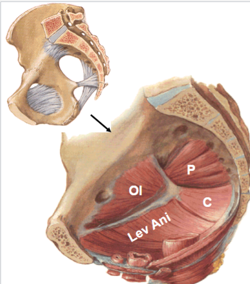
23
what makes up the pelvic diaphragm?
levator anti m and coccygeous m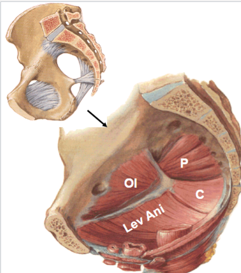
24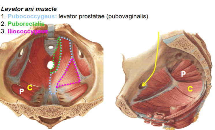
Image occlusion: name the hidden labels
25
Image occlusion: name the hidden labels
26
Image occlusion: name the hidden labels
27
O and I levator anti m
O: body of pubis, arch of obtyrator fascia, ischial spine
I: perineal body, coccyx and anococcygeal ligaments, walls of prostate (vagina) rectum and anal canal
I: perineal body, coccyx and anococcygeal ligaments, walls of prostate (vagina) rectum and anal canal
28
Innervation levator ani m
nerve to levator ani (S4)
29
muscle that maintains the anorectal angle and is imp for defication
puborectalis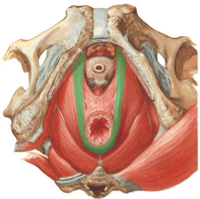
30
O and I coccygeus m
Proximal attachment: ischial spine
Distal attachment: coccyx
Distal attachment: coccyx
31
innervation coccygeus m
sacral plexyus (s4 and 5)
32
what can weaknesses of the pelvic floor cause?
• Prolapse: cystocele, rectocele, uterine prolapse, enterocele
• Stress incontinence
• Stress incontinence
33
a condition where the bladder prolapses into the vagina. This occurs when the muscles and tissues that support the bladder weaken, causing it to bulge downward. 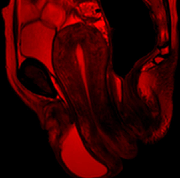
cystocele
34
a condition where the rectum (the lower part of the large intestine) bulges into the vagina, creating a pouch-like structure. 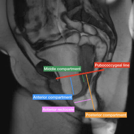
rectocele
35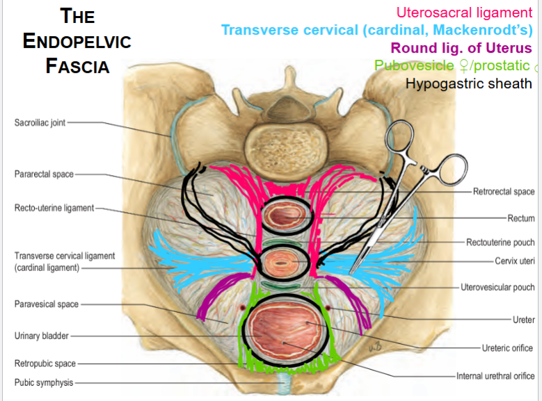
Image occlusion: name the hidden labels
36
Image occlusion: name the hidden labels
37
Image occlusion: name the hidden labels
38
Image occlusion: name the hidden labels
39
Image occlusion: name the hidden labels
40
what is to the R and L of the internal iliac a
• Right: terminal loops of the ilium
• Left: sigmoid colon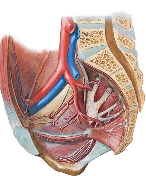
• Left: sigmoid colon
41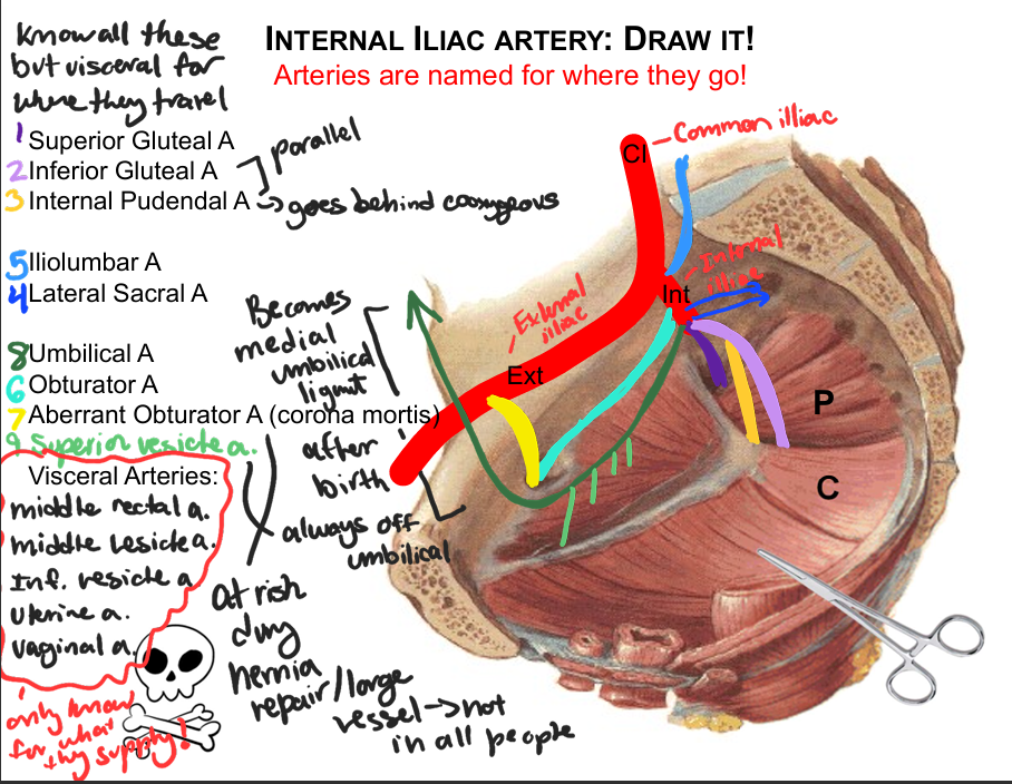
Image occlusion: name the hidden labels
42
Image occlusion: name the hidden labels
43
Image occlusion: name the hidden labels
44
Image occlusion: name the hidden labels
45
Image occlusion: name the hidden labels
46
Image occlusion: name the hidden labels
47
Image occlusion: name the hidden labels
48
Image occlusion: name the hidden labels
49
Image occlusion: name the hidden labels
50
what are the visceral arteries of the internal iliac a
middle rectal a
middle vesical a
inferior vesicle a
uterine a
vaginal a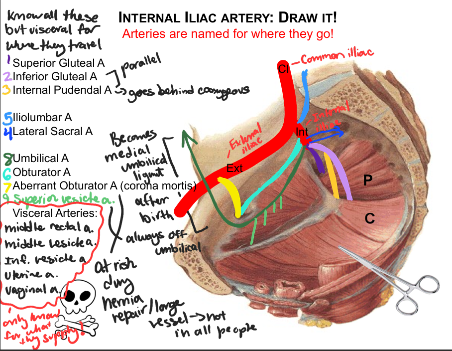
middle vesical a
inferior vesicle a
uterine a
vaginal a
51
what arteries supply the anal canal?
above pectinate line from superior and middle rectal arteries,
below the pectinate line middle and inferior rectal artery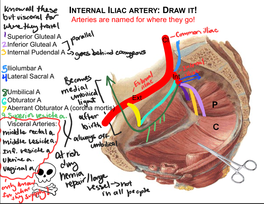
below the pectinate line middle and inferior rectal artery
52
what arteries supply the ureters
: renal, testicular/ovarian, common iliac, branches from the internal iliac,
inferior vesical and uterine arteries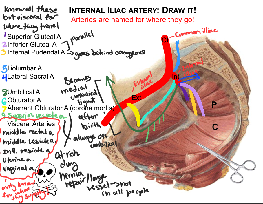
inferior vesical and uterine arteries
53
what arteries supply the bladder
superior (1-4), middle and inferior vesical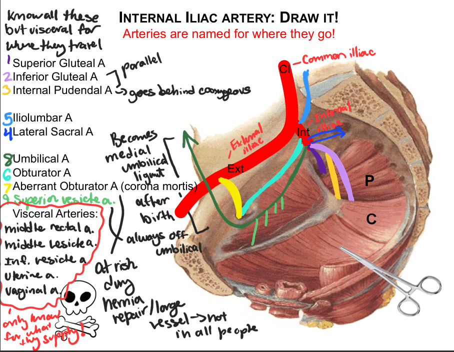
54
what arteries supply the ductus deferens
artery of the ductus deferens (from superior vesical artery)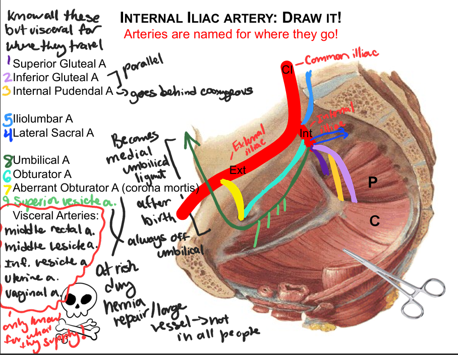
55
what arteries supply the esminal vesical
middle and inferior vesical, middle rectal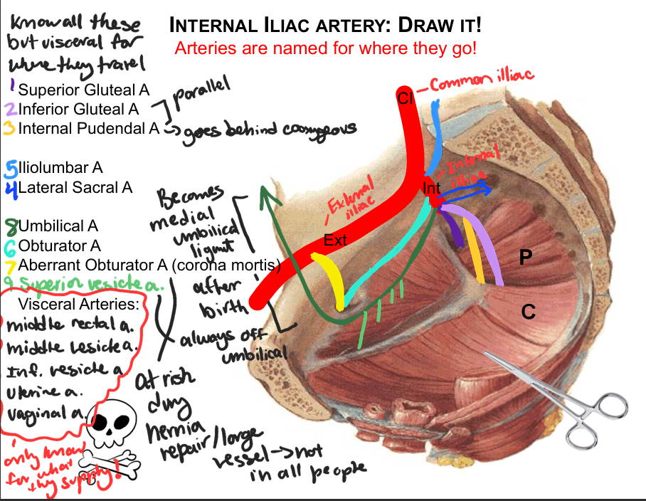
56
what arteries supply the prostate
inferior vesical, middle rectal, internal pudendal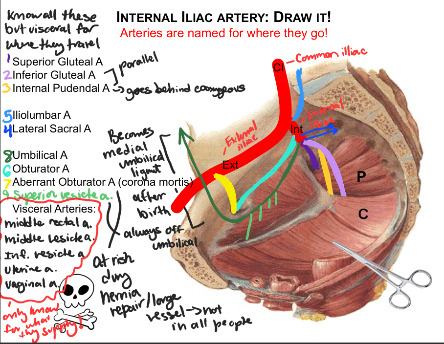
57
what arteries supply the urethra (membranous and spongy)
: internal pudendal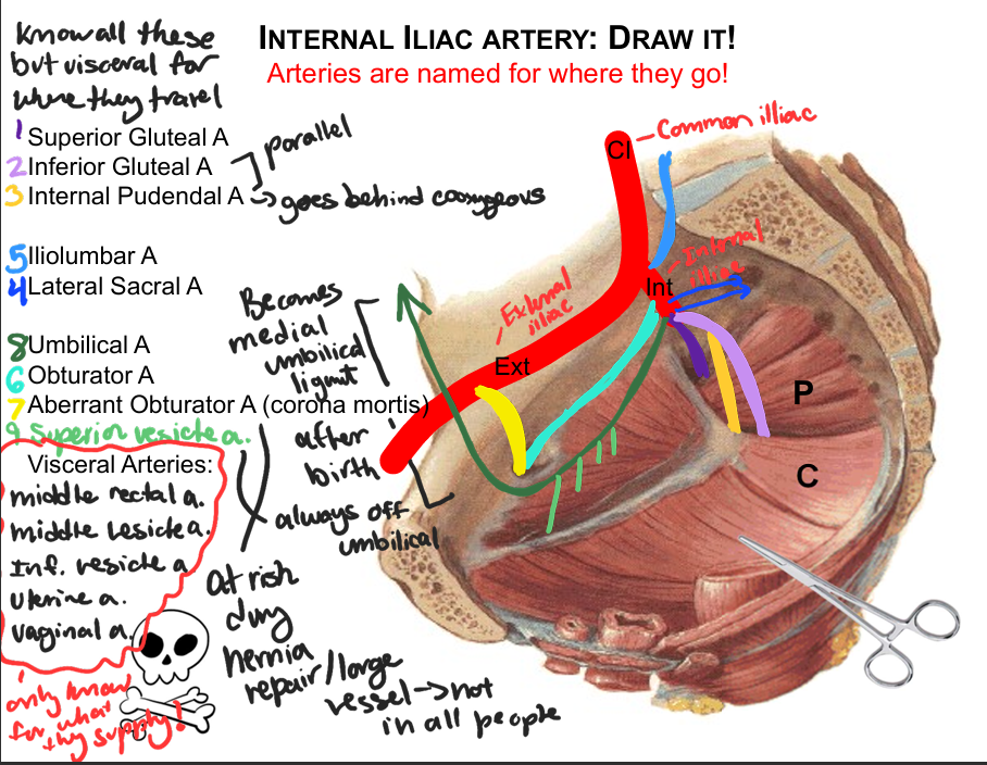
58
what arteries supply the penis
internal pudendal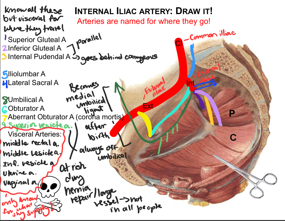
59
what arteries supply the ovary
ovarian (aorta)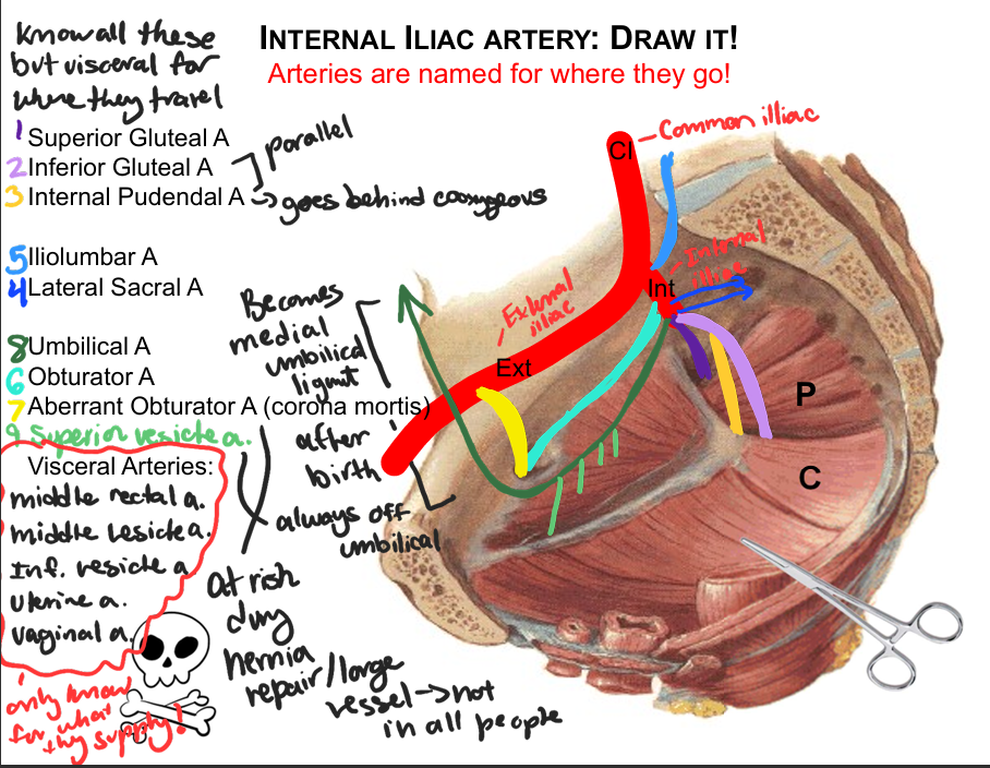
60
what arteries supply the uterune tube
ovarian and uterine 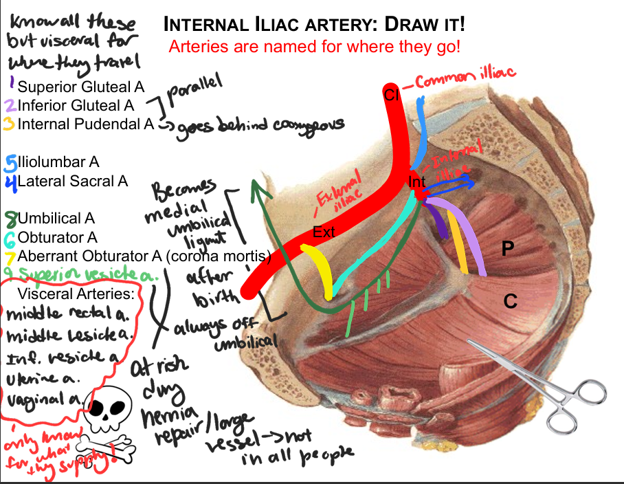
61
what arteries supply the uterus
: uterine and ovarian vessels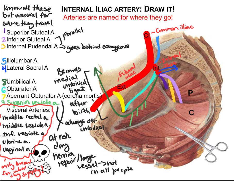
62
what arteries supply the vagina
uterine, vaginal artery & internal pudendal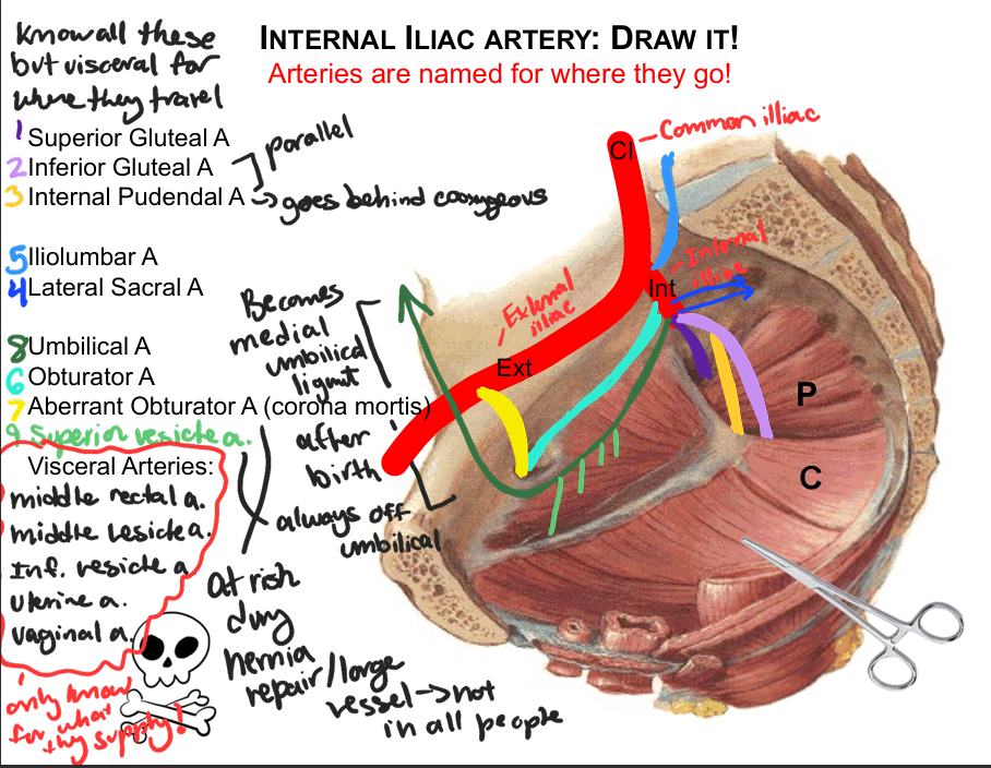
63
qhat arteries supply the urethra (female)
inferior vesical and internal pudendal 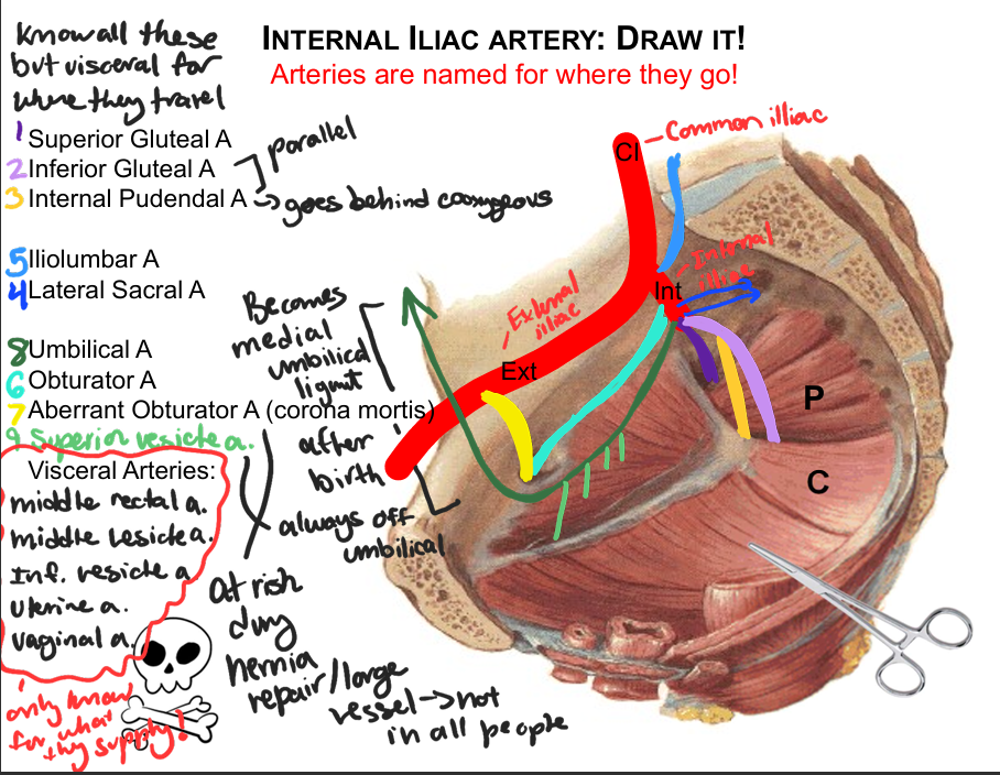
64
what arteries supply the external genitalia female
internal pudendal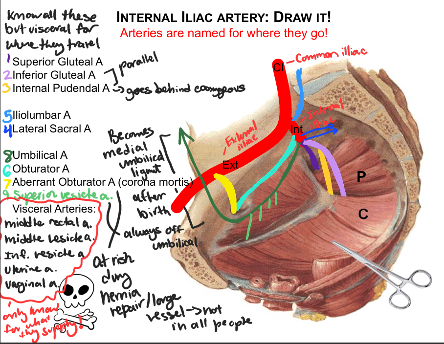
65
what arteries anastomoses rectum and anal canal?
• Inferior mesenteric (aorta)
• Middle rectal (internal iliac)
• Inferior rectal (internal iliac)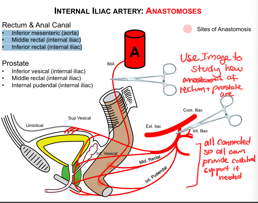
• Middle rectal (internal iliac)
• Inferior rectal (internal iliac)
66
what arteries anastomoses in the prostate
• Inferior vesical (internal iliac)
• Middle rectal (internal iliac)
• Internal pudendal (internal iliac)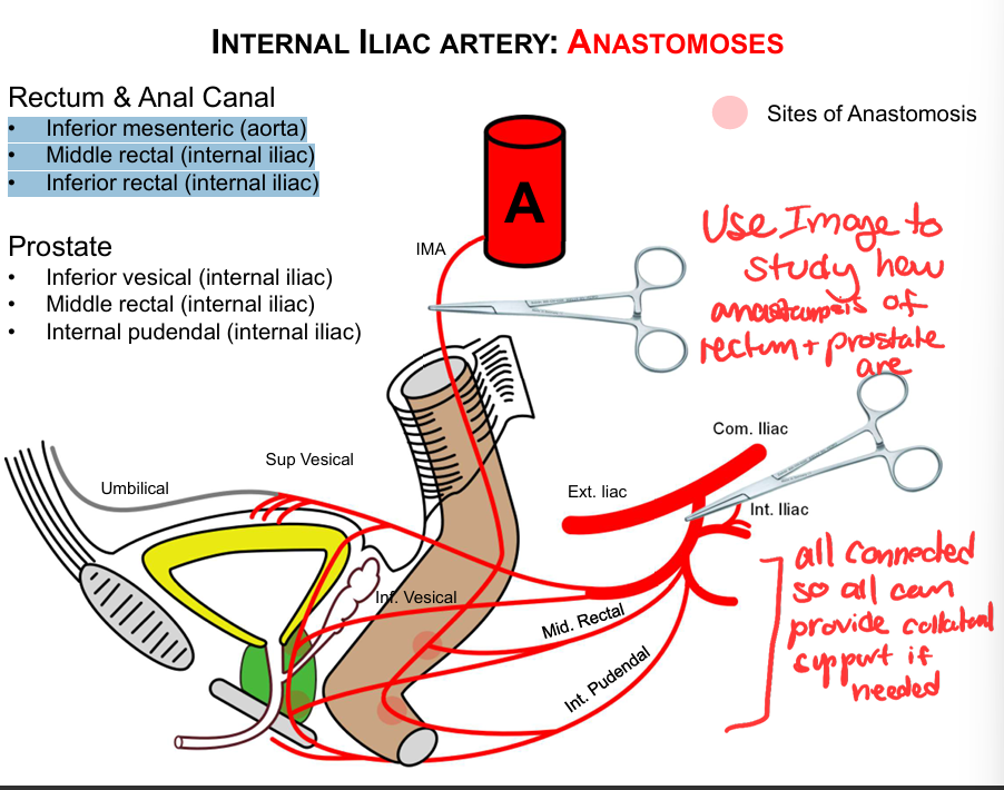
• Middle rectal (internal iliac)
• Internal pudendal (internal iliac)
67
what are the anastomoses of the female reproductive tract
-Ovarian (aorta)
• Uterine (internal iliac)
• Vaginal (internal iliac)
• Internal pudendal (internal iliac)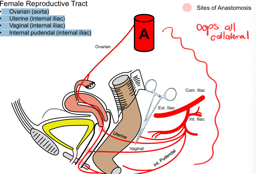
• Uterine (internal iliac)
• Vaginal (internal iliac)
• Internal pudendal (internal iliac)
68
what runs inferior to the uterine a?
ureter; in danger if uterine a clamped
69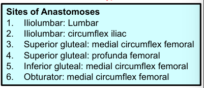
Sites of anastomoes of the internal iliac a (aboveelow)
70
what runs through the pudental canal?
perineal and dorsal n
internal pudental a and v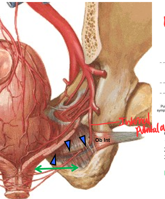
internal pudental a and v
71
what do the superficial dorsal veins drain?
drain the skin and prepuce of the penis/clitoris 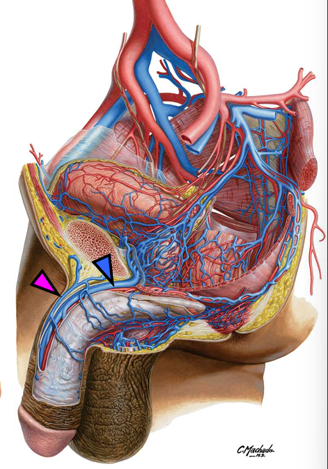
72
what do the deep dorsal veins drain?
drains the glans and corpora cavernosa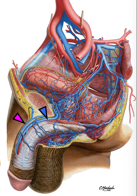
73
what forms the sacral plexus
the lumbosacral trunk and by S1, S2 and S3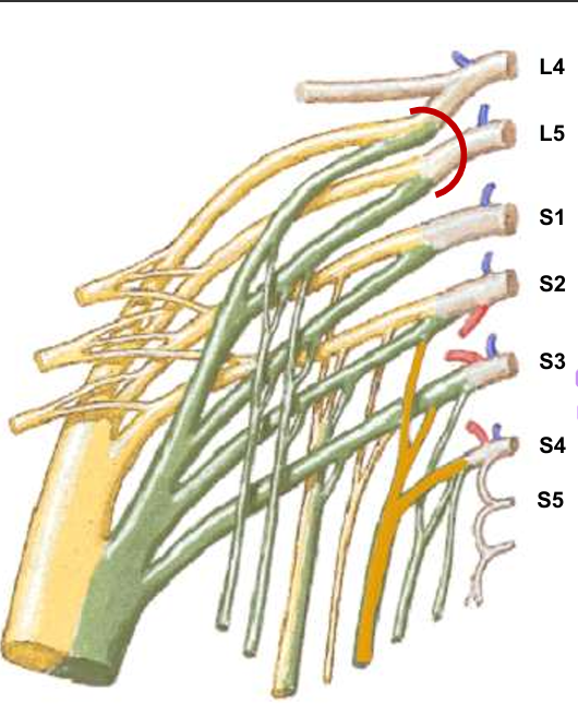
74
postganglionic
sympathetic GVE + GVA
sympathetic GVE + GVA
gray rami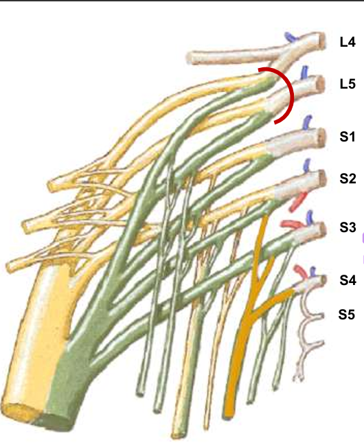
75
preganglionic parasympathetic
GVE + GVA (S2-4)
GVE + GVA (S2-4)
pelvic splanchnic nerves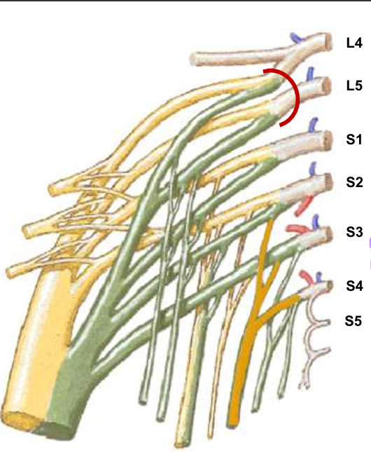
76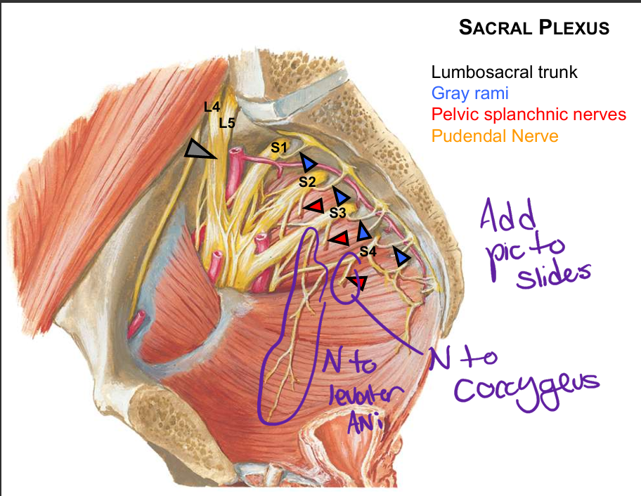
Image occlusion: name the hidden labels
77
Image occlusion: name the hidden labels
78
Image occlusion: name the hidden labels
79
Image occlusion: name the hidden labels
80
what does the inferior rectal n supply
external anal sphincter
and overlying skin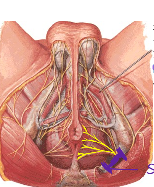
and overlying skin
81
symptoms of inferior rectal n damage
incontenencxe, saddle anestesia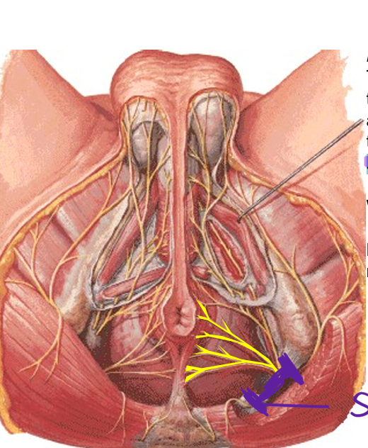
82
what does the deep branch of perineal n innervate?
corpus spongiosum
and mucus membrane of the
urethrae.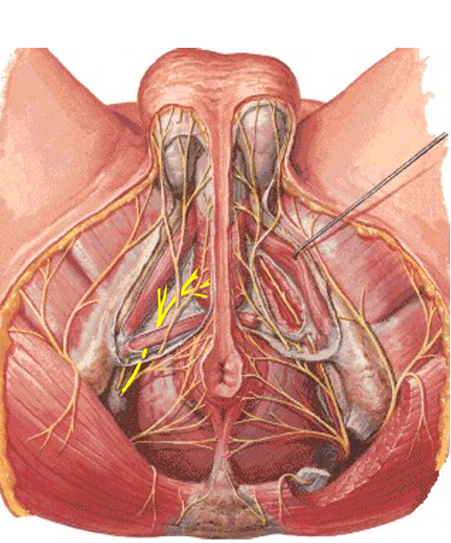
and mucus membrane of the
urethrae.
83
what does the superficial branch of perineal n innervate?
distributes to skin of the
scrotum (labia majora) as
posterior scrotal/labial
nerve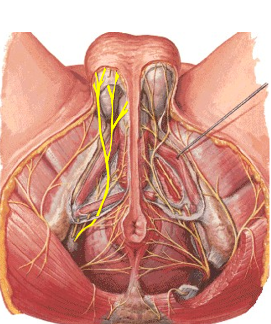
scrotum (labia majora) as
posterior scrotal/labial
nerve
84
what does the dorsal nerve of the penis (clitoris) innervate
gives off a branch to the corpus
cavernosum
-breaks up into small filaments that
distribute to the glans
• supplies no skeletal muscle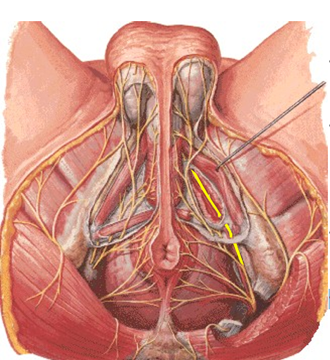
cavernosum
-breaks up into small filaments that
distribute to the glans
• supplies no skeletal muscle
85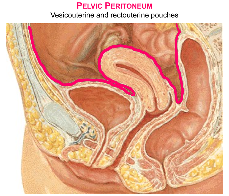
Image occlusion: name the hidden labels
86
Image occlusion: name the hidden labels
87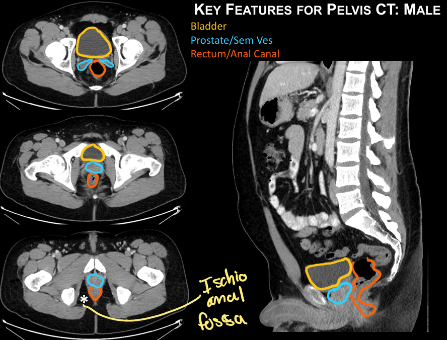
Image occlusion: name the hidden labels
88
Image occlusion: name the hidden labels
89
Image occlusion: name the hidden labels
90
Image occlusion: name the hidden labels
91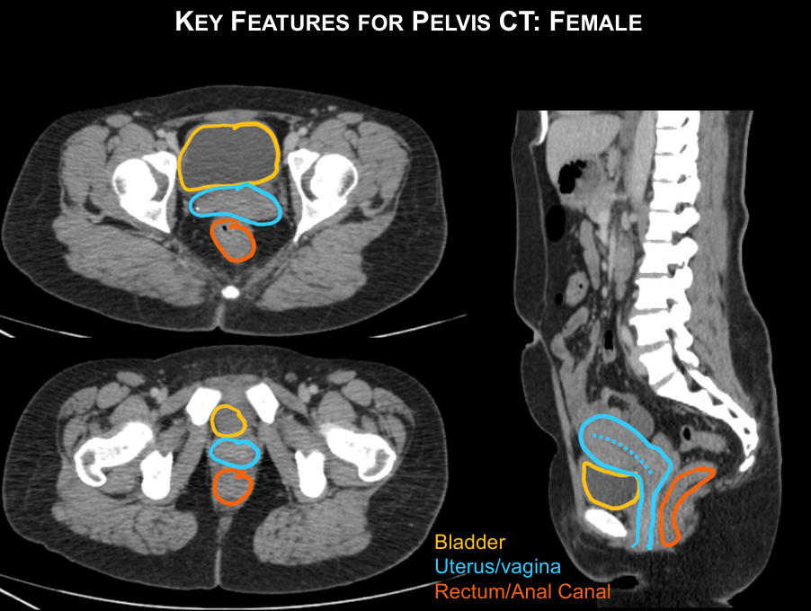
Image occlusion: name the hidden labels
92
Image occlusion: name the hidden labels
93
Image occlusion: name the hidden labels
94
how do you surgically aproach the bladder?
above pubic symphysis through anterior wall
95
what is a pelvic fracture anle to damage (thinking urinary system)
bladder b/cause it is in direct contact with apex of bladder
96
what holds the bladder neck; posteriorly? to umbilicus?
puboprostatic/pubovesical; rectovesical ligaments; median umbilical ligament
97
smooth muscle coat of the bladder that expands/stretches as the bladder fills
and contracts by parasympathetic
stimulation.
and contracts by parasympathetic
stimulation.
detursor m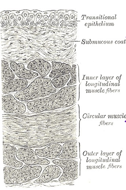
98
how does camcer from the bladder spread (x3)
local spread: peritoneum, pelvic organs
lymph: ext & int iliac nodes
Veins (caval): lungs, vertebra…liver
lymph: ext & int iliac nodes
Veins (caval): lungs, vertebra…liver
99
-herniation of the bladder along the
anterior wall of the vagina
occurs with injury or weakness of
pelvic and urogenital diaphragm or
vaginal support (transverse cervical
ligament, uterosacral ligament
anterior wall of the vagina
occurs with injury or weakness of
pelvic and urogenital diaphragm or
vaginal support (transverse cervical
ligament, uterosacral ligament
cystocele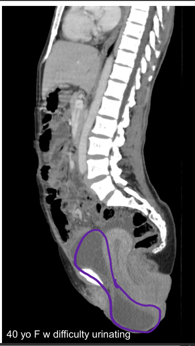
100
4 parts of male urethra
1. Preprostatic
2. Prostatic – widest and most dilatable; receives the ejaculatory and prostatic
ducts
3. Membranous – surrounded by the sphincter urethra (external urethral
sphincter)
4. Spongy – within the corpus spongiosum, receives openings of the
bulbourethral glands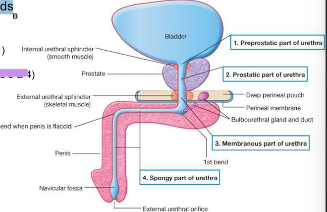
2. Prostatic – widest and most dilatable; receives the ejaculatory and prostatic
ducts
3. Membranous – surrounded by the sphincter urethra (external urethral
sphincter)
4. Spongy – within the corpus spongiosum, receives openings of the
bulbourethral glands
101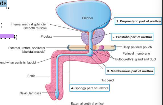
Image occlusion: name the hidden labels
102
Image occlusion: name the hidden labels
103
Image occlusion: name the hidden labels
104
Image occlusion: name the hidden labels
105
arterial supply to the male yrethra
• inferior vesical (~part 1)
• middle rectal (~part 2)
• internal pudendal (parts 3 & 4)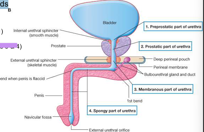
• middle rectal (~part 2)
• internal pudendal (parts 3 & 4)
106
what happens when the ductus deferens enters the posterior aspect of the bladder?
loses its peritoneal covering
forms an ampulla before joining the duct of the seminal
vesicle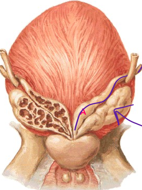
forms an ampulla before joining the duct of the seminal
vesicle
107
the seminal vesicle and ductus deferens join to form...
ejaculatory duct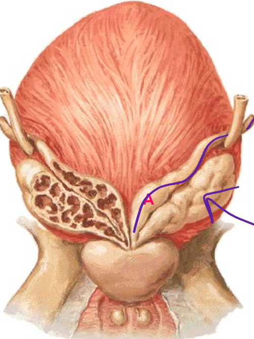
108
arterial support fdor the seminal vesicles comes from (x2)
• inferior vesical artery
• middle rectal artery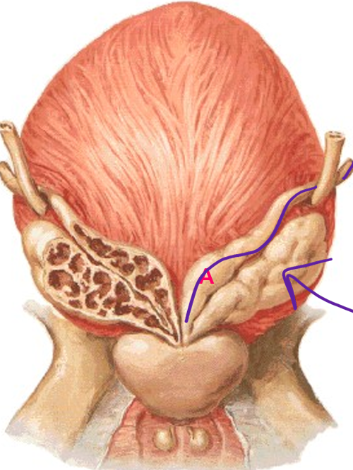
• middle rectal artery
109
when do the ejaculatory ducts terminate?
opening of seminal colliculus (blue)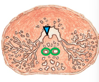
110
what makes up the central zone of the prostate
periurethral mucosal glands; ductless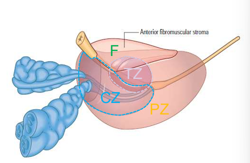
111
what makes up the transitional zone of prostate
submucosal with short ducts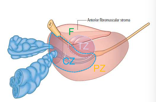
112
what makes up the periphera;l zone of prostate
oter main prostate glands have long ducts 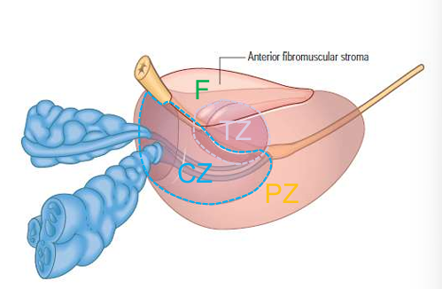
113
• proliferation of stroma & glands in the inner periurethral zone (CZ)
• s/s = urinary hesitance, urgency, nocturia, poor urinary stream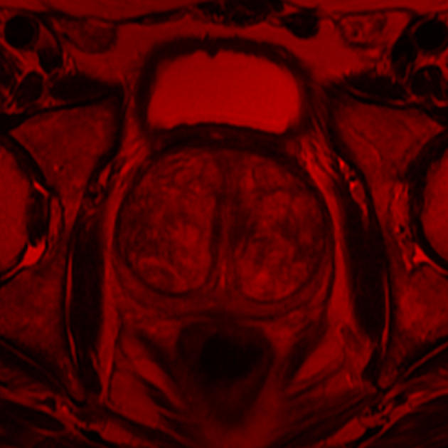
• s/s = urinary hesitance, urgency, nocturia, poor urinary stream
benign prostatic hyperstrophy
114
where do most adrenocarcinomas of the prostate occur?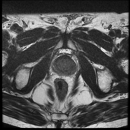
peripheral zone
115
The sheath of the prostate is derived from the ___________ and is continuous with the
puboprostatic ligaments; _______________ is imbedded within the sheath
puboprostatic ligaments; _______________ is imbedded within the sheath
pelvic fasci; prostatic veins and the prostatic nerve plexus
116
prostate cancer can spread by (x3)
local growth: pelvic organs,
peritoneum
lymph: int iliac >> sacral
veins:
• Batson’s: vertebra & brain, lungs
• Caval: bones, liver, adrenal
gland
peritoneum
lymph: int iliac >> sacral
veins:
• Batson’s: vertebra & brain, lungs
• Caval: bones, liver, adrenal
gland
117
Removal/ablation of the prostate
endangers the _____________
which carries both ______________] and
____________to the penis
endangers the _____________
which carries both ______________] and
____________to the penis
prostatic nerve plexus;
sympathetic [via
hypogastric nerves] and
parasympathetic axons [via pelvic
splanchnic nerves]
sympathetic [via
hypogastric nerves] and
parasympathetic axons [via pelvic
splanchnic nerves]
118
arterial supply to the prostate
inferior
vesical a, internal pudendal a, middle
rectal a
vesical a, internal pudendal a, middle
rectal a
119
– embryological
remnant of the paramesonephric
ducts
remnant of the paramesonephric
ducts
prostatic utricle
120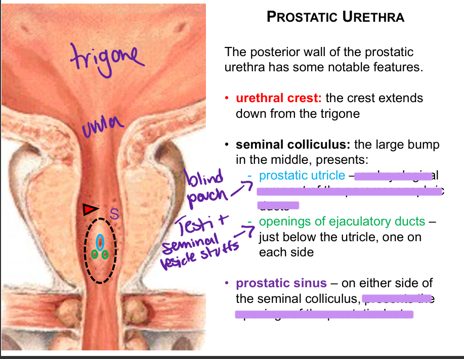
Image occlusion: name the hidden labels
121
Image occlusion: name the hidden labels
122
Image occlusion: name the hidden labels
123
Image occlusion: name the hidden labels
124
Image occlusion: name the hidden labels
125
Image occlusion: name the hidden labels
126
Image occlusion: name the hidden labels
127
where are the bulbourethral glands located
within the deep peritineal pouch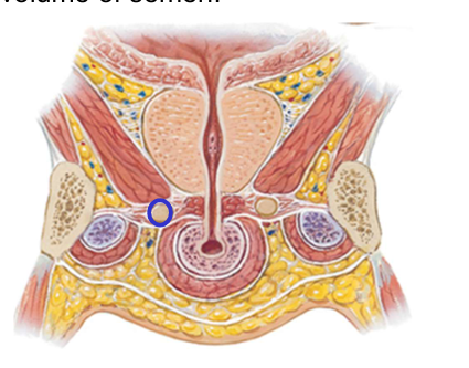
128
mesentary of the uterine tube
mesosalrina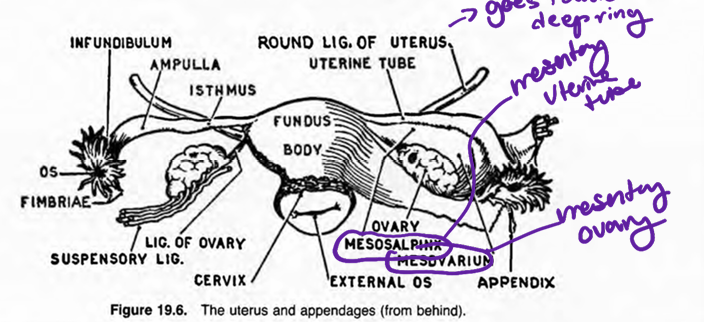
129
mesentary of the ovary
mesovarium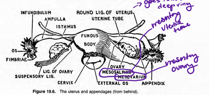
130
what ligaments support the ovary/hold it in place
ovarian, suspensory, mesovarium
131
where does the ovary lie in woman who hacent had a pregnancy or baby
ovarian fossa
132
borders of the ovary
Anterior: external iliac
artery & vein
Inferior: broad
ligament
Posterior: ureter,
internal iliac artery &
vein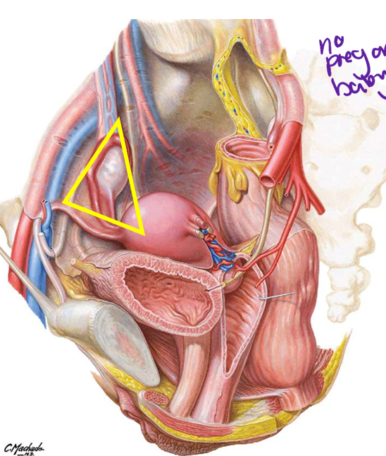
artery & vein
Inferior: broad
ligament
Posterior: ureter,
internal iliac artery &
vein
133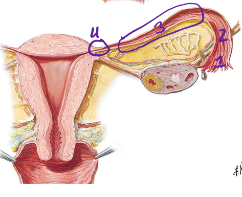
Image occlusion: name the hidden labels
134
Image occlusion: name the hidden labels
135
Image occlusion: name the hidden labels
136
Image occlusion: name the hidden labels
137
the uterine tubes are hollow tubes that open into the __________ and empoty into the ________
peritoneal cavity; uterus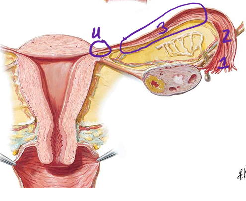
138
venous drainage to uterine tubes
ovarian and uterine veins
139
where do hydatid cyst of morgagni occur?
broad ligament cyst in epophoron.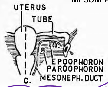
140
inflamation of the peritoneal cavity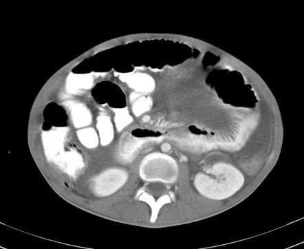
peritonitis
141
a pregnancy that happens outside of your uterus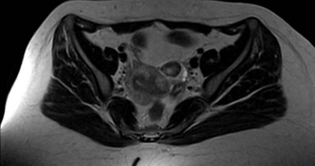
ectopic pregnancy
142
test to test the patency of te uetrine tubes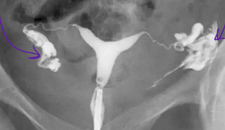
hysterosalpingogram
143
what is the normal position of the uterus?
antrior and flexed over the bladdere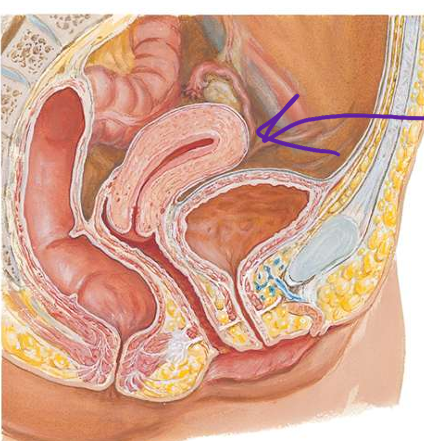
144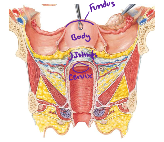
Image occlusion: name the hidden labels
145
Image occlusion: name the hidden labels
146
Image occlusion: name the hidden labels
147
Image occlusion: name the hidden labels
148
the external Os is a common site of _______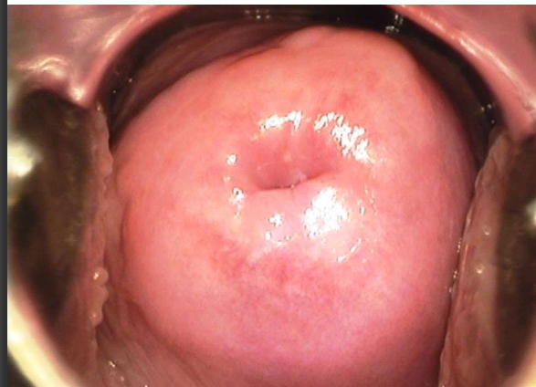
cervical cancer
149
benign tumors of the myometrium
leiomyomas
150
malignant tumors of the myometrium
leiomyosarcoma
151
what runs through the suspensory ;ligament
ovarian VAN, lymph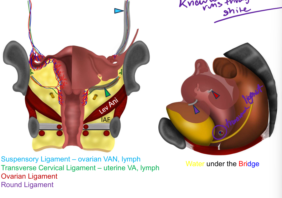
152
what runs through the transverse cervucal ligament
uterine VA, lymph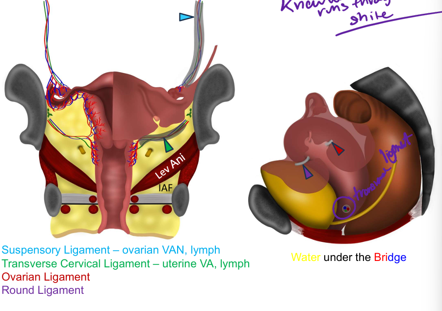
153
what holds the uterus in place (x4)
1. Levator ani
2. Urogential diaphragm
3. Other pelvic organs
(bladder, rectum)
4. Ligaments and parts of
the endopelvic fascia
2. Urogential diaphragm
3. Other pelvic organs
(bladder, rectum)
4. Ligaments and parts of
the endopelvic fascia
154
what is the metastasis of uterine cancer
local spread: out uterine tubes to peritoneum (peritoneal mesothelioma)
lymph: pelvic nodes, lumbar/para-aortic, superficial inguinal (via vessels
along the round ligament)
vascular: lungs, liver, bone, brain
lymph: pelvic nodes, lumbar/para-aortic, superficial inguinal (via vessels
along the round ligament)
vascular: lungs, liver, bone, brain
155
what is the spread of ovarian neoplasm?
local: obturator n!
lymph: lumbar/para-aortic lungs
vascular: Batson’s plexus, IVC
lungs, brain, liver, spleen (see Sister
Mary Joseph nodule)
lymph: lumbar/para-aortic lungs
vascular: Batson’s plexus, IVC
lungs, brain, liver, spleen (see Sister
Mary Joseph nodule)
156
what 4 muscles serve as vaginal sphincters
Pubovaginalis (levator ani)
2. External urethral sphincter
3. Urethrovaginal sphincter (compressor urethrae)
4. Bulbospongiosus
2. External urethral sphincter
3. Urethrovaginal sphincter (compressor urethrae)
4. Bulbospongiosus
157
what drains the uterus?
uterus plexus yto internal iliac v
158
what drains the fundus of uterus
lumbar and external iliac
159
what drains the body of the uterus
external iliac
160
what drains the lymph cervix of the uteryus
internal iliac and sacral nodes
161
what drains the vagina (lymph)
• the upper two-thirds drain to internal and external iliac nodes
• the lower third drains to superficial inguinal nodes
• the lower third drains to superficial inguinal nodes
162
what forms the boundaries of the perineum
roof is formed by the pelvic & urogenital diaphragms + corresponding
fascia; the
floor of the perineum is skin and fascia.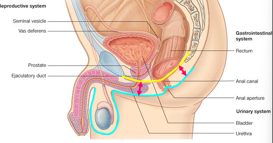
fascia; the
floor of the perineum is skin and fascia.
163
Image occlusion: name the hidden labels
164
Image occlusion: name the hidden labels
165
Image occlusion: name the hidden labels
166
Image occlusion: name the hidden labels
167
Image occlusion: name the hidden labels
168
Image occlusion: name the hidden labels
169
Image occlusion: name the hidden labels
170
what are he muscles of the superficial pouch (perineum) what do they attach to: what innervates them
Muscles attaching to the perineal body:
• Superficial Transverse Perineal
• Bulbospongiosus
• External anal sphincter
• Pubococcygeus
Doesn’t attach to perineal body:
• Ischiocavernosus
ALL INNERVATED BY PUDENTAL N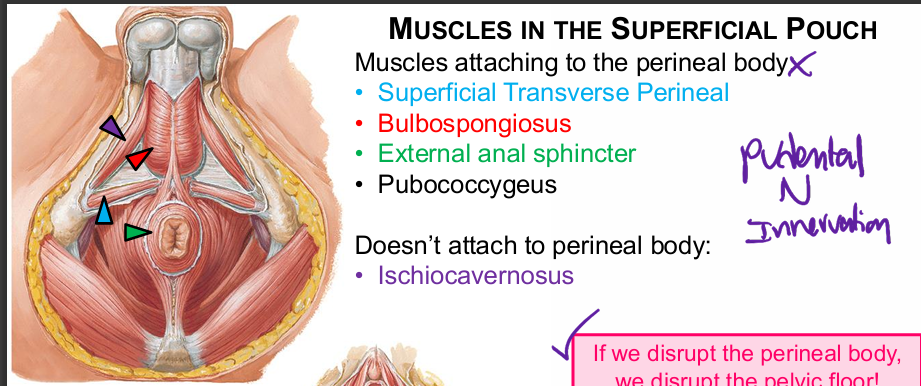
• Superficial Transverse Perineal
• Bulbospongiosus
• External anal sphincter
• Pubococcygeus
Doesn’t attach to perineal body:
• Ischiocavernosus
ALL INNERVATED BY PUDENTAL N
171
muscles of the erectile tissue
corpus spongiosum and cavernosum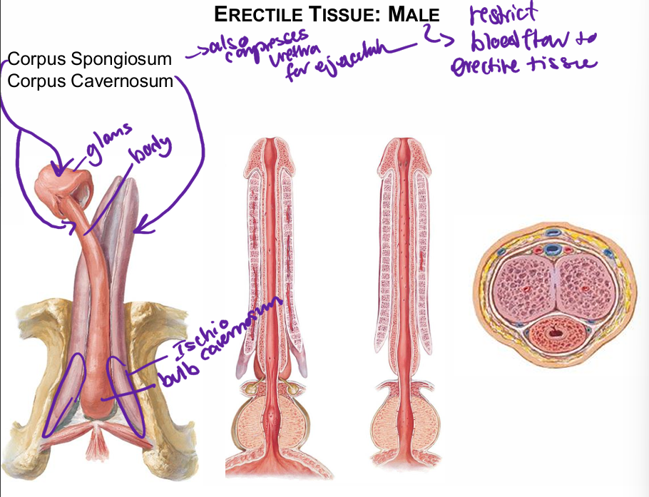
172
fx o erectile muscle
restrict blood flow to erectile tissue; spongiosum also compresses uretgra fdor ejaculation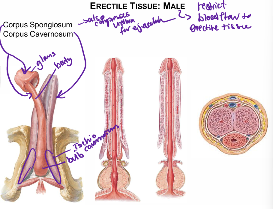
173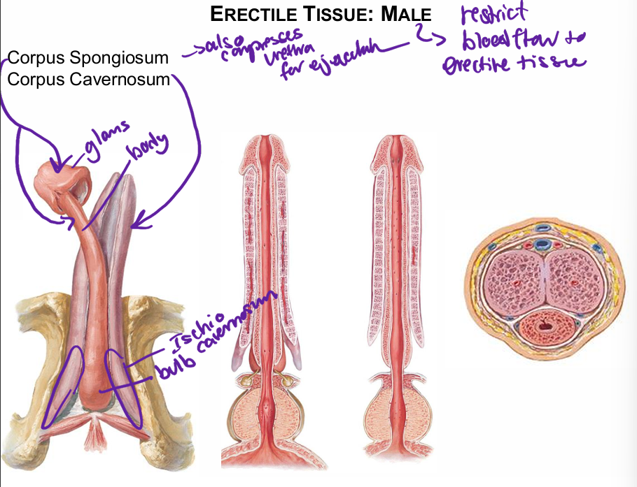
Image occlusion: name the hidden labels
174
Image occlusion: name the hidden labels
175
Image occlusion: name the hidden labels
176
Image occlusion: name the hidden labels
177
Image occlusion: name the hidden labels
178
Image occlusion: name the hidden labels
179
Image occlusion: name the hidden labels
180
a condition that causes a curvature of the penis during erection. It is caused by the formation of fibrous plaques (scar tissue) within the penis. 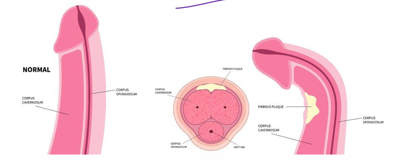
peyr
181
muscle that serves as the central tendon of the perineum
perineal membrane
182
reinforces the pelvic diaphragm at
the genital hiatus and provides support for
pelvic viscer
the genital hiatus and provides support for
pelvic viscer
urogenital diaphragm
183
structures in the deep perineal pouch
Nerves: Dorsal nerve of the Penis (Clitoris)
and Deep Perineal Nerve
Vessels: Internal Pudendal artery and vein
Misc:
• membranous part of urethra
• male: bulbourethral glands
• female: vagina
and Deep Perineal Nerve
Vessels: Internal Pudendal artery and vein
Misc:
• membranous part of urethra
• male: bulbourethral glands
• female: vagina
184
muscles of the deep perineal pouch (x2)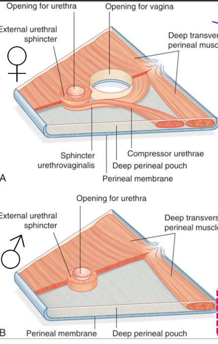
deep tramsverse perineal m
external urethral sphincter
females: sphincter urethrovagialis, compressor urethrae
external urethral sphincter
females: sphincter urethrovagialis, compressor urethrae
185
stabilizes the perineal body to support te sphincter m and levator an
deep transverse perineal m
186
close the urethra abd in malescompress the bulbourethral glabds
external urethral sphincter
187
where does urine go in a superficial bladder tear
peritoneal cavityt
188
where does urine go in a intrapelvic rupture
pelvic cavity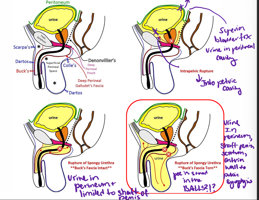
189
what happens in a rupture of the spongy urethra
urine in perineum limited to shaft of penis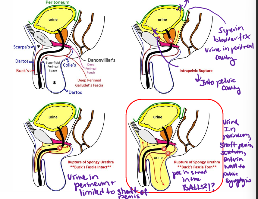
190
wheree does the urine go in a ripture of the spongy urethra where the bucks fascia is torn
urine in perineum, shaft of penis and scrotum to anterior wall to pubic symphysis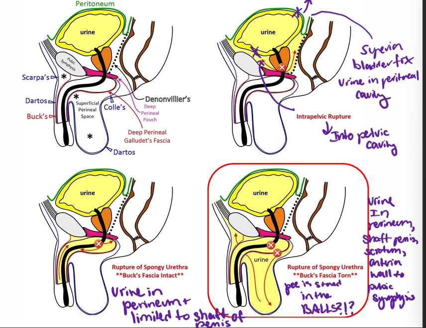
191
what layers are pierced in an injection to the ischioanal fossa
skin and fat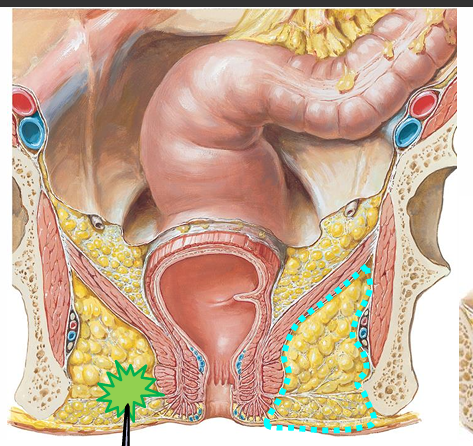
192
what is at risk in an injection to the ischioanal fossa or an infection to the ischioabal fossa
inferior rectal VAN
193
lymph to rectum above pectinate line
internal iliacx n
194
arterial support to rectum above pectinate line
IMA and internal pudenytal a
195
venous drainage to rectum above pectinate line
portal system
196
lymph to rectum below pectinate line
superficial inguinal nodes
197
arterial support to rectum below pectinate line
pudenta;l a
198
what is the venous support to the rectu below the pectinate line
caval system
199
what muscle is responsible for the anal wink reflex
subcutaneoyus part of external sphincter
200
wat are the ana;l canal myscles
external and internal sphincter
201
related to internal rectal
plexus and breakdown of muscularis mucosa;
prolapse of mucosa and dilated veins which can
become strangulated or ulcerated; involves
visceral part of canal and are not painful (GVA
innervation)
plexus and breakdown of muscularis mucosa;
prolapse of mucosa and dilated veins which can
become strangulated or ulcerated; involves
visceral part of canal and are not painful (GVA
innervation)
internal hemroids
202
involve blood clots within
veins of the external rectal plexus; covered by skin
(GSA → painful!)
veins of the external rectal plexus; covered by skin
(GSA → painful!)
external hemroids
203
Android pelvic inlet shape
heart shaped, more commonly male
204
Anthropoid pelvic inlet shape
rounded to oval in shape, more commonly male
205
Gynecoid pelvic inlet shape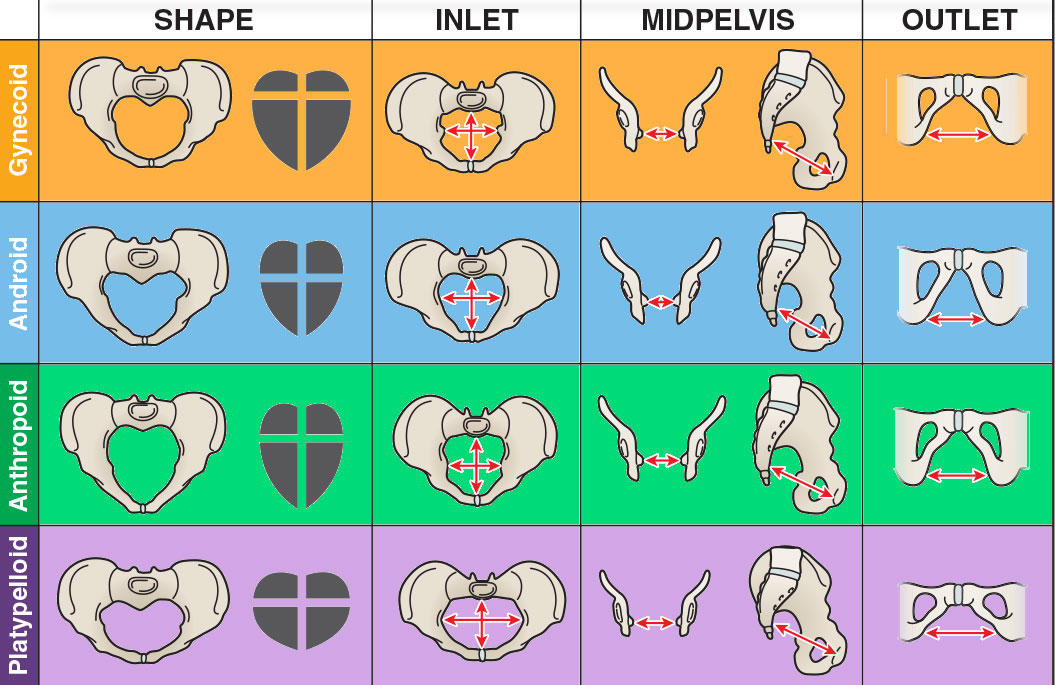
rounded, oval shape, wide transverse diameter, most common female type
206
Platypelloid pelvic inlet shape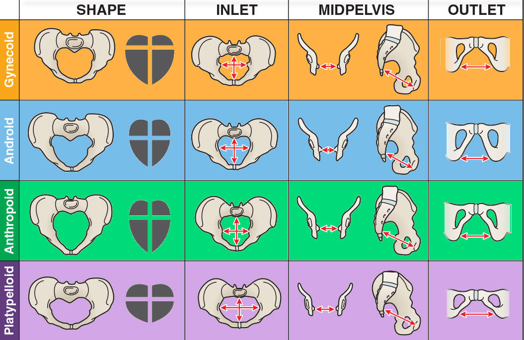
oval and wide, uncommon
207
Obstetric conjugate
extends from the posterior aspect of the middle of the pubic symphysis to sacral promontory
208
Diagonal conjugate
extends from the inferior aspect of the pubic symphysis to sacral promontory; can be estimated during manual pelvic exam
209
Anatomic conjugate
extends from the posterior aspect of the superior aspect of the pubic symphysis to sacral promontory; unaffected by relaxation of pelvic ligaments
210
Interspinous distance
distance between ischial spines
211
Sacro-iliac joint type
a plane, synovial joint
212
Pubic symphysis joint type
secondary cartilaginous; includes a fibro-cartilaginous disc
213
an aberrant obturator artery that arises from the external iliac artery and crosses over the superior pubic ramus, at high risk of injury during hernia repairs
Arterial corona mortis ("crown of death")
214
herniation of the uterus into the vagina, potentially through the external orifice, due to pelvic floor muscle dysfunction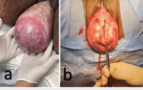
Uterine prolapse
215
collapse of the bladder along the anterior wall of the vagina
Cystocele
216
anterior and inferior protrusion of the rectum along the posterior wall of the vagina
Rectocele
217
anterior and inferior protrusion of the rectovaginal pouch (with gut loops) along the posterior wall of the vagina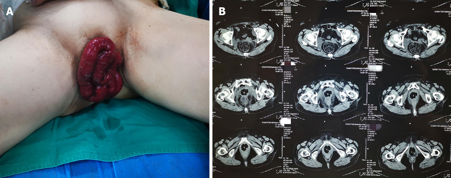
Enterocele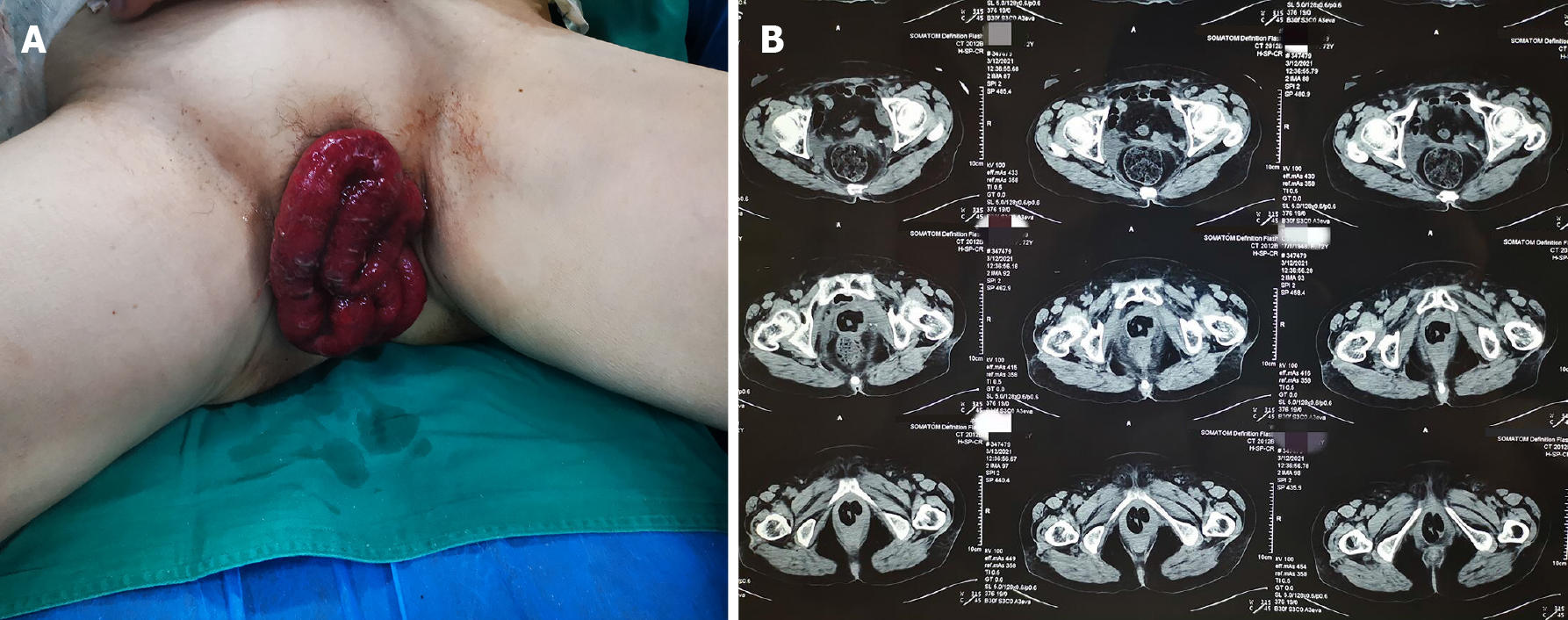
218
When is the ureter in danger of injury during a hysterectomy?
when it travels inferior (under) the uterine artery
219
Laceration between prostatic and membranous urethra results in urine spilling into what space?
the true pelvis, inferior to the peritoneum
220
Laceration of spongy urethra with Buck's fascia torn results in urine leaking into what space?
the superficial pouch (scrotum), along the shaft of the penis and anterior abdominal wall
221
a condition where the foreskin cannot be retracted over the glans penis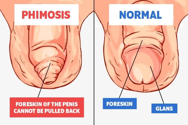
Phimosis
222
constriction of the glans penis by a tight band of foreskin, which can lead to ischemia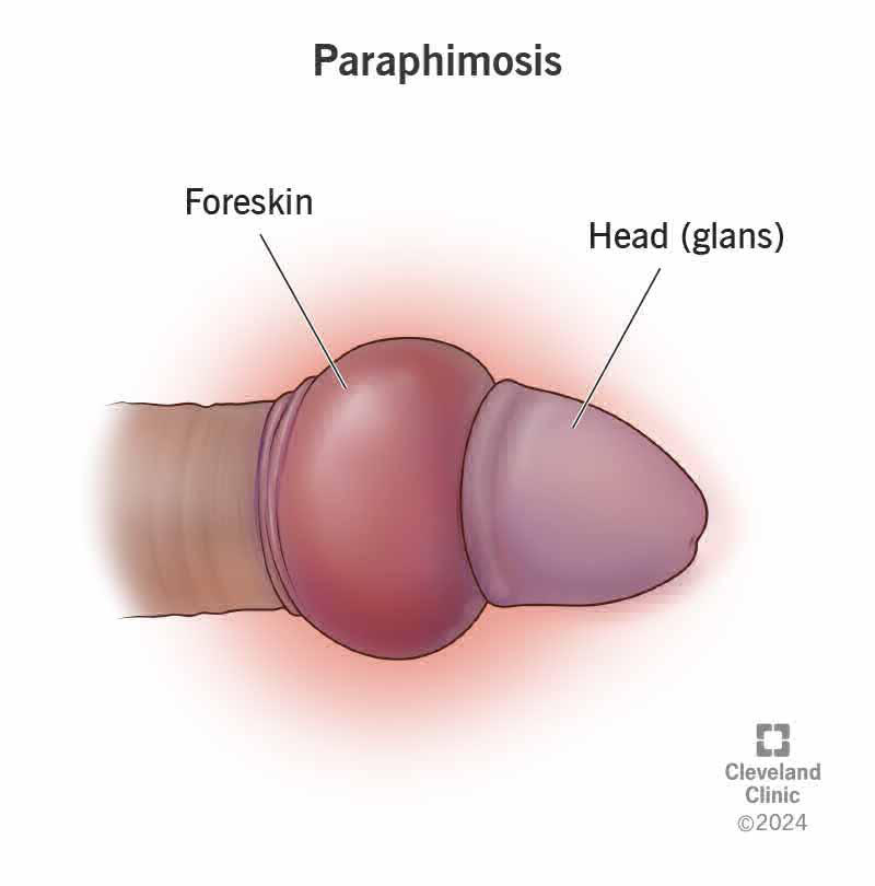
Paraphimosis
223
Peyronie's disease
a buildup of scar tissue within the tunica albuginea of the penis resulting in an abnormal (painful) curvature during erection
224
Why are bladder infections more common in females?
the female urethra is shorter, more distendable and opens to the vaginal vestibule
225
Rectouterine pouch (pouch of Douglas)
the lowest point of the female abdominal cavity, where fluid like blood or ascites can accumulate
226
a mass detected on digital rectal or vaginal exam indicating metastasis within the pouch of Douglas
Blumer's shelf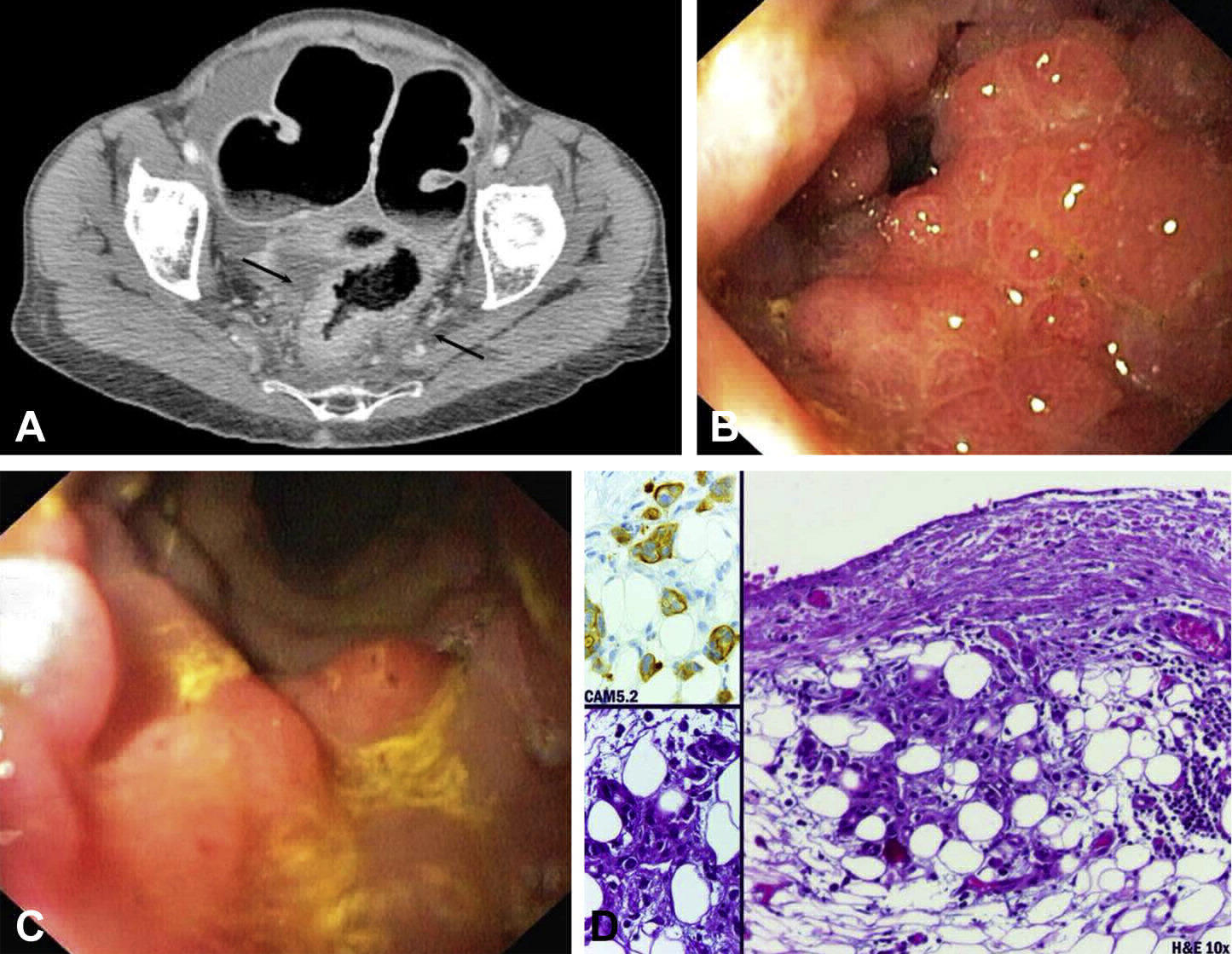
227
What nerve traverses the ischioanal fossa and can result in fecal incontinence if damaged?
the inferior rectal nerve
228
Batson's venous plexus
valveless veins providing a route for metastasis of cancer or spread of infections from pelvic organs to the vertebral column, skull and brain
229
What are the classic signs of benign prostatic hypertrophy (BPH)?
nocturia, dysuria and urgency
230
From which lobe does prostate cancer most commonly arise?
the posterior lobe
231
What nerve plexus is endangered during removal of the prostate, potentially causing erectile dysfunction?
the prostatic nerve plexus
232
Autonomic control for erection
parasympathetic (S2-4) for vascular smooth muscle; pudendal (S2-4) for perineal muscles
233
Autonomic control for emission
sympathetic (L1-2) for contraction of smooth muscle in ductus deferens
234
Autonomic control for ejaculation
sympathetic (L1-2) for internal urethral sphincter closure;
parasympathetic (S2-4) for urethral muscle contraction;
pudendal (S2-4) for perineal muscle contraction
parasympathetic (S2-4) for urethral muscle contraction;
pudendal (S2-4) for perineal muscle contraction
235
Leading cause of incontinence in young females
stress incontinence
236
Leading cause of incontinence in older patients
urge incontinence
237
Normal position of the uterus
anteverted and anteflexed
238
Main passive support of the cervixterus
transverse cervical (cardinal) ligament
239
Most common site for ectopic pregnancy
tubal implantation
240
rotation of the ovary and uterine tube around the suspensory ligament of the ovary, which carries its blood supply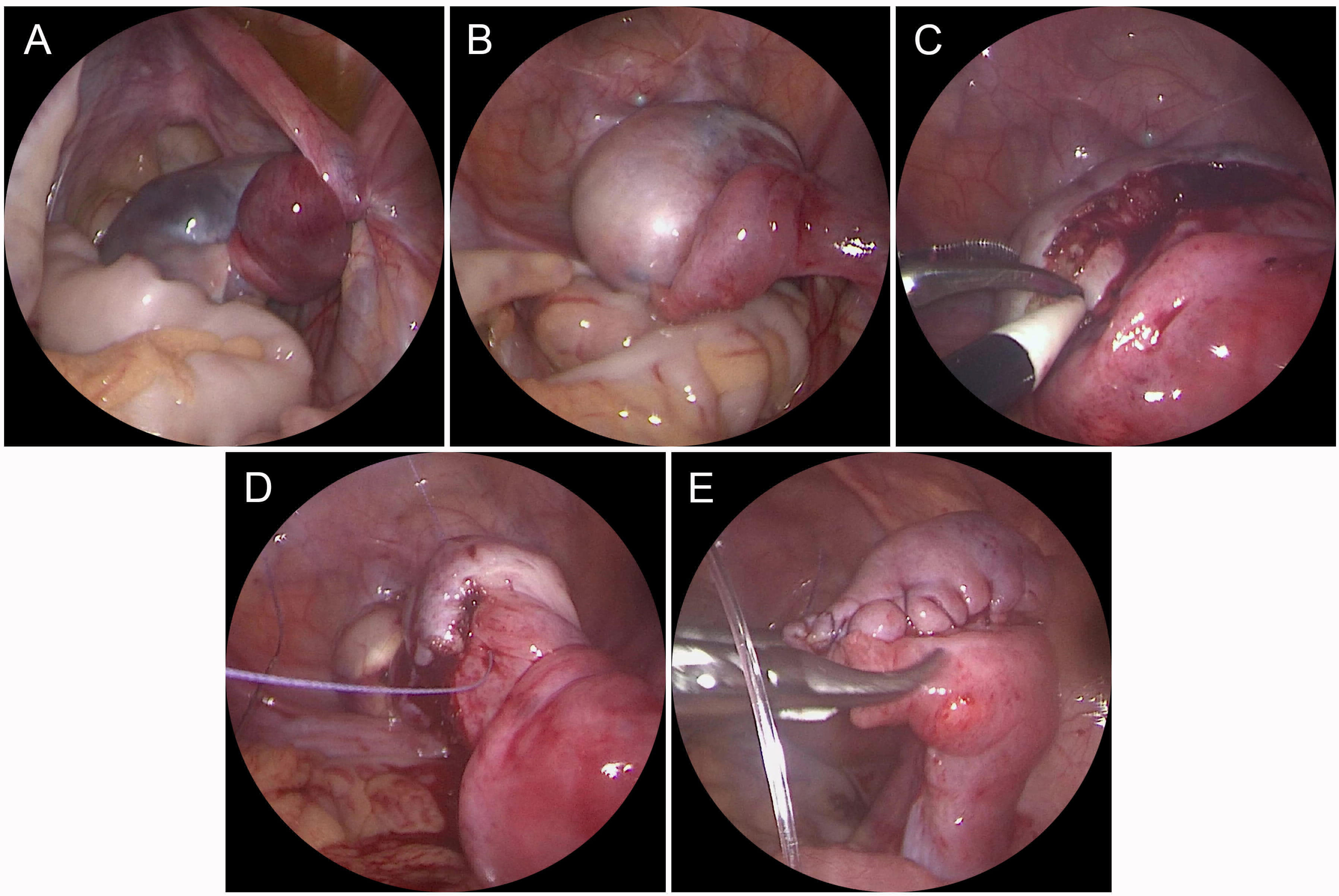
Ovarian torsion
241
Anesthesia that numbs the uterus, vagina, pelvic floor, and lower extremities, where the patient cannot feel contractions
Spinal anesthesia
242
Anesthesia that numbs the cervix, vagina, and pelvic floor, but the patient can still feel uterine contractions
Caudal epidural
243
Anesthesia that numbs the lower vagina and pelvic floor only
Pudendal nerve block
244
The uterine artery is a branch of which artery?
internal iliac artery
245
The ovarian artery is a branch of which artery?
abdominal aorta
246
Mnemonic for the relationship between the ureter and uterine artery
"water under the bridge" (ureter is inferior to the artery)
247
Landmark for differentiating internal and external hemorrhoids
pectinate line
248
Innervation of internal hemorrhoids
autonomic (visceral), not painful
249
Innervation of external hemorrhoids
somatic, painful
250
Lymphatic drainage of the testes/ovary
lumbar (para-aortic) nodes
251
Lymphatic drainage of the lower 1/3 of the vagina and vulva
superficial inguinal nodes
252
Sensory innervation to the anterior aspect of the scrotum
ilioinguinal nerve and genital branch of the genitofemoral nerve
253
Sensory innervation to the posterior aspect of the scrotum
pudendal nerve
254
Do pelvic splanchnic nerves carry pain information from pelvic viscera?
Yes, unlike the vagus nerve. Referred pain is to S2-4 dermatomes.
255
what in the perineum follows the pathway of pattern 1 synpathetic nervous sysytem
pudental n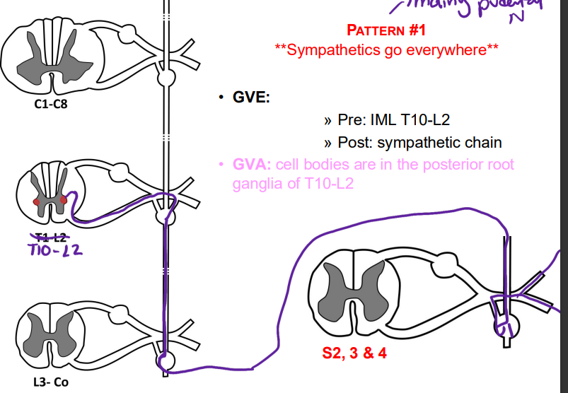
256
what follows pattern 3 in the perineum/pelvis of sympathetic innervation
lumbar splanchnics and sacral splancnics
257
what does the hypogastric plexus contain (x6)
1. Pre and
2. Post ganglionic Sympathetic axons (GVE)
3. GVA pain (from T10 -L2)
4. Pre and
5. Post Ganglionic Parasympathetic axons
(GVEp)
6. GVA interoceptive and pain from S2-4
2. Post ganglionic Sympathetic axons (GVE)
3. GVA pain (from T10 -L2)
4. Pre and
5. Post Ganglionic Parasympathetic axons
(GVEp)
6. GVA interoceptive and pain from S2-4
258
autonomic both sympathetic and
parasympathetic innervation (GVE & GVA)
to the erectile tissue
parasympathetic innervation (GVE & GVA)
to the erectile tissue
cavernous n
259
what is responssinle for emission in the mechanism of etrection
a. Sacral Splanchnic Nerves (via hypogastric plexus)
b. Peristaltic movement of semen through the reproductive tract
b. Peristaltic movement of semen through the reproductive tract
260
what is responsible for ejaculation in the mevchanism of erection
a. Closure of the vesical sphincter (sympathetics)
b. Contraction of urethral muscles (parasympathetic)
c. Contraction of bulbospongiosus (pudendal)
b. Contraction of urethral muscles (parasympathetic)
c. Contraction of bulbospongiosus (pudendal)
261
what do sympayjeytocs do in the control of the bladder
L1-2) – promote storage
(holding urine) = relaxes the detrusor/contracts
the internal sphincter
(holding urine) = relaxes the detrusor/contracts
the internal sphincter
262
what do parasympathetics do in the control of the bladder
(S2-4) – promote release
of urine = contracts the detrusorelaxes the
internal sphincter
of urine = contracts the detrusorelaxes the
internal sphincter
263
what is the pudental n role in control of the bladder
(S2-4) – LMNs in Onuf’s nucleus;
voluntary relaxation of the sphincter urethrae
voluntary relaxation of the sphincter urethrae
264
how does filloing phase work ina the nladder (sympathetics and pudental n)
Sympathetics:
▪ Inhibit detrusor
▪ Keeps the internal sphincter closed
Pudendal Nerve
▪ Keeps the external sphincter closed
▪ Inhibit detrusor
▪ Keeps the internal sphincter closed
Pudendal Nerve
▪ Keeps the external sphincter closed
265
▪ Leading cause of incontinence in young
females
▪ S/S: leakage of small volume of urine with
Valsalva maneuvers
▪ Cause: weak voluntary sphincters
– Pudendal n, spinal stenosis
– Trauma to pelvic floor (vaginal delivery
females
▪ S/S: leakage of small volume of urine with
Valsalva maneuvers
▪ Cause: weak voluntary sphincters
– Pudendal n, spinal stenosis
– Trauma to pelvic floor (vaginal delivery
stress incontinence
266
▪ S/S: involuntary emptying/leaking of an
overfull bladder + painful retention
▪ Causes:
– Impaired parasympathetic function
(diabetic neuropathy, autonomic
neuropathy, MS, spinal cord injury):
weak detrusor
– Structural: chronic outlet obstruction
(prostate)
overfull bladder + painful retention
▪ Causes:
– Impaired parasympathetic function
(diabetic neuropathy, autonomic
neuropathy, MS, spinal cord injury):
weak detrusor
– Structural: chronic outlet obstruction
(prostate)
overflow incontinence
267
what is everything above the blue line innervated by (pain)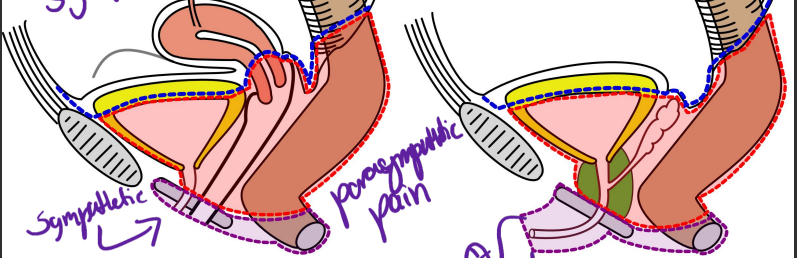
GVA sympathetic (lumbar and sacral splancnic n)
268
what is everything in the red innervated by (pain)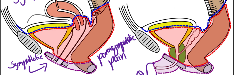
GVA parasympathetic (S2-4) prlvic splacnic N
269
what is the purple innervated y (pain)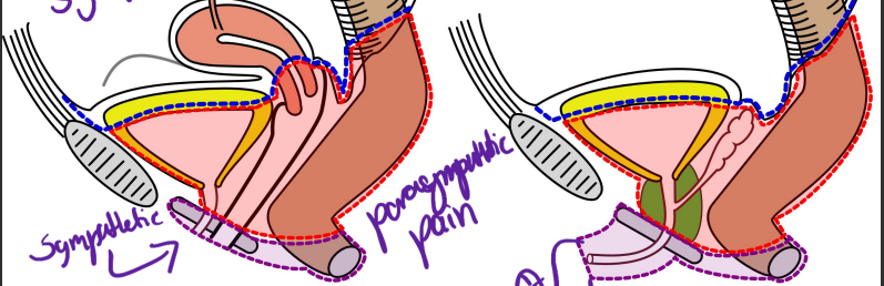
GSA pudental n (s2-4)
270
the skin of the perineum drains to...
superficial inguinal nodes
271
what are the layers to a pudental n block at the ischial spine
skin
272
what are the layers to penitrate doing a fluid drain from the pouch of douglas through the vaginal canal
layers of vagina, visceral peritoneum