First 300 of 333 cards
1
4 compartments of the neck
investing, visceral, vascular, vertebral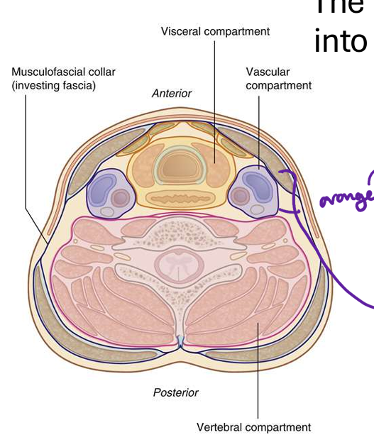
2
the anterior structures of the neck (and anterir triangke) pass throug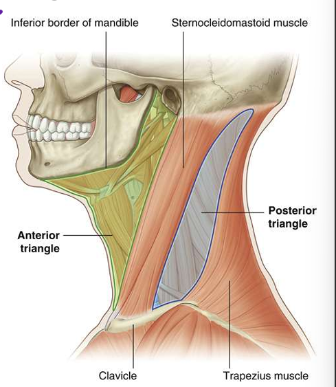
tjorax
3
the pisterior structures and triangles pass into 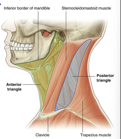
upper limb
4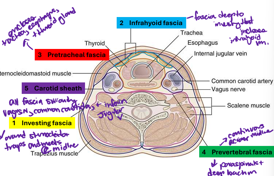
Image occlusion: name the hidden labels
5
Image occlusion: name the hidden labels
6
Image occlusion: name the hidden labels
7
Image occlusion: name the hidden labels
8
Image occlusion: name the hidden labels
9
what are the 4 deep cervicak fascia layers
investing, infrahyoud, pretracheal, prevertebral, carotid sheath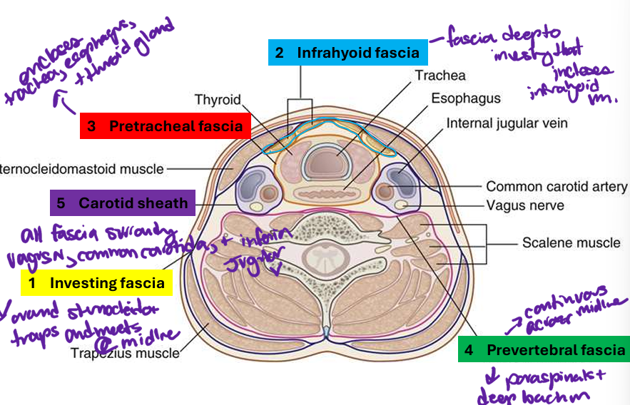
10
completely surrounds the neck; arises from skull and mandible and
extends inferiorly to the sternum, clavicles, manubrium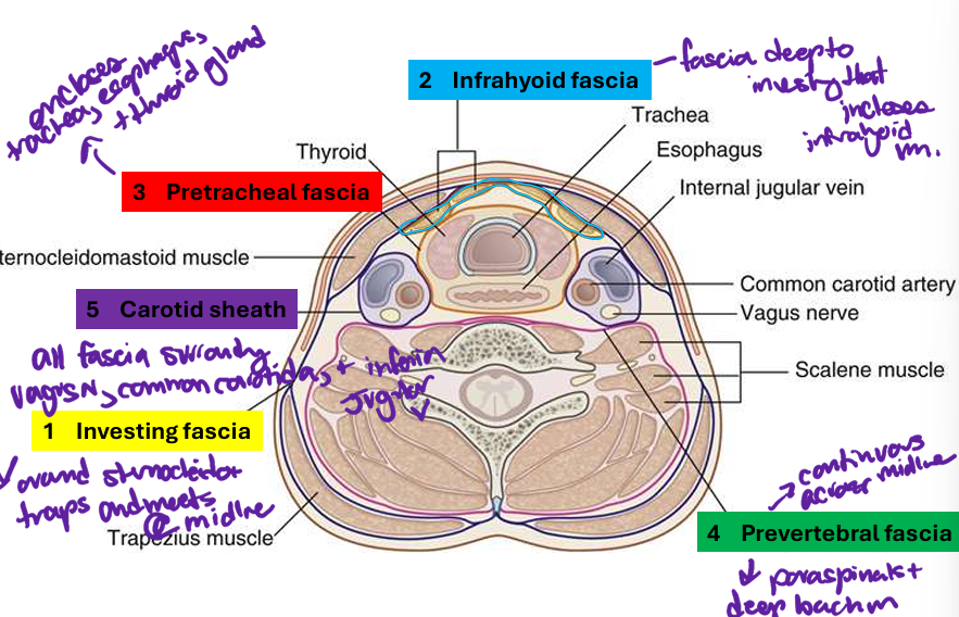
extends inferiorly to the sternum, clavicles, manubrium
investing fascia
11
begins at hyoid bone, forms an investing fascia for the infrahyoid muscles; superficial and deep
layers; continuous across the midline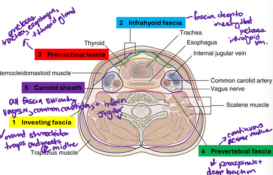
layers; continuous across the midline
infrahyoid fascia
12
a continuous fascial tube that surrounds the viscera in the neck:
a. Pre-tracheal proper – attaches to the hyoid bone above and fibrous pericardium below
b. Buccopharyngeal– attaches to base of the skull, covering the buccinator and
pharyngeal constrictors, fuses with pre-tracheal, fuses with adventitia of esophagus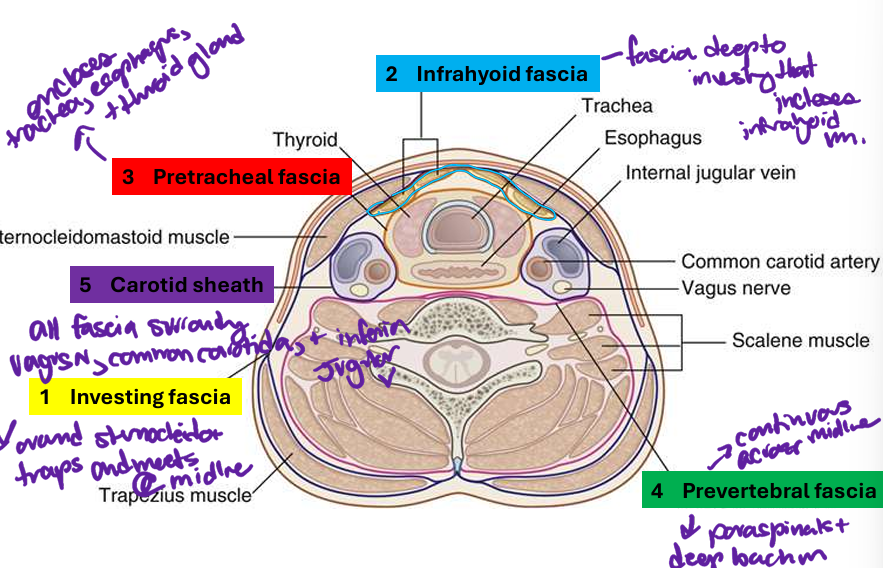
a. Pre-tracheal proper – attaches to the hyoid bone above and fibrous pericardium below
b. Buccopharyngeal– attaches to base of the skull, covering the buccinator and
pharyngeal constrictors, fuses with pre-tracheal, fuses with adventitia of esophagus
pretracheal fascia
13
attaches to base of skull and vertebrae and encloses the deep
muscles, has specializations: alar fascia, axillary sheath and Sibson’s fascia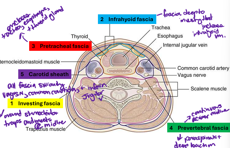
muscles, has specializations: alar fascia, axillary sheath and Sibson’s fascia
prevertebral fascia
14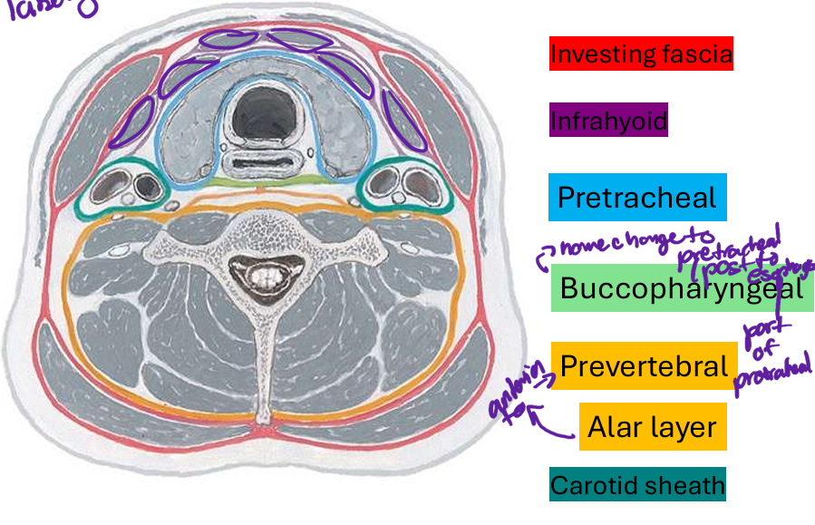
Image occlusion: name the hidden labels
15
Image occlusion: name the hidden labels
16
Image occlusion: name the hidden labels
17
Image occlusion: name the hidden labels
18
Image occlusion: name the hidden labels
19
Image occlusion: name the hidden labels
20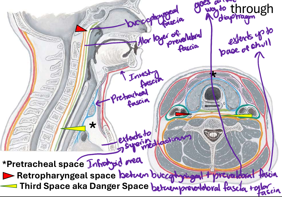
Image occlusion: name the hidden labels
21
Image occlusion: name the hidden labels
22
Image occlusion: name the hidden labels
23
Image occlusion: name the hidden labels
24
Image occlusion: name the hidden labels
25
Image occlusion: name the hidden labels
26
Image occlusion: name the hidden labels
27
space between ucoopharyngeal and prevertebral fascia; extends to base of skull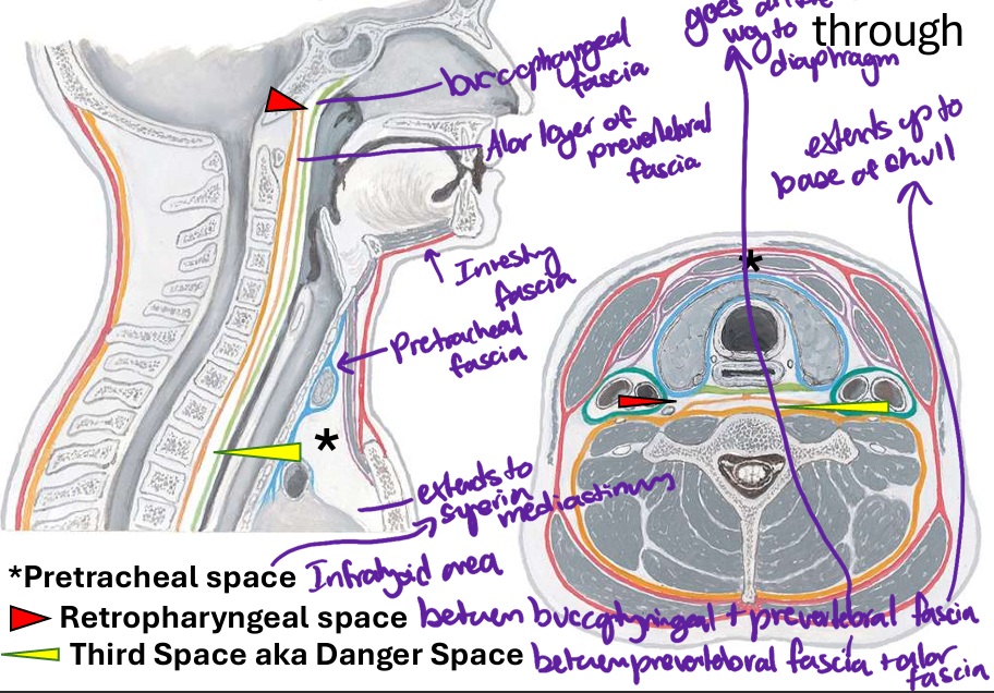
retropharyngeal space
28
space between preverteral fascia and alar fascia; goes to diaphragm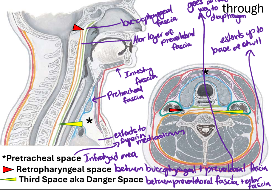
third space; danger space
29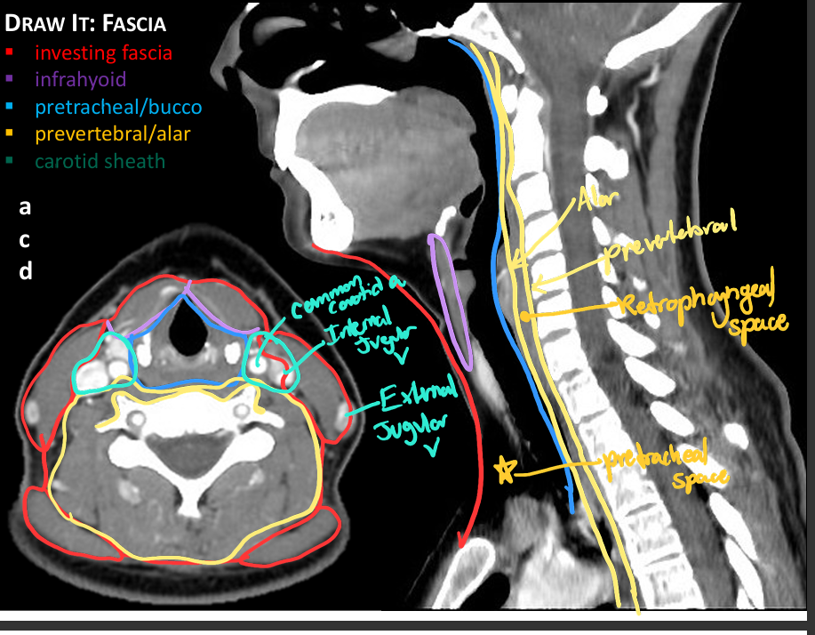
Image occlusion: name the hidden labels
30
Image occlusion: name the hidden labels
31
Image occlusion: name the hidden labels
32
Image occlusion: name the hidden labels
33
Image occlusion: name the hidden labels
34
Image occlusion: name the hidden labels
35
Image occlusion: name the hidden labels
36
Image occlusion: name the hidden labels
37
Image occlusion: name the hidden labels
38
Image occlusion: name the hidden labels
39
Image occlusion: name the hidden labels
40
Image occlusion: name the hidden labels
41
what does this show?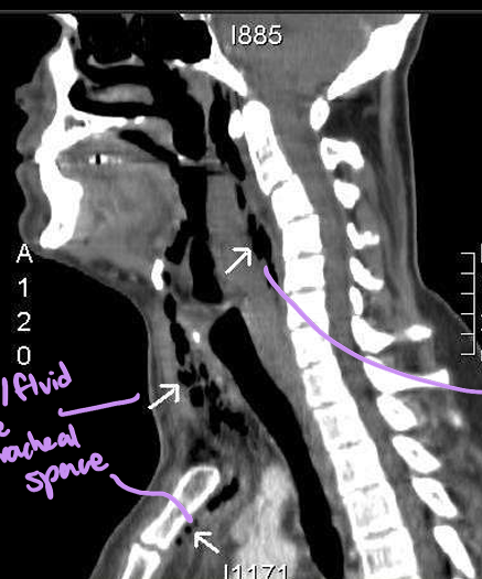
air and or fluid in the pretracheal space and retropharyngeal space
42
what innervates the platysma m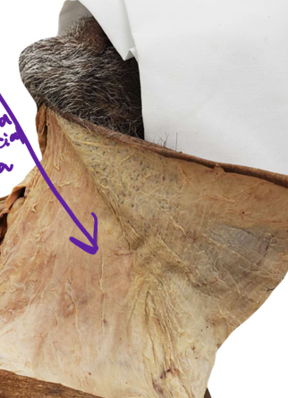
cervical branch of facial n CN VII for motor
transverse cervical n for proprioception
transverse cervical n for proprioception
43
muscles that reside aboce the hyoid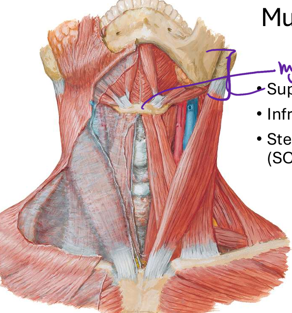
suprahyoud muscles
44
musckes below the hyoid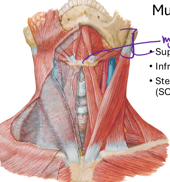
infrahyoid muscles
45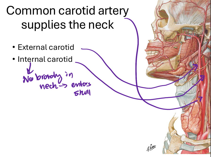
Image occlusion: name the hidden labels
46
Image occlusion: name the hidden labels
47
Image occlusion: name the hidden labels
48
the anterior jugular v drains unto _- which drains into __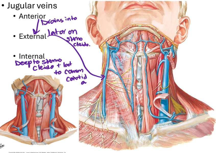
external jugular v; internal jugular v
49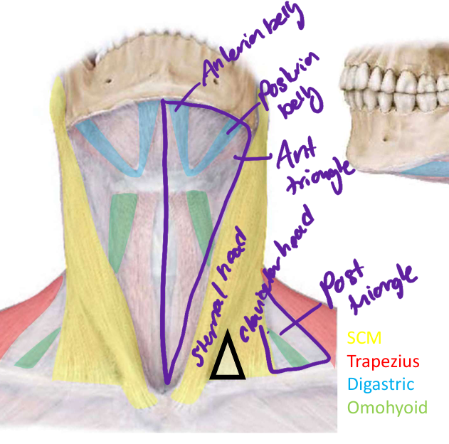
Image occlusion: name the hidden labels
50
Image occlusion: name the hidden labels
51
Image occlusion: name the hidden labels
52
Image occlusion: name the hidden labels
53
Image occlusion: name the hidden labels
54
Image occlusion: name the hidden labels
55
Image occlusion: name the hidden labels
56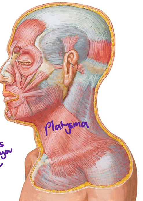
Image occlusion: name the hidden labels
57
innervation of SCM and trapezius m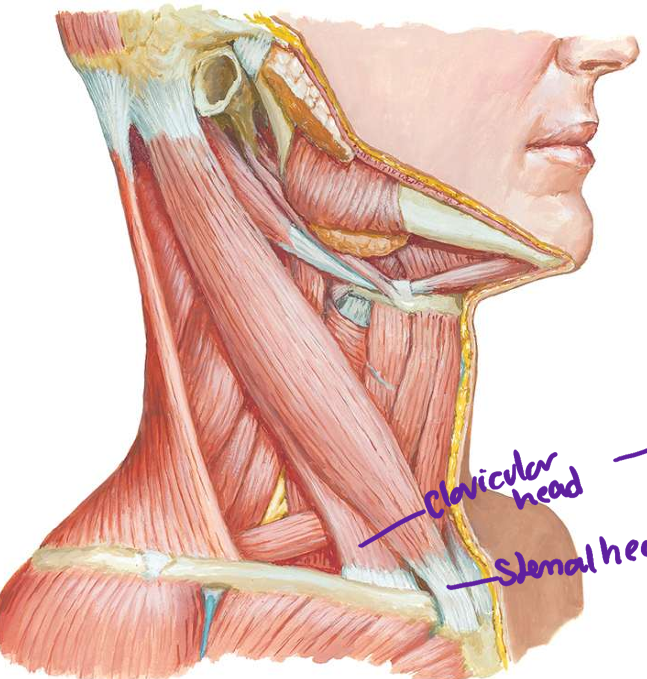
spinal accessory n CN XI
58
fx SCM
turn head to oppsoite side and sidebend head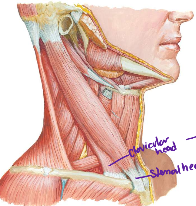
59
how do yo gain access to internal jugular v for procedure/surgery
enter needle between clavicular and sternal head of SCM
60
what layers are traversed in a internal jugular vein injection
skin, superficial fascia, plastysmus, investing fascoa, infrahyoid fascia, carotid sheath, vein
61
shortening of the SCM muscle. head turned toward effected side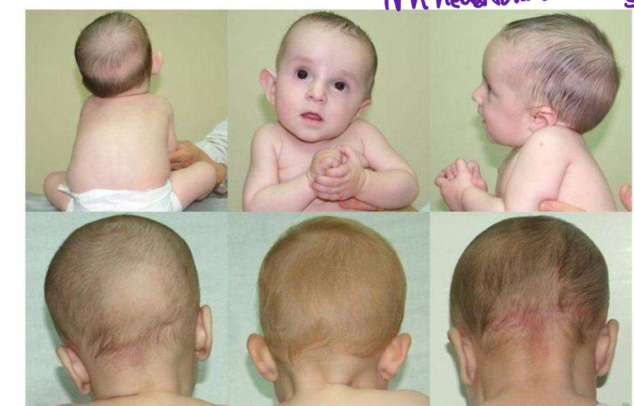
congenital torticollis
62
attachmets of anterior digastric m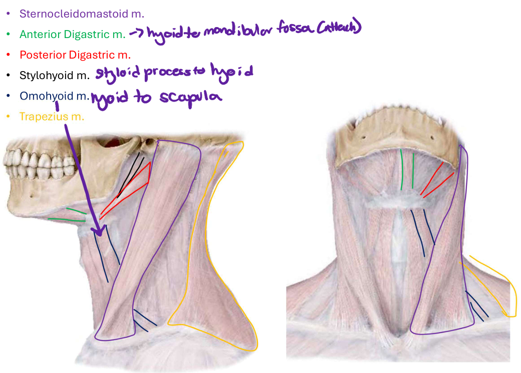
hyoid to mandibular fossa
63
attachments of styohyoid m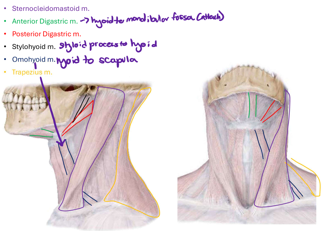
styloid process to hyoid bone
64
attachments of omohyoid m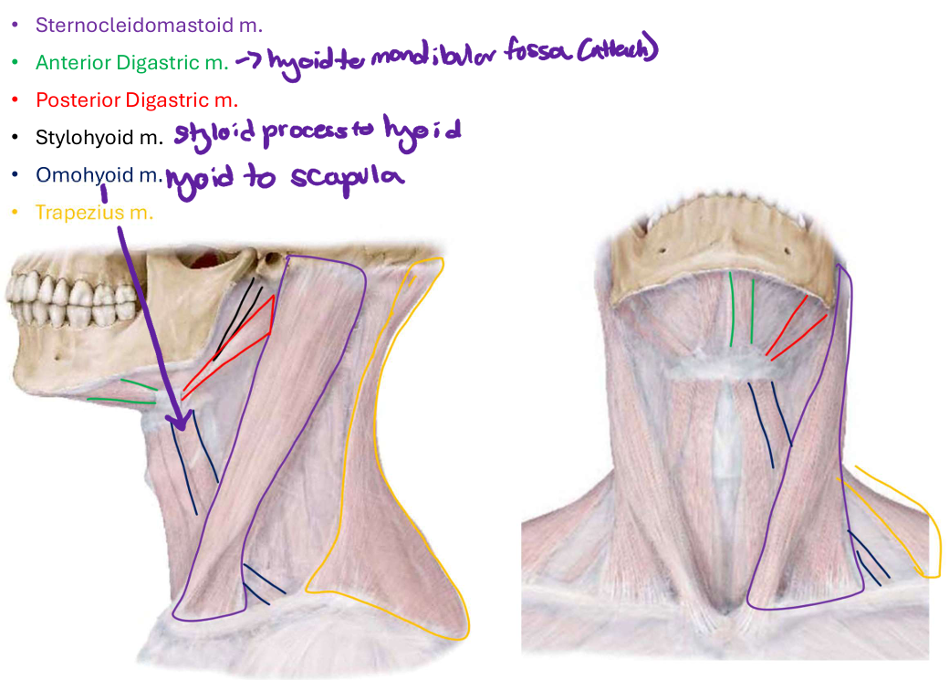
hyoid to scapula
65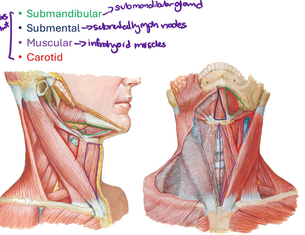
Image occlusion: name the hidden labels
66
Image occlusion: name the hidden labels
67
Image occlusion: name the hidden labels
68
Image occlusion: name the hidden labels
69
innervation anterior belly of digastric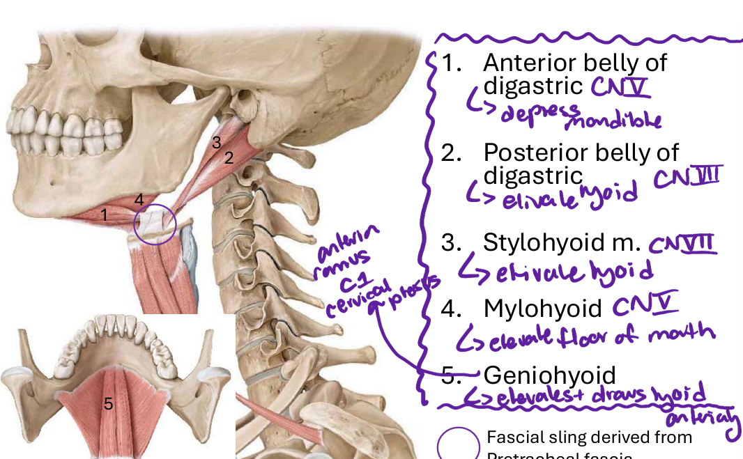
CN V
70
fx anterior belly of digastric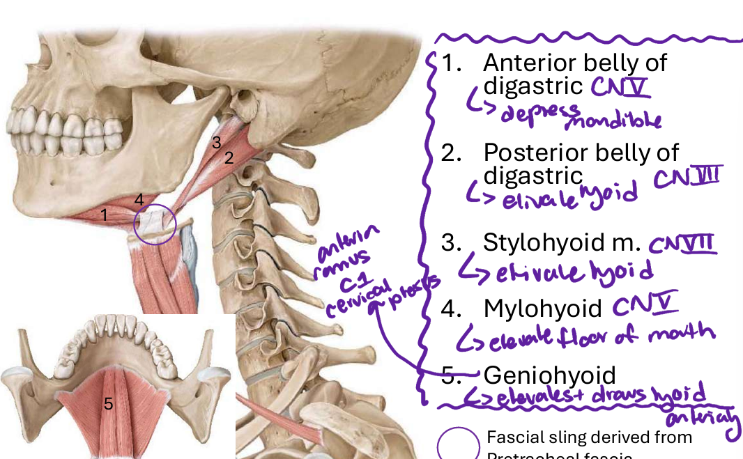
depress mandible
71
innervation posteror belly of digastric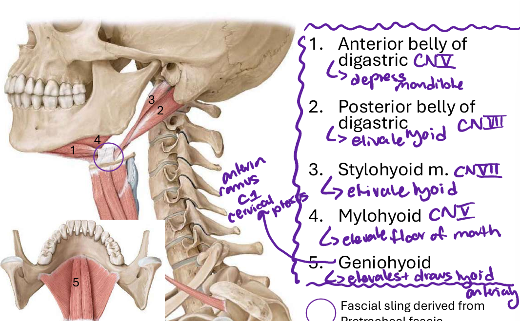
CN VII
72
fx posterior belly of digastric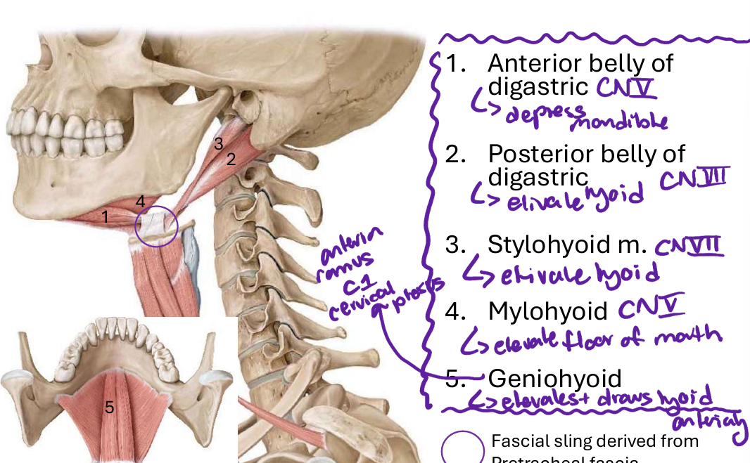
elevate hyoid bone
73
fx stylohyoid m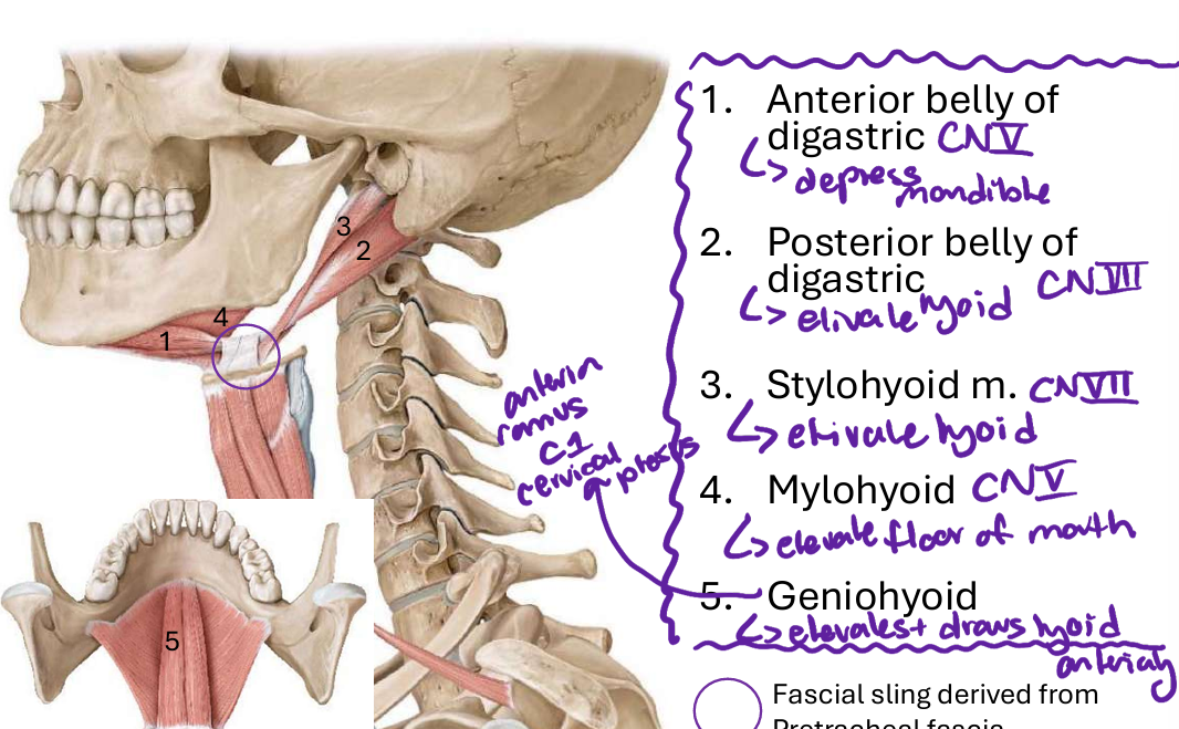
elevate hyoid
74
innetvation stylohyoid m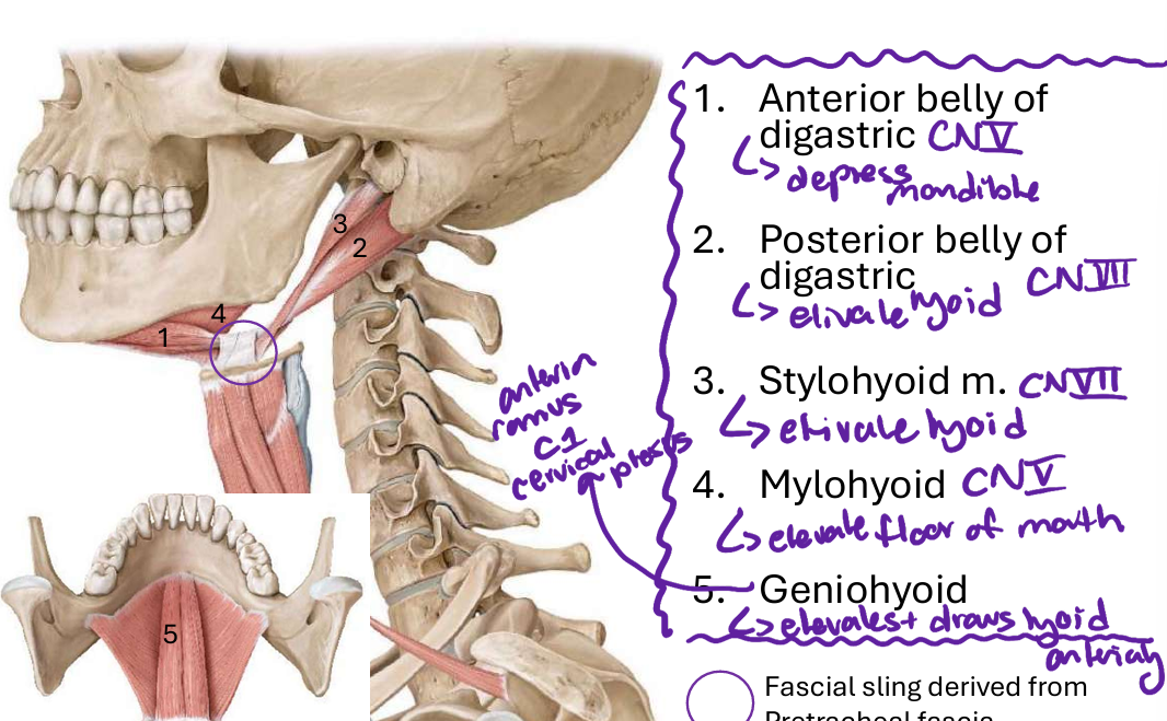
CN VII
75
innervation mylohyoid m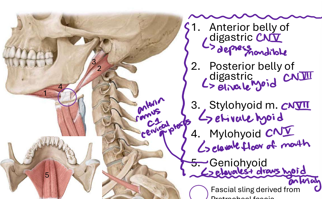
CN V
76
fx mylohyoid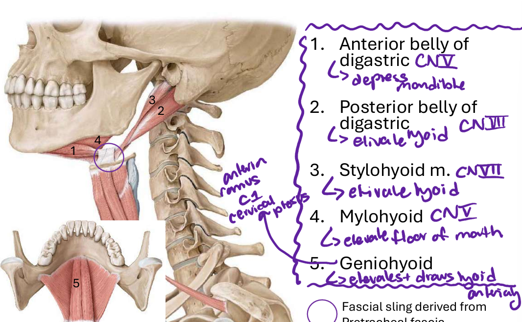
elevate floor of mouth
77
innervation geniohyoid m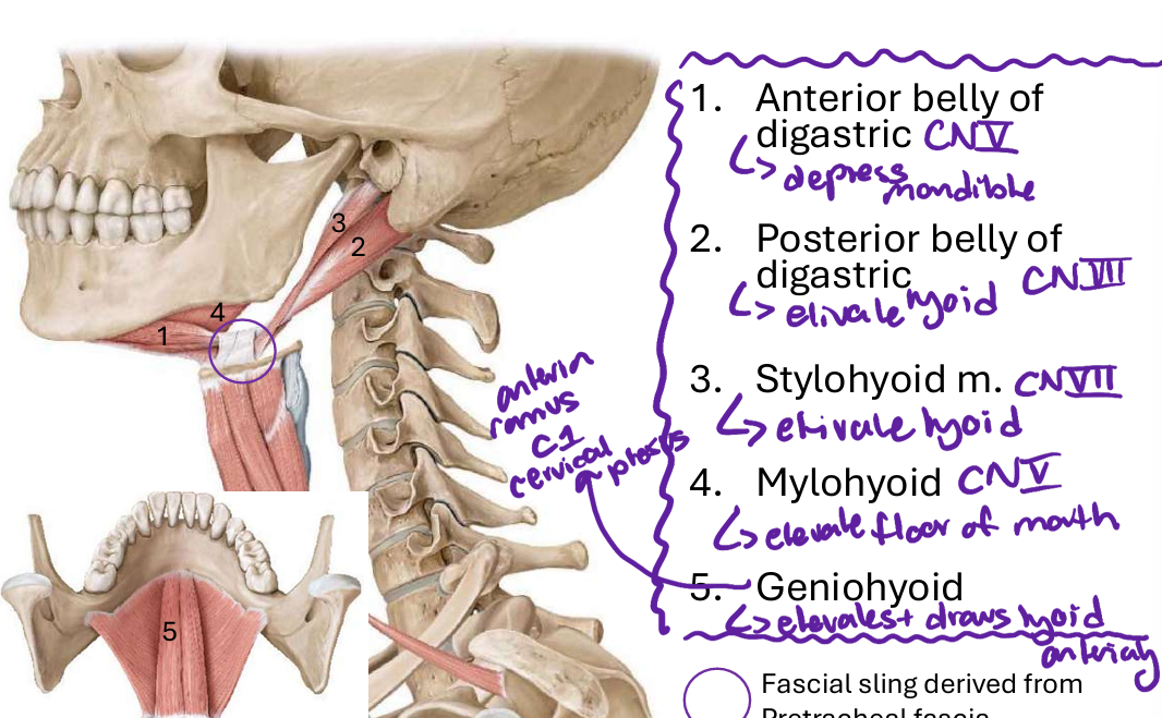
a nterior rami of C1
78
fx geniohyoid m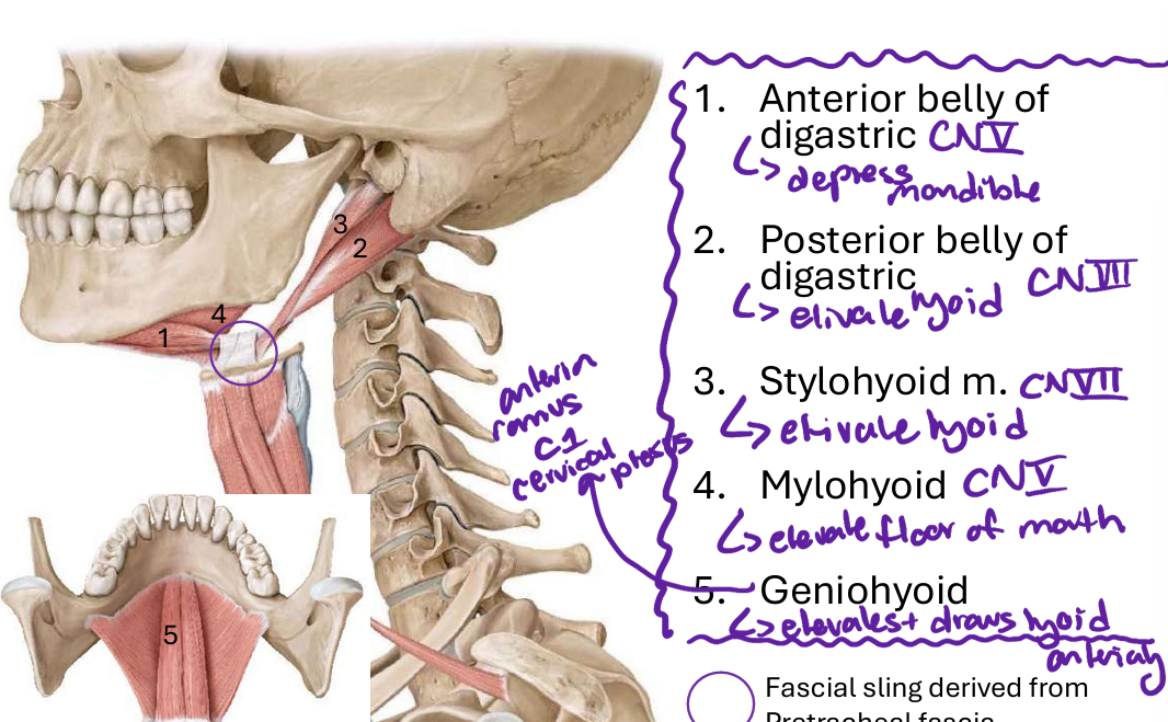
elevcates and draws hyoid anteriorly
79
fx sternohyoid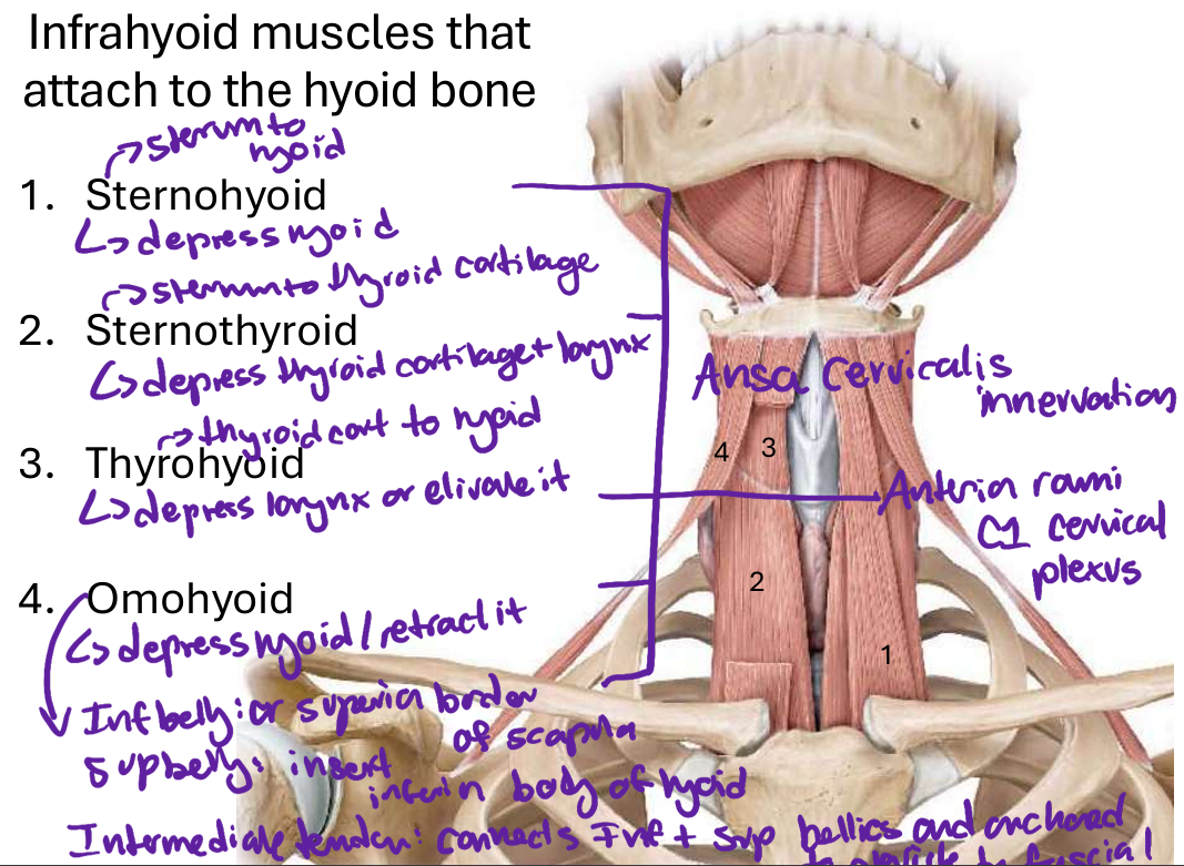
depress hyoid
80
fx sternothyroid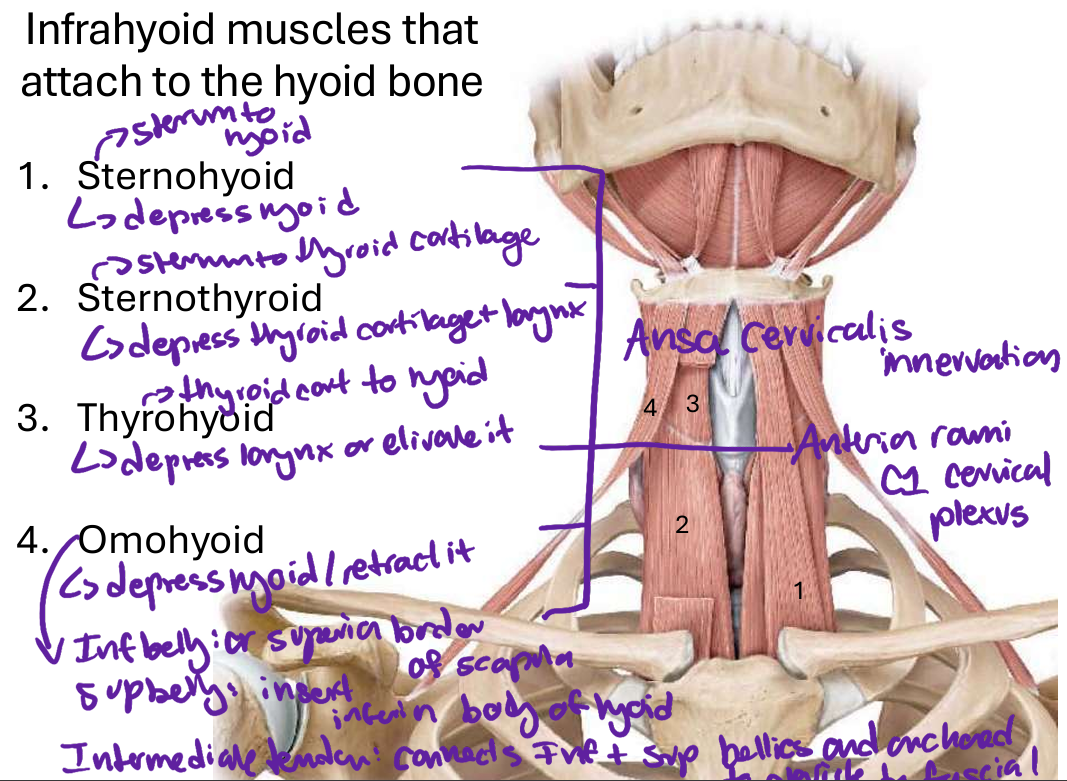
depress thyroid cartilage and larynx
81
fx thyrohyoid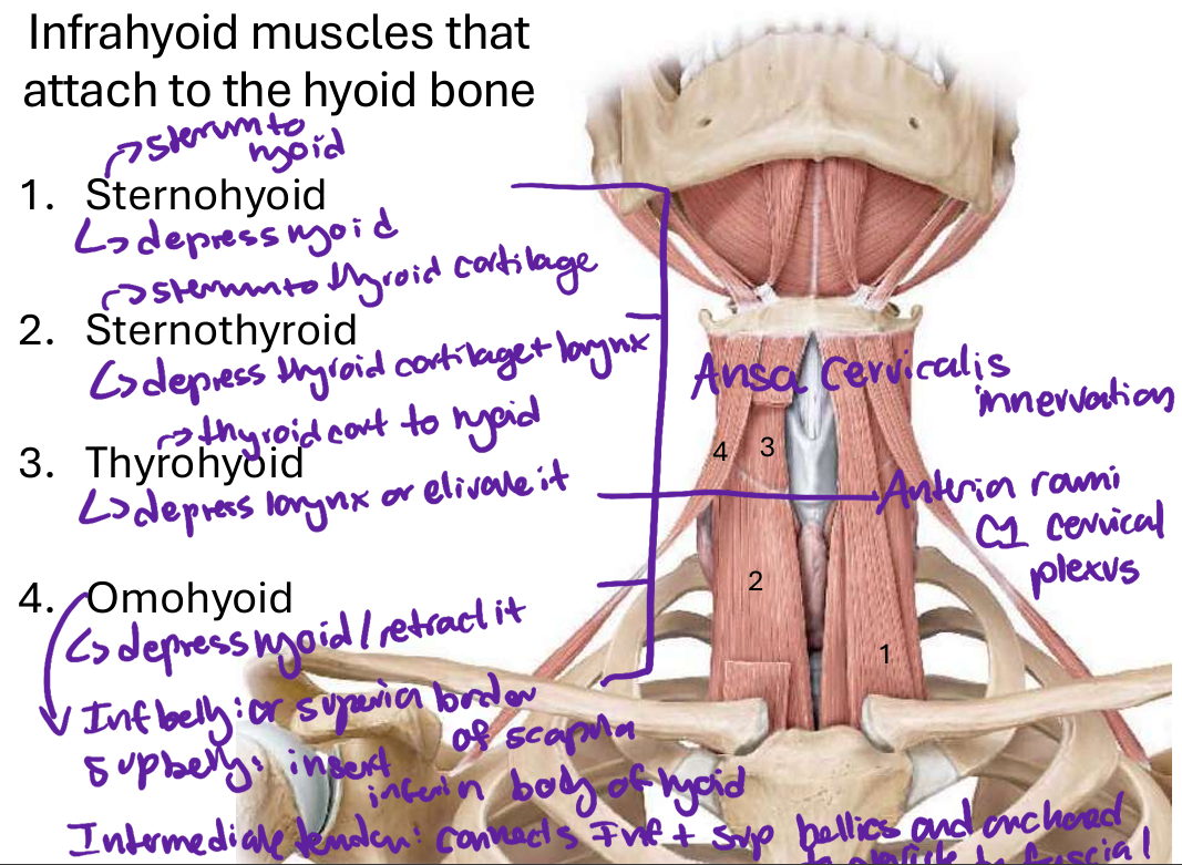
depress larynx or elivate it
82
fx omohyoid m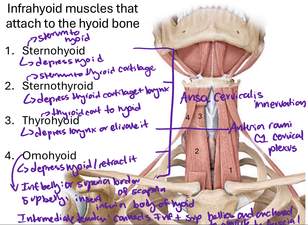
depress hyoid and retract it
83
innervation to sternohyoid, sternothyroid, and omohyoid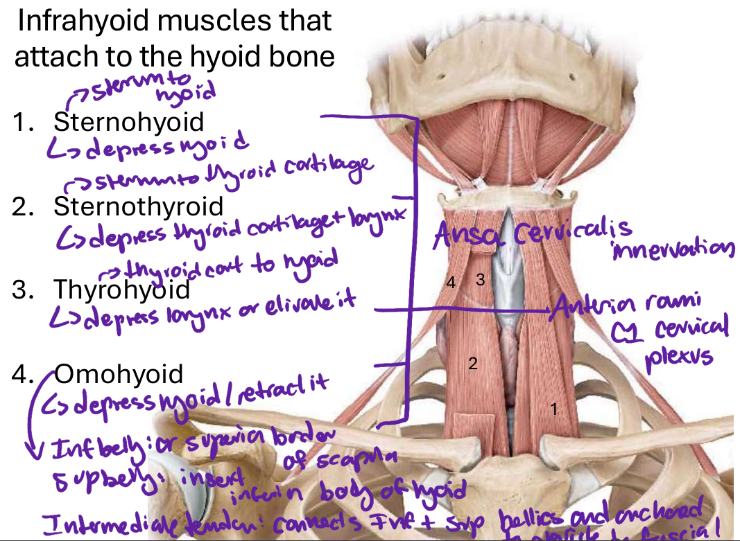
ansa cervicalis
84
thyrohyoid innervation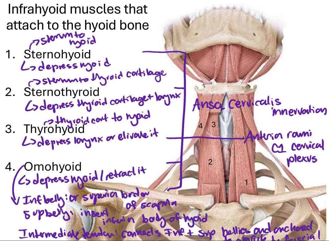
anterior rami C1
85
rectus capitis lateralis fx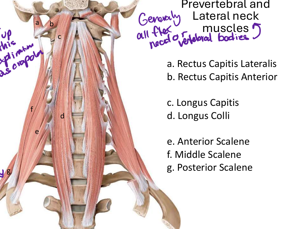
primarily responsible for lateral flexion of the head to the same side and stabilizing the atlanto-occipital joint during movement
86
fx rectus capitis anterior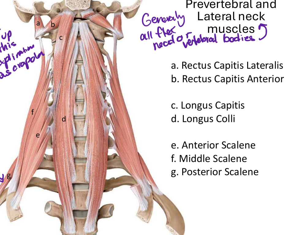
responsible for flexing the head on the neck at the atlantooccipital joint. It plays a crucial role in stabilizing the atlantooccipital joint and maintaining proper alignment between the upper cervical vertebrae and occiput
87
function longus capitis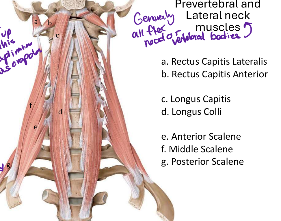
Function: It acts as a weak flexor of the head and cervical spine, working synergistically with other muscles like longus colli and scalene muscles.
88
innervation longus capitus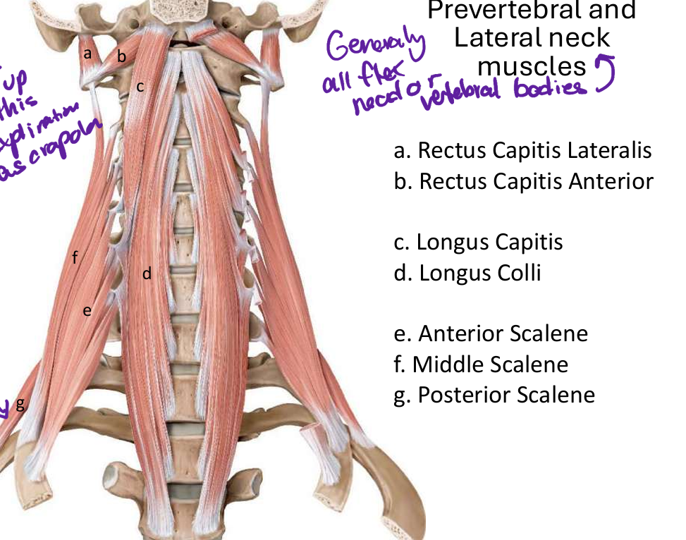
anterior ramu C1-C3 spinal n
89
longus colli fx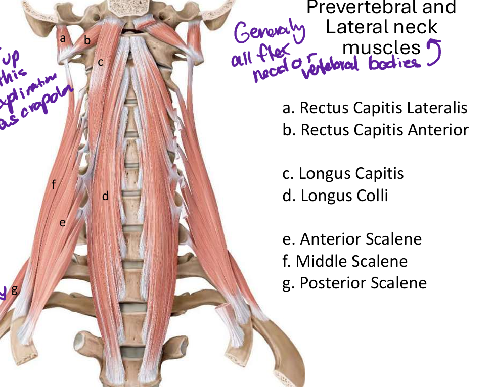
produce neck flexion, Longus Colli has been shown to have a postural function on cervical curvature, counteracting the lordosis increment related to the weight of the head and to the contraction of the posterior cervical muscles
90
longus coli innervation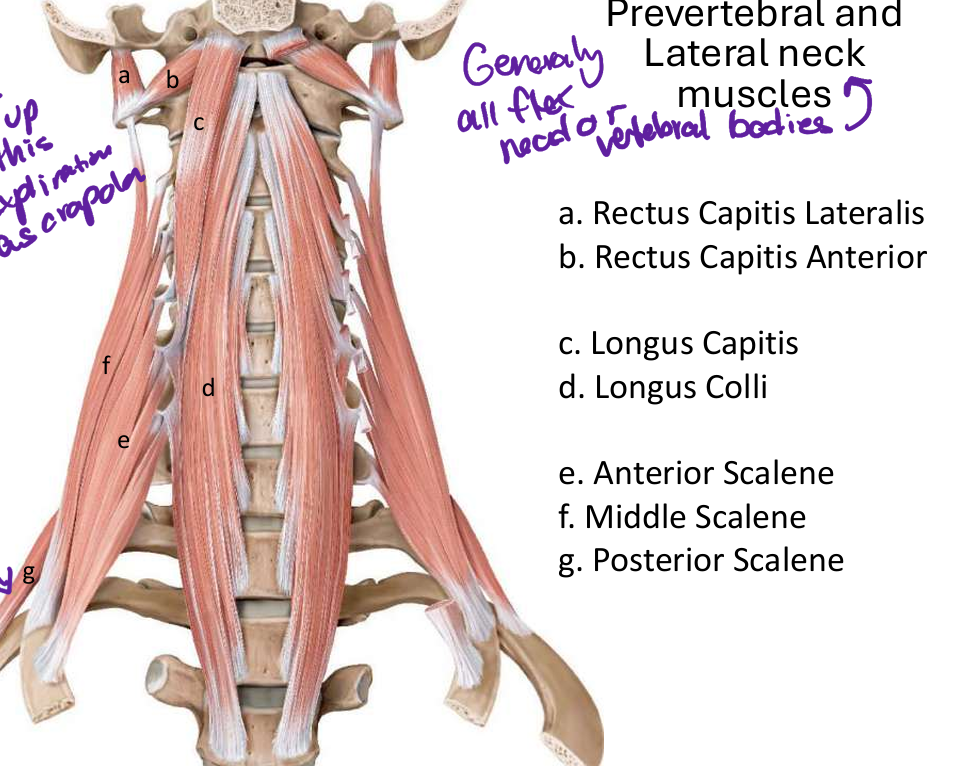
anterior rami C2-6
91
anterior scalene m fx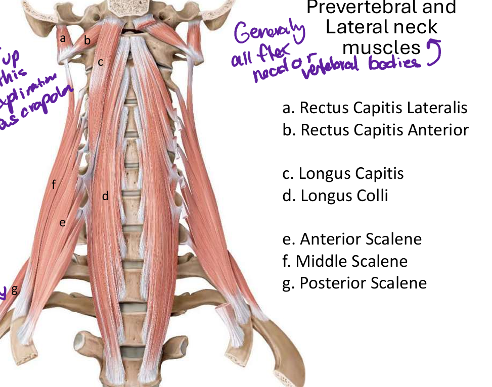
bending neck forward and laterally, rotate the neck to opposite side; assists first rib in respiration
92
anterior scalene m innervation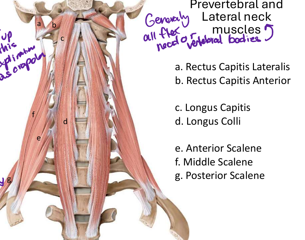
anterior rami C4-6
93
middle scalene m innervation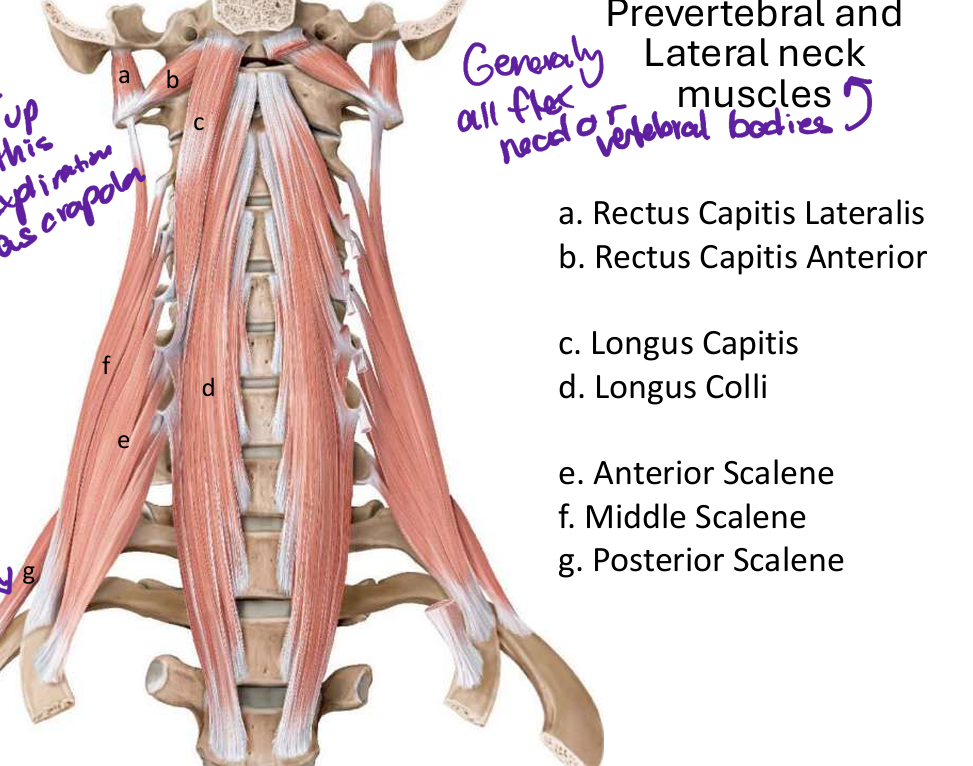
elevate first rib during inhalation
lateral flexion of neck
lateral flexion of neck
94
innervation middle scalene m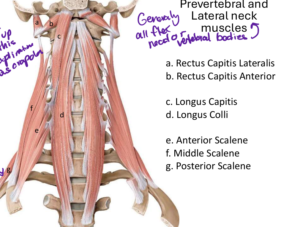
anterior rami C3-8
95
fx posterior scalene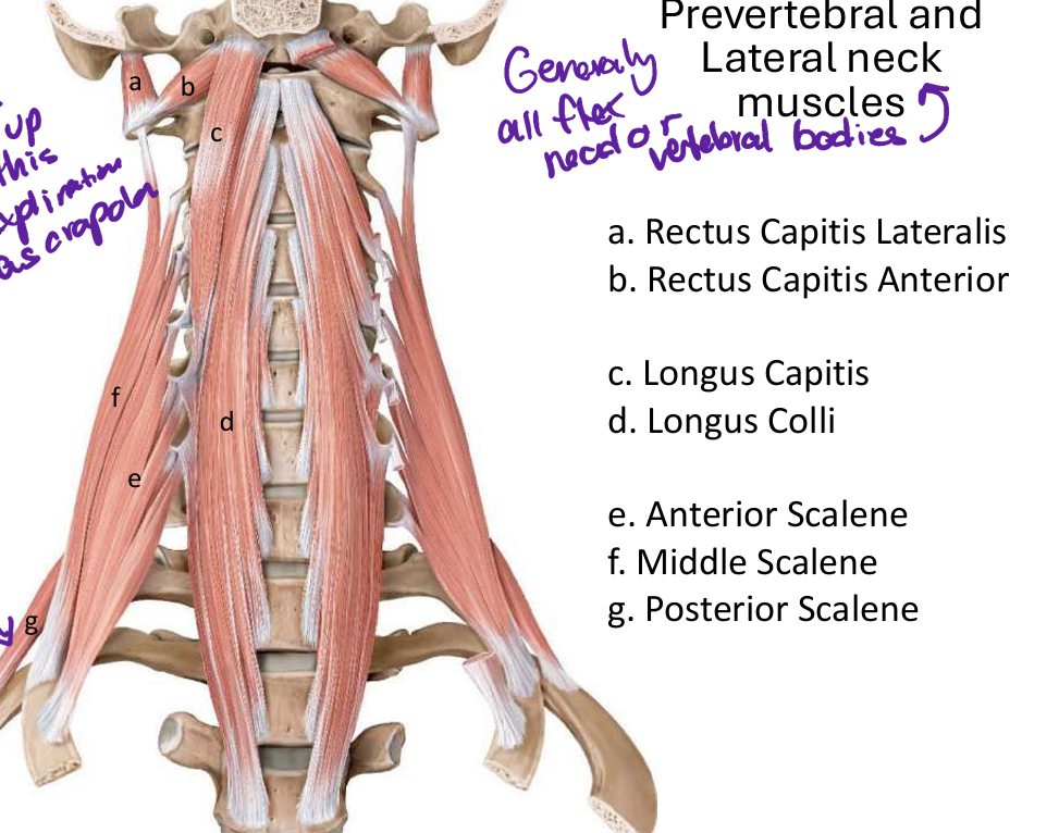
elevate second rib and ipsilateral lateral flexion of neck
96
innervation posterior scalene m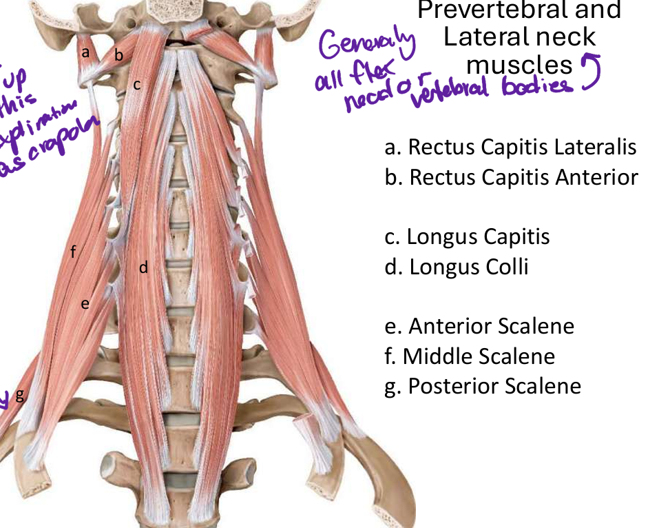
anterior rami C5-8
97
branches of the subclavian a (L/R)
1. Vertebral
2. Internal thoracic
3. Thyrocervical
trunk
4. Costocervical
trunk
5. Descending
(Dorsal) Scapular – 50%
2. Internal thoracic
3. Thyrocervical
trunk
4. Costocervical
trunk
5. Descending
(Dorsal) Scapular – 50%
98
branches subclavian p1
nternal thoracic a
thyrocervical trunk
vertebral a
thyrocervical trunk
vertebral a
99
branches of thyrocervicak trunk
inferior thtroid
ascending cervical
transverse cerival
suprascapular
ascending cervical
transverse cerival
suprascapular
100
what does the inferior thyrpid a supply
thyroid gland
101
what does the asceding cervical a supply
deep muscles of the neck, such as the anterior scalene muscle, and sends spinal branches through the intervertebral foramina to provide blood to the cervical spinal cord and its surrounding meninges.
102
what adoes the transverse cervical a suppky
trapexiys m
103
wat does the suprascapular a supply
supraspinatus and infraspinatus
104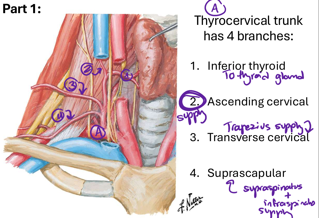
Image occlusion: name the hidden labels
105
Image occlusion: name the hidden labels
106
Image occlusion: name the hidden labels
107
Image occlusion: name the hidden labels
108
Image occlusion: name the hidden labels
109
what branches off part 2 of the subclavuan a
costocervical trunk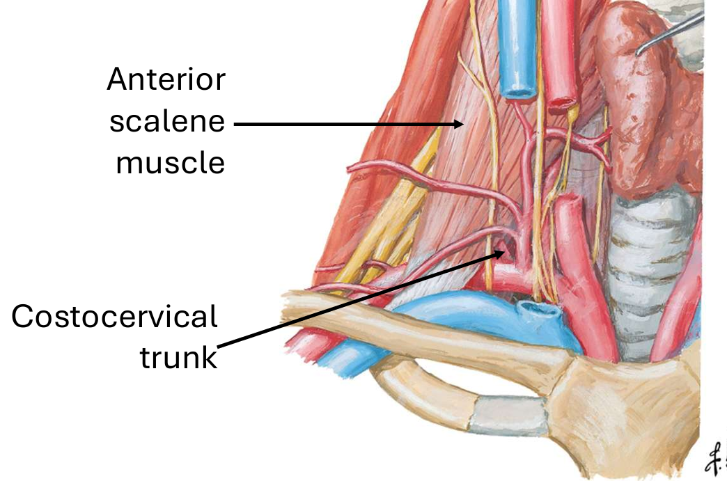
110
what does the costocervical trunk supply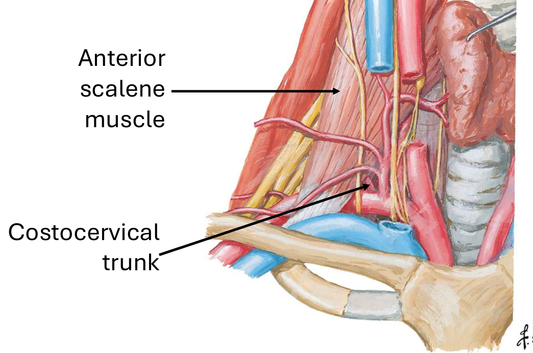
blood to the deep cervical muscles of the back and the upper thorax, specifically the first and second intercostal spaces
111
what are the branches of the costocervicak trunk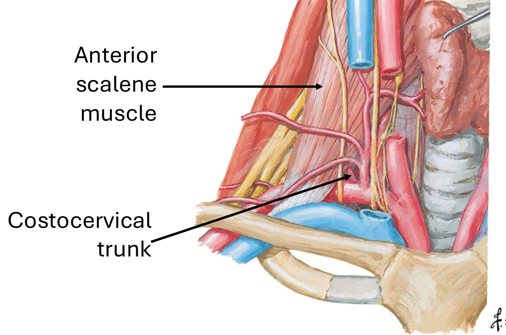
deep cervical a and superior intercostal a
112
what does the deep cervical a supply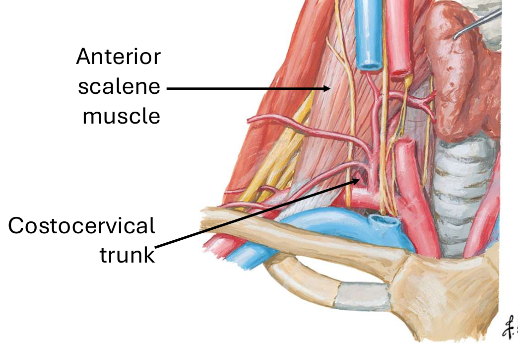
deep back muscles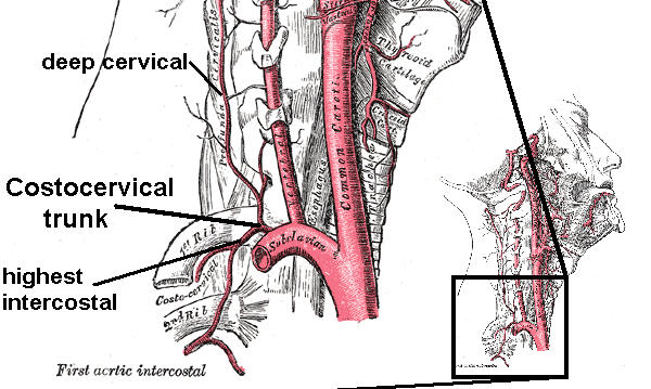
113
what does the superiorintercostal a supply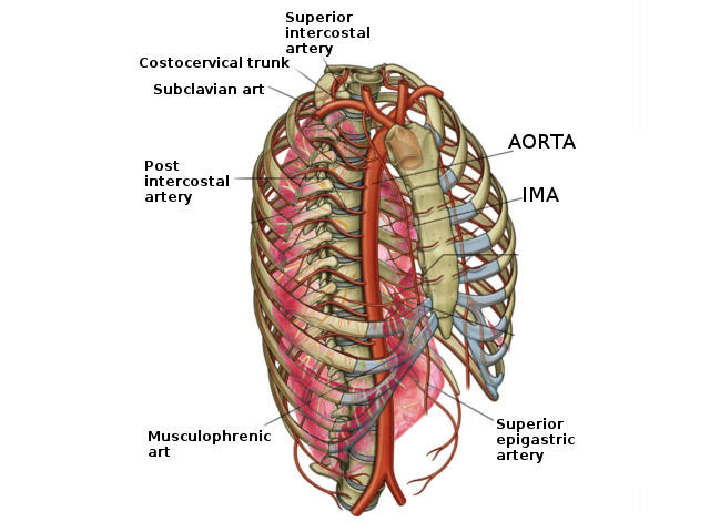
upper 2 posterior intercostal spaces
114
what possibly can branch from the third part of the subclavian a (L/R)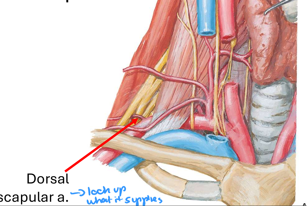
dorsal scapular a
115
of this artery exists, the dorsal scapular a does not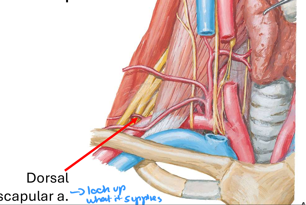
deep branch of transverse cervical
116
what are the common variations of the thyrocervical trunk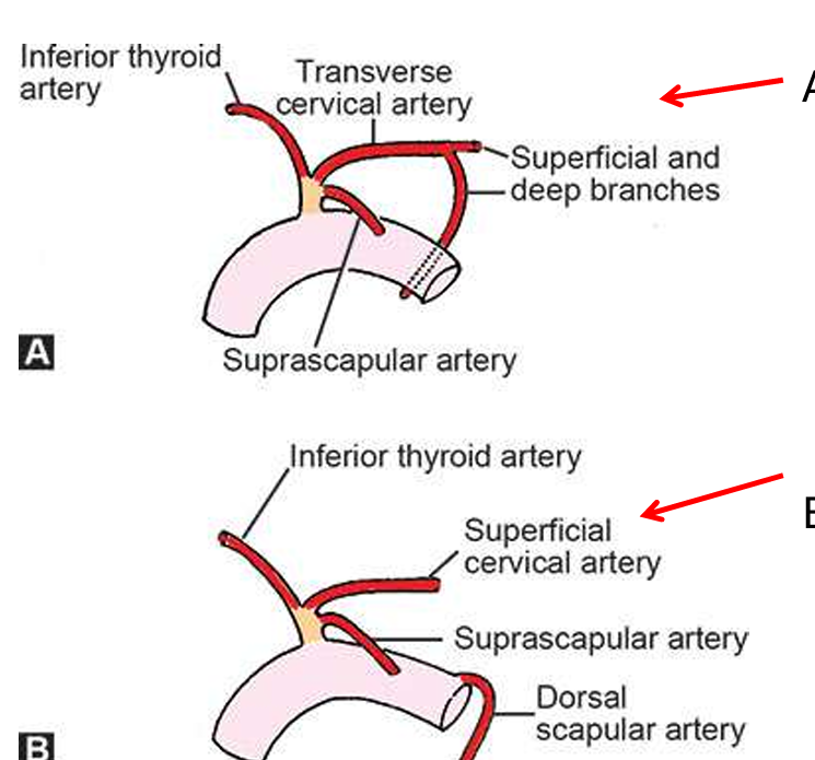
one where the transverse cervical has a superficial and deep branch
OR
one where the superficial cervical a ad dorsal scapular a branch from the tyrocervicak trunk
OR
one where the superficial cervical a ad dorsal scapular a branch from the tyrocervicak trunk
117
what spinal level does the common carotid a typically split at
C4
118
what are the branches of the external carotid?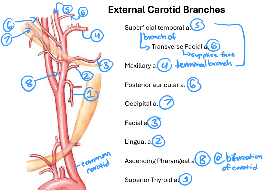
Superior thyroid, lingual, facial, maxillary, superficial tenmporal, posterior auricular, occipital, ascending pharyngeal
(Some little fat man stole papa's only apple)
(Some little fat man stole papa's only apple)
119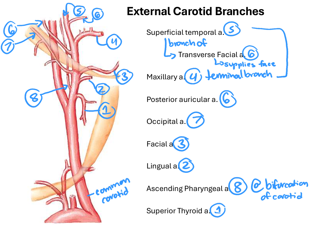
Image occlusion: name the hidden labels
120
Image occlusion: name the hidden labels
121
Image occlusion: name the hidden labels
122
Image occlusion: name the hidden labels
123
Image occlusion: name the hidden labels
124
Image occlusion: name the hidden labels
125
Image occlusion: name the hidden labels
126
Image occlusion: name the hidden labels
127
Image occlusion: name the hidden labels
128
what does the superficial temporal a supply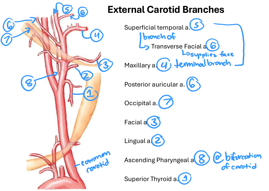
forehead, temporal and parietal regions of the head, the temporalis muscle, and parts of the parotid gland
129
what are the branches of the superficial temporal a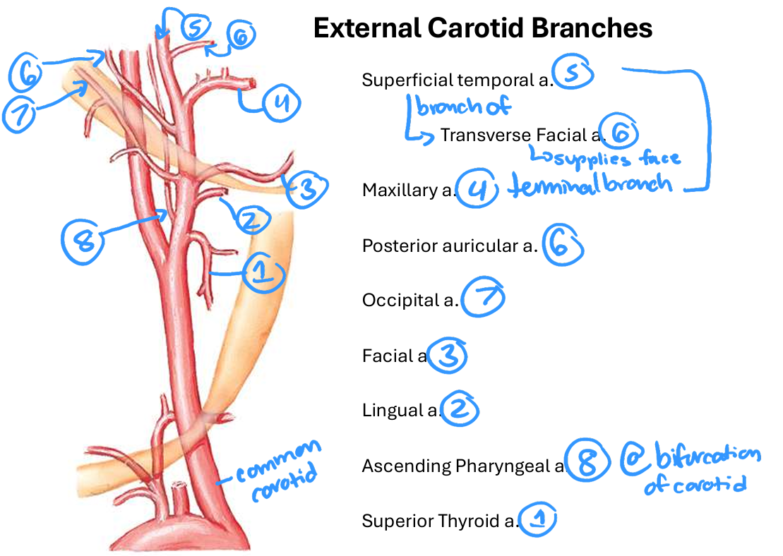
frontal brancj and parietal branchand transverse facial a
130
what does the frontal branch of superficial temporal a supply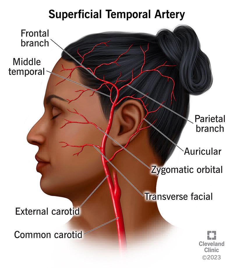
forehead
131
what foes the parietal branch of the superficial temporal a supply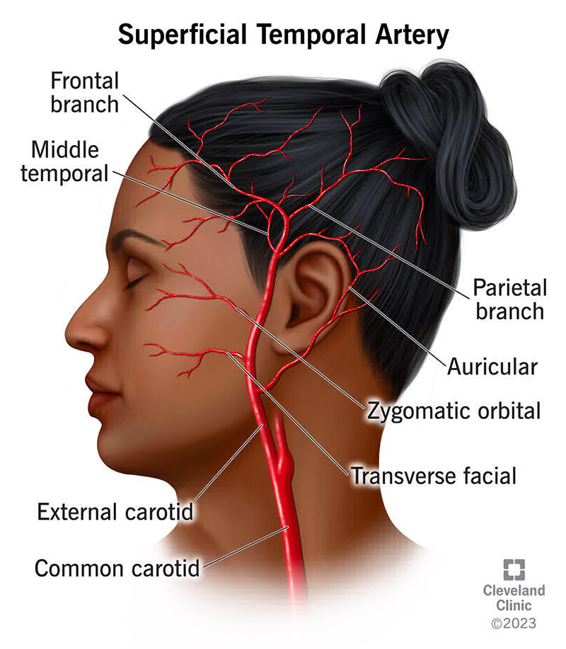
side and back of scalp
132
what does the transverse facial a supply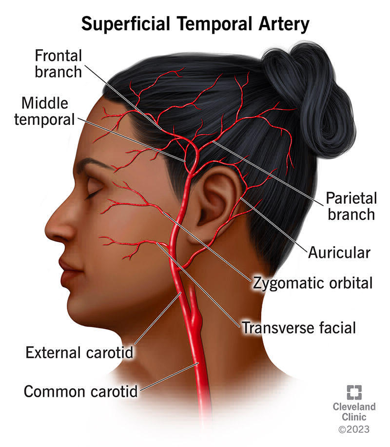
parotif gland and cheek
133
what does the maxillary a supply
Gives off numerous branches that supply:
Muscles of mastication
Teeth and gums
Nasal cavity
Dura mater
Middle ear
Muscles of mastication
Teeth and gums
Nasal cavity
Dura mater
Middle ear
134
what does the lingual a supply
oral cavity
135
what does the superior thyroid a supply
thyroid
136
what is the arerial support to the tonsils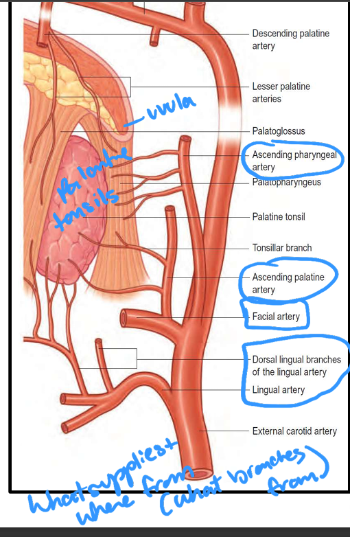
ascendng pharyngeal a
ascending palantine a from facial a
dorsal lingual branches of lungual a from lingual a
ascending palantine a from facial a
dorsal lingual branches of lungual a from lingual a
137
what n runs with the occipital a
CN XII
138
what n runs with the superior thyroid a
external laryngeal n (innervates cricthyroid m)
139
what n runs with the superior laryngeal a
internal laryngeal n
140
branches of the superior thyroid a that anastomose across the midline
infrahyoid a abd superior laryngeal a
141
branches od lungual a that anastomose acrpss the midline
suprahypid, dorsal lingual, sublinguial a
142
branches of facial a that anastomose across the midline
ascending palatine a
143
branches of occipital a that anastomose acrpss the midline
occipital branches
144
fx as a baroreceptor, consists of dilation ofd lower end of internal carotid a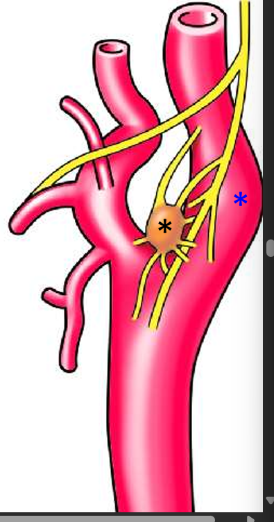
carotid sinus (blue)
145
small redisj vrwn chemorecepor innervated by CN IX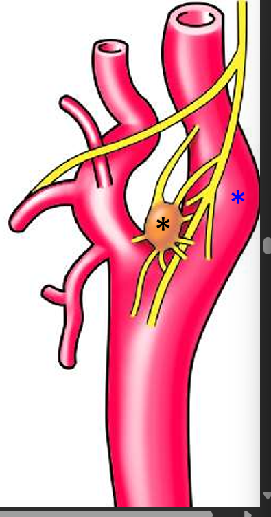
carotid bpdy (black)
146
the superficial neck is drained by these veins
anterior and external jugular v
147
veins that dumb into the internal jugular v
Retromandibular vein
• Common facial vein
• Middle thyroid vein
• Additional branche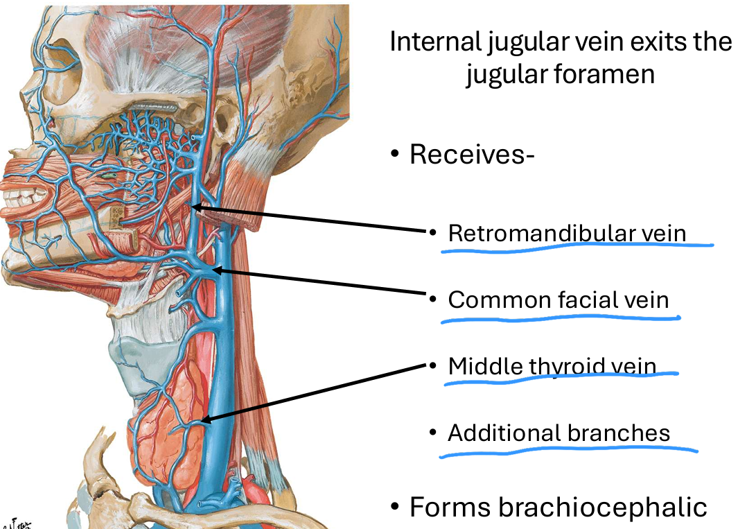
• Common facial vein
• Middle thyroid vein
• Additional branche
148
Image occlusion: name the hidden labels
149
Image occlusion: name the hidden labels
150
Image occlusion: name the hidden labels
151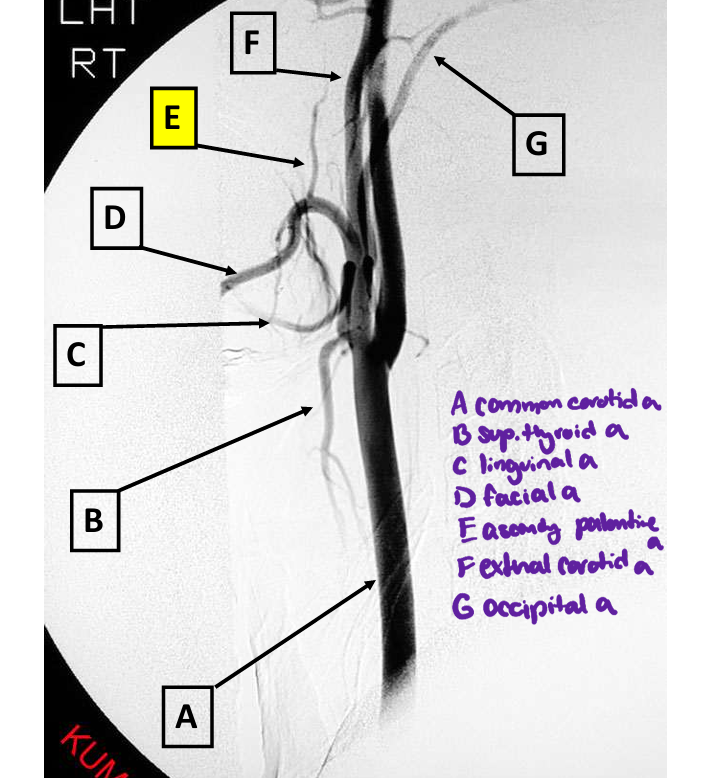
Image occlusion: name the hidden labels
152
Image occlusion: name the hidden labels
153
Image occlusion: name the hidden labels
154
Image occlusion: name the hidden labels
155
Image occlusion: name the hidden labels
156
Image occlusion: name the hidden labels
157
Image occlusion: name the hidden labels
158
superficial structures of the facse drain to thes elymph nodes
superficial cervical nodes
159
the level 1 lymoh nodes drain to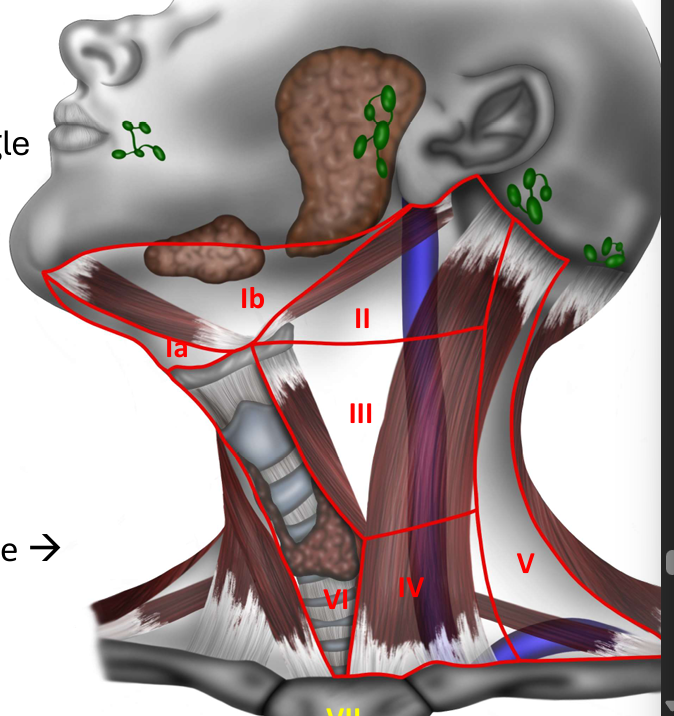
levels II and III
160
the level II lymph nodes drai to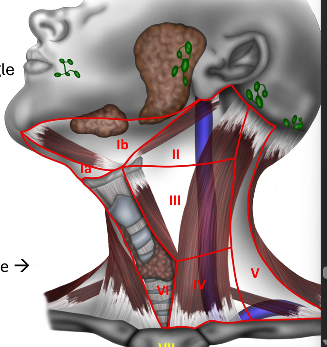
Levels III-V
161
the level III lymph nodes drain to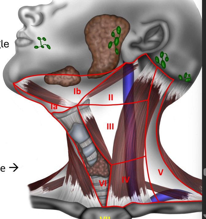
level IV
162
the level IV lymph nodes drain to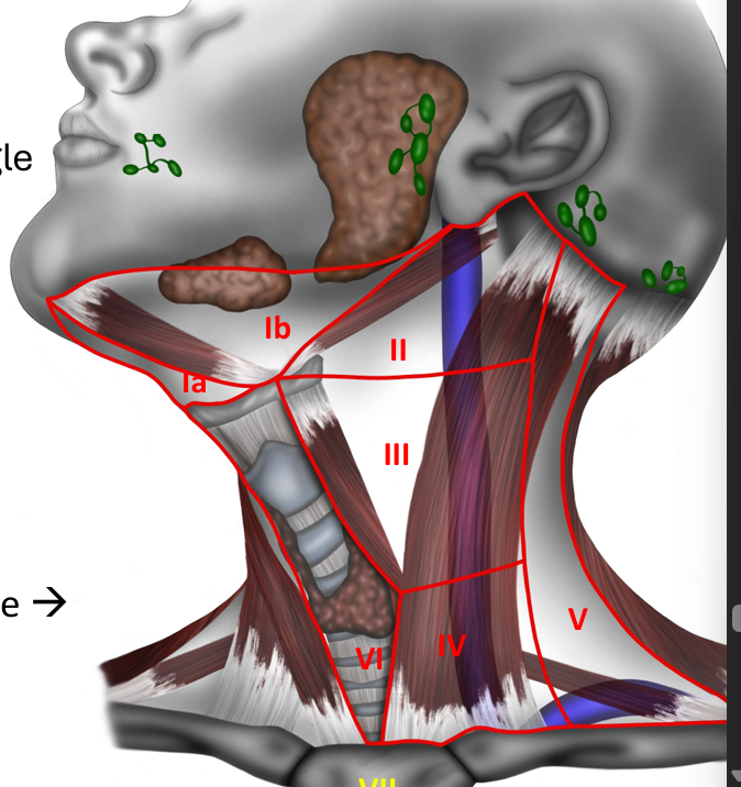
thoracuc duct or right lymphatic duct
163
what do the level V lumph nodes drain to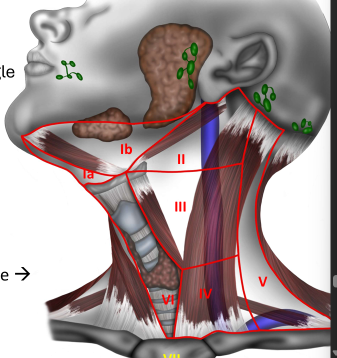
posterior triangle to level IV
164
what do the level VI lymph nodes drain to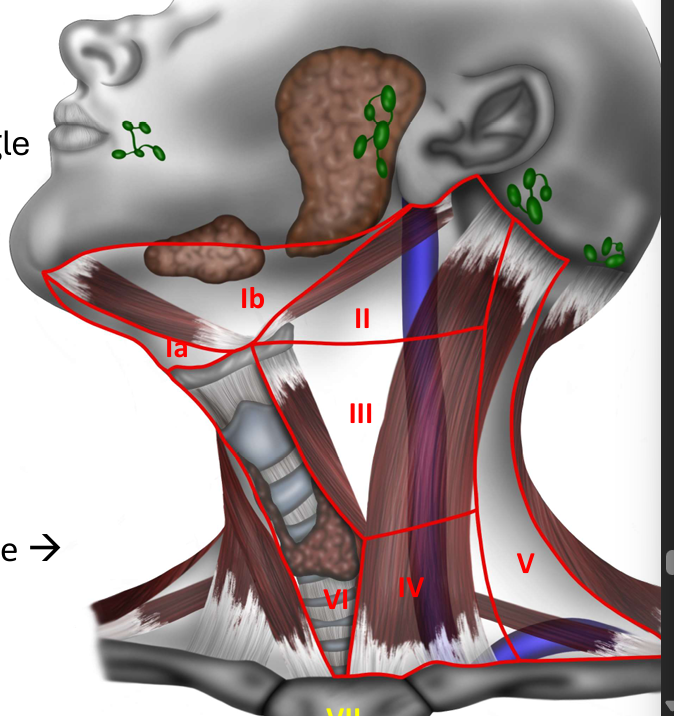
levels II to IV
165
what does the level VII lymph nodes drain to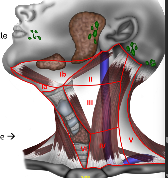
sperior mediastinum
166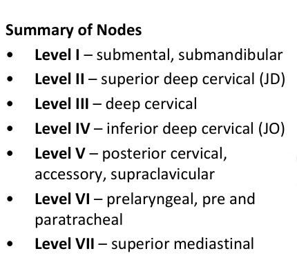
Image occlusion: name the hidden labels
167
Image occlusion: name the hidden labels
168
Image occlusion: name the hidden labels
169
Image occlusion: name the hidden labels
170
Image occlusion: name the hidden labels
171
Image occlusion: name the hidden labels
172
Image occlusion: name the hidden labels
173
where does CN I arise?
forebrain
174
where does CN II arise
diencephalon
175
where do CNIII-XII arise
brainstem
176
where does CN XI arrise
spinal cord
177
pseudounipolar cell bodies in sensory ganglia
afferent
178
innervate skeletal muscle of viscera
efferent
179
which cranial nerves carry parasympathetic fibers
CN III, VII, IX, and X
180
taste and olfaction
special visceral afferent (SVA)
181
viscion, balanxe and hearing
special somatic afferentr (SSA)
182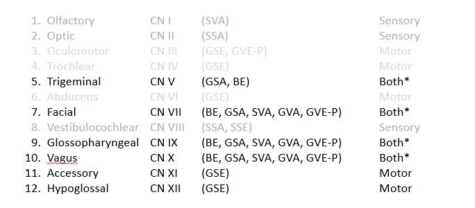
Image occlusion: name the hidden labels
183
Image occlusion: name the hidden labels
184
Image occlusion: name the hidden labels
185
Image occlusion: name the hidden labels
186
Image occlusion: name the hidden labels
187
Image occlusion: name the hidden labels
188
Image occlusion: name the hidden labels
189
Image occlusion: name the hidden labels
190
Image occlusion: name the hidden labels
191
Image occlusion: name the hidden labels
192
Image occlusion: name the hidden labels
193
Image occlusion: name the hidden labels
194
Image occlusion: name the hidden labels
195
Image occlusion: name the hidden labels
196
Image occlusion: name the hidden labels
197
Image occlusion: name the hidden labels
198
Image occlusion: name the hidden labels
199
Image occlusion: name the hidden labels
200
Image occlusion: name the hidden labels
201
Image occlusion: name the hidden labels
202
Image occlusion: name the hidden labels
203
Image occlusion: name the hidden labels
204
Image occlusion: name the hidden labels
205
Image occlusion: name the hidden labels
206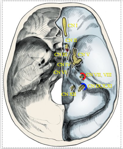
Image occlusion: name the hidden labels
207
Image occlusion: name the hidden labels
208
Image occlusion: name the hidden labels
209
Image occlusion: name the hidden labels
210
Image occlusion: name the hidden labels
211
Image occlusion: name the hidden labels
212
Image occlusion: name the hidden labels
213
Image occlusion: name the hidden labels
214
Image occlusion: name the hidden labels
215
these cranial n innervate the extra ocular muscles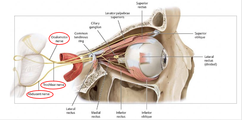
CN III, IV, and VI
216
pathway of GVE oculomotor III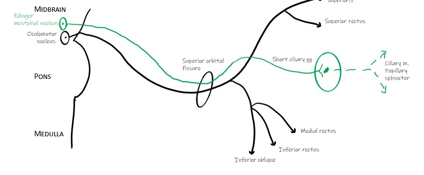
edwnger westphal nucleus to suerior orbital fissure to short ciliary m (synapse) to cilliary m, pupillary sphincter m
217
what does the oculomotor n (III) innervate in the ocular m (GSE)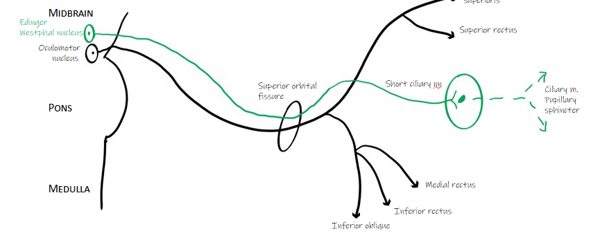
levator palpebrae superioris m, superior rectus m, medial rectus m, inferior rectus m, inferior oblique m
218
what important eye m does the trochear n innervate (GSE)
suprior oblique m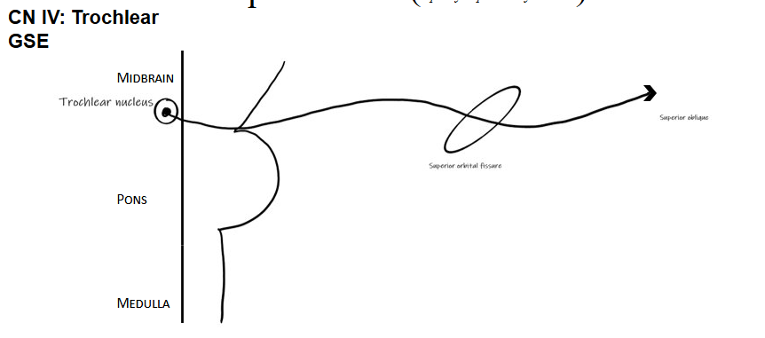
219
what m does the abducens innervate (imp eye muscle) (GSE)
lateral rectus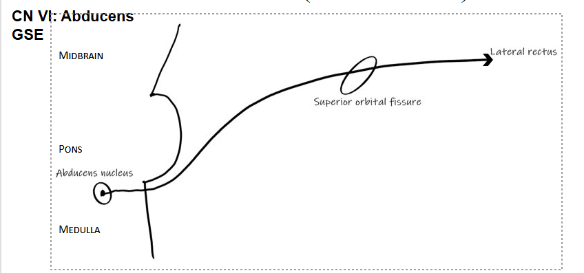
220
what is the fx of the trigeminal n CNV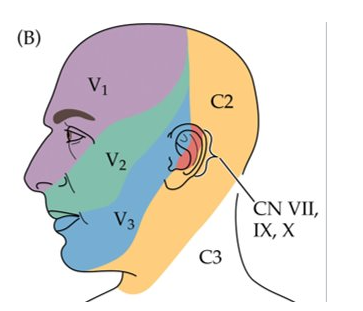
senspry innervation (GSA) to face and innervates muscles of mastication
221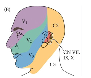
Image occlusion: name the hidden labels
222
Image occlusion: name the hidden labels
223
Image occlusion: name the hidden labels
224
Image occlusion: name the hidden labels
225
Image occlusion: name the hidden labels
226
Image occlusion: name the hidden labels
227
what is the pathway for V1 trigeminal CNV
GSA from pons through trigeminal ganglion to superior orbital fissure and V1 innervation (also hits ciliary ganglion)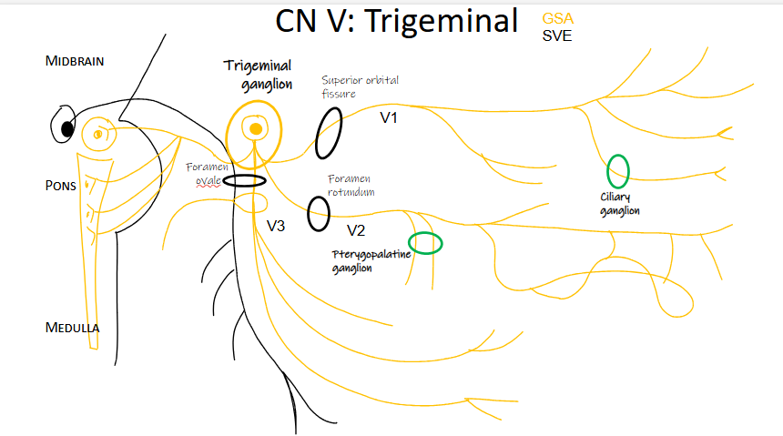
228
what is the pathway for V2 trigemial CNV
GSA fdrom pons and medulla go through trigeminal ganglion and fotamen rotundum to innervate V2 sensory. also hits pterygopalatine ganglion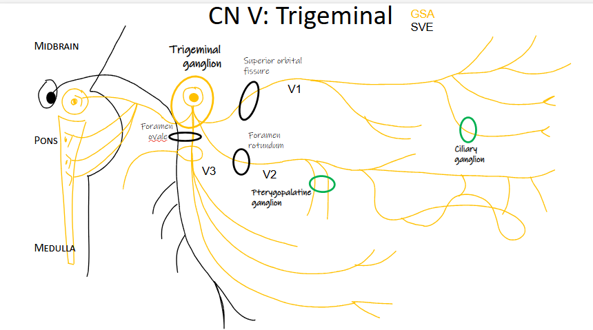
229
what is the pathway for V3 trigeminal CN V
GSA and SVE pons and medulla through trigeminal ganglion through foramen ovale to V3 innervation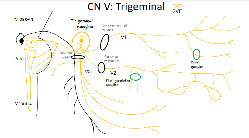
230
what is innervated by the V1 branch of CN V
GSA to frontal and lacrimal
GSA to nasociliary than long ciliary to eye and short ciliary through ciliary ganglion
GSA through nasociliary branching ti posterior and anterior ethmoidal and infratrochlear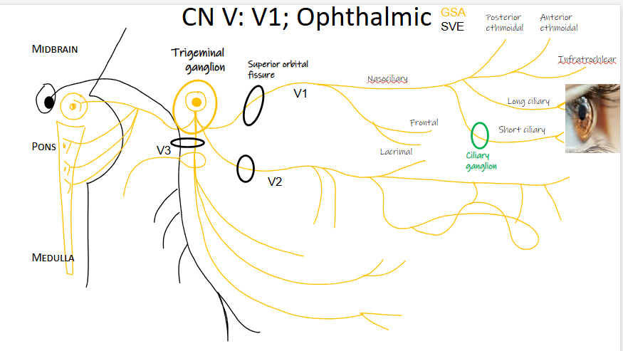
GSA to nasociliary than long ciliary to eye and short ciliary through ciliary ganglion
GSA through nasociliary branching ti posterior and anterior ethmoidal and infratrochlear
231
what is innervated by the CN V at V2 maxillary
GSA thrugh foramen rotundum to descending palantine and nasopalantine.
GSA through foramen rotundum and inferior orbital fissure to zygomatic
GSA posterior to middle superiuor alveolar m at dental plexus
GSA through infraorbital foramen (anterior) to infraorbital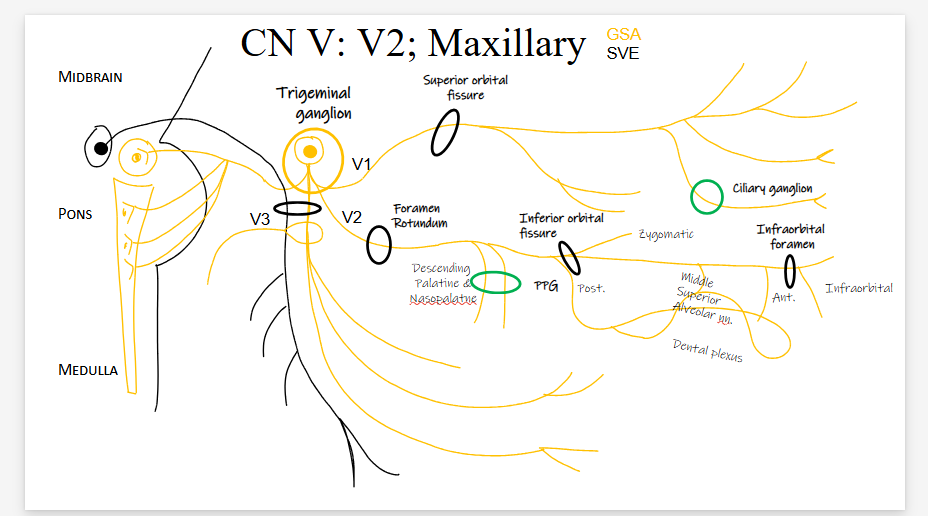
GSA through foramen rotundum and inferior orbital fissure to zygomatic
GSA posterior to middle superiuor alveolar m at dental plexus
GSA through infraorbital foramen (anterior) to infraorbital
232
CN V V3 mandibular branch innervation GSA
through foramen ovale to buccal and ligual
through foramen ovale and mandibular foramen to inferior alveolar and mental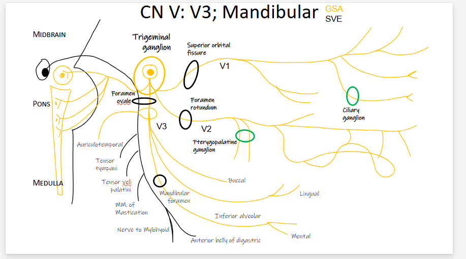
through foramen ovale and mandibular foramen to inferior alveolar and mental
233
CN V V3 mandibular innervation SVE
through foramen ovale to tensir tympani, tensor veli palatini, mm of mastication, nerve to mylohyoid, and anterior belly of digastric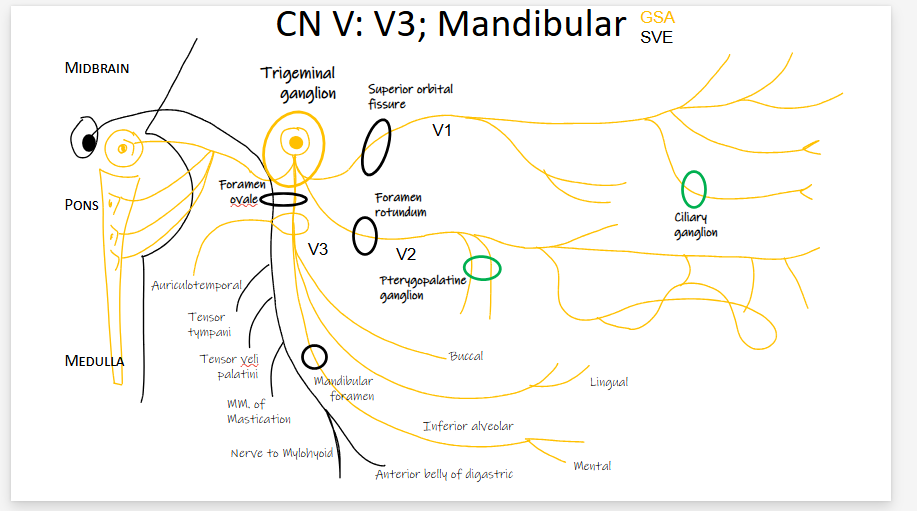
234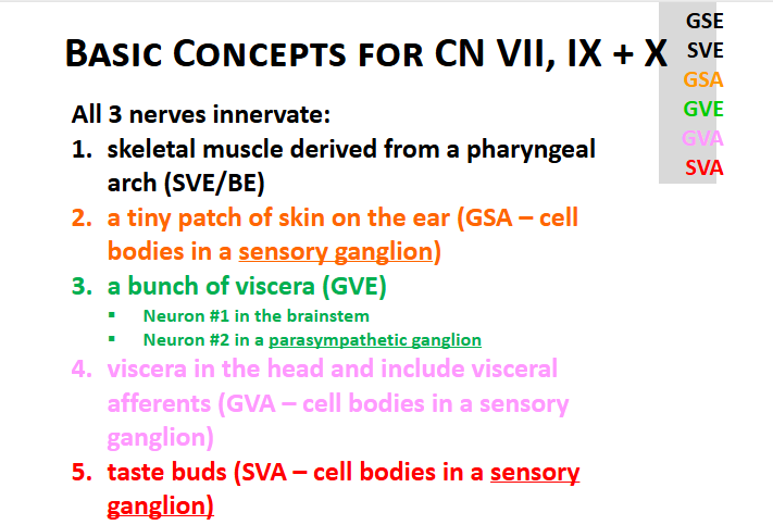
what do nerves CN VII, IX, and X innervate basically
235
what does the cranial nerve innervate Facial VII
-muscles of facial expression through internal auditory meatus and stylomastoid foramen
-glands to oral cavity (GVE)
-taste anterior 2/3 tongue (SVA)
-glands on palate, nasal cavity and paranasal sinuses (GVE)
-lacrimal gland (GVE)
-fdeep petrosal n (GVA)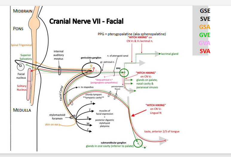
-glands to oral cavity (GVE)
-taste anterior 2/3 tongue (SVA)
-glands on palate, nasal cavity and paranasal sinuses (GVE)
-lacrimal gland (GVE)
-fdeep petrosal n (GVA)
236
Image occlusion: name the hidden labels
237
Image occlusion: name the hidden labels
238
Image occlusion: name the hidden labels
239
Image occlusion: name the hidden labels
240
Image occlusion: name the hidden labels
241
Image occlusion: name the hidden labels
242
Image occlusion: name the hidden labels
243
Image occlusion: name the hidden labels
244
Image occlusion: name the hidden labels
245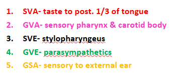
glossopharyngeal n fx
246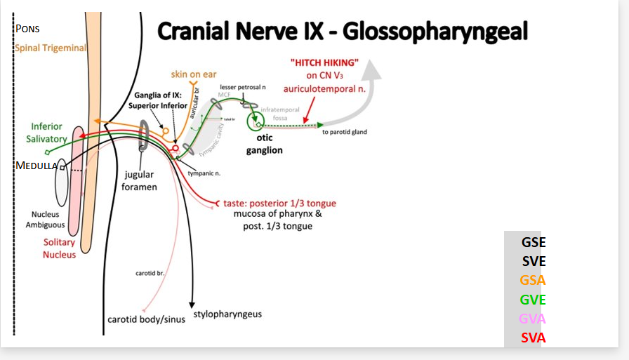
Image occlusion: name the hidden labels
247
Image occlusion: name the hidden labels
248
Image occlusion: name the hidden labels
249
Image occlusion: name the hidden labels
250
Image occlusion: name the hidden labels
251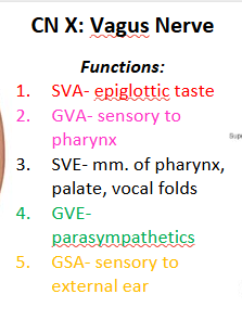
CN X vagus n fx
252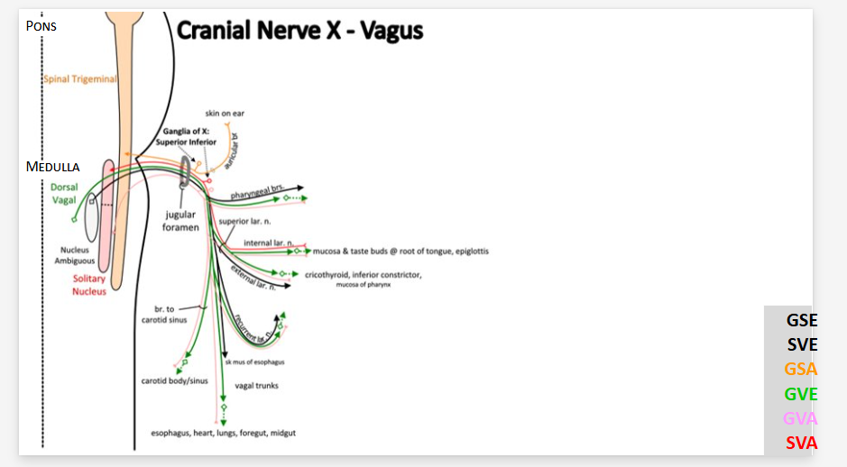
Image occlusion: name the hidden labels
253
Image occlusion: name the hidden labels
254
Image occlusion: name the hidden labels
255
Image occlusion: name the hidden labels
256
Image occlusion: name the hidden labels
257
what do the 4 parasympathetic ganglia hitchhike on?
CN V branches
258
what are the 4 parasympathetic ganglia
ciliary (CN III), Otic (CN IX), pterygopalatine (CN VII), submanibular (CN VII)
259
what are the 4 sensory ganglia in cranial N
trigeminal (CN V). geniculate (CN VII), inferior (CN IX and X), and superior (CN IX and X)
260
functions of the spinal accesory n
Elevating and rotating the head contralaterally (opposite side)
Flexing the neck
Tilting the head towards the side of the contracting muscle
Assists in breathing by elevating the ribs during deep inspiration
Contributes to swallowing by innervating some muscles of the pharynx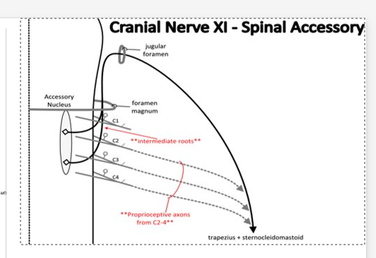
Flexing the neck
Tilting the head towards the side of the contracting muscle
Assists in breathing by elevating the ribs during deep inspiration
Contributes to swallowing by innervating some muscles of the pharynx
261
functions of the hypoglossal n (CN XII)
controls all the muscles of the tongue, enabling movements for speech and swallowing. It also contains sympathetic fibers that supply blood vessels and glands in the mouth.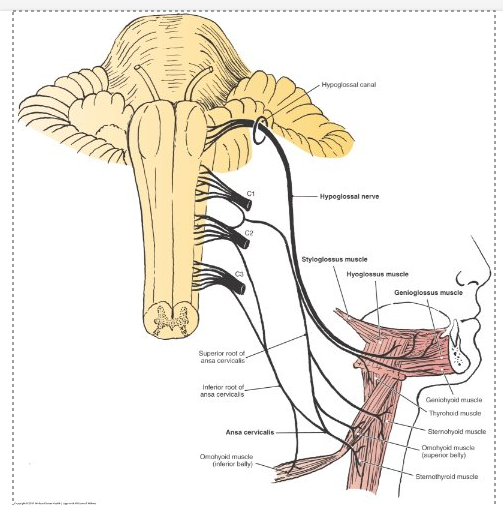
262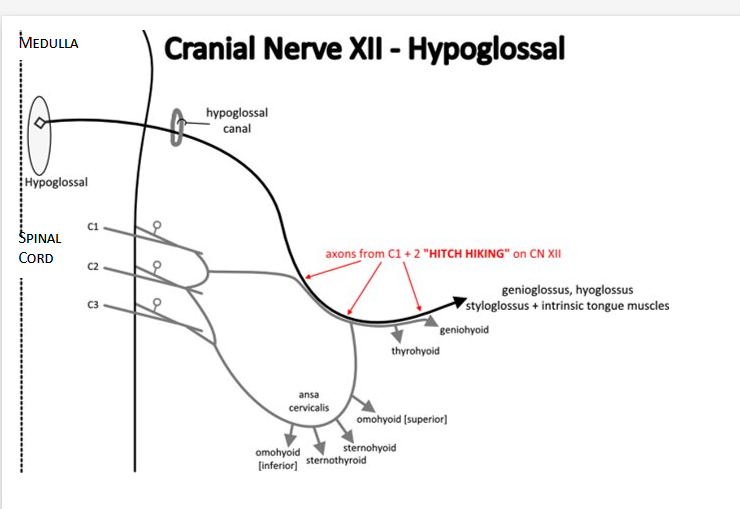
Image occlusion: name the hidden labels
263
Image occlusion: name the hidden labels
264
Image occlusion: name the hidden labels
265
Image occlusion: name the hidden labels
266
Image occlusion: name the hidden labels
267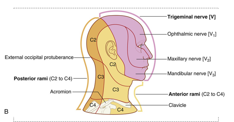
Image occlusion: name the hidden labels
268
Image occlusion: name the hidden labels
269
Image occlusion: name the hidden labels
270
Image occlusion: name the hidden labels
271
Image occlusion: name the hidden labels
272
Image occlusion: name the hidden labels
273
Image occlusion: name the hidden labels
274
motor n in cervical plexus
ansa cervicalis
C1 to thyrohyoid
phrenic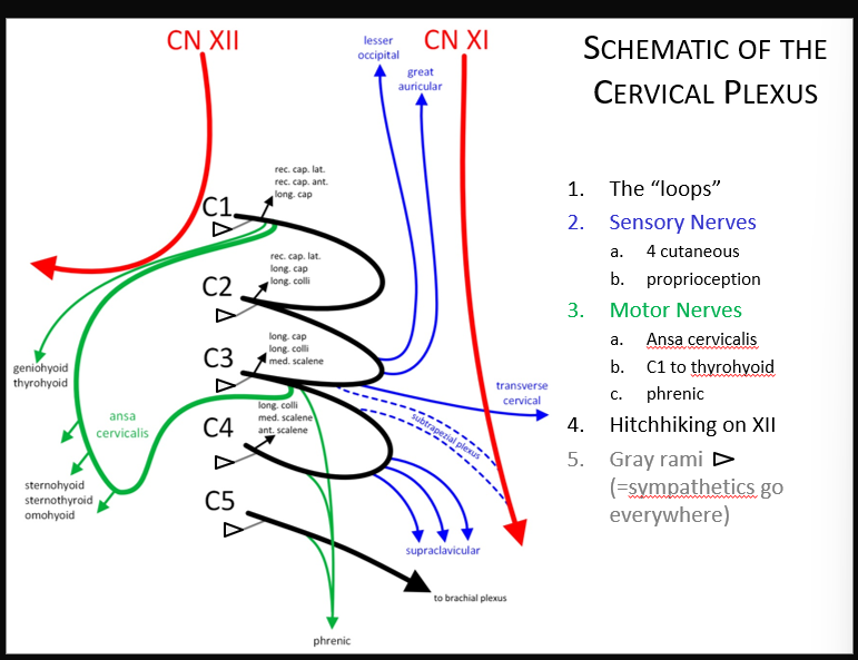
C1 to thyrohyoid
phrenic
275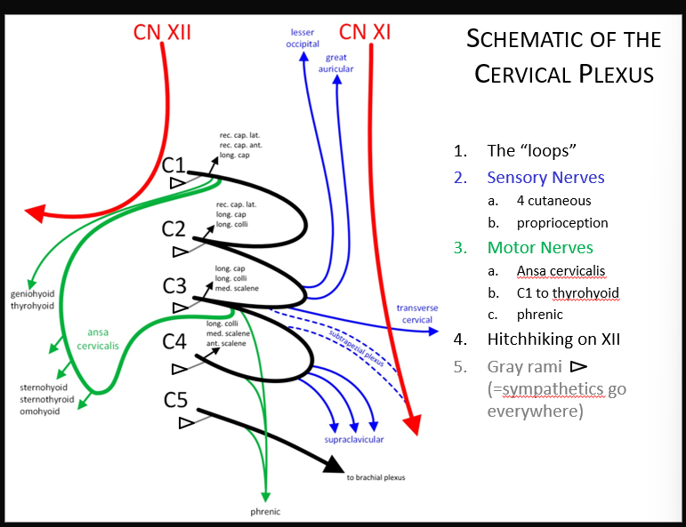
Image occlusion: name the hidden labels
276
Image occlusion: name the hidden labels
277
Image occlusion: name the hidden labels
278
Image occlusion: name the hidden labels
279
Image occlusion: name the hidden labels
280
Image occlusion: name the hidden labels
281
Image occlusion: name the hidden labels
282
Image occlusion: name the hidden labels
283
what spinal rami form the cervical plexus
C1-4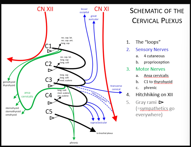
284
what does the ansa cervicalis (c1-3) supplu
motor innervation to the infrahyoid m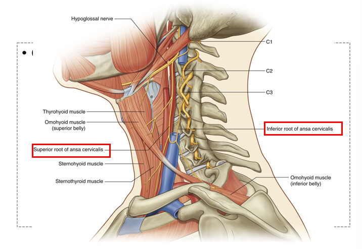
285
the roots of the brachial plexus emerge between these two nexk muscles
middle and anterior scalene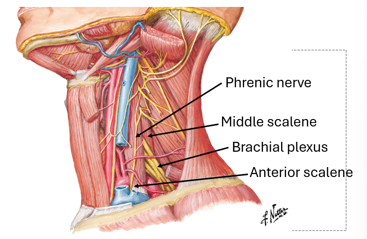
286
the SVE/BE for skeletal muscle are derived from...
oharyngeal arch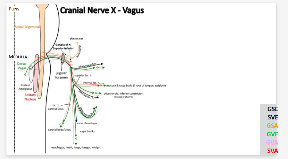
287
what does the vagus n course through as it travels through the neck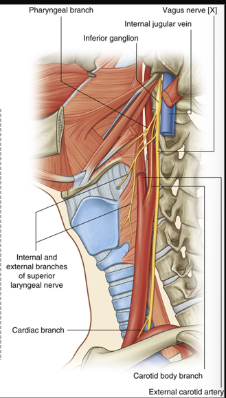
carotid sheath
288
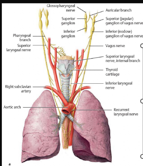
vagus n branches in the neck (x3)
Pharyngeal branches
Superior laryngeal n.
--Internal laryngeal n.
--External laryngeal n.
Recurrent/Inferior laryngeal n.
Superior laryngeal n.
--Internal laryngeal n.
--External laryngeal n.
Recurrent/Inferior laryngeal n.
289
Image occlusion: name the hidden labels
290
Image occlusion: name the hidden labels
291
Image occlusion: name the hidden labels
292
Image occlusion: name the hidden labels
293
Image occlusion: name the hidden labels
294
Image occlusion: name the hidden labels
295
what is the s/s of a vagus n injiury at the jugular fpramen or a brainstem lesion
Il dysphagia, sagging palate, horseness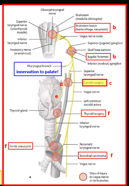
296
what is ine s/s of a vagus n inhjury at the superior laryngeal n post caotid surgery
diminished coughing reflex, aspiration, monotonous vpice, IL deviation of thyrpid cartilage, IL vocal cord is slacjk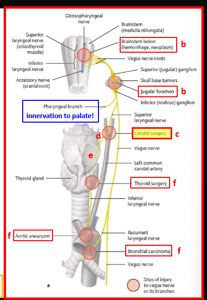
297
what is the s/s of an internal larynheal n surgery and what can cause it
diminished cpugh reflex
-traima, lumphadeopathy, retrieval of fpreign object in priform recess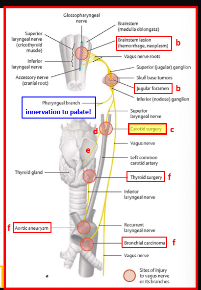
-traima, lumphadeopathy, retrieval of fpreign object in priform recess
298
what is the s/s of an external laryngel n injury and what can cause it
-monotonous voice, Il devoation of thyrpid cartilage and Il vocal cord is slack
-ligation of the superior thyroid a
-it oinnervates the cricothyrud and inferior constrictor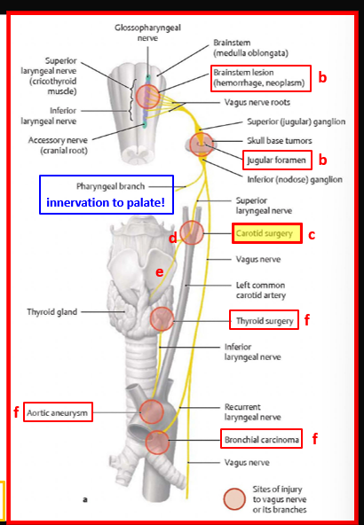
-ligation of the superior thyroid a
-it oinnervates the cricothyrud and inferior constrictor
299
what are the s/s and wjat can cause injury to the recurrent larungeal n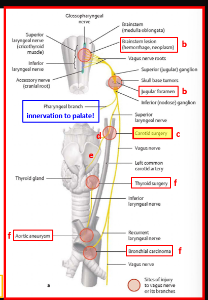
horseness, paralyzed vocal cord, innervation to all laryngeal miuscles except cricothyroid and sensory below vocal cords
-aorric arch aneurysm, surgical repair of PDA, ligation of inferior thyroid a
-aorric arch aneurysm, surgical repair of PDA, ligation of inferior thyroid a
300
what is the fusion of the superior cervical ganglia?
C1-4
33 more cards in OutLearn. Open the full deck